Citation:
GONG Liaokuo, SONG Ying, SHEN Nannan, ZHANG Bo, WU Zhaofeng, HUANG Xiaoying. A Fluorescent Magnesium-Based Metal-Organic Framework with a Sensitive Sensing Property for Carbon Disulfide[J]. Chinese Journal of Applied Chemistry,
2017, 34(9): 1059-1065.
doi:
10.11944/j.issn.1000-0518.2017.09.170189

一例对二硫化碳具有荧光传感性能的Mg-金属有机框架化合物
摘要:
通过溶剂热合成了一例Mg-MOF化合物[Mg4(1,4-NDC)4(DMA)2(CH3OH)2(H2O)2]·DMA·CH3OH(1,1,4-H2NDC=1,4-萘二酸,DMA=N,N'-二甲基乙酰胺),并对其结构表征及荧光性能进行了研究。单晶X射线研究结果表明,化合物结晶于P21/c空间群,其晶体学数据为a=2.06090(12)nm,b=2.21014(13)nm,c=1.50385(10)nm,β=111.399(3)°,V=6.3776(7)nm3,Z=4,Dc=1.403 g/cm3,F(000)=2824,R=0.0596,wR=0.1225(I>2σ(I))。化合物1中,二核的镁作为次级构筑单元通过桥连配体1,4-NDC连接形成沿c轴方向拓展的一维链。一维链间进一步通过配体连接形成3D框架的化合物。荧光性能研究表明,化合物1对CS2具有灵敏的荧光传感性能,在0.4%的体积分数条件下可引起CS2荧光的完全淬灭。此外,化合物1的热稳定性也通过热重分析进行了研究,发现其可稳定到140℃左右。
English
A Fluorescent Magnesium-Based Metal-Organic Framework with a Sensitive Sensing Property for Carbon Disulfide
Abstract:
Presented here are the solvothermal synthesis, structural characterization and fluorescent properties of a magnesium metal-organic framework(Mg-MOF), namely[Mg4(1, 4-NDC)4(DMA)2(CH3OH)2(H2O)2]·DMA·CH3OH(1, 1, 4-H2NDC=1, 4-naphthalene dicarboxylic acid, DMA=N, N'-Dimethylacetamide). Single-crystal X-ray diffraction studies revealed that compound 1 crystallized in the monoclinic space group P21/c(No.14) with a=2.06090(12) nm, b=2.21014(13) nm, c=1.50385(10) nm, β=111.399(3)°, V=6.3776(7) nm3, Z=4, Dc=1.403 g/cm3, F(000)=2824, R=0.0596 and wR=0.1225(I>2σ(I)). The structure of compound 1 features a three-dimensional(3D) network constructed from the 1, 4-NDC ligands as bridging linkers and binuclear magnesium clusters as the secondary building units, with cages occupied by different solvent molecules of DMA and CH3OH. Notably, fluorescence studies revealed that compound 1 demonstrated sensitive sensing towards carbon disulfide(CS2); remarkably, the fluorescence intensity of compound 1 could be almost completely quenched at the low concentration of 0.4%(volume fraction) of CS2. Thermal stability was investigated by thermogravimetric analysis which indicated that compound 1 could be stable up to 140℃.
-
Key words:
- magnesium
- / metal-organic framework
- / fluorescence sensor
- / carbon disulfide
-
With growing concerns over the major ecosystem and health risk, the detection of harmful volatile organic compounds(VOCs) has attracted tremendous attention over the decades. Acting as a typically toxic molecule of VOCs, carbon disulfide(CS2) can trigger severe multiorgan diseases when people are continuously exposed to it even in extremely low concentrations[1-2]. Currently, well-trained canines, sophisticated analytical instruments, chemiluminescence-based methods and nanoprobes are used for CS2 sensing[3-6]. However, the above detection methods have different degrees of weakness, such as low sensitivity, high costs and high complexity. Hence, it is a significant but challenging task to explore new sensing materials for rapid and selective detection of the CS2.
Fluorescent metal-organic-frameworks(MOFs) have received much attention due to their potential application in sensitive and selective detection of hazardous substances[7-11]. As a new type of chemosensor, the fluorescent detection based on MOFs can be conveniently monitored by using changes in fluorescent properties caused by host-guest interactions as interpretable signals[12]. Magnesium as a candidate metal ion for the construction of MOFs has caused the climax of researches over the past few years because of its low-cost, nontoxicity and especially the unique 3d0 electron configuration favoring ligand-centered emission[13-15]. Some Mg-MOFs have been explored as fluorescence sensors for typical VOCs and water, such as [Mg2(BINDI)2(DMF)2] H2O(H4BINDI=N, N′-bis(5-isophthalic acid) naphthalenediimide)[14], [NH2(CH3)2][Mg3(NDC)2.5(HCO2)2(DMF)0.75(H2O)0.25]·1.25DMF·0.75H2O(H2NDC=1, 4-naphthalene dicarboxylic acid)[16], Mg5(OH)2(BTEC)2(H2O)4·11H2O(H4BTEC=1, 2, 4, 5-benzenetetracarboxylic acid)[17] and [Mg(H2dhtp)(H2O)2]·DMA(H4dhtp=2, 5-dihydroxy-terepthalic acid)[18]. Herein, we present the solvothermal synthesis, crystal structure and characterizations of a new fluorescent Mg-MOF named as [Mg4(1, 4-NDC)4(DMA)2(CH3OH)2(H2O)2]·DMA·CH3OH(1). Fluorescence measurements demonstrated that the title compound displayed a purple light emission(λem=385 nm) and further fluorescent study indicated that compound 1 exhibited a selective and sensitive sensing property for CS2 with a low concentration.
1 Experimental
1.1 Reagent and Instrument
All reagents and chemicals were purchased from commercial sources and used without further purification. (Mg(NO3)2·6H2O(≥99%, Tianjin BoDi Chemical Co., Ltd.); 1, 4-H2NDC(≥95%, Beijing HWRK Chem Co., Ltd.); DMA(≥99%, Shanghai Titan Chemical Co., Ltd.); methanol anhydrous(≥99.5%, Shanghai Titan Chemical Co., Ltd.). Powder X-ray diffraction(PXRD) patterns were recorded on a Rigaku Miniflex Ⅱ diffractometer using CuKα radiation(λ=0.154178 nm). Elemental analyses(EA) of C, N, H were performed on a German Elementary Vario Ⅲ instrument. Thermogravimetric analysis(TGA) was carried out with a NETZACH STA 449F3 unit at a heating rate of 10 ℃/min under a nitrogen atmosphere. Emission and excitation spectra of compound 1 in the solid state and at a suspension system were recorded on a Perkin-Elmer LS55 luminescence spectrometer at room temperature.
1.2 Synthesis of compound 1
A mixture of 1 mmol Mg(NO3)2·6H2O(0.256 g) and 1 mmol 1, 4-H2NDC(0.216 g) in 4 mL DMA(N, N-dimethylacetamide) and 1 mL anhydrous methanol was sealed into a stainless steel reactor with a 20 mL teflon-lined bomb. The mixture was heated at 130 ℃ for 3 days and then was slowly cooled to room temperature. The colorless block-shaped crystals were obtained after being filtered and air-dried. Yield, 84.3%(0.284 g) based on Mg. Anal.(calc.) for compound 1:C 55.90%(56.16%), H 4.82%(5.01%), N 3.08%(3.12%).
1.3 Determination of crystal structure
A suitable single crystal of compound 1 was carefully selected under an optical microscope and glued to a thin glass fiber. The intensity data were collected on a SuperNova CCD diffractometer with MoKα radiation(λ=0.071073 nm) at 100(2) K. The structure was solved by direct methods and refined by full-matrix least-squares on F2 using the SHELX-2016 program package[19]. All the non-hydrogen atoms were refined anisotropically, and the hydrogen atoms bonded to carbon were located by geometrical calculations, while those for O atoms were located from difference-Fourier maps and refined with restrained O-H distances. The empirical formula of compound 1 was further confirmed by the EA and TGA results. The crystallographic data and details of structural refinements for compound 1 are listed in Table 1.
 Table 1.
Crystallographic data and structural refinement details for compound 1
Table 1.
Crystallographic data and structural refinement details for compound 1
Empirical formula C63H67Mg4N3O24 Formula mass 1 347.431 00(2) T/K 100(2) Crystal system Monoclinic Space group P21/c a/nm 2.060 90(12) b/nm 2.210 14(13) c/nm 1.503 85(10) α/(°) 90 β/(°) 111.399(3) γ/(°) 90 V/nm3 6.377 6(7) Z 4 Dcalc./(g·cm-3) 1.403 λ/nm 0.071 073 μ/mm-1 0.142 F(000) 2 824 Reflections measured 28 773 Independent reflections 12 199 No. of parameters 878 GOF on F2 1.033 R1[I > 2σ(I)]a 0.059 6 wR2[I > 2σ(I)]a 0.122 5 R1[all data] 0.089 9 wR2[all data] 0.146 1 CCDC 1552691 a.R1=Σ‖Fo|-|Fc‖/Σ|Fo|, wR2={Σw[(Fo)2-(Fc)2]2/Σw[(Fo)2]2}1/2. 1.4 Fluorescence detection measurements
The fluorescence properties of compound 1 and the 1, 4-H2NDC ligand were investigated in the solid state at room temperature and the fluorescent spectra were recorded. Then 2 mg of compound 1(the as-made crystalline sample of compound 1 was loaded into an agate mortar and was manually ground with the pestle to afford a fine powder) was dispersed in 2 mL of CS2, CH3OH, acetone, DMA and acetonitrile, respectively. After ultrasonic treatment for a few seconds, the suspension was placed in a quartz cell of 1 cm width for fluorescence detection. Detailed detections were carried out by gradually adding the CS2 as quenchers in an incremental fashion with a pipette. The corresponding fluorescent spectra were recorded at 298 K. For all the measurements, the dispersed suspensions of compound 1 were excited at λex=345 nm(λem=385 nm) and the corresponding emission wavelengths were monitored from 360 nm to 650 nm.
2 Results and discussion
2.1 Crystal structure descriptions
Single-crystal X-ray diffraction analysis reveals that compound 1 crystallizes in the monoclinic space group P21/c. The crystallographic asymmetric unit contains one formula unit. All the Mg2+ ions are six-coordinated except that there are some differences in the coordinated modes. The Mg(1) and Mg(3) atoms have similar coordination modes that are coordinated by five carboxylic O atoms from four 1, 4-NDC2- ligands(one carboxylic group adopts a chelating coordination mode) and one O atom from a water molecule(Fig. 1A); while the Mg(2) and Mg(4) are both coordinated by four carboxylic O atoms from four 1, 4-NDC2- ligands in a monodentate way and one O atom from a methanol molecule and one O atom from a DMA molecule(Fig. 1A). The Mg—O bond lengths range from 0.1991(3) nm to 0.2206(3) nm, which are comparable to those in the reported magnesium-carboxylate compounds[20-23]. As shown in Fig. 1B, the 1, 4-NDC ligands(L1, L2, L3, L4) adopt different coordination modes which can be depicted as (k1-k1-μ2)-(k1-k1-μ2)-μ4(L1 and L3) and (k1-k1-μ2)-(k1-k2-μ2)-μ4(L2 and L4), respectively.
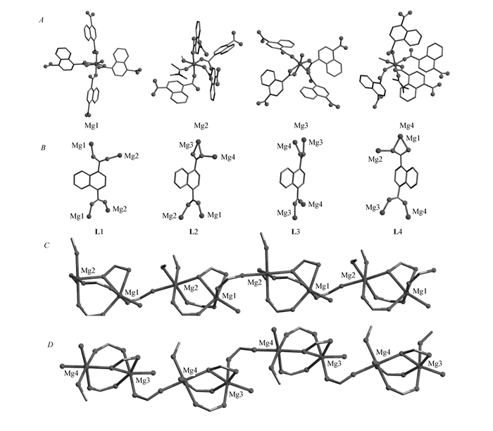 图1
The coordination environments of Mg atoms(A), coordination modes of the four crystallographically independent 1, 4-NDC2- ligands(B), and the 1D chains in compound 1 extended along the c axis(C, D)
Figure1.
The coordination environments of Mg atoms(A), coordination modes of the four crystallographically independent 1, 4-NDC2- ligands(B), and the 1D chains in compound 1 extended along the c axis(C, D)
图1
The coordination environments of Mg atoms(A), coordination modes of the four crystallographically independent 1, 4-NDC2- ligands(B), and the 1D chains in compound 1 extended along the c axis(C, D)
Figure1.
The coordination environments of Mg atoms(A), coordination modes of the four crystallographically independent 1, 4-NDC2- ligands(B), and the 1D chains in compound 1 extended along the c axis(C, D)
As shown in Fig. 1C and Fig. 1D, there exist corner-shared coordination polyhedra of dinuclear [Mg1Mg2] and [Mg3Mg4] units in the structure which can be viewed as the secondary building units(SBU) for compound 1. Then, the adjacent [Mg1Mg2] and [Mg3Mg4] units are, respectively, interconnected by the COO- groups of ligands L1 and L3 to form one-dimensional(1D) infinite chains of [—Mg1—Mg2—Mg1—Mg2—] and [—Mg3—Mg4—Mg3—Mg4—] along the c-axis. Further, each [—Mg1—Mg2—Mg1—Mg2—] chain connects to four adjacent [—Mg3—Mg4—Mg3—Mg4—] chains by the ligands L3 and L4 to form a 3D skeleton. The coordinated and free DMA, methanol, water molecules settle in the cages of the 3D framework(Fig. 2A). The solvent accessible volume is 43.6% if all the solvent molecules are removed according to the calculation performed by PLATON analysis. Topologically, when regarding the 1, 4-NDC2- ligands and each dinuclear secondary building unit as 3-connected and 5-connected nodes respectively, the structure of compound 1 could be simplified as a typical fsc-3, 5-C2/c topology, Fig. 2B.
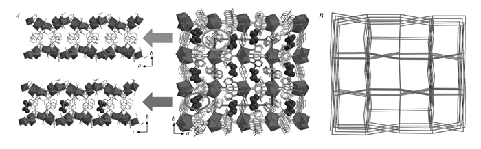 图2
View of the 3D framework of compound 1 along the c axis showing the cages in which the DMA and methanol molecules are filled(A) and the topology of compound 1(B)
Figure2.
View of the 3D framework of compound 1 along the c axis showing the cages in which the DMA and methanol molecules are filled(A) and the topology of compound 1(B)
图2
View of the 3D framework of compound 1 along the c axis showing the cages in which the DMA and methanol molecules are filled(A) and the topology of compound 1(B)
Figure2.
View of the 3D framework of compound 1 along the c axis showing the cages in which the DMA and methanol molecules are filled(A) and the topology of compound 1(B)
2.2 Thermal stability
The phase purity of compound 1 was confirmed by PXRD(see Fig.S1 in Supporting Information) carried out with the polycrystalline sample of compound 1. Thermogravimetric analysis of compound 1 was performed under a N2 atmosphere from 25 to 800 ℃ with a heating rate of 10 ℃ /min on pure powdered sample. The thermogravimetric curve for compound 1 is shown in Fig. 3. The 9.01% mass loss from room temperature(RT) to ~180 ℃ should be attributed to the loss of the free guests(calcd. 8.84%); the 19.72% mass loss of compound 1 from 180 to 500 ℃ corresponds to the departure of the coordinated water, CH3OH and DMA molecules in the structure(calcd. 20.36%). The mass of the samples remained nearly constant from 600 to 800 ℃ and the characterization of PXRD suggested that the residual powder was MgO(see Fig.S2, in Supporting Information).
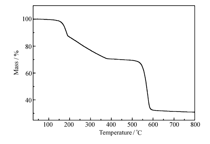 图3
TG curve for compound 1
Figure3.
TG curve for compound 1
图3
TG curve for compound 1
Figure3.
TG curve for compound 1
2.3 Fluorescence detection properties
As shown in Fig. 4, the fluorescent spectra of compound 1 in the solid state exhibited a purple emission band with an intense peak maximum at 385 nm upon excitation at 345 nm at room temperature. Compared with the emission band of the free 1, 4-H2NDC ligand(λem=485 nm monitored at λex=390 nm), compound 1 showed blue-shift emission. The title compound should exhibit a ligand-centered emission due to the unique electron configuration of Mg2+. Further fluorescence measurement for compound 1 has been carried out to identify whether compound 1 has a luminescent response to volatile organic molecules. As shown in Fig. 5A, compound 1 was dispersed in five kinds of typically used solvents and it was interesting to see that the fluorescence intensities of compound 1 were heavily dependent on the identity of the organic solvent molecules.
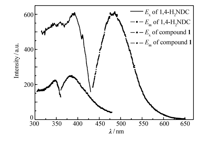 图4
Solid state fluorescence spectra of compound 1 and 1, 4-H2NDC
Figure4.
Solid state fluorescence spectra of compound 1 and 1, 4-H2NDC
图4
Solid state fluorescence spectra of compound 1 and 1, 4-H2NDC
Figure4.
Solid state fluorescence spectra of compound 1 and 1, 4-H2NDC
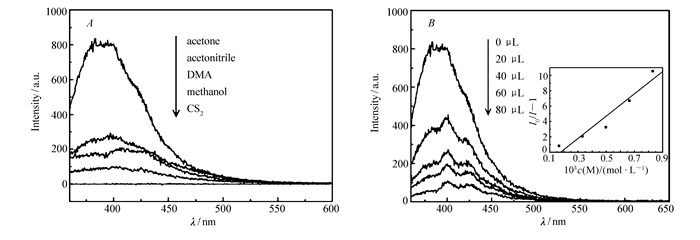 图5
Emission spectra of compound 1 dispersed in different solvents(A) and emission spectra of compound 1 dispersed in the acetone with various contents of CS2(inset is the SV plot for the quenching of compound 1 by CS2)(B)
Figure5.
Emission spectra of compound 1 dispersed in different solvents(A) and emission spectra of compound 1 dispersed in the acetone with various contents of CS2(inset is the SV plot for the quenching of compound 1 by CS2)(B)
图5
Emission spectra of compound 1 dispersed in different solvents(A) and emission spectra of compound 1 dispersed in the acetone with various contents of CS2(inset is the SV plot for the quenching of compound 1 by CS2)(B)
Figure5.
Emission spectra of compound 1 dispersed in different solvents(A) and emission spectra of compound 1 dispersed in the acetone with various contents of CS2(inset is the SV plot for the quenching of compound 1 by CS2)(B)
Notably, we found that compound 1 exhibited a significant quenching of fluorescence when dispersed in CS2. Since compound 1 demonstrated the strongest fluorescence emission in acetone, the acetone was chosen as the dispersed solvent to find the potential fluorescence detection of compound 1 for CS2. The sensing sensitivity towards CS2 was examined in detail through gradually increasing CS2 contents into the emulsions of compound 1 dispersed in acetone to monitor the emissive response. As depicted in Fig. 5B, the fluorescent intensity of compound 1 was almost completely quenched when only 80 μL CS2(0.4%(volume fraction)) was added, indicating that compound 1 was a benign candidate for selective sensing of CS2. Stern-Volmer equation(SV plot):I0/I=1+Ksv×[M] was applied to judge the quenching effect(I0 and I are the suspension luminescence intensity of compound 1 without and with addition of quencher, and [M] is the molarity of quencher and Ksv is the quenching constant)[24]. As shown in Fig. 5B, the SV plot displays a good linear behavior and the Ksv constant calculated from the experimental data is 1.45×10³ L/mol. Compared to the former reports detecting CS2 based on the Mg-MOF luminescence intensity, the quenching concentration of CS2 for the title compound is slightly higher[17]. This could be attributed to the larger channels in the former reported Mg-MOFs, which facilitate the interactions between CS2 and MOFs[16-17]. Further investigation is still required for exploring the mechanism of fluorescence quenching in compound 1.
3 Conclusions
In summary, a novel 3D Mg-MOF, namely [Mg4(1, 4-NDC)4(DMA)2(CH3OH)2(H2O)2]·DMA·CH3OH has been synthesized under solvothermal conditions and characterized. Fluorescence study indicates that the title compound shows a highly sensitive fluorescent response for CS2 with a low concentration. Future work will continue to study the construction of fluorescent Mg-MOFs, explore their sensing properties towards the harmful volatile organic compounds and aim at a deep understanding of the relationship of structure and property.
Supporting Information [PXRD patterns] is available free of charge on the website of Chinese Journal of Applied Chemistry(http://yyhx.ciac.jl.cn/).
-
-
[1]
Chuang W L, Huang C C, Chen C J. Carbon Disulfide Encephalopathy:Cerebral Microangiopathy[J]. Neurotoxicology, 2007, 28(2): 387-393. doi: 10.1016/j.neuro.2006.10.008
-
[2]
Wang S, Irving G, Jiang L L. Oxidative Stress Mediated Hippocampal Neuron Apoptosis Participated in Carbon Disulfide-Induced Rats Cognitive Dysfunction[J]. Neurochem Res, 2017, 42(2): 583-594. doi: 10.1007/s11064-016-2113-8
-
[3]
Ciaffoni L, Peverall R, Ritchie G A D. Laser Spectroscopy on Volatile Sulfur Compounds:Possibilities for Breath Analysis[J]. J Breath Res, 2011, 5(2): 024002. doi: 10.1088/1752-7155/5/2/024002
-
[4]
Furton K G, Myers L J. The Scientific Foundation and Efficacy of the Use of Canines as Chemical Detectors for Explosives[J]. Talanta, 2001, 54(3): 487-500. doi: 10.1016/S0039-9140(00)00546-4
-
[5]
Lu W, Xiao P, Liu Z Z. Reaction-Driven Self-Assembled Micellar Nanoprobes for Ratiometric Fluorescence Detection of CS2with High Selectivity and Sensitivity[J]. ACS Appl Mater Interfaces, 2016, 8(31): 20100-20109. doi: 10.1021/acsami.6b06472
-
[6]
Zhang R K, Li G K, Hu Y F. Simple and Excellent Selective Chemiluminescence-Based CS2 On-Line Detection System for Rapid Analysis of Sulfur-Containing Compounds in Complex Samples[J]. Anal Chem, 2015, 87(11): 5649-5655. doi: 10.1021/acs.analchem.5b00722
-
[7]
Heine J, Muller-Buschbaum K. Engineering Metal-Based Luminescence in Coordination Polymers and Metal-Organic Frameworks[J]. Chem Soc Rev, 2013, 42(24): 9232-9242. doi: 10.1039/c3cs60232j
-
[8]
Hu Z C, Deibert B J, Li J. Luminescent Metal-Organic Frameworks for Chemical Sensing and Explosive Detection[J]. Chem Soc Rev, 2014, 43(16): 5815-5840. doi: 10.1039/C4CS00010B
-
[9]
Banerjee D, Hu Z C, Li J. Luminescent Metal-Organic Frameworks as Explosive Sensors[J]. Dalton Trans, 2014, 43(28): 10668-10685. doi: 10.1039/C4DT01196A
-
[10]
Kreno L E, Leong K, Farha O K. Metal-Organic Framework Materials as Chemical Sensors[J]. Chem Rev, 2012, 112(2): 1105-1125. doi: 10.1021/cr200324t
-
[11]
Cui Y J, Yue Y F, Qian G D. Luminescent Functional Metal-Organic Frameworks[J]. Chem Rev, 2012, 112(2): 1126-1162. doi: 10.1021/cr200101d
-
[12]
Meyer L V, Schoenfeld F, Zurawski A. A Blue Luminescent MOF as a Rapid Turn-Off/Turn-On Detector for H2O, O-2 and CH2Cl2, MeCN:∞3Ce(Im)3ImH ·ImH[J]. Dalton Trans, 2015, 44(9): 4070-4079. doi: 10.1039/C4DT03578J
-
[13]
Xu F, Wang H, Teat S J. Synthesis, Structure and Enhanced Photoluminescence Properties of Two Robust, Water Stable Calcium and Magnesium Coordination Networks[J]. Dalton Trans, 2015, 44(47): 20459-20463. doi: 10.1039/C5DT03705K
-
[14]
Jayaramulu K, Kanoo P, George S J. Tunable Emission From a Porous Metal-Organic Framework by Employing an Excited-State Intramolecular Proton Transfer Responsive Ligand[J]. Chem Commun, 2010, 46(42): 7906-7908. doi: 10.1039/c0cc02069a
-
[15]
Brown J W, Henderson B L, Kiesz M D. Photophysical Pore Control in an Azobenzene-Containing Metal-Organic Framework[J]. Chem Sci, 2013, 4(7): 2858-2864. doi: 10.1039/c3sc21659d
-
[16]
Wu Z F, Tan B, Feng M L. A Magnesium-Carboxylate Framework Showing Luminescent Sensing for CS2 and Nitroaromatic Compounds[J]. J Solid State Chem, 2015, 223: 59-64. doi: 10.1016/j.jssc.2014.06.018
-
[17]
Wu Z F, Tan B, Feng M L. A Magnesium MOF as a Sensitive Fluorescence Sensor for CS2 and Nitroaromatic Compounds[J]. J Mater Chem A, 2014, 2(18): 6426-6431. doi: 10.1039/C3TA15071B
-
[18]
Douvali A, Tsipis A C, Eliseeva S V. Turn-On Luminescence Sensing and Real-Time Detection of Traces of Water in Organic Solvents by a Flexible Metal-Organic Framework[J]. Angew Chem Int Ed, 2015, 54(5): 1651-1656. doi: 10.1002/anie.201410612
-
[19]
Sheldrick G M. Crystal Structure Refinement with SHELXL[J]. Acta Crystallogr, 2015, 71: 3-8.
-
[20]
Zhai Q G, Lin Q P, Wu T Z. Induction of Trimeric[Mg3(OH)(CO2)6] in a Porous Framework by a Desymmetrized Tritopic Ligand[J]. Dalton Trans, 2012, 41(10): 2866-2868. doi: 10.1039/c2dt12215d
-
[21]
Guo Z Y, Li G H, Zhou L. Magnesium-based 3D Metal-Organic Framework Exhibiting Hydrogen-Sorption Hysteresis[J]. Inorg Chem, 2009, 48(17): 8069-8071. doi: 10.1021/ic901056d
-
[22]
Zhai Q G, Bu X, Zhao X. Advancing Magnesium-Organic Porous Materials Through New Magnesium Cluster Chemistry[J]. Cryst Growth Des, 2016, 16(3): 1261-1267. doi: 10.1021/acs.cgd.5b01297
-
[23]
Rood J A, Noll B C, Henderson K W. Synthesis, Structural Characterization, Gas Sorption and Guest-Exchange Studies of the Lightweight, Porous Metal-Organic Framework Alpha-[Mg3(O2CH)6][J]. Inorg Chem, 2006, 45(14): 5521-5528. doi: 10.1021/ic060543v
-
[24]
Lakowicz J R. Principles of Fluorescence Spectroscopy[M]. New York:Plenum Press, 1983:260-266.
-
[1]
-

Figure 1 The coordination environments of Mg atoms(A), coordination modes of the four crystallographically independent 1, 4-NDC2- ligands(B), and the 1D chains in compound 1 extended along the c axis(C, D)

Figure 2 View of the 3D framework of compound 1 along the c axis showing the cages in which the DMA and methanol molecules are filled(A) and the topology of compound 1(B)

Figure 5 Emission spectra of compound 1 dispersed in different solvents(A) and emission spectra of compound 1 dispersed in the acetone with various contents of CS2(inset is the SV plot for the quenching of compound 1 by CS2)(B)
Table 1. Crystallographic data and structural refinement details for compound 1
Empirical formula C63H67Mg4N3O24 Formula mass 1 347.431 00(2) T/K 100(2) Crystal system Monoclinic Space group P21/c a/nm 2.060 90(12) b/nm 2.210 14(13) c/nm 1.503 85(10) α/(°) 90 β/(°) 111.399(3) γ/(°) 90 V/nm3 6.377 6(7) Z 4 Dcalc./(g·cm-3) 1.403 λ/nm 0.071 073 μ/mm-1 0.142 F(000) 2 824 Reflections measured 28 773 Independent reflections 12 199 No. of parameters 878 GOF on F2 1.033 R1[I > 2σ(I)]a 0.059 6 wR2[I > 2σ(I)]a 0.122 5 R1[all data] 0.089 9 wR2[all data] 0.146 1 CCDC 1552691 a.R1=Σ‖Fo|-|Fc‖/Σ|Fo|, wR2={Σw[(Fo)2-(Fc)2]2/Σw[(Fo)2]2}1/2.  下载: 导出CSV
-


 扫一扫看文章
计量
- PDF下载量: 1
- 文章访问数: 1035
- HTML全文浏览量: 88
 下载:
下载:
