Table 1.
Selected Bond Lengths (Å) and Bond Angles (°)
Citation:
Yang LIU, Xue-Li CAO, Wei WANG, Guo-Ling LI, Shun LI, You-Gui HUANG. A Linear CeⅢ Complex Based on in-situ Generated N-hydroxy-1, 8-naphthalenedicarboximide[J]. Chinese Journal of Structural Chemistry,
2021, 40(11): 1475-1481.
doi:
10.14102/j.cnki.0254-5861.2011-3190

A Linear CeⅢ Complex Based on in-situ Generated N-hydroxy-1, 8-naphthalenedicarboximide
English
A Linear CeⅢ Complex Based on in-situ Generated N-hydroxy-1, 8-naphthalenedicarboximide
Abstract:
A CeⅢ linear complex of N-hydroxy-1, 8-naphthalenedicarboximide (HL) has been synthesized with acenaphthoquinone dioxime (acndH2) under solvothermal conditions, in which L is generated by a series of in-situ reactions starting from acndH2. X-ray crystallography reveals the coordination chains of CeⅢ ions are assembled into a 3D supramolecular framework via π…π interactions. The title complex has been characterized by IR and UV-Vis spectra and thermogravimetric analysis (TGA). Furthermore, the fluorescent properties study demonstrated a ligand-based emission of the CeⅢ complex with ~6 nm blue-shift compared with the emisision of the HL ligand.
-
1. INTRODUCTION
Rare earth coordination complexes have attracted much attention because of their potential applications on catalysis[1, 2], luminescence[3], magnetism[4], and bio-imaging or sensing[5, 6]. As hard Lewis acids, rare earth ions can easily coordinate with multidentatehard base ligands containing O atoms[7, 8]. Therefore, various multidentate O-containing ligands such as phenol[9], quinine[10], carboxylic acid[11-15], and oxime[16, 17] have been used to prepare rare earth complexes, and numerous complexes with structural diversities ranging from discrete molecule to three-dimensional (3D) framework have been obtained[18-21]. As an O-containing multidentate ligand, acenaphthoquinone dioxime (acndH2) comprising four oximate-based donor sites is a promising ligand for rare earth ions, and several 3d-4f complexes based on this ligand have been synthesized[22, 23]. For example, a series of structures based on the {Cu6ⅡLn2Ⅲ} building unit have been reported by Stamatatos et al[23]. However, homometallic rare earth coordination complexes of acndH2 are still elusive. Therefore, we intend to obtain such rare earth complexes, and Ce attracts our interest because of its two stable oxidation states of CeⅢ and CeⅣ[24, 25]. Surprisingly, acndH2 undergoes a series of reactions under solvothermal condition. The solvothermal reaction of CeCl3 and acndH2 in the presence of MnCl2·4H2O and Co(SCN)2 leads to a CeⅢ chain complex [Ce(L)2Cl]n·nEtOH (1) (EtOH = ethanol) with N-hydroxy-1, 8-naphthalenedicarboximide (HL) as ligand which is in-situ generated from acndH2. A reasonable route of the formation of L ligand is proposed. To support the proposed route, crystals of 1, 8-naphthalimide were also obtained through crystallization of the resulting filtrate from the solvothermal reaction. Herein, we report the structure of complex 1 and propose a possible generation mechanism of L ligand from acndH2. The thermal stability and the UV-Vis and IR spectra of the complex were investigated. Furthermore, the fluorescent properties were also studied.
2. EXPERIMENTAL
2.1 Materials and methods
All reagents were purchased from commercial suppliers and used without further purification unless otherwise specified. IR spectrum was recorded on a Thermo Nicolet is 50 FT-IR spectrometer using the KBr pellet method in the range of 400~4000 cm−1 and processed with the OMNIC 6.0 software package. Powder X-ray diffraction (PXRD) pattern was performed on a Rigaku Miniflex 600 diffractometer with Cu-Kα radiation using flat plate geometry: the scan rate was 5 °/min and the 2θ scan range is 5~50°. Thermogravimetric analysis (TGA) was performed on a Mettler-Toledo TGA/DSC-1 at 30~800 ℃ in argon atmosphere at a heating rate of 10 °/min. UV-vis spectrum was recorded on a Cary 5000 at room temperature. Fluorescence spectra were measured at room temperature on a FLS980 system.
2.2 Synthesis of the title complex
A mixture containing acndH2 (10.0 mg, 0.047 mmol), CeCl3 (25.0 mg, 0.101 mmol), MnCl2·4H2O (30.0 mg, 0.152 mmol), Co(SCN)2 (10.0 mg 0.057 mmol), ethanol (10 mL), and distilled H2O (0.5 mL) was sealed in a 25 mL Teflon-lined autoclave and heated to 160 ℃ in 5 h. After maintaining at 160 ℃ for 20 h, the autoclave was cooled to room temperature in 8 h. Yellow crystals of complex 1 were obtained (yield: 42% based on acndH2). Crystallization of the green filtrate led to the crystals of 1, 8-naphthalimide (2).
2.3 X-ray structure determination
The single-crystal X-ray diffraction data were collected at 200 K on a Bruker D8 Venture diffractometer with MoKα (λ = 0.71073 Å) radiation. Data reduction, integration, and scaling were performed with a Bruker APEX3 software package. Crystal structures were solved by direct methods and refined by full-matrix least-squares technique on F2 with the SHELXTL 2014 program[26, 27]. Hydrogen atoms on C atoms were located using the geometric method, while those on O and N atoms were found by Fourier map. Non-hydrogen atoms were refined with anisotropic thermal parameters.
3. RESULTS AND DISCUSSION
3.1 Crystal structure
Single-crystal X-ray analysis reveals that complex 1 crystallizes in triclinic space group P
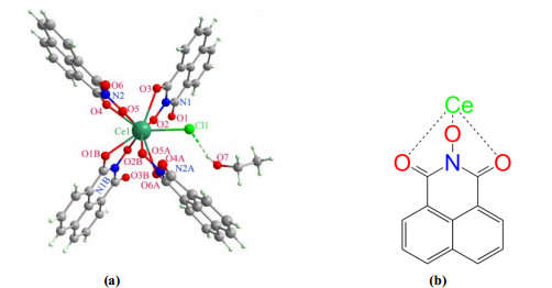
$ \overline 1 $ , with a = 8.3852(3), b = 12.1240(6), c = 12.1565(6) Å, α = 101.993(2)°, β = 91.757(2)° and γ = 102.412(2)°. Its asymmetric unit contains a CeⅢ ion, a Cl– ion, two L ligands, and a lattice ethanol molecule. Each CeⅢ ion is nono-coordinated by one Cl– ion and four L ligands in a tricapped trigonal prismatic geometry (Fig. 1a). The Ce–Cl bond length is 2.819 Å and the Ce–O bond distances fall in the range of 2.427~2.670 Å (Table 1). The L ligand adopts a ŋ1: ŋ2: ŋ1: µ3 binding mode bridging two CeⅢ ions (Fig. 1b).Table 1

 DownLoad:
CSV
DownLoad:
CSV
Bond Dist. Bond Dist. Bond Dist. Ce(1)–Cl(1) 2.8188(8) Ce(1)–O(3) 2.670(2) Ce(1)–O(6)#2 2.668(2) Ce(1)–O(5) 2.427(2) Ce(1)–O(4) 2.583(2) Ce(1)–O(2)#1 2.475(2) Ce(1)–O(2) 2.450(2) Ce(1)–O(5)#2 2.507(2) Ce(1)–O(1)#1 2.558(2) Angle (°) Angle (°) Angle (°) O(5)#2–Ce(1)–O(1)#1 69.20(6) O(2)–Ce(1)–O(2)#1 59.82(7) O(5)–Ce(1)–O(2)#1 134.64(7) O(5)#2–Ce(1)–O(4) 116.35(6) O(5)–Ce(1)–O(5)#2 62.46(7) O(2)–Ce(1)–O(5)#2 156.68(7) O(1)#1–Ce(1)–O(4) 72.97(7) O(5)–Ce(1)–O(4) 61.95(6) O(2)#1–Ce(1)–O(1)#1 61.46(6) O(5)–Ce(1)–O(6)#2 121.74(6) O(2)#1–Ce(1)–O(3) 116.21(6) O(5)–Ce(1)–O(1)#1 79.93(6) O(2)–Ce(1)–O(6)#2 100.22(6) O(5)#2–Ce(1)–O(3) 126.12(6) O(2)–Ce(1)–O(1)#1 119.40(6) O(5)#2–Ce(1)–O(6)#2 59.77(6) O(1)#1–Ce(1)–O(3) 145.22(7) O(2)–Ce(1)–O(4) 86.86(7) O(1)#1–Ce(1)–O(6)#2 73.77(7) O(4)–Ce(1)–O(3) 72.33(7) O(1)#1–Ce(1)–Cl(1) 141.22(5) O(4)–Ce(1)–O(6)#2 145.00(7) O(6)#2–Ce(1)–O(3) 140.62(7) O(4)–Ce(1)–Cl(1) 142.12(6) O(5)–Ce(1)–O(3) 81.98(6) O(5)–Ce(1)–Cl(1) 101.55(5) O(3)–Ce(1)–Cl(1) 71.65(5) O(2)–Ce(1)–O(3) 60.73(6) O(2)–Ce(1)–Cl(1) 85.67(5) O(2)#1–Ce(1)–O(5)#2 117.59(6) O(2)#1–Ce(1)–O(6)#2 71.29(7) O(2)#1–Ce(1)–Cl(1) 123.28(5) O(6)#2–Ce(1)–Cl(1) 72.86(5) O(5)–Ce(1)–O(2) 137.80(6) O(5)#2–Ce(1)–Cl(1) 77.22(5) O(2)#1–Ce(1)–O(4) 83.47(7) Symmetry codes: (#1) –x+1, –y+1, –z+1; (#2) –x, –y+1, –z+1 Figure 1
 Figure 1. (a) Coordination environment of CeⅢ in complex 1. Symmetry codes: A: –x, 1–y, 1–z; B: 1–x, 1–y, 1–z. (b) Coordination mode of L ligand in complex 1
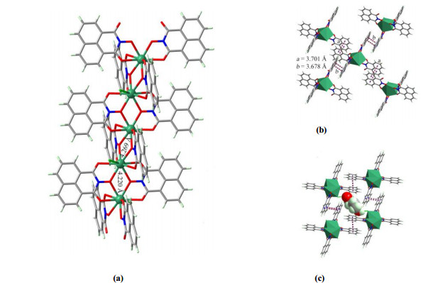
The CeⅢ ions are doublely bridged by the L ligands, resulting in a linear structure with alternative Ce–Ce distances of 4.220 and 4.269 Å (Fig. 2a), respectively. Each chain associates with its four neighbors through two types of strong offset π…π stacking interactions. The two interassociated naphthalene moieties are parallel with each other with centroid-centroid distances of 3.701 and 3.678 Å, respectively (Fig. 2b). As a result of π…π interactions, a three-dimensional (3D) porous supramolecular structure with channels along the [1 0 0] direction is generated. The lattice ethanol molecules hydrogen-bond to the Cl– ions (OH…Cl: 2.370 Å) and fill the channels (Fig. 2c).
Figure 2
 Figure 2. (a) Linear CeⅢ chain of complex 1. (b) Structural illustration of the π…π interactions between adjacent chains of complex 1. (c) 3D porous supramolecular structure showing the ethanol filled channel
3.2 PXRD, TGA, UV-Vis, and FT-IR analyses
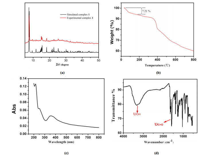
The powder X-ray diffraction pattern (PXRD) of the synthesized sample of complex 1 matches well with the simulated one, confirming the phase purity of the as-synthesized sample (Fig. 3a). Thermogravimetric analysis (TGA) demonstrates an initial weight loss of ~6.57% from 30 to 180 ℃ which corresponds to the departure of lattice ethanol molecules (calcd. weight loss: 7.12%) (Fig. 3b). UV-Vis spectrum of the solid sample of complex 1 reveals a broad absorption at ~370 nm (Fig. 3c). The FT-IR spectrum of complex 1 was also recorded in the 4000~400 cm–1 range. The absorption at ~3329 cm–1 is characteristic of ν(-OH), confirming the existence of ethanol in the crystal structure. The absorption at ~1653 and 1585 cm–1 is attributed to the C=O groups in the L ligand (Fig. 3d).
Figure 3
 Figure 3. (a) PXRD patterns, (b) TGA, (c) UV-Vis spectrum, and (d) IR spectrum of complex 1
3.3 Fluorescent properties
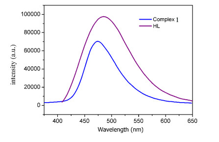
Although CeⅢ is nonfluorescent, as a 1, 8-naphthalimide derivative, the ligand HL is expected to be luminescent[28]. To give insights into whether the coordination of CeⅢ influences the luminescence of HL, the fluorescence of both the ligand and complex 1 was studied at room temperature. Excited at 248 nm, the HL ligand yields blue fluorescence with a broad emission band at 480 nm. Excited at the same wavelength, complex 1 exhibits a broad emission band at 474 nm, which is weaker and ~6 nm blue-shifted compared with that of the HL ligand. The blue luminescence behaviors of the HL ligand and complex 1 are due to the π → π* transition of the naphthalene core[29], while the blue-shift can be attributed to the destabilizing effect on the π* orbitals of the naphthalene core induced by CeⅢ ions, therefore increasing the energy of the intraligand π→π* transition[29].
Figure 4
 Figure 4. Photoluminescent spectra of the ligand (HL) and complex 1 (λex = 248 nm)
3.4 Proposed route of the generation of L ligand
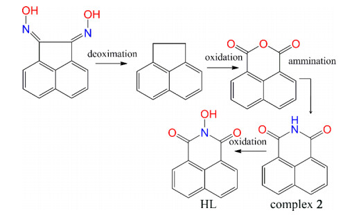
The most surprising is the finding of L ligand in the structure of complex 1, considering that the added ligand is acndH2. Therefore, acndH2 must undergo a series of in-situ reactions under the hydrothermal condition. The proposed route of the generation of L ligand is illustrated in Scheme 1. Initially, acndH2 underwent deoximation forming acenaphthene, which then was further oxidized into 1, 8-naphthalic anhydride[30]. The generating 1, 8-naphthalic anhydride could undergo ammination[31] and then be oxidized leading to the ligand L[32]. To support the proposed route, we tried to obtain some intermediate states. Fortunately, some crystals of 1, 8-naphthalimide (2) molecules were isolated from the filtrate after the solvothermal reaction for the synthesis of complex 1. Complex 2 crystallizes in space group of P21/n (Table 1), wherein 1, 8-naphthalimide molecules exist as hydrogen bonded dimers, as found previously[33].
Scheme 1
 Scheme 1. Proposed route of the synthesis of ligand HL (N-hydroxy-1, 8-naphthalenedicarboximide) using ancdH2 as the reactant
4. CONCLUSION
In summary, a linear CeⅢ complex based on in-situ generated L ligand has been synthesized using ancdH2 under solvothermal conditions. The complex exhibits a ligand-based emission with a broad band at 474 nm, which is ~6 nm blue-shifted compared with the emisision of the HL ligand. It was proposed that ancdH2 undergoes a series of in-situ reactions of deoximation/oxidation/ammination/oxida-tion forming L ligand which further coordinates with CeⅢ ions in the resulting linear structure. Our study demonstrates a new avenue for coordination complexes based on N-hydroxy-1, 8-naphthalenedicarboximide. Following this strategy, some new coordination complexes based on N-hydroxy-1, 8-naphthalenedicarboximide can be obtained.
-
-
[1]
Lin, F.; Liu, Z. H.; Wang, T. T.; Cui, D. M. Highly 2, 3-selective polymerization of phenylallene and its derivatives with rare-earth metal catalysts: from amorphous to crystalline products. Angew. Chem. Int. Ed. 2017, 56, 14653−14657. doi: 10.1002/anie.201707601
-
[2]
Yadav, M.; Bhunia, A.; Jana, S. K.; Roesky, P. W. Manganese- and lanthanide-based 1D chiral coordination polymers as an enantioselective catalyst for sulfoxidation. Inorg. Chem. 2016, 55, 2701−2708. doi: 10.1021/acs.inorgchem.5b02234
-
[3]
Hasegawa, M.; Ishii, A. Thin-film formation for promoting the potential of luminescent lanthanide coordination complexes. Coord. Chem. Rev. 2020, 421, 213458−17. doi: 10.1016/j.ccr.2020.213458
-
[4]
Gould, C. A.; McClain, K. P.; Yu, J. M.; Groshens, T. J.; Furche, F.; Harvey, B. G.; Long, J. R. Synthesis and magnetism of neutral, linear metallocene complexes of terbium(Ⅱ) and dysprosium(Ⅱ). J. Am. Chem. Soc. 2019, 141, 12967–12973. doi: 10.1021/jacs.9b05816
-
[5]
Wei, C.; Ma, L.; Wei, H. B.; Liu, Z. W.; Bian, Z. Q.; Huang, C. H. Advances in luminescent lanthanide complexes and applications. Sci. China Tech. Sci. 2018, 61, 1265–1285. doi: 10.1007/s11431-017-9212-7
-
[6]
Piccinelli, F.; Bettinelli, M.; Melchior, A.; Grazioli, C.; Tolazzi, M. Structural, optical and sensing properties of novel Eu(Ⅲ) complexes with furan- and pyridine-based ligands. Dalton Trans. 2015, 44, 182–192. doi: 10.1039/C4DT02326A
-
[7]
Huang, Y. G.; Wu, B. L.; Yuan, D. Q.; Xu, Y. Q.; Jiang, F. L.; Hong, M. C. New lanthanide hybrid as clustered infinite nanotunnel with 3D Ln–O–Ln framework and (3, 4)-connected net. Inorg. Chem. 2007, 46, 1171−1176. doi: 10.1021/ic0615271
-
[8]
Huang, Y. G.; Jiang, F. L.; Hong, M. C. Magnetic lanthanide-transition-metal organic-inorganic hybrid materials: From discrete clusters to extended frameworks. Coord. Chem. Rev. 2009, 253, 2814–2834. doi: 10.1016/j.ccr.2009.05.007
-
[9]
Arnold, P. L.; Wang, K.; Gray, S. J.; Moreau, L. M.; Booth, C. H.; Curcio, M.; Wells, J. A. L.; Slawin, A. M. Z. Dicerium letterbox-shaped tetraphenolates: f-block complexes designed for two-electron chemistry. Dalton Trans. 2020, 49, 877−884. doi: 10.1039/C9DT03291F
-
[10]
Ishikawa, R.; Michiwaki, S.; Noda, T.; Katoh, K.; Yamashita, M.; Kawata, S. Series of chloranilate-bridged dinuclear lanthanide complexes: kramers systems showing field-induced slow magnetic relaxation. Magnetochem. 2019, 5, 5020030−12.
-
[11]
Huang, Y. G.; Jiang, F. L.; Yuan, D. Q.; Wu, M. Y.; Gao, Q.; Wei, W.; Hong, M. C. Intricate 3D lanthanide-organic frameworks with mixed nodes nets. J. Solid State Chem. 2009, 182, 215−222. doi: 10.1016/j.jssc.2008.09.021
-
[12]
Huang, Y. G.; Jiang, F. L.; Wu, M. Y.; Gao, Q.; Wei, W.; Hong, M. C. A chain structure based on homodinuclear scandium units: [Sc(µ-OH)(2, 5-pydc)(H2O)]n. Chin. J. Struct. Chem. 2009, 182, 215−222.
-
[13]
Chen, Y. M.; Huang, L. C.; Gao, R.; Chen, Y. H.; Huang, Z.; Zhang, W. J. Dy(Ⅲ) and Sm(Ⅲ) coordination polymers based on 2, 4-pyridinedicarboxylic acid: synthesis, structures, luminescence and magnetism. J. Cluster Sci. 2020, 31, 1013−1019. doi: 10.1007/s10876-019-01706-5
-
[14]
Li, Y. Y.; Ren, N.; He, S. M.; Wang, S. P.; Zhang, J. J. Synthesis, structures, thermal behavior, luminescence and magnetic properties of lanthanide complexes constructed from 2, 6-dimethylbenzoic acid and 5, 5΄-dimethyl-2, 2΄-bipyridine. Appl. Organomet. Chem. 2020, 34, e5418–15.
-
[15]
She, S. X.; Gu, X. Y.; Yang, Y. Field-induced single molecule magnet behavior of a three-dimensional Dy(Ⅲ)-based complex. Inorg. Chem. Commun. 2019, 110, 107584−9. doi: 10.1016/j.inoche.2019.107584
-
[16]
Xi, L.; Sun, J.; Wang, K.; Lu, J.; Jing, P.; Li, L. C. Slow magnetic relaxation in Co–Ⅱ–Ln(Ⅲ) heterodinuclear complexes achieved through a functionalized nitronyl nitroxide biradical. Dalton Trans. 2020, 49, 1089–1096. doi: 10.1039/C9DT04036F
-
[17]
Yang, H.; Meng, Y. X.; Tian, H. Q.; Li, D. C.; Zeng, S. Y.; Song, Y.; Dou, J. M. Investigating the effect of lanthanide radius and diamagnetic linkers on the framework of metallacrown complexes. Dalton Trans. 2020, 49, 1955–1962. doi: 10.1039/C9DT04383G
-
[18]
Wang, J.; Wu, Z. L.; Yang, L. R.; Xue, M. M.; Fang, Z. X.; Luo, S. C.; Wang, W. M. Two lanthanide-based dinuclear clusters (Gd-2 and Dy-2) with Schiff base derivatives: synthesis, structures and magnetic properties. Inorg. Chim. Acta 2020, 514, 120015−6.
-
[19]
Liu, S.; Li, L. L.; Li, H.; Gao, H. L.; Cui, J. Z.; Cheng, P. Slow magnetic relaxation in a lanthanide helix chain compound [Dy(HNA)(NA)2(NO3)]n (HNA = nicotinic acid). Dalton Trans. 2015, 44, 6169–6174. doi: 10.1039/C5DT00205B
-
[20]
Li, Z. Y.; Zhang, C.; Zhang, F. L.; Zhang, F. Q.; Zhang, X. F.; Li, S. Z.; Cao, G. X.; Zhai, B. Two novel 2D lanthanide sulfate frameworks: syntheses, structures, and luminescence properties. J. Mol. Struc. 2016, 1108, 516–520. doi: 10.1016/j.molstruc.2015.12.058
-
[21]
Liu, J. H.; Zhang, R. T.; Zhang, J.; Zhao, D.; Li, X. X.; Sun, Y. Q.; Zheng, S. T. A series of 3D porous lanthanide-substituted polyoxometalate frameworks based on rare hexadecahedral {Ln6W8O28} heterometallic cage-shaped clusters. Inorg. Chem. 2019, 58, 14734−14740. doi: 10.1021/acs.inorgchem.9b02413
-
[22]
Richardson, P.; Gagnon, K. J.; Teat, S. J.; Lorusso, G.; Evangelisti, M.; Tang, J. K.; Stamatatos, T. C. New dioximes as bridging ligands in 3d/4f-metal cluster chemistry: one-dimensional chains of ferromagnetically coupled {Cu6Ln2} clusters bearing acenaphthenequinone dioxime and exhibiting magnetocaloric properties. Cryst. Growth Des. 2017, 17, 2486−2497. doi: 10.1021/acs.cgd.7b00011
-
[23]
Alaimo, A. A.; Worrell, A.; Gupta, S. D.; Abboud, K. A.; Lampropoulos, C.; Christou, G.; Stamatatos. T. C. Structural and magnetic variations in a family of isoskeletal, oximate-bridged {Mn2ⅣMⅢ} complexes (MⅢ = Mn, Gd, Dy). Chem. Eur. J. 2018, 24, 2588–2592. doi: 10.1002/chem.201706098
-
[24]
Wemer, D.; Deacon, G. B.; Junk, P. C.; Anwander, R. Pyrazolates advance cerium chemistry: a Ce-Ⅲ/Ce-Ⅳ redox equilibrium with benzoquinone. Dalton Trans. 2017, 46, 6265–6277. doi: 10.1039/C7DT00635G
-
[25]
Levin, J. R.; Dorfner, W. L.; Dai, A. X.; Carroll, P. J.; Schelter, E. J. Density functional theory as a predictive tool for cerium redox properties in nonaqueous solvents. Inorg. Chem. 2016, 55, 12351−12659.
-
[26]
Sheldrick, G. M. SHELXTL-integrated space-group and crystal-structure determination. Acta Cryst C. 2015, 71, 3−8. doi: 10.1107/S2053229614024218
-
[27]
Spek, A. L. Single-crystal structure validation with the program PLATON. J. App. Cryst. 2003, 36, 7−13. doi: 10.1107/S0021889802022112
-
[28]
Shen, B. X.; Qian, Y. A novel fluorescent dye naphthalene imide-fluorine boron two pyrrole: synthesis, fluorescence resonance energy transfer and cell imaging. Chin. J. Org. Chem. 2016, 36, 774–781. doi: 10.6023/cjoc201510028
-
[29]
Diamantis, S. A.; Hatzidimitriou, A.; Plessas, A. K.; Pournara A.; Manos, M. J.; Papaefstathiou, G. S.; Lazarides, T. Alkaline earth-organic frameworks with amino derivatives of 2, 6-naphthalene dicarboxylates: structural studies and fluorescence properties. Dalton Trans. 2020, 49, 16736–16744. doi: 10.1039/D0DT03325A
-
[30]
Li, Y. G.; Yue, K. X.; Li, L. R.; Niu, J.; Liu, H. F.; Ma, J.; Xie, S. Q. A Pt(Ⅳ)-based mononitro-naphthalimide conjugate with minimized side-effects targeting DNA damage response via a dual-DNA-damage approac-h to overcome cisplatin resistance. Bioorg. Chem. 2020, 101, 104011−17. doi: 10.1016/j.bioorg.2020.104011
-
[31]
Streciwilk, W.; Terenzi, A.; Lo Nardo, F.; Prochnow, P.; Bandow, J. E.; Keppler, B. K.; Ott, I. Synthesis and biological evaluation of organometallic complex bearing bis-1, 8-naphthalimide ligands. Eur. J. Inorg. Chem. 2018, 3104−3112.
-
[32]
Bezuglyi, M.; Ivaniuk, K.; Volyniuk, D.; Gražulevičius, J. V.; Bagdžiūnas, G. An approach to discovering novel exciplex supramolecular complex based on carbazole-containing 1, 8-naphthalimide. Dyes Pigm. 2018, 149, 298−305. doi: 10.1016/j.dyepig.2017.10.013
-
[33]
Sobolev, A. N.; Chetkina, L. A.; Golder, G. A.; Fedorov, Y. G.; Zavodnik, V. E. X-ray structure investigation of naphthaloimide. Kristallografiya. 1973, 18, 1157−1161.
-
[1]
-
Figure 1 (a) Coordination environment of CeⅢ in complex 1. Symmetry codes: A: –x, 1–y, 1–z; B: 1–x, 1–y, 1–z. (b) Coordination mode of L ligand in complex 1
Figure 2 (a) Linear CeⅢ chain of complex 1. (b) Structural illustration of the π…π interactions between adjacent chains of complex 1. (c) 3D porous supramolecular structure showing the ethanol filled channel
Figure 3 (a) PXRD patterns, (b) TGA, (c) UV-Vis spectrum, and (d) IR spectrum of complex 1
Scheme 1 Proposed route of the synthesis of ligand HL (N-hydroxy-1, 8-naphthalenedicarboximide) using ancdH2 as the reactant
Table 1. Selected Bond Lengths (Å) and Bond Angles (°)
Bond Dist. Bond Dist. Bond Dist. Ce(1)–Cl(1) 2.8188(8) Ce(1)–O(3) 2.670(2) Ce(1)–O(6)#2 2.668(2) Ce(1)–O(5) 2.427(2) Ce(1)–O(4) 2.583(2) Ce(1)–O(2)#1 2.475(2) Ce(1)–O(2) 2.450(2) Ce(1)–O(5)#2 2.507(2) Ce(1)–O(1)#1 2.558(2) Angle (°) Angle (°) Angle (°) O(5)#2–Ce(1)–O(1)#1 69.20(6) O(2)–Ce(1)–O(2)#1 59.82(7) O(5)–Ce(1)–O(2)#1 134.64(7) O(5)#2–Ce(1)–O(4) 116.35(6) O(5)–Ce(1)–O(5)#2 62.46(7) O(2)–Ce(1)–O(5)#2 156.68(7) O(1)#1–Ce(1)–O(4) 72.97(7) O(5)–Ce(1)–O(4) 61.95(6) O(2)#1–Ce(1)–O(1)#1 61.46(6) O(5)–Ce(1)–O(6)#2 121.74(6) O(2)#1–Ce(1)–O(3) 116.21(6) O(5)–Ce(1)–O(1)#1 79.93(6) O(2)–Ce(1)–O(6)#2 100.22(6) O(5)#2–Ce(1)–O(3) 126.12(6) O(2)–Ce(1)–O(1)#1 119.40(6) O(5)#2–Ce(1)–O(6)#2 59.77(6) O(1)#1–Ce(1)–O(3) 145.22(7) O(2)–Ce(1)–O(4) 86.86(7) O(1)#1–Ce(1)–O(6)#2 73.77(7) O(4)–Ce(1)–O(3) 72.33(7) O(1)#1–Ce(1)–Cl(1) 141.22(5) O(4)–Ce(1)–O(6)#2 145.00(7) O(6)#2–Ce(1)–O(3) 140.62(7) O(4)–Ce(1)–Cl(1) 142.12(6) O(5)–Ce(1)–O(3) 81.98(6) O(5)–Ce(1)–Cl(1) 101.55(5) O(3)–Ce(1)–Cl(1) 71.65(5) O(2)–Ce(1)–O(3) 60.73(6) O(2)–Ce(1)–Cl(1) 85.67(5) O(2)#1–Ce(1)–O(5)#2 117.59(6) O(2)#1–Ce(1)–O(6)#2 71.29(7) O(2)#1–Ce(1)–Cl(1) 123.28(5) O(6)#2–Ce(1)–Cl(1) 72.86(5) O(5)–Ce(1)–O(2) 137.80(6) O(5)#2–Ce(1)–Cl(1) 77.22(5) O(2)#1–Ce(1)–O(4) 83.47(7) Symmetry codes: (#1) –x+1, –y+1, –z+1; (#2) –x, –y+1, –z+1  下载: 导出CSV
-


 扫一扫看文章
计量
- PDF下载量: 2
- 文章访问数: 714
- HTML全文浏览量: 3
 下载:
下载:
