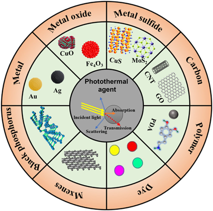
Figure 1.
Types of photothermal agents.
Multifunctional photothermal hydrogels: Design principles, various functions, and promising biological applications
Zikang Hu , Hengjie Zhang , Zhengqiu Li , Tianbao Zhao , Zhipeng Gu , Qijuan Yuan , Baoshu Chen

Hydrogels with three-dimensional network, are composed of hydrophilic polymers by various covalent and noncovalent interactions (i.e., hydrogen bonding electrostatic interactions, and polymeric chemical bonds) [1-3]. The unique network structure endows hydrogels with many excellent properties, which is widely beneficial for biomedicine, electronic devices, resource processing and other applications [4,5]. As we all know, hydrogels can be classified into conventional hydrogels and stimulus-responsive hydrogels from the perspective of smart responses [6]. After swelling balance is reached in aqueous medium, the equilibrium swelling of conventional hydrogels will not be changed under the temperature, pH, light, electric field or other stimulus. However, the equilibrium swelling of stimulus-responsive hydrogels will be changed according to various external stimuli [7,8]. Physical stimuli are caused by light [9], temperature [10], electric [11], sound field [12], magnetic [13] and so on. Chemical stimuli include ionic strength, pH [14,15], specific molecular recognition (i.e., glucose) [16] and so on. Among the various stimulus-responsive hydrogels, photothermal hydrogel is a special category with photo-thermal conversion ability under photo irradiation, has been developed for various applications owing to their simple and rapid response, remote control, low invasiveness, and high space-time selectivity [17,18].
Photothermal hydrogels have been widely documented in recent years, which are formed by photothermal agent and hydrogel matrix materials. Photothermal agents include metal (Au, Ag), metal sulfide/oxide (MoS2, CuS), carbon-based (carbon nanotubes (CNTs), graphene oxide (GO), reduced graphene oxide (rGO)), conjugate polymer (polydopamine (PDA)), dyes (Prussian blue, indocyanine green), black phosphorus, and MXene [19], which have different photothermal conversion mechanisms. The photo-thermal conversion mechanisms of different materials are mainly classified into three categories (i.e., local plasmon resonance of metals, nonradiative relaxation of semiconductors, and molecular thermal vibrations of polymers) [20,21]. The in situ generated heat in photothermal hydrogel after the irradiation of light is of great importance in many research fields, such as tissue repair [22-24], photothermal therapy [25], drug release [26], bone repair [27-29] and tumor therapy [30]. In the future, photothermal conversion hydrogels will certainly be widely promoted as a new therapeutic method.
In recent years, photothermal hydrogels developed rapidly worldwide due to their excellent photo-thermal conversion ability, and potential application perspectivity, and a lot of reviews have been reported on photothermal hydrogels [31,32]. However, these reviews mainly focused on one aspect of photothermal hydrogels. Here, to better introduce multifunctional photothermal hydrogels, the design principles, various functions, and promising applications have been documented in this review carefully. Firstly, the classification, preparation methods, and photothermal conversion mechanisms of photothermal hydrogels with various photothermal agents have been fully illustrated. Then, the various properties (i.e., general and specific functions) of photothermal hydrogels have been summarized. Finally, the various applications in biomedicine applications have been introduced, and the future opportunities and challenges of photothermal hydrogels have been briefly discussed. We believe that this review could provide new opportunities and strategies for new and comprehensive photothermal hydrogels.
Photothermal hydrogels is prepared by loading photothermal agents on hydrogel matrix materials, and its photothermal conversion function mainly depends on photothermal agents [33]. In this chapter, the classification, preparation, and mechanism of photothermal hydrogels are introduced by introducing different photothermal agents, including metal, metal sulfide/oxide, MXene, carbon-based, dyes, black phosphorus, polymer (Fig. 1). In addition, the photothermal conversion efficiency of different photothermal agents was compared (Table S1 in Supporting information).
Metal photothermal hydrogels and photothermal materials have developed rapidly in recent years. Local surface plasmon resonance effect (LSPR) of metal photothermal agents (i.e., Au, Ag, Pt) endows hydrogels good photothermal properties [34,35]. Gold nanoparticles (Au NPs), as a promising photothermal agent, have been utilized in the field of photothermal hydrogels owning to its unique advantages, including wonderful photothermal conversion efficiency, low toxicity, and good biocompatibility [36]. Qin et al. and Lin et al. modified Au NPs and Au nanorods (Au NRs) to prepare photothermal crosslinkers by dynamic covalent bond (RS-Au bond) interaction using the multi-functional crosslinker N, N′-bis(acryloyl)cystamine (BACA). Then, the intelligent Au NPs/Au NRs photothermal hydrogel was prepared by poly(N-isopropylacrylamide) (PNIPAM) and BACA-modified Au NPs/Au NRs free radical polymerization [37,38]. As shown in Fig. 2a and Fig. S1 (Supporting information), with the photothermal properties of Au NPs, the coordination mode can be reversibly turned on/off in response to external stimuli, such as near-infrared lasers, and then rebuilt the polymer network between the damaged parts, offering the possibility of self-healing.
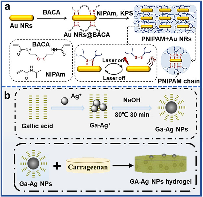
Although Au-photothermal agents have been widely studied, it is relatively expensive, which seriously limits its further application. Recently, silver nanoparticles (Ag NPs) are increasingly used in various applications due to its numerous advantages, including high photothermal conversion ability, chemical stability, and lower cost. For example, Amatya et al. prepared Ag NPs coated with bovine serum protein (BSA) through solution reduction, and then the BSA/Ag NPs were dispersed in gelatin to form photothermal hydrogels (Fig. S2 in Supporting information) [39]. The hydrogels showed unique antibacterial properties and excellent photothermal properties owning to the addition of Ag NPs, leading to successful ablation of skin cancer cells with local laser irradiation of the tumor region. Additionally, Liu et al. used a one-pot method to prepare GA-Ag NPs by reducing Ag+ with gallic acid in alkaline environment, and then embedded them into a natural polysaccharide to prepare GA-Ag NPs hydrogel [40]. The unique antibacterial properties and photothermal conversion properties of Ag NPs give hydrogels excellent broad-spectral sterilization performance under near infrared light (NIR) irradiation (Fig. 2b).
Metal sulfide nanomaterials (MeSNs) are a new class of photothermal agents composed of metal ions and sulfur compounds [41]. Compared to metal-based photothermal hydrogels, they have a lot of advantages, including wider absorption in near-infrared light, lower cost, stable performance, and greater thermal conversion capability [42]. Additionally, unlike the metal-based photothermal conversion mechanism, metal sulfides perform photothermal conversion by utilizing the non-radiative relaxation effect of semiconductors [43]. CuS nanoparticles, as a typical MeSNs, are commonly used in photothermal hydrogel because of their advantages, including low cost, easy preparation, and small surface modification volume [44]. For instance, Xie et al. dispersed CuS nanoparticles uniformly on the lignin skeleton, and then co-mixed with glutaraldehyde (GA) in polyvinyl alcohol (PVA) solution to obtain LS-CuS@PVA composite hydrogel, which has excellent antibacterial properties (Fig. S3 in Supporting information) [45]. Additionally, MoS2, as a new two-dimensional sulfide, is a great photothermal agent with high photothermal conversion efficiency and few side effects, which can be used as good drug and/or gene carrier [46]. Li et al. used the coordination of dopamine and molybdenum disulfide nanosheets to synthesize MP with high catalytic and photothermal properties. Then, in the presence of triethylamine, acryloyl chloride, Pluronic 127 (PF127) as a crosslinking agent was prepared by reaction in methylene chloride. Finally, MoS2-PDA hydrogel (MPH) was prepared by copolymerizing acrylamide (AM), n-isopropylacrylamide and PF127-DA in water using MoS2-PDA (MP) as functional component, which has excellent antibacterial properties (Fig. 3a) [47].
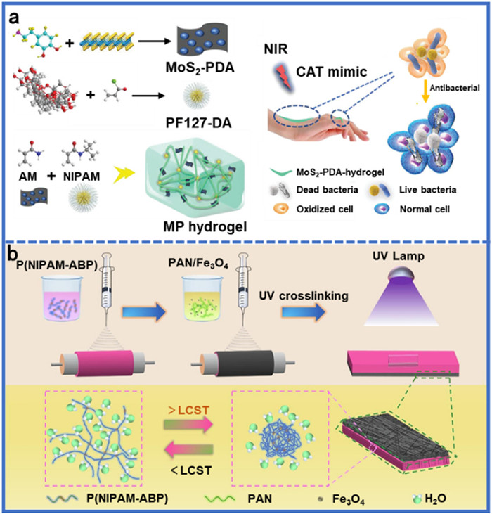
Recently, metal oxide nanomaterials (MeONs) have attracted wide attention due to their excellent photoactivity, high stability, and high-cost effectiveness [48]. For example, Wei et al. prepared a bilayer structure by doping nano Fe3O4 and copolymer (P(NIPAM-ABP) of 4-allylbenzophenone-n-isopropylacrylamide) into polyacrylonitrile (PAN) solution (Fig. 3b) [49]. The Fe3O4/PAN-P(NIPAM-ABP) composite hydrogel (FPP hydrogel) with great photothermal conversion ability was obtained by cross-linking P(NIPAM-ABP) fiber layer by ultraviolet light (UV) initiated polymerization. Additionally, Wang et al. designed and prepared a novel multifunctional hydrogel dressing for hyperglycemia adjustment, adhesion, oxygen generation and biofilm resistance by introducing GOx and nano MnO2 into PDA/AM hydrogel, which had excellent antibacterial, compression, adhesive, hemostatic, and wound healing ability (Fig. S4 in Supporting information) [50].
MXene, as a novel two-dimensional (2D) material with excellent photothermal conversion performance derived from electromagnetic interference shielding effect and LSPR effect, were widely used to prepare photothermal conversion hydrogels [51-53]. For example, Sun et al. reported a thermally responsive conductive anisotropic double layer hydrogel for soft manipulators and sensitive strain sensors [54]. As the first layer, ternary nanocomposite (TN) hydrogel was formed by loading MXene nanosheets, into PNIPAM hydrogel matrix. The second layer is prepared by the same method without MXene nanocomposite (NC) hydrogel. Then, the two-layer TN/NC hydrogel actuators were obtained by bonding tightly through hydrogen bonds between the polymer chain and the laponite nanosheet (Fig. S5 in Supporting information). MXene could generate heat under NIR irradiation, which makes TN hydrogel shrink, resulting in inconsistent shrinkage of the two layers and bending, thus achieving the purpose of grasping objects. This study will provide a new method for the preparation of photothermal responsive conductive hydrogels for remote near-infrared photocontrolled actuators and wearable electronic devices. Moreover, Tao et al. used p-Ti3C2Tx NPs prepared by ultrasonic assisted mild etching method as initiator to generate high concentration of N-isopropyl proacrylamide monomer polymerization to form PNIPAM-based nanocomposite hydrogel under deoxidation condition (Fig. S6 in Supporting information) [55]. The prepared NC hydrogels were sensitive to temperature and NIR, which could be used to regulate transmittance, volume, and conductivity. They also have excellent photothermal properties and electrical conductivity, which could be used as remote photo-controlled "smart" windows, fluid valves and photodetectors.
Carbon-based materials (i.e., CNT [56,57], GO [58,59], and rGO [60,61]), are widely used in photothermal hydrogels due to their high photothermal conversion efficiency and excellent light absorption capacity [62]. Graphene, including GO and rGO, is a two-dimensional material composed of honeycomb carbon atoms. Due to its high surface area, high light absorption, and chemical stability, it could be applied to photothermal hydrogels in various fields [63]. For example, Qi et al. reported an injectable self-healing hydrogel for the treatment of breast cancer [64]. Injectable self-healing hydrogels (PEG-CMC hydrogels) were successfully prepared by cross-linkable aldehyde-modified polyethylene glycol (PEG) with carboxymethyl chitosan (CMC) through dynamic chemical reaction between aldehyde group and amino group in relative component, loading with a certain size of GO or needle-like nano-hydroxyapatite (HAP) as tumor inhibitor, which had obvious effect on the treatment of breast tumor (Fig. 4a). Additionally, rGO, as a graphene derivative with a highly intact aromatic graphene ring structure, has been found it have six times higher photothermal conversion efficiency than GO [65]. Wang et al. reported a rGO-based hydrogel with good photothermal and antibacterial properties, which were constructed by schiff base reaction between carboxymethyl chitosan (CMCS) and oxypectin (OP) (Fig. S7 in Supporting information) [66]. In addition to graphene, carbon nanotubes are also being studied as materials for photothermal hydrogel. For example, Hao et al. used low-cost biomass-derived sodium lignosulfonate (SLS) as the starting material to build an efficient PVA/SLS-CNT hydrogel (PSCH) solar evaporator with good antibacterial properties and high evaporation rate (Fig. 4b) [67].

Small molecule materials have several advantages in biomedical application due to their simple synthesis process, good biocompatibility, and great degradation. Cyanine dye molecules, such as indocyanine green (ICG) [68], IR825 [69], IR780 and Cypate [70] have excellent photothermal conversion ability and have been widely used in photothermal therapy [71]. For example, Kuang et al. developed an injectable multifunctional hydrogel, in situ cured via the coordination of calcium phosphate nanoparticles with poly(dimethyl aminoethyl methacrylate-co-2-hydroxyethyl methacrylate) (DHCP) [72]. The polymer microsphere (MS) was synthesized by self-assembly of the thermosensitive polymer poly(n-acryloylglycine-coacrylamide) (PNAm) and ICG, which were mixed to form DHCP-10PIP/d hydrogel. A coordinated osteoblast/osteoclast balance was achieved through an appropriate concentration of parathyroid hormone (PTH) in DHCP-10PIP/d hydrogel, which could promote osteoporotic bone regeneration in vivo (Fig. 5a). In contrast, Prussian blue, another organic dye, has better photostability and photothermal conversion ability. Tong et al. prepared a further supramolecular photothermal hydrogels, containing hollow mesoporous Prussian blue nanoparticles (HMPB NPs) loading doxorubicin (HMPB-DOX@GEL) [73]. The as-prepared hydrogel could achieve the controlled release of local drugs and have great reactive oxygen species (ROS) scavenging ability (Fig. S8 in Supporting information).
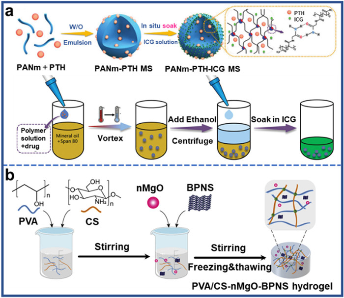
Black phosphorus, as a new photothermal agent in recent years, has been widely studied due to its good biocompatibility and excellent phototherapy effect [74]. For example, Shao et al. combined black phosphorus nanosheets (BPNS) with a thermosensitive hydrogel [poly(d, l-lactide)-poly(ethylene glycol)-poly(d, l-lactide) (PDLLA-PEG-PDLLA: PLEL)] to design a sprayable photothermal therapy (PTT) system for postoperative photothermal therapy of cancer (Fig. S9 in Supporting information) [75]. BP@PLEL hydrogel quickly formed through physical crosslinking under near-infrared irradiation, which could not only eliminate residual tumor tissue, but also prevent wound infection. Altogether, it has tremendous clinical potential in cancer treatment. Phosphorus is one of the essential elements for human body, which plays an important role in bones [76]. The degradation products of BPNS could be transformed into P-based agents for enhancing the osteogenesis process [77,78]. For example, Qing et al. prepared a hydrogel composed of PVA and chitosan encapsulating MgO and BPNS using freeze-thaw methods (Fig. 5b) [79]. The release of Mg2+ and PO43− from the hydrogel could promote the recruitment of mesenchymal stem cell (MSCs), osteogenic differentiation, and biomineralization. The exceptional osteoinductivity of the PVA/CS-MgO-BPNS hydrogel could provide an opportunity for in-situ bone regeneration, making it suitable for large-scale bone defects treatment in clinical practice.
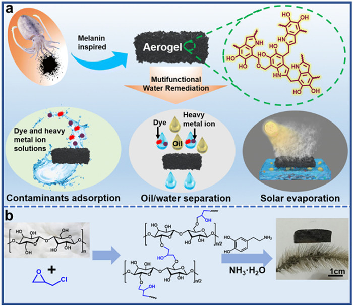
Melanin is a class of biopolymers widely distributed in natural organisms (such as animals, plants, bacteria, and fungi), and has been widely studied for its excellent light-collecting and photothermal conversion ability [80-83]. For instance, inspired by melanin, Yang et al. prepared a bionic hydrogel evaporator through a simple one-pot condensation copolymerization of 5,6-dihydroxyindole (DHI) and formaldehyde, which could achieve 91% evaporation efficiency under a single sunlight irradiation, and at the same time remove various organic dyes and heavy metal ions in wastewater (Fig. 6a) [84]. PDA is a biomimetic polymer material inspired by mussel protein, which is considered as a kind of typical artificial melanin material because of its similar chemical and physical properties to natural melanin [85,86]. In addition to the ability of light collection and photothermal conversion like natural melanin, PDA also has the advantages of structural design and adjustable particle size [87,88]. Zou et al. designed and prepared a low-cost polydopamine-filled cellulose aerogel (PDA-CA) inspired by mussels, which has a photothermal conversion efficiency of 86% under sunlight irradiation, in addition to good long-term water evaporation anti-fouling ability, and effective adsorption of organic dye pollutants (Fig. 6b) [89].
In addition to the above 7 categories of photothermal hydrogels, there are several other photothermal hydrogels. For example, Xiong et al. synthesized a bio-based photothermal hydrogel using cuttlefish juice as photothermal agent, which could effectively resist the solid barrier of foreign bacteria and protect the damage [90]. Additionally, Ma et al. synthesized a silicate photothermal hydrogel (FMS/SA), which had good biocompatibility and could promote skin wound healing after skin tumor resection [91].
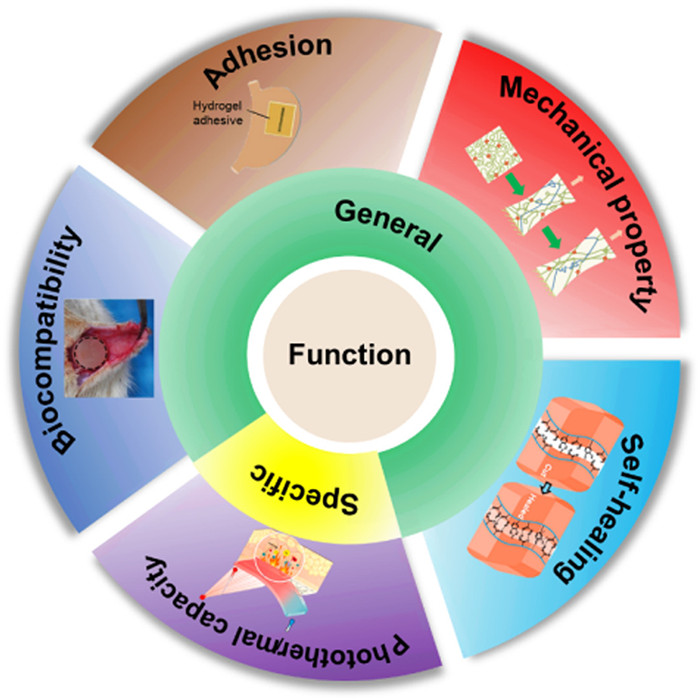
To expand the application of photothermal hydrogel in biological field, in addition to its unique photothermal conversion ability, it is necessary to solve other practical problems by their general properties, such as biocompatibility, adhesion, mechanical properties and self-healing properties (Fig. 7).
Biocompatibility mainly includes histocompatibility and blood compatibility [92]. Good histocompatibility requires hydrogel to not cause any irritation, inflammation, necrosis, functional decline of tissues and cells after administration in vivo. And it could ensure that the performance of photothermal hydrogels meets the requirements for safe usage. For example, Chen et al. used gallic acid grafted chitosan and oxidized Bletilla striata polysaccharide as scaffolds to form a double-crosslinked dynamic photothermal hydrogel (GCOBFe hydrogel) by Schiff base reaction and pyrotrocinol-Fe3+ chelation [93]. According to the results of cell viability and live/dead staining experiments, the cells maintained good morphology after 48 h and there were almost no observed dead cells (Fig. 8a). In addition, it is also necessary to evaluate the blood compatibility of photothermal hydrogels. Zhao et al. prepared a hydrogel (rGB/QCS/PDA-PAM) based on the cross-linking system of PDA and polyacrylamide (PAM), and further mixed the photothermal 3-aminobenzobenboric acid modified reduced graphene oxide (rGB) sheet and QAS-modified carboxymethyl chitosan (QCS), which was assessed to have good blood compatibility (Fig. 8b) [94].
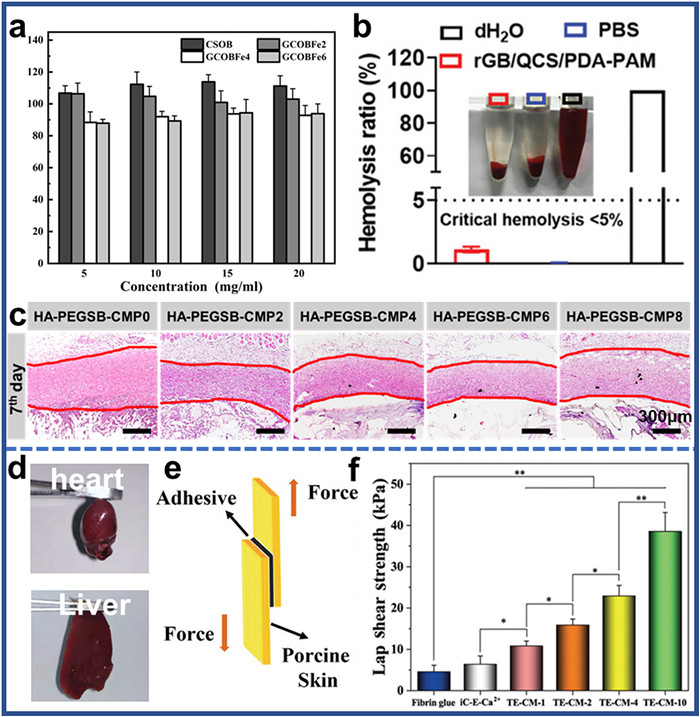
Further, subcutaneous implantation experiments in organisms are also needed to evaluate the biocompatibility and degradability of biomaterials in vivo. For instance, Li et al. prepared a series of tissue adhesion, antioxidant, self-healing and photothermal antibacterial hydrogels (HA-PEGSB-CMP gels) based on the Schiff base network of hyaluronic acid (HA), polysebacin (PEGSB) and cuttlefish melanin nanoparticles (CMP), which was proven good biocompatibility by subcutaneous implantation in Sprague Dawley (SD) rats (Fig. 8c) [95].
The strong bio-adhesion of photothermal hydrogels is of great significance in various applications, particularly in tissue repair, wound management, and connecting wearable electronics [96,97]. However, bio-adhesion technology of hydrogels has faced challenges for a long time. For example, various surfaces such as moist tissue surface, dense cuticle of skin, and mucous membrane on the surface of internal organs can restrict the penetration and result in poor bio-adhesives [12]. So, researchers continue to explore innovative ways to overcome the limitations of bio-adhesion technology and develop new photothermal hydrogels that exhibit strong adhesion for various applications. For example, Fu et al. developed tannin-europium ligand cross-linked mussel citrate photothermal bio-adhesive (TE-CMBAs) with tissue adhesion strength up to 38.5 kPa, which could be used for wound healing applications (Figs. 8d–f) [98]. We believe that the design principles of this work can be generally extended to inspire the development of smart therapeutic tissue adhesives.
Photothermal hydrogels are prone to rupture under excessive external stimuli or experience fatigue after prolonged use, which limits their widespread application in various fields [99]. To address this issue, self-healing capacity is explored and integrated to photothermal hydrogel to recover their intact hydrogel structure after damage through dynamic bonds [100]. The self-healing mechanism is mainly based on dynamic reversible covalent bonds such as imine bonds (Schiff bases) [93], Diels-Alder (DA) reaction [101], borate bond [56], coordination bond [102], and physical non-covalent interactions including hydrogen bond [103], ionic bond [104], host–guest interaction [105]. For example, Qin et al. constructed a double-crosslinked network nanocomposite hydrogel by two water-soluble polymers (polyacryla (PAA) and polyvinyl alcohol (PVA)) interacted with calcium ions and gold nanorods to under UV photopolymerization, which had suitable fatigue resistance, excellent self-healing properties [106]. The as-prepared PCR-2 hydrogel had the unique capability to reconnect itself within 150 s through ion and hydrogen bonds when cut into two parts under NIR irradiation. Moreover, the mechanical properties and cyclic energy loss of these gels barely change after the self-healing, which indicated that the self-healing ability did not compromise the quality and functionality of these photothermal hydrogels (Figs. 9a and b). We believe that this research could provide meaningful approaches for designing high-performance and self-healing hydrogels, as well as new strategies for developing innovative photothermal materials.
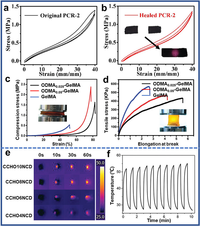
Suitable strength and toughness of photothermal hydrogel for kinetic body sites is crucial for its successful application. To address the mechanical limitation, proper modification is required to improve the mechanical strength of the hydrogels [107,108]. To solve this problem, there are two ways to improve the mechanical strength of photothermal hydrogels. First, increase the number of entanglements in polymer materials, thus increasing the degree of molecular interaction. Second, cross-linking polymers through hydrogen bonds, covalent bonds could also improve the mechanical strength of the photothermal hydrogels [109]. For example, Gan et al. used dopamine methacrylate (ODMA) oligomers to insert into the Gel-MA chain, which reduced the tangling density of the Gel-MA chain, and introduced additional physical crosslinking, making the hydrogel toughness and elasticity [110]. As shown in Figs. 9c and d, it could be found that the tensile and compressive properties of the hydrogel were steadily improved after ODMA oligomers were added into the Gel-MA chain. We believe that this work will provide a feasible way to further develop the preparation of high-performance photothermal hydrogels.
As photothermal hydrogels, it usually has very excellent photothermal conversion performance which refers to the process of concentrating illumination radiation energy through absorption, then converting it into heat, increasing the hydrogel and environment temperature [111]. Photothermal hydrogels with high efficiency could be applied to biomedical materials, solar steam, and other fields. For instance, Chen et al. developed a method to prepare an injectable hydrogel by using the Schiff base reaction between acylamide-modified carbon dots (NCD) and aldehyde-modified cellulose nanocrystals (CCHO), which had great photothermal therapeutic properties [112]. NCDs exhibited high photothermal conversion efficiency of 77.6%, and the CCHO10NCD hydrogel rapidly heated up when exposed to 660 nm light emitting diode (LED) light, such as its surface temperature increasing from room temperature to 55 ℃ within 1 min (Fig. 9e). In addition, it also has good optical stability, and the photothermal performance will not be weakened by photobleaching due to repeated irradiation (Fig. 9f). This sample method, which was direct reaction of photothermal agents with the matrix, not only provided a new strategy for the preparation of photothermal hydrogels, but also paved a new way for advanced cancer treatment.
In addition to the fundamental properties mentioned above, some photothermal hybrid hydrogels were endowed with unique features that make them useful in different application fields. For example, some photothermal hybrid hydrogels possess electrical conductivity through proper design in gel fabrication, enabling their use in electronic and sensor applications. For example, Dong et al. prepared a tough and conductive hydrogel by copolymerization of polyaniline (PANI) and PAM, which had light-stimuli-responsive, excellent electrical conductivity, similar mechanical properties to nerve tissue and good biocompatibility [113].
In recent years, photothermal hydrogels have been widely used in medical. In the medical field, photothermal hydrogels could be used for wound healing, antibacterial treatments, drug-controlled release, bone repair, and tumor therapy treatment, and achieved good therapeutic effect. In the future, photothermal conversion hydrogels will certainly appear as a new therapeutic method in clinical treatment (Fig. 10).
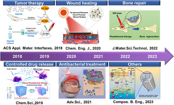
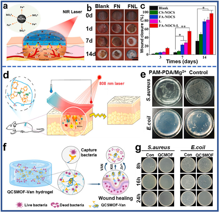
Skin is the largest organ of human body, which plays a vital role in maintaining homeostasis and protecting human body [120,121]. As a protective barrier against the external environment, skin could be damaged in some extreme conditions or accidents [95]. Skin damage could also happen resulting from some diseases. Wound healing consists of four continuous processes: hemostasis, inflammation, proliferation, and remodeling [122,123]. Most skin defects can be quickly and effectively recovered within one to two weeks [124,125], but a few blocked healing processes have become chronic wounds [126,127], which seriously affects life quality and results in disability or death [128,129]. Photothermal hydrogels could accelerate wound healing through hyperthermia therapy base on the photo-thermal conversion capacity. According to studies, local mild heating (about 40 ℃) could stimulate local microcirculation blood flow, promote cell proliferation, angiogenesis, and wound healing [130]. For example, Sheng et al. designed a new bioactive photothermal hydrogel with "hot spring effect" using ferridolomite and N, O-carboxymethyl chitosan (NOCS), which could release bioactive ions and product heating function to form a thermionic environment in the wound area to promote wound healing (Figs. 11a–c) [131]. Altogether, photothermal hydrogel is effective to promote wound healing, accelerating the rapid repair of wounds.
In modern society, infectious diseases caused by pathogenic bacteria continue to pose a significant threat to human life and health [132]. The current strategy for treating bacterial infections involves the use of antibiotics. However, overuse and misuse of antibiotics would give rise to antibiotic resistance, reducing the antibacterial effect [133], so it is particularly important to develop new antibacterial treatments that could inactivate bacteria without causing resistance [134]. One promising strategy is the use of photothermal hydrogels, which could kill bacteria directly through the photothermal effect [135,136]. Unlike antibiotics, photothermal hydrogels do not rely on specific targets within the bacterial cell, avoiding antibiotic resistance happening.
So photothermal hydrogels could offer a potential solution to the problem of antibiotic resistance, and could help reduce the incidence of bacterial infections [137]. For example, Guo et al. designed a composite antibacterial PDA-PAM/Mg2+ hydrogels (Fig. 11d) [138], which had excellent photothermal effect, good cytocompatibility, and excellent antibacterial ability. After NIR irradiation, the survival rate of S. aureus and E. coli was only 5.29% and 7.06%, respectively (Fig. 11e). Huang et al. prepared a curcumin-based organic framework (QCSMOF-Van) loaded with vancomycin (Van) and coated with quaternary ammonium chitosan (QCS) [139]. Then, the multifunctional hydrogels could be prepared by free radical polymerization and Schiff base reaction, which could realize the efficient capture and rapid killing of bacteria (Figs. 11f and g). This approach could limit systemic exposure and reduce the minimum effective dose compared to systemic administration, thereby reducing the potential risk of bacterial resistance [140].
Traditional drug delivery hydrogels mainly release drugs by means of drug diffusion or material degradation [141]. The release process is uncontrollable and could not achieve the expected effect of drug delivery [142,143]. Via proper structure designation, hydrogels with controlled release capacity could address these problems, avoiding boosting release in blood, reducing the side effect of drugs [144].
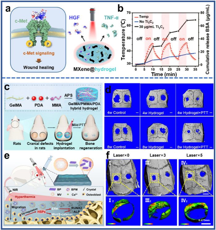
Various photothermal hydrogels have been developed to solve the problems mentioned above, realizing controlled drug release. For example, Qiu et al. designed a BP@Hydrogel drug release system, which can be used to regulate the release of anticancer drugs under NIR irradiation (Fig. S10a in Supporting information) [145]. BP PTA could convert light into heat and raise the temperature of the hydrogel, thereby controlling the rate at which the drug diffuses from the hydrogel to the environment. As shown in Fig. S10b (Supporting information), it could be found that the absorption spectrum of DOX at 480 nm gradually increased, which indicated that the concentration of DOX released gradually increased with the passage of irradiation time. Additionally, the hydrogels could be further hydrolyzed and melted at enhanced laser power, and eventually degrade into oligomers, which were excreted in urine after treatment. Additionally, Wang et al. prepared a multifunctional NIR photo-responsive MXene@hydrogel/protein complex system by packaging photothermal agent Ti3C2 MXene and therapeutic protein drugs into agarose hydrogels (Figs. 12a and b) [146]. Ti3C2 MXene could convert NIR light energy into heat energy, induce reversible phase transformation of hydrogels, release preloaded protein drugs, activate cell signaling pathway mediated by specific receptors, and achieve the therapeutic purpose of enhancing skin wound healing and tumor eradication. The successful preparation of these photothermal hydrogels will provide a feasible strategy for further solving the drug release problem.
Bone is a highly dynamic connective tissue that forms the structural framework of the body and participates in movement, mineral homeostasis, and protection of the viscera [147,148]. Bone injury can recruit and differentiate osteoblasts through a variety of cytokines and growth factors [149,150], but for severe injury or other conditions, the self-healing process is limited and external intervention is required to promote bone regeneration [151-153]. While traditional bone graft treatments have some limitations, bone tissue engineering is a new approach that promotes bone repair and regeneration through scaffolds implanted with cells or bioactive growth factors [154-156]. Photothermal conversion hydrogels combined the benefits of conventional hydrogels with their effect on tissue temperature stimulation, where mild local heating improved the expression of various genes that promote tissue repair, thereby promoting cell proliferation and differentiation, and ultimately accelerating bone regeneration [157].
For instance, Wu et al. prepared gelatin-methyl methacrylate/polymethyl methacrylate/polydopamine (Gel-MA/PMMA/PDA) hydrogels by free radical polymerization, which could promote the activity of alkaline phosphatase (ALP), the formation of extracellular calcification nodules, and the regeneration of mouse skull under PTT condition (Figs. 12c and d) [158]. Additionally, Tan et al. designed a composite extracellular matrix (ECM)-mimicking chitosan/collagen-based hydrogels, which could be remotely activated, containing black phosphorus (BP) encapsulated by the membrane of MSCs (Fig. 12e) [159]. Under NIR light, the MSC membrane-encapsulated BP nanosheets could induce a mild photothermal effect, promote osteoblast recruitment through the activation of matrix metalloproteinases (MMPs) and the ERK-Wnt/β-catenin-RUNX2 axis mediated by heat shock proteins (HSPs). Simultaneously, the thermal decomposition could release phosphate ions into the surrounding medium, which was beneficial for osteoblast migration and differentiation. After implanting the hydrogel into the cranial defects of SD rats, it could be found that the local bone density and new bone formation were significantly increased. These ECM-mimicking hydrogels with BP incorporation could enhance osteoblast migration/differentiation, stimulate the biomineralization process under remote NIR activation, and promote bone healing, providing a new opportunity for clinical cranial defect repair (Fig. 12f).
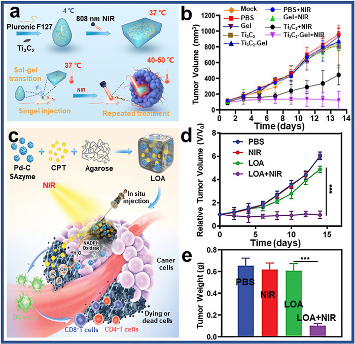
The increasing incidence and mortality of cancer is a serious threat to the health of people all over the world [160,161]. At present, there are mainly three traditional clinical treatment methods for tumors: surgical resection, radiotherapy, and chemotherapy, but they have various shortcomings, such as strong side effects, obvious trauma [162-165]. In recent years, PTT has been a promising cancer treatment that relies on a specific laser to shine heat necrosis/apoptosis on cancer cells. Compared to traditional treatment methods, PTT has the advantages of high therapeutic efficiency, less invasive, and large penetration depth [166]. PTT therapy could be divided into two therapeutic methods, one was to use photothermal gel to absorb the heat generated by light energy to make tumor cell proteins and so on to kill cancer cells. For example, Yao et al. blended Ti3C2 and Pluronic F127 to form Ti3C2-gel by one-step synthesis [167]. In vitro and in vivo results showed that the hydrogel system could effectively inhibit tumor growth when exposed to NIR irradiation (Figs. 13a and b). The other was to use the sensitivity of the photothermal gel to temperature, and the heat generated by the photothermal gel will degrade itself, to release the drugs in it at a fixed point to achieve the purpose of tumor treatment, Zhu et al. designed a photocontrolled oxidative stress amplification system to enhance synergistic anti-tumor activity through autogeneration of H2O2 and transformation of "cold" tumors (Fig. 13c) [168]. In this nano-enzyme hydrogel system, nanoenzymes in the Pd-C unit convert NIR laser to heat, resulting in azgarose-degradation and subsequent camptothecin release, which could enhance the H2O2 level in tumor by activating niacinamide adenine dinucleotide phosphate oxidase, improve the catalytic performance of single atom nanozymes with peroxide-like activity, and improve the anti-tumor effect (Figs. 13d and e).
In this paper, the classification, preparation, functions, and applications of photothermal hydrogels were summarized comprehensively. Firstly, the classification and preparation methods of photothermal hydrogels doped with seven different kinds of photothermal agents (metals, metal sulfides/oxides, carbon-based, conjugated polymers, small molecular dyes, black phosphorus, MXene) were discussed. Secondly, five functions of photothermal hydrogels were introduced in detail: biocompatibility, adhesion, self-healing, mechanical strength, and photothermal properties. In addition, photothermal hydrogels with good sustainable use, biocompatibility and safety were applied to medical field, which contain wound healing, antibacterial treatments, drug-controlled release, bone repair, and cancer treatment (Fig. 14).
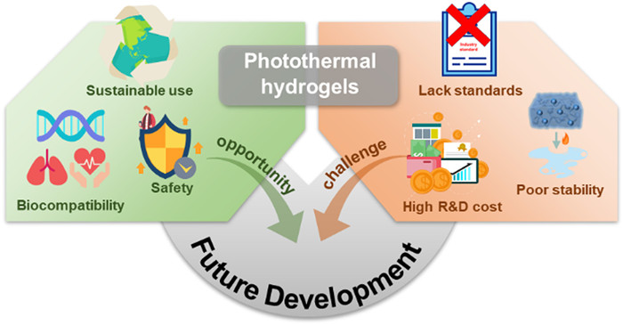
However, there are some main problems in photothermal hydrogels: photothermal conversion efficiency and stability. According to current research, the photothermal conversion efficiency of most photothermal hydrogel is below 70%, resulting in partial energy loss. Persistent effort is needed to improve the photothermal conversion efficiency of photothermal hydrogels, either through the modification of photothermal agents or improving hydrogel fabrication strategies. Since most of the photothermal hydrogels are formed by the hybrid of nanoparticles and hydrogels, the compatibility between nanoparticles and hydrogels is crucial for photo-heat conversion in application. Therefore, surface modification of nanoparticles is needed to improve the binding force with the hydrogels, which should be taken in consideration for further investigation. Photothermal hydrogels will certainly solve the current problems in the future, and could cut a conspicuous figure in various fields (Fig. 14).
The authors declare that they have no known competing financial interests or personal relationships that could have appeared to influence the work reported in this paper.
This article was completed with the support of the General Project of Sichuan Natural Science Foundation (No. 2022NSFSC0349), and National Natural Science Foundation of China Youth Fund Project (No. 5180316).
Supplementary material associated with this article can be found, in the online version, at doi:
K.H. Shen, C.H. Lu, C.Y. Kuo, et al., J. Mater. Chem. B 9 (2021) 7100–7116. doi: 10.1039/d1tb00980j
Y.S. Zhang, A. Khademhosseini, Science 356 (2017) eaaf3627. doi: 10.1126/science.aaf3627
W. Ji, Q. Wu, X. Han, et al., Sci. China Life Sci. 63 (2020) 1813–1828. doi: 10.1007/s11427-019-1710-8
B.G. Wu, W.J. Yang, D.Y. Niu, et al., Chin. J. Polym. Sci. 38 (2020) 1107–1116. doi: 10.1007/s10118-020-2443-5
A. Motealleh, N.S. Kehr, Adv. Healthc. Mater. 6 (2017) 1600938. doi: 10.1002/adhm.201600938
M. Bustamante-Torres, D. Romero-Fierro, B. Arcentales-Vera, et al., Gels 7 (2021) 182. doi: 10.3390/gels7040182
P. Sikdar, M.M. Uddin, T.M. Dip, et al., Mater. Adv. 2 (2021) 4532–4573. doi: 10.1039/d1ma00193k
F. Ullah, M.B.H. Othman, F. Javed, et al., Mater. Sci. Eng. C 57 (2015) 414–433. doi: 10.1016/j.msec.2015.07.053
A. Osman, E.T. Oner, M.S. Eroglu, Carbohydr. Polym. 165 (2017) 61–70. doi: 10.1016/j.carbpol.2017.01.097
Y.H. Choi, J.S. Hwang, S.H. Han, et al., Adv. Funct. Mater. 31 (2021) 2100782. doi: 10.1002/adfm.202100782
H. Luo, K. Wu, Q. Wang, et al., J. Membr. Sci. 593 (2020) 117406. doi: 10.1016/j.memsci.2019.117406
Z. Ma, C. Bourquard, Q. Gao, et al., Science 377 (2022) 751–755. doi: 10.1126/science.abn8699
S. Zhang, H. Gao, P. Guo, et al., Colloids Surf. A 603 (2020) 125280. doi: 10.1016/j.colsurfa.2020.125280
J. Kang, X. Yang, X. Yang, et al., Chin. Chem. Lett. 35 (2024) 109297. doi: 10.1016/j.cclet.2023.109297
J. Li, Z. Wang, H. Han, et al., Chin. Chem. Lett. 33 (2022) 1936–1940. doi: 10.1016/j.cclet.2021.10.058
D.N. Hu, X.J. Ju, X.Q. Pu, et al., Ind. Eng. Chem. Res. 60 (2021) 8147–8158. doi: 10.1021/acs.iecr.1c01277
C. Li, Y. Cheng, D. Li, et al., Int. J. Mol. Sci. 23 (2022) 7909. doi: 10.3390/ijms23147909
L. Cao, Y. Wu, Y. Shan, et al., Bioact. Mater. 17 (2022) 2022008.
H.S. Han, K.Y. Choi, Biomedicines 9 (2021) 305. doi: 10.3390/biomedicines9030305
P. Cheng, D. Wang, P. Schaaf, Adv. Sustain. Syst. 6 (2022) 2200115. doi: 10.1002/adsu.202200115
J. Li, W. Zhang, W. Ji, et al., J. Mater. Chem. B 9 (2021) 7909–7926. doi: 10.1039/d1tb01310f
G. Liu, Y. Zhou, Z. Xu, et al., Chin. Chem. Lett. 34 (2023) 107705. doi: 10.1016/j.cclet.2022.07.048
G. Liu, Z. Bao, J. Wu, Chin. Chem. Lett. 31 (2020) 1817–1821. doi: 10.1016/j.cclet.2020.03.005
T. Liu, G. Liu, J. Zhang, et al., Chin. Chem. Lett. 33 (2022) 1880–1884. doi: 10.1016/j.cclet.2021.10.022
L. Jin, X. Guo, D. Gao, et al., Bioact. Mater. 16 (2022) 162–172.
M.A. Kim, S.D. Yoon, C.M. Lee, Int. J. Biol. Macromol. 103 (2017) 839–844. doi: 10.1016/j.ijbiomac.2017.05.139
B. Liu, X. Gu, Q. Sun, et al., Adv. Funct. Mater. 31 (2021) 2010779. doi: 10.1002/adfm.202010779
L. Wang, L. Chen, J. Wang, et al., Chin. Chem. Lett. 33 (2022) 1956–1962. doi: 10.1016/j.cclet.2021.10.070
Y. Peng, Y. Zhuang, Y. Liu, et al., Exploration 3 (2023) 20210043. doi: 10.1002/EXP.20210043
J. Fu, B. Wu, M. Wei, et al., Acta Pharm. Sin. B 9 (2019) 604–614. doi: 10.1016/j.apsb.2018.12.005
G. Xie, X. Wang, M. Mo, et al., Macromol. Biosci. 23 (2022) e2200378.
P. Le Minh Tu, V. Thuy Anh Thu, H. Thi Xoan, et al., Nanomaterials 11 (2021) 906. doi: 10.3390/nano11040906
F. Ding, L. Zhang, X. Chen, et al., Front. Bioeng. Biotechnol. 10 (2022) 1066617. doi: 10.3389/fbioe.2022.1066617
E. Petryayeva, U.J. Krull, Anal. Chim. Acta 706 (2011) 8–24. doi: 10.1016/j.aca.2011.08.020
K.M. Mayer, J.H. Hafner, Chem. Rev. 111 (2011) 3828–3857. doi: 10.1021/cr100313v
P. Deveci, J. Incl. Phenom. Macrocycl. Chem. 99 (2021) 23–31. doi: 10.1007/s10847-020-01035-5
H. Qin, T. Zhang, H.N. Li, et al., Chem 3 (2017) 691–705. doi: 10.1016/j.chempr.2017.07.017
S. Lin, S. Jiang, Y. Zhang, et al., Gold Bull. 54 (2021) 59–67. doi: 10.1007/s13404-021-00293-6
R. Amatya, S. Hwang, T. Park, et al., Pharm. Res. 38 (2021) 873–883. doi: 10.1007/s11095-021-03038-4
Y. Liu, F. Li, Z. Guo, et al., Chem. Eng. J. 382 (2020) 122990. doi: 10.1016/j.cej.2019.122990
W. Fei, M. Zhang, X. Fan, et al., J. Nanobiotechnol. 19 (2021) 93. doi: 10.1186/s12951-021-00839-y
R. Marin, A. Skripka, L.V. Besteiro, et al., Small 14 (2018) 1803282. doi: 10.1002/smll.201803282
A. Iglesias-Juez, F. Fresno, J.M. Coronado, et al., Curr. Opin. Green Sustain. Chem. 37 (2022) 100652. doi: 10.1016/j.cogsc.2022.100652
L. Li, L.H. Rashidi, M. Yao, et al., Photodiagn. Photodyn. Ther. 19 (2017) 5–14. doi: 10.1007/978-3-319-72395-2_1
Y. Xie, C. Gan, Z. Li, et al., ACS Biomater. Sci. Eng. 8 (2022) 560–569. doi: 10.1021/acsbiomaterials.1c01406
J. Shi, J. Li, Y. Wang, et al., J. Mater. Chem. B 8 (2020) 5793–5807. doi: 10.1039/d0tb01018a
Y. Li, P. Yu, J. Wen, et al., Adv. Funct. Mater. 32 (2022) 2110720. doi: 10.1002/adfm.202110720
K.W. Tan, C.M. Yap, Z. Zheng, et al., Adv. Sustain. Syst. 6 (2022) 2100416. doi: 10.1002/adsu.202100416
X. Wei, L. Chen, Y. Wang, et al., Chem. Eng. J. 433 (2022) 134258. doi: 10.1016/j.cej.2021.134258
Q. Wang, W. Qiu, M. Li, et al., Biomater. Sci. 10 (2022) 4796–4814. doi: 10.1039/d2bm00433j
K. Huang, Z. Li, J. Lin, et al., Chem. Soc. Rev. 47 (2018) 5109–5124. doi: 10.1039/c7cs00838d
D. Xu, Z. Li, L. Li, et al., Adv. Funct. Mater. 30 (2020) 2000712. doi: 10.1002/adfm.202000712
J. Meng, Z. An, Y. Liu, et al., J. Appl. Phys. 55 (2022) 374003. doi: 10.1088/1361-6463/ac79d9
Z. Sun, C. Song, J. Zhou, et al., Macromol. Rapid Commun. 42 (2021) 2100499. doi: 10.1002/marc.202100499
N. Tao, D. Zhang, X. Li, et al., Chem. Sci. 10 (2019) 10765–10771. doi: 10.1039/c9sc03917a
P. Deng, X. Liang, F. Chen, et al., Appl. Mater. Today 26 (2022) 101362. doi: 10.1016/j.apmt.2022.101362
L. Zhao, Z. Yang, J. Wang, et al., Chem. Eng. J. 451 (2023) 138676. doi: 10.1016/j.cej.2022.138676
C. Gong, W. Guan, X. Liu, et al., Adv. Mater. 34 (2022) 2206134. doi: 10.1002/adma.202206134
S. Ramesh, J. Davis, A. Roros, et al., ACS Appl. Mater. Interfaces 14 (2022) 42558–42567. doi: 10.1021/acsami.2c13280
Y. Li, J. He, J. Zhou, et al., Biomater. Sci. 10 (2022) 1326–1341. doi: 10.1039/d1bm01937f
B.L. Melo, R. Lima-Sousa, C.G. Alves, et al., Biochem. Eng. J. 185 (2022) 108529. doi: 10.1016/j.bej.2022.108529
G. Xue, K. Liu, Q. Chen, et al., ACS Appl. Mater. Interfaces 9 (2017) 15052–15057. doi: 10.1021/acsami.7b01992
C. Tan, X. Cao, X.J. Wu, et al., Chem. Rev. 117 (2017) 6225–6331. doi: 10.1021/acs.chemrev.6b00558
Y. Qi, Z. Qian, W. Yuan, et al., J. Mater. Chem. B 9 (2021) 9734–9743. doi: 10.1039/d1tb01753e
J.T. Robinson, S.M. Tabakman, Y. Liang, et al., J. Am. Chem. Soc. 133 (2011) 6825–6831. doi: 10.1021/ja2010175
S. Wang, W. Zhang, J. Yan, et al., Macromol. Mater. Eng. 307 (2022) 2100878. doi: 10.1002/mame.202100878
L. Hao, N. Liu, H. Bai, et al., J. Colloid Interface Sci. 608 (2022) 840–852. doi: 10.1016/j.jcis.2021.10.035
S. Zhu, S. Wang, C. Liu, et al., Front. Oncol. 12 (2022) 918416. doi: 10.3389/fonc.2022.918416
P. Pham Thi My, H. Jhon, I. In, et al., Biomater. Sci. 7 (2019) 4800–4812. doi: 10.1039/C9BM00734B
I.J. Sabino, R. Lima-Sousa, C.G. Alves, et al., Int. J. Pharm. 600 (2021) 120510. doi: 10.1016/j.ijpharm.2021.120510
X. Song, Q. Chen, Z. Liu, Nano Res. 8 (2014) 340–354. doi: 10.7315/CADCAM.2014.340
L. Kuang, J. Huang, Y. Liu, et al., Adv. Funct. Mater. 31 (2021) 2105383. doi: 10.1002/adfm.202105383
Z. Tong, Q. Guo, G. Xu, et al., Compos. B: Eng. 237 (2022) 109872. doi: 10.1016/j.compositesb.2022.109872
D. Fan, Y. Lu, H. Zhang, et al., Appl. Catal. B 295 (2021) 120285. doi: 10.1016/j.apcatb.2021.120285
J. Shao, C. Ruan, H. Xie, et al., Adv. Sci. 5 (2018) 1700848. doi: 10.1002/advs.201700848
S.T. Itkonen, H.J. Rita, E.M. Saarnio, et al., Nutr. Res. 37 (2017) 58–66. doi: 10.1016/j.nutres.2016.12.009
B. Yang, J. Yin, Y. Chen, et al., Adv. Mater. 30 (2018) 1705611. doi: 10.1002/adma.201705611
K. Huang, J. Wu, Z. Gu, ACS Appl. Mater. Interfaces 11 (2019) 2908–2916. doi: 10.1021/acsami.8b21179
Y. Qing, H. Wang, Y. Lou, et al., Mater. Today Chem. 24 (2022) 100894. doi: 10.1016/j.mtchem.2022.100894
W. Bai, P. Xiang, H. Liu, et al., Macromolecules 55 (2022) 6426–6434. doi: 10.1021/acs.macromol.2c01440
L. Guo, T. Wang, Z. Li, et al., Chem. Mater. 35 (2023) 5420–5432. doi: 10.1021/acs.chemmater.3c00758
W. Bai, P. Yang, H. Zhang, et al., Macromolecules 56 (2023) 4566–4574. doi: 10.1021/acs.macromol.3c00852
W. Bai, P. Yang, H. Liu, et al., Macromolecules 55 (2022) 3493–3501. doi: 10.1021/acs.macromol.2c00506
P. Yang, W. Bai, Y. Zou, et al., Mater. Horiz. 10 (2023) 1020–1029. doi: 10.1039/d2mh01474b
Y. Xu, J. Hu, J. Hu, et al., Prog. Polym. Sci. 146 (2023) 101740. doi: 10.1016/j.progpolymsci.2023.101740
Y. Zou, X. Chen, P. Yang, et al., Sci. Adv. 6 (2020) eabb4696. doi: 10.1126/sciadv.abb4696
X. Wang, L. Yang, P. Yang, et al., Sci. China Chem. 63 (2020) 1295–1305. doi: 10.1007/s11426-020-9797-3
C. Huang, X. Wang, P. Yang, et al., Macromol. Rapid Commun. 44 (2023) 2100916. doi: 10.1002/marc.202100916
Y. Zou, J. Zhao, J. Zhu, et al., ACS Appl. Mater. Interfaces 13 (2021) 7617–7624. doi: 10.1021/acsami.0c22584
Y.H. Xiong, L. Zhang, Z. Xiu, et al., Acta Biomater. 148 (2022) 119–132. doi: 10.1016/j.actbio.2022.06.018
W. Ma, H. Zhang, H. Ma, et al., Prog. Nat. Sci. 32 (2022) 171–178. doi: 10.1016/j.pnsc.2022.01.012
L. Ghasemi-Mobarakeh, D. Kolahreez, S. Ramakrishna, et al., Curr. Opin. Biomed. Eng. 10 (2019) 45–50. doi: 10.1016/j.cobme.2019.02.004
C. Chen, P. Zhou, C. Huang, et al., Carbohydr. Polym. 273 (2021) 118557. doi: 10.1016/j.carbpol.2021.118557
P. Zhao, Y. Zhang, X. Chen, et al., Adv. Sci. 10 (2023) 2206585. doi: 10.1002/advs.202206585
M. Li, Y. Liang, Y. Liang, et al., Chem. Eng. J. 427 (2022) 132039. doi: 10.1016/j.cej.2021.132039
W. Zhang, X. Zhang, Matter 5 (2022) 4114–4115. doi: 10.1016/j.matt.2022.11.013
Y. Xiong, X. Zhang, X. Ma, et al., Polym. Chem. 12 (2021) 3721–3739. doi: 10.1039/d1py00282a
M. Fu, Y. Zhao, Y. Wang, et al., Small 19 (2022) 2205489.
G. Su, S. Yin, Y. Guo, et al., Mater. Horiz. 8 (2021) 1795–1804. doi: 10.1039/d1mh00085c
Y. Li, X. Wang, Y. Wei, et al., Chin. Chem. Lett. 28 (2017) 2053–2057. doi: 10.1016/j.cclet.2017.09.004
Z. Yang, F. Zhao, W. Zhang, et al., Chem. Eng. J. 419 (2021) 129520. doi: 10.1016/j.cej.2021.129520
W. Wang, L. Cao, Q. Li, et al., J. Colloid Interface Sci. 630 (2023) 511–522. doi: 10.1016/j.jcis.2022.10.089
Y.H. Yeo, K. Chathuranga, J.S. Lee, et al., Carbohydr. Polym. 277 (2022) 118834. doi: 10.1016/j.carbpol.2021.118834
S. Guo, M. Yao, D. Zhang, et al., Adv. Healthc. Mater. 11 (2022) 2101808. doi: 10.1002/adhm.202101808
R. Yu, M. Li, Z. Li, et al., Adv. Healthc. Mater. 11 (2022) 2102749. doi: 10.1002/adhm.202102749
M. Qin, Y. Guo, F. Su, et al., Chem. Eng. J. 455 (2023) 140854. doi: 10.1016/j.cej.2022.140854
J. Kim, G. Zhang, M. Shi, et al., Science 374 (2021) 212–216. doi: 10.1126/science.abg6320
N. Bosnjak, M.N. Silberstein, Science 374 (2021) 150–151. doi: 10.1126/science.abl6358
D.M. Fitzgerald, Y.L. Colson, M.W. Grinstaff, Nat. Rev. Mater. 8 (2022) 5.
D. Gan, T. Xu, W. Xing, et al., J. Mater. Chem. B 7 (2019) 1716–1725. doi: 10.1039/c8tb01664j
Q. Zhang, W. Xu, X. Wang, Sci. China Mater. 61 (2018) 905–914. doi: 10.1007/s40843-018-9250-x
T. Chen, T. Yao, H. Peng, et al., Adv. Funct. Mater. 31 (2021) 2106079. doi: 10.1002/adfm.202106079
M. Dong, B. Shi, D. Liu, et al., ACS Nano 14 (2020) 16565–16575. doi: 10.1021/acsnano.0c05197
H. Pan, C. Zhang, T. Wang, et al., ACS Appl. Mater. Interfaces 11 (2018) 2782–2789. doi: 10.1364/ome.8.002782
C. Ruan, C. Liu, H. Hu, et al., Chem. Sci., 2019, 10, 4699–4706. doi: 10.1039/c9sc00375d
G. Xie, N. Zhou, Y. Gao, et al., Chem. Eng. J. 403 (2021) 126353. doi: 10.1016/j.cej.2020.126353
S. Wang, B. Chen, L. Ouyang, et al., Adv. Sci. 8 (2021) 2004721. doi: 10.1002/advs.202004721
C. Wei, X. Jin, C. Wu, et al., J. Mater. Sci. Technol. 118 (2022) 64–72. doi: 10.1016/j.jmst.2021.10.053
G. Li, L. Zhang, Q. Han, et al., Compos. B. Eng. 254 (2023) 110551. doi: 10.1016/j.compositesb.2023.110551
A. Maleki, J. He, S. Bochani, et al., ACS Nano 15 (2021) 18895–18930. doi: 10.1021/acsnano.1c08334
H. Chen, R. Cheng, X. Zhao, et al., NPG Asia Mater. 11 (2019) 3. doi: 10.1038/s41427-018-0103-9
Y. Liang, H. Xu, Z. Li, et al., NanoMicro Lett. 14 (2022) 1851. doi: 10.1109/embc48229.2022.9871167
Y. Huang, X. Zhao, Z. Zhang, et al., Chem. Mater. 32 (2020) 6595–6610. doi: 10.1021/acs.chemmater.0c02030
Y. Liang, M. Li, Y. Yang, et al., ACS Nano 16 (2022) 3194–3207. doi: 10.1021/acsnano.1c11040
B. Guo, R. Dong, Y. Liang, et al., Nat. Rev. Chem. 5 (2021) 773–791. doi: 10.1038/s41570-021-00323-z
X. Zhao, B. Guo, H. Wu, et al., Nat. Commun. 9 (2018) 2784. doi: 10.1038/s41467-018-04998-9
P. Xin, S. Han, J. Huang, et al., Chin. Chem. Lett. 34 (2023) 108125. doi: 10.1016/j.cclet.2022.108125
G.C. Gurtner, S. Werner, Y. Barrandon, et al., Nature 453 (2008) 314–321. doi: 10.1038/nature07039
Z. Xu, G. Liu, Q. Li, et al., Nano Res. 15 (2022) 5305–5315. doi: 10.1007/s12274-022-4192-y
B.D. Zheng, M.T. Xiao, Carbohydr. Polym. 299 (2023) 120228. doi: 10.1016/j.carbpol.2022.120228
L. Sheng, Z. Zhang, Y. Zhang, et al., Biomaterials 264 (2021) 120414. doi: 10.1016/j.biomaterials.2020.120414
H. Li, J. Zhang, L. Yang, et al., Adv. Funct. Mater. 33 (2023) 2212193. doi: 10.1002/adfm.202212193
X. Li, H. Bai, Y. Yang, et al., Adv. Mater. 31 (2019) e1805092. doi: 10.1002/adma.201805092
Z. Cao, Y. Luo, Z. Li, et al., Macromol. Biosci. 21 (2021) 2000252. doi: 10.1002/mabi.202000252
S. Cheng, M. Pan, D. Hu, et al., Chin. Chem. Lett. 34 (2023) 108276. doi: 10.1016/j.cclet.2023.108276
X. Huang, W. Zhang, G. Guan, et al., Acc. Chem. Res. 50 (2017) 2529–2538. doi: 10.1021/acs.accounts.7b00294
U.S. Malik, Q. Duan, M.B.K. Niazi, et al., Chin. Chem. Lett. 34 (2023) 108071. doi: 10.1016/j.cclet.2022.108071
Z. Guo, Z. Zhang, N. Zhang, et al., Bioact. Mater. 15 (2022) 203–213.
K. Huang, W. Liu, W. Wei, et al., ACS Nano 16 (2022) 19491–19508. doi: 10.1021/acsnano.2c09593
C. Fiorica, F.S. Palumbo, G. Pitarresi, et al., Int. J. Pharm. 610 (2021) 121231. doi: 10.1016/j.ijpharm.2021.121231
S. Adepu, S. Ramakrishna, Molecules 26 (2021) 5905. doi: 10.3390/molecules26195905
Y. Tian, Z. Gao, N. Wang, et al., J. Am. Chem. Soc. 144 (2022) 18419–18428. doi: 10.1021/jacs.2c06877
Q. Luo, W. Sun, Z. Li, et al., Biomaterials 303 (2023) 122368. doi: 10.1016/j.biomaterials.2023.122368
C. Xian, Q. Yuan, Z. Bao, et al., Chin. Chem. Lett. 31 (2020) 19–27. doi: 10.1016/j.cclet.2019.03.052
M. Qiu, D. Wang, W. Liang, et al., Proc. Natl. Acad. Sci. U.S.A. 115 (2018) 501–506. doi: 10.1073/pnas.1714421115
S. Wang, Z. Zhang, S. Wei, et al., Acta Biomater. 130 (2021) 138–148. doi: 10.1016/j.actbio.2021.05.027
X. Xue, Y. Hu, Y. Deng, et al., Adv. Funct. Mater. 31 (2021) 2009432. doi: 10.1002/adfm.202009432
K. Huang, J. Huang, J. Zhao, et al., Chin. Chem. Lett. 33 (2022) 1941–1945. doi: 10.1016/j.cclet.2021.10.073
X. Bai, M. Gao, S. Syed, et al., Bioact. Mater. 3 (2018) 401–417. doi: 10.1016/J.BIOACTMAT.2018.05.006
Y. Qiao, X. Liu, X. Zhou, et al., Adv. Healthc. Mater. 9 (2020) 2070001. doi: 10.1002/adhm.202070001
A. Forlino, J.C. Marini, Lancet 387 (2016) 1657–1671. doi: 10.1016/S0140-6736(15)00728-X
K. Huang, G. Liu, Z. Gu, et al., Chin. Chem. Lett. 31 (2020) 3190–3194. doi: 10.1016/j.cclet.2020.07.002
X. Hao, X. Zhang, Y. Hu, et al., Chin. Chem. Lett. 34 (2023) 107965.
T. Zhu, M. Jiang, M. Zhang, et al., Bioact. Mater. 9 (2022) 446–460.
Z. Liu, J. Zhang, C. Fu, et al., Asian J. Pharm. 18 (2023) 100774.
D. Tang, R.S. Tare, L.Y. Yang, et al., Biomaterials 83 (2016) 363–382. doi: 10.1016/j.biomaterials.2016.01.024
J.P. Ribeiro, R.M.A. Domingues, P.S. Babo, et al., Carbohydr. Polym. 292 (2022) 119638. doi: 10.1016/j.carbpol.2022.119638
Y. Wu, X. Zhang, B. Tan, et al., Mater. Sci. Eng. C 133 (2022) 112641.
L. Tan, Y. Hu, M. Li, et al., Chem. Eng. J. 431 (2022) 133382. doi: 10.1016/j.cej.2021.133382
H. Sung, J. Ferlay, R.L. Siegel, et al., CA Cancer J. Clin. 71 (2021) 209–249. doi: 10.3322/caac.21660
Y. Wang, X. Weng, L. Wang, et al., J. Clin. Investig. 128 (2018) 5235–5250. doi: 10.1172/jci99974
R. Oun, Y.E. Moussa, N.J. Wheate, Dalton Trans. 47 (2018) 6645–6653. doi: 10.1039/c8dt00838h
S. Tohme, R.L. Simmons, A. Tsung, Cancer Res. 77 (2017) 1548–1552. doi: 10.1158/0008-5472.CAN-16-1536
S. Liu, Z. Wang, Q. Wei, et al., J. Control. Rel. 363 (2023) 721–732. doi: 10.1016/j.jconrel.2023.09.039
L. Zhai, Y. Shi, Y. Yan, et al., Chin. Chem. Lett. 34 (2023) 108104. doi: 10.1016/j.cclet.2022.108104
B. Liu, F. Jiang, J. Sun, et al., J. Mater. Chem. B 9 (2021) 7007–7022. doi: 10.1039/d1tb00725d
J. Yao, C. Zhu, T. Peng, et al., Front. Bioeng. Biotechnol. 9 (2021) 791891. doi: 10.3389/fbioe.2021.791891
D. Zhu, H. Chen, C. Huang, et al., Adv. Funct. Mater. 32 (2022) 2110268. doi: 10.1002/adfm.202110268


Figure 3 (a) MP hydrogel with antibacterial and antioxidant dual functions for closely fitting and wound healing in movable parts. Copied with permission [47]. Copyright 2020, Elsevier B.V. (b) Preparation process and bending mechanism of Fe3O4/PAN-P(NIPAM-ABP) composite hydrogel actuator. Copied with permission [49]. Copyright 2020, Elsevier B.V.

Figure 4 (a) The preparation process of the injectable self-healing hydrogel by the reaction of DF-PEG with CMC (formation of the Schiff base bond) loading HAP and GO, and a schematic of the synergistic inhibition-therapy and photothermal therapy processes from the injectable PEG–CMC/HAP/GO self-healing nanocomposite hydrogel. Copied with permission [64]. Copyright 2021, Royal Society of Chemistry. (b) Scheme of preparing PVA/SLS-CNT hydrogel hybrid solar evaporator. Copied with permission [67]. Copyright 2021, Elsevier B.V.

Figure 5 (a) Schematic illustration of the fabrication of the NIR (808 nm) light-responsive PANm-PTH-ICG MSs based on W/O emulsion. Copied with permission [72]. Copyright 2021, John Wiley & Sons, Inc. (b) Schematic diagram of the preparation of PVA/CS-nMgO-BPNS hydrogel. Copied with permission [79]. Copyright 2021, John Wiley & Sons, Inc.


Figure 8 (a) The L929 mouse fibroblasts cytotoxicity of hydrogels. Copied with permission [93]. Copyright 2023, John Wiley & Sons, Inc. (b) Hemolysis test of the rGB/QCS/PDA-PAM hydrogel, compared to positive control (dH2O) and negative control (PBS). Copied with permission [94]. Copyright 2022, John Wiley & Sons, Inc. (c) In vivo biocompatibility of the hydrogels. Scale bar: 300 µm. Copied with permission [95]. Copyright 2021, Elsevier B.V. (d) Photographs of TE-CMBAs adhered to various wet tissues. (e) Schematic diagram of lap shear strength test of the adhesive to porcine skin tissue. (f) Lap shear strength of the adhesive with different groups to porcine skin tissue. Copied with permission [98]. Copyright 2022, John Wiley & Sons, Inc.

Figure 9 Successive two times loading–unloading experiments of (a) original PCR-2 hydrogel and (b) healed sample. Copied with permission [106]. Copyright 2023, Elsevier B.V. (c) Typical compression stress–strain curves of the hydrogel with different ODMA concentration. (d) Typical tensile stress–strain curves of the hydrogel with different ODMA concentration. Copied with permission [110]. Copyright 2021, Elsevier B.V. (e) Infrared thermal images of CCHONCD hydrogels at different time intervals. (f) Photothermal stability of CCHO10NCD hydrogel under LED irradiation for ten on/off cycles. Copied with permission [112]. Copyright 2021, John Wiley & Sons, Inc.

Figure 10 Biological applications of photothermal hydrogels [114-119]. Copied with permission [114]. Copyright 2018, American Chemical Society. Copied with permission [115]. Copyright 2019, Elsevier B.V. Copied with permission [116]. Copyright 2020, Elsevier B.V. Copied with permission [117]. Elsevier B.V. Copied with permission [118]. Copyright 2021, John Wiley & Sons. Copied with permission [117]. Copyright 2022, Elsevier B.V. Copied with permission [119]. Copyright 2023, Elsevier B.V.

Figure 11 (a) Applications of the FA-NOCS hydrogel with "hot spring effect" for enhancing wound healing. (b) Gross observation of wound healing on day 0, 3, 7, 14 with different treatments. Scale bar: 5 mm. (c) Quantification of wound closure rate. Copied with permission [131]. Copyright 2021, Elsevier B.V. (d) Schematic diagram of mechanism and application of PDA-PAM/Mg2+ hydrogels. (e) Survival of S. aureus and E. coli treated with different hydrogels after NIR irradiation. Copied with permission [138]. Copyright 2021, Elsevier B.V. (f) Schematic diagram of the antibacterial ability and mechanism of QCSMOF-Van hydrogels. (g) Images of S.aureus and E.coli of hydrogels after NIR irradiation. Copied with permission [139]. Copyright 2022, American Chemical Society.

Figure 12 (a) Schematic illustration of the mechanism of the injectable and NIR light-controllable protein-releasing system. (b) NIR light-modulated temperature rise and Rho-BSA release from MXene@hydrogels. Copied with permission [146]. Copyright 2017, National Academy of Science. (c) Illustration of GelMA/PMMA/PDA hybrid hydrogel with mild PTT for skull regeneration. (d) Micro-CT reconstruction of different groups after 4 and 8 weeks. Copied with permission [158]. Copyright 2022, Elsevier B.V. (e) The composite hydrogel with great photothermal effect promote bone regeneration ability. (f) The hydrogel-induced bone repair after different treatment for 8 weeks. Scale bar: 0.475 mm. Copied with permission [159]. Copyright 2021, Elsevier B.V.

Figure 13 (a) Schematic illustration of the fabrication of the injectable hydrogel system with excellent photo–heat transition capacity. (b) Growth curves of tumors in all mice after treatment. Copied with permission [167]. Copyright 2021, Frontiers Media S.A. (c) Schematic illustration of H2O2 self-producing single-atom nanozyme hydrogels as light-controlled oxidative stress amplifier for enhanced synergistic therapy. (d) Changes in primary tumor volume over time in response to the indicated treatments. (e) Average primary tumor weights following the indicated treatments. Copied with permission [168]. Copyright 2021, John Wiley & Sons, Inc.

 扫一扫看文章

扫一扫关注我们
 DownLoad:

 下载:
下载:


 下载:
下载: