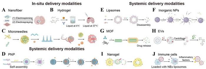
Figure 1.
The schematic diagrams summarize the drug loading and release mechanisms of both local and systemic delivery systems.

Engineering therapeutics for glioma: Multiscale drug delivery strategies from in-situ drug depots to advanced systemic approaches
Huarong Lai , Fenglin Xu , Mingjie Song , Yun Chen , Yi Jin , Jianping Zhou , Yang Ding , Huaqing Zhang


Among various tumors, glioma is the most common and fatal primary tumor in the central nervous system and one of the most aggressive tumors [1,2]. Standard glioma management involves maximal safe resection followed by radiotherapy and temozolomide (TMZ) chemotherapy [3]. Due to the high heterogeneity and invasiveness of glioma, it shows the proliferation of endothelial cells and angiogenesis around the tumor, and the boundary between the tumor and healthy brain tissue is not clear, surrounded by peritumoral edema and inflammatory cells [4,5]. While this multimodal approach extends survival to a certain extent, its efficacy remains severely confined: over 90% of gliomas recur within 2 years postresection due to infiltrative residual tumor cells [6,7]. Worse still, TMZ can only achieve subtherapeutic brain concentrations (< 20% plasma levels) owing to ATP-binding cassette (ABC) transporter-mediated efflux at the blood-brain barrier (BBB). Moreover, most tumor cells exhibit intrinsic TMZ resistance via O6-methylguanine-DNA methyltransferase (MGMT) overexpression [8,9]. For radiotherapy, radiation efficacy is further limited by radioresistant hypoxic niches and dose-limiting neurotoxicity, which might be ascribed to the complex tumor microenvironment (TME) and BBB [10,11]. These anatomical and molecular constraints collectively sustain a median survival of merely 14–16 months for glioma patients, thereby underscoring the urgency for advanced drug delivery solutions that can overcome the limitation of BBB and the TME [12].
Beyond these clinical constraints, intrinsic biological barriers critically undermine drug delivery efficacy [13]. The BBB and blood-brain tumor barrier (BBTB) constitute dual gatekeepers that systematically exclude therapeutics [14,15]. The BBB is composed of tightly joined endothelial cells (sealing 99% of paracellular pathways), astrocytic end feet, and efflux transporters [16,17]. Hence, > 98% of small-molecule drugs and virtually all macromolecular therapeutics are excluded by BBB [18,19]. More critically, in glioma, the BBTB exhibits pathophysiological duality [20]. More specifically, the focal hyperpermeability of BBTB in necrotic cores allows partial drug entry via enhanced permeation and retention effect (EPR) [21,22]. While aberrant functionality persists with upregulated ABC transporters and heterogeneous pore sizes [23]. And, interstitial fluid pressure surges to 40–60 mmHg, which is > 4-fold higher than the normal brain, creating convective counterflows that expel 92% of drugs [24,25]. Furthermore, efflux pumps highly expressed on various cell membranes, such as P-glycoprotein (P-gp), actively pump hydrophobic drugs (e.g., paclitaxel (PTX)) back into the bloodstream using energy-dependent transport mechanisms, drastically reducing drug concentrations in the brain [26]. Additionally, the high expression of cytochrome P450 in endothelial cells leads to the decomposition and inactivation of drugs before they can enter the brain tissue [27].
Moreover, glioma progression orchestrates a pathologically evolved microenvironment that actively subverts therapeutic efficacy through multifaceted mechanisms [28]. The immunosuppressive landscape that is dominated by tumor-associated macrophages (TAMs), constituting 30%–50% of the cellular mass and myeloid-derived suppressor cells (MDSCs), secretes interleukin (IL)-10 and transforming growth factor-beta (TGF-β) to establish an immunologically "cold" microenvironment [29-31]. In this immunosuppressive microenvironment, cytotoxic T-cell infiltration is reduced to < 5% of the tumor volume while checkpoint ligands are elevated. Concurrently, profound hypoxia upregulates hypoxia-inducible factor-1α (HIF-1α), inducing vascular endothelial growth factor (VEGF)-mediated aberrant angiogenesis and enhancing MGMT-driven chemoresistance [32,33]. This oxygen deprivation synergizes with lactate accumulation to trigger "ion trapping" of weak-base chemotherapeutics [34]. Critically, perivascular niches enriched with fibronectin and laminin shield therapy-resistant glioma stem cells (GSCs), exhibit 30-fold higher drug efflux capacity via ATP-binding cassette subfamily G member 2 (ABCG2) overexpression. This dynamically hostile microenvironment not only compromises conventional chemo-radiotherapy but also imposes barriers to drug delivery. Moreover, the glioma TME presents additional obstacles, including severe hypoxia, acidic pH, and a dense extracellular matrix (ECM), which further diminishes drug efficacy and limits penetration into the tumor core [35].
For overcoming physiological and pathological barriers, existing studies have proposed strategies to enhance drug delivery to brain tumor cells without relying on the circulatory system, completely bypassing the BBB to improve drug delivery to the brain tumor site [36,37]. These delivery techniques can provide a treatment plan with high in-situ drug activity concentration and low systemic concentration, thereby eliminating the limitations imposed by systemic toxicity and providing new tumor-selective large-molecule delivery strategies. In contrast, systemic delivery of drugs to the targeted site of glioma is highly challenging. Despite this, systemic delivery is an optimal approach for patients with tumors that cannot be surgically removed and can be repeated with more convenient dosing regimens [38,39]. Compared to invasive in-situ delivery, systemic delivery is easier to use. However, due to its physical and chemical properties and the heterogeneity of BBB and BBTB, few drugs can reach the glioma tumor site. These drugs alone are not sufficient to treat malignant glioma and require concurrent radiotherapy for combined treatment. Overall, existing drug delivery methods and postoperative rehabilitation treatment strategies for glioma present significant challenges. Glioma drug delivery therapy urgently requires more precise and effective approaches.
To overcome the multiple barriers hindering drug penetration across the BBB, recent research has focused on developing various novel systemic drug delivery systems (DDS) or local in-situ delivery approaches. Among these, local therapies bypass the BBB entirely by delivering drugs directly into the tumor cavity. Systemic delivery strategies involve loading glioma therapeutics onto carriers that employ "biomimetic" tactics to engage the BBB's physiological transport pathways and precisely circumvent its defense systems [40]. Nanoparticle (NP)-based DDS, for instance, leverage the enhanced permeability and retention (EPR) effect to increase drug accumulation at the brain tumor site. Furthermore, utilizing receptor-mediated transcytosis and active brain-targeting drug delivery strategies aims to enhance drug penetration across the BBB and achieve precise localization within the tumor. In summary, the core mechanisms by which nanocarriers breach the BBB include: designing carriers with an appropriate size to activate receptor-mediated transport or mimic the size of natural ligands; modifying the carrier surface charge to open tight junctions via adsorptive-mediated transcytosis; and inhibiting drug efflux pumps and protecting drugs from degradation. Additionally, numerous studies employ various surface modifications to enhance the drug's ability to cross the BBB and target tumor tissue, effectively delivering therapeutics to the diseased site while reducing systemic toxicity. Surface modification of carriers (e.g., ligand targeting, charge modulation) is a key strategy for regulating their interaction with the BBB and exploiting physiological pathways. Moreover, the size of the carrier is crucial for blood circulation and passive diffusion.
As the most prevalent and lethal primary intracranial malignancies, gliomas persistently defy conventional therapeutic paradigms of maximal resection, radiotherapy, and TMZ chemotherapy. And, these approaches are all critically compromised by the BBB and BBTB. To dismantle these pharmacological fortresses, this review critically deciphers next-generation delivery strategies through dual vectors: In-situ delivery modalities and advanced systemic delivery systems. By synthesizing advances across biodegradable implants, convection-enhanced delivery (CED), biomimetic NPs, and stimuli-responsive platforms, we establish a multiscale delivery framework addressing spatial challenges from macroscopic tumors to disseminated infiltrates. Furthermore, we delineate underdeveloped frontiers for clinical translation, this work ultimately provides a roadmap for engineered delivery solutions to breach the neuro-oncological survival plateau.
Direct in-situ drug delivery to the brain is a strategy to bypass the blood-brain barrier, which can increase the in-situ drug concentration in the tumor site and reduce or avoid systemic side effects, opening up new avenues for the precise delivery of more drug molecules [41]. In-situ delivery strategies include intracavitary implantation of various types of stents after tumor resection, direct intratumoral injection of active drugs, and in-situ convection-enhanced drug delivery therapy [42-44]. In recent years, researchers have drawn lessons from clinical trials and developed appropriate materials for drug delivery, focusing on the main factors contributing to tumor recurrence [45,46]. Given that up to 95% of glioma tumors rapidly recur within 2 cm around the resection cavity [47], in-situ drug delivery within the resection cavity has become one of the effective treatment options for glioma after surgery.
Various treatment methods have been developed, such as implanting drug-loaded hydrogels, nanofibers, or wafers in the tumor resection cavity. These DDS should possess features such as facilitating clinical translation, being easy to operate in clinical settings, being suitable for filling the brain microenvironment, having good biocompatibility, and reasonable dosing. Theoretically, bypassing BBB for in-situ drug delivery should act on all tumor cells, most in-situ delivery systems rely on the diffusion pathways of drugs to achieve effective drug concentration in the brain parenchyma, and the diffusion mode only covers a few millimeters around the delivery site [48]. Moreover, the lower drug diffusion rate may lead to higher local toxicity at the in-situ administration site [49]. Therefore, in addition to being able to cross BBB/BBTB, new delivery strategies should be able to enable drug diffusion in the brain tissue for effective treatment.
Despite its advantages, in-situ delivery modalities pose significant risks. Considering the metabolism of tumor cells and the clearance effect of chemotherapy drugs by brain tissue, in-situ administration requires a higher drug concentration, which may cause normal cells to be in a high-toxicity environment. This chapter will introduce the existing in-situ delivery modalities for glioma, and briefly describe their research status, advantages and disadvantages, and clinical translation applications from the structural and functional perspectives.
In-situ drug injection allows drugs to bypass the BBB and reduce systemic side effects. However, to date, most strategies for direct in-situ injection of drugs are still in clinical trials, primarily due to the brain's unique structural constraints and clinical implementation challenges [50,51]. The viscoelastic parenchyma generates backflow pressures during infusion, forcing > 40% of therapeutics to reflux along the catheter tract, while intraoperative brain shift exceeds the submillimeter precision required for safe drug distribution. Clinically, only 15%–20% of gliomas occupy anatomically accessible locations, and heterogeneous drug dispersion (coefficient of variation > 60%) prevents standardized therapeutic protocols.
Direct injection into the in-situ tumor cavity requires strict control of drug release. The special and complex brain structure and environment greatly affect the efficacy and safety of direct in-situ injection [52,53]. The glioma tissue area is complex, and drugs cannot evenly diffuse throughout the entire tumor area, especially the infiltrative growth edge area [54]. Hence, carriers need to be developed to enhance the penetration and sustained-release ability of drugs, or combined with tumor treatment fields to promote drug diffusion to the tumor edge [55]. Through the design of long-acting sustained-release preparations and responsive drug-release systems, drugs are prevented from being rapidly metabolized and cleared. In conclusion, direct in-situ injection treatment of drugs requires collaborative innovation through strategies such as optimizing delivery systems and upgrading precise drug delivery technology to break through the bottleneck of in-situ delivery modalities and ultimately achieve a more efficient and less toxic treatment mode.
Due to the limitations of systemic drugs and the technological progress in materials science, the research on in-situ drug delivery to the brain has become one of the promising strategies for treating glioma. As is well known, glioma cells have high invasiveness and infiltration ability and can penetrate brain tissues several centimeters away from the tumor tissue. Surgical resection cannot completely eliminate tumor cells [56]. The recurrence of glioma occurs at the tumor resection site after surgery, so in-situ drug delivery in the tumor resection cavity is of vital importance [57]. Moreover, the main components in the postoperative cavity are GSCs, reactive astrocytes, tumor-associated macrophages, microglia, and glioma-associated stromal cells. These different cells can interact with each other and drive the proliferation and migration of glioma cells [58].
High molecular weight biodegradable materials have good biocompatibility and can be degraded after implantation in the body and excreted through normal metabolic pathways, avoiding the risk of reoperation to remove the implanted materials [59]. And, high molecular weight biodegradable materials can be modified to change their surface properties or introduce bioactive substances to enable them to have specific biological functions [60]. As drug carriers, high molecular weight biodegradable materials can achieve controlled and targeted drug delivery, achieving the purpose of efficient and low-toxic drug delivery [61]. Hence, polymer-based biodegradable materials have been used to deliver drugs to the tumor resection cavity of glioma surgery to kill tumor cells, effectively prolonging the effect and duration of action of the drug at the target site and improving the therapeutic effect.
Implantable wafer represents an advanced class of programmable, long-acting platforms designed for sustained and localized drug delivery and release [62]. The implantable wafer is a biodegradable sheet that is surgically implanted into the tumor resection cavity. Usually, the polymer is used as the carrier, and the active drug is evenly dispersed in the polymer matrix to achieve local sustained release of chemotherapy drugs.
Direct delivery of chemotherapeutic agents into the surgical resection cavity of the tumor to achieve high in-situ activity and low systemic toxicity has become an important method for the treatment of glioma. Carmustine (BCNU) can be used in patients with newly diagnosed glioma and has been identified as the best candidate for local chemotherapy in recurrent glioma. BCNU wafers are polycarboxyphenoxy propane/sebacic anhydrides (PCPP: SA) wafers containing 3.85% BCNU that are placed on the surface of the tumor resection cavity to slowly release BCNU [63]. The wafer architecture protects carmustine from degradation and controls drug release, with slow release into the tumor cavity over 3 weeks [64]. Carmustine sheet (GliadelⓇ), which is implanted into the tumor cavity after surgery to remove the glioma tissue. Clinical data have shown that GliadelⓇ wafers are effective in prolonging the median survival of patients with glioma compared with placebo and are well tolerated without serious adverse effects [65,66]. Brem et al. found that topical delivery of TMZ via PCPP: SA sheets was superior to oral TMZ in a rodent glioma model, with an improved median survival [67]. This proves that the chip can provide local sustained drug delivery that needs further clinical exploration.
As a classic technique for in-situ drug delivery in glioma treatment, wafer implants have seen GliadelⓇ carmustine slow-release wafers used clinically for nearly 30 years. However, future clinical application still faces challenges and opportunities for evolution. Current therapeutic limitations include the restricted drug diffusion range, reaching only 3–5 mm around the implant, and the significant rigidity of the wafers, potentially contributing to postoperative complication risks. The future clinical application urgently needs to solve its rigidity problem, which may focus on multi-drug loaded wafers, stimuli-responsive smart wafers, and customized implantation using three-dimensional (3D) printing technology for long-acting drugs [68].
Nanofibers are microfibrous materials with a diameter of the nanometer scale (usually 1–1000 nm). They can form thin films or 3D scaffolds with extremely high specific surface area, controllable porosity, and good biomimetic ECM structure. Typically fabricated via electrospinning of biocompatible polymers, nanofiber-based implantable systems offer a versatile platform for localized and sustained drug delivery.
The commonly used methods for preparing nanofibers include the template method, tensile method, direct deposition method, and electrospinning [69-71]. Among them, the commonly used electrospinning technology can be used to produce polymers into ultrafine fibers with a diameter of nanometers to micrometers, which has a wide range of applications in biomedicine and drug delivery [72-74]. Therapeutic agents can be incorporated into electrospun nanofibers by blending, coaxial, and emulsion electrospinning. Studies over the years have shown that nanofiber-based delivery of drugs such as TMZ, carmustine, doxorubicin, and PTX can significantly reduce systemic side effects while delivering sufficient drug doses to precise tumor sites [75]. Furthermore, the engineering of nanofibers could allow the co-delivery of multiple therapeutic agents to meet the clinical needs of treating malignant glioma.
Composite nanofiber mesh can achieve safe and sustained drug release in the in-situ tumor tissue, can efficiently encapsulate chemotherapy drugs or active drugs, and simultaneously realize chemotherapy, radiotherapy, and molecular targeted therapy [76,77]. Li et al. proposed a biodegradable composite nanofiber (NFM) loaded with TMZ and 17-allylamino-17-demethoxygeldanamycin (17 AAG) simultaneously for chemotherapy and radiotherapy sensitization, which showed synergistic cytotoxic effects [78]. In addition, in-situ application of NFM could deliver drugs to the tumor site multiple times, resulting in long-term sustained drug release, which was expected to solve the problem of rapid postoperative recurrence of glioma.
Nanofibers in glioma treatment hold clinical promise due to their unique biomimetic 3D structure and precise drug release control, potentially overcoming the diffusion limitations of traditional implants. Constructed via techniques like electrospinning, nanofiber scaffolds can mimic the brain's ECM. Their drug loading capacity is significantly higher than wafers, enabling co-loading of multiple drugs for synergistic effects or cytokines to remodel the immune microenvironment, converting "cold tumors" into "hot tumors". Future clinical application requires improved operability during implantation and surface modification to address long-term inflammatory responses, positioning them as a promising next-generation local implant delivery technology.
Hydrogel implants exploit their tunability and biocompatibility as versatile drug depots for glioma therapy. The materials for synthesizing hydrogels mainly include natural materials such as gelatin, hyaluronic acid (HA), alginate, and dextran, as well as synthetic hydrophilic polymer materials such as polyethylene glycol (PEG), poloxamers, etc., and polyacrylamide (PAM) [79,80]. Hydrogels have pore structures and microchannel structures, enabling them to transport drugs or deliver cells [81,82]. Hydrogels form stable networks via chemical cross-linking (covalent bonds) or non-covalent interactions (e.g., ionic, hydrophobic, hydrogen bonding) [83-85]. Their high-water content mimics brain tissue elasticity, enhancing interfacial integration while enabling sustained release of chemotherapeutics) and biologics directly to residual tumor beds post-resection [86,87].
Hydrogel grids can be decomposed in response to external stimuli, as well as internal physiological factors such as glucose, pH value, enzyme activity, temperature, and redox potential to release drugs controllably [88-90]. Furthermore, hydrogels offer minimally invasive injection conforming to irregular margins, TME-triggered responsive release, and synergistic co-delivery to overcome treatment resistance [91-93].
After the surgery of primary glioma, a hydrogel implant can be placed in the surgical cavity to inhibit the recurrence of glioma. Dong et al. developed a hydrogel drug delivery system encapsulated with intracavitary sprayable nano-regulators (NRs) for regulating the cholesterol metabolism of glioma-supporting macrophages (GSMs) [94]. The sprayable hydrogel provided great application advantages in the postoperative tumor cavity, enabling the direct and high-throughput administration of drugs to the uneven surfaces of the entire target site. This research on the treatment in both in-situ glioma models and post-recurrence models showed good anti-tumor effects, proposed a novel drug delivery strategy, and is worthy of further clinical trials. Detailed introduction and illustrations refer to Section 2.3.3 and Fig. S1 (Supporting information).
Hydrogels in glioma treatment offer significant clinical potential primarily because of their "injectable, smart, sustained-release" properties. Their ability to gel in-situ after injection enables precise spatiotemporal drug control, potentially solving the diffusion and rigid-trauma issues of traditional implants. The injectable liquid precursors allow hydrogels to perfectly conform to irregular tumor cavities, greatly improving drug coverage. Hydrogel networks can also diffuse up to 1 cm into the peritumoral area. Key advantages include the design of smart stimuli-responsive release systems and the capacity for chemo-immunotherapy synergy, significantly extending patient survival. Hydrogels already in clinical trials for postoperative cavities suggest utility in treating recurrent glioma, potentially replacing repeat craniotomies. Hydrogels are poised to become a highly promising first-choice local therapeutic strategy for glioma [95].
MNs are miniaturized biocompatible devices (50–1000 µm length) that enable precise glioma therapy post-resection [96,97]. In recent years, they have received widespread attention in the application of in-situ delivery modalities for glioma. By loading drugs onto biodegradable materials, MNs are implanted into the brain cavity after tumor resection [98]. Using biodegradable materials (such as HA, and gelatin methacryloyl hydrogel) can avoid the need for a second surgery to remove them and reduce the risk of infection [99,100]. Glioma is a malignant tumor, and a combined treatment strategy can effectively improve the median survival time of patients.
MNs can serve as multifunctional carriers, simultaneously loading chemotherapy drugs, immune checkpoint inhibitors (such as anti-programmed cell death 1 antibodies (aPD-1)), and photothermal conversion agents (such as gold nanorods), achieving a synergistic effect of chemotherapy-immunotherapy-photothermal therapy [101,102]. Experimental studies have shown that near-infrared light-activated photothermal MNs can induce in-situ hyperthermia ablation of tumors and release immunoadjuvants, activating tumor-specific T-cell responses.
Wang et al. developed a heterogeneous, heterogeneous, and biodegradable microneedle patch (SMN) based on silk protein for the treatment of glioma. This patch could simultaneously carry three drugs, loading two clinically relevant chemotherapy drugs (thrombin and TMZ) and a targeted drug (bevacizumab) into the SMN patch, releasing them directly to the tumor site by bypassing the BBB [96]. The SMN patch has the function of on-demand multi-drug delivery and has the potential for combined treatment of glioma. Detailed introduction and illustrations refer to Section 2.3.4 and Fig. S2 (Supporting information).
The MN technology provides new ideas for the treatment of glioma by offering precise in-situ drug delivery and minimally invasive operation. However, there are still some limitations, such as the relatively short length of MNs, which makes it difficult to penetrate the blood-brain barrier or reach deeper tumor-infiltrated areas. There are also hardware technical limitations for treating tumor cavities after surgery, and MNs are difficult to cover all tumor cells, resulting in more tumor cells remaining and leading to the recurrence of glioma. Although currently mainly laboratory research, its potential in improving efficacy and reducing toxicity has been preliminarily verified. Future research needs to further optimize its penetration depth and drug loading efficiency and verify its long-term safety through clinical translation, potentially becoming an important tool in neuro-oncology.
Implantable MNs for glioma treatment offer minimally invasive barrier penetration and multi-dimensional control capabilities. By penetrating the blood-brain barrier to target deep infiltrative lesions, they achieve precise delivery and real-time monitoring unattainable with traditional implants. They directly penetrate peritumoral tissue to treat deep-seated lesions, and their biodegradable nature avoids the need for secondary removal surgery, offering distinct advantages for deep and recurrent gliomas [103].
MOF is an extended porous network material composed of metal nodes (metal ions or metal clusters) and an organic skeleton from the bottom up [104]. MOF can encapsulate cargo and protect cargo in the self-assembly process. Or by mesoporous permeation, covalent or noncovalent processes in mesoporous MOF structures that bind the cargos to the surface, allowing the cargos to remain active under denaturant, high temperature, unnatural pH, and organic solvent conditions [105].
In addition, other materials can be introduced into the cargos-MOF composite to form a multicomponent composite. Yalamandala et al. investigated and developed an implantable self-cascade catalytic antigen-capture scaffold (CAS) that combines the catalytic activity of MOFs with the antigen-capture ability of polyurethane-polyethylene glycol-polypropylene glycol (PU-EO-PO) [106]. This implantable self-cascade catalytic antigen capture scaffold was equivalent to an in-situ-formed personalized vaccine that stimulates the body's immune response for long-term antigen release and sustained immune enhancement with excellent therapeutic potential. Detailed introduction and illustrations refer to Section 2.3.5 and Fig. S3 (Supporting information).
MOFs face rigidity-related technical challenges in glioma treatment, requiring breakthroughs via material design and engineering strategies. The mismatch between the crystalline brittleness of MOFs and the extreme softness of brain tissue can cause lattice fracture during injection, leading to premature drug leakage. Their rigidity also risks compressing brain tissue and triggering inflammation. Current research focuses on flexibility modification and using composite materials to improve rigidity, gradually progressing from the laboratory towards the clinic [107].
Based on the aforementioned delivery issues faced by glioma treatment, various schemes have been proposed to deliver drugs to brain tissue and tumor sites. Among them, CED is an in-situ drug delivery technology that directly delivers drugs to the target tissues [108,109]. CED technology was first proposed by Bobo et al. in 1994 [110]. It bypassed the BBB by inserting a catheter and delivering drugs directly to the target area without relying on the circulatory system. It accurately delivered drugs to the target area and used a hydraulic device for continuous drug delivery. Under the combined drive of pressure gradient and concentration gradient, the drug distribution in brain tissue was enhanced [111]. In recent years, studies have shown that compared with systemic treatment, local CED significantly enhances the efficacy of drugs, macromolecules, and nanocarriers delivered to the brain.
In recent years, enhanced perfusion delivery has been applied in the treatment of glioma with clinical practice protocols. Annick et al. evaluated a phase I clinical trial using CED for intratumoral delivery of recombinant immunotoxin D2 C7-IT, and the results indicated that CED delivery might become a treatment option that could effectively prolong patients' survival [112]. In addition, Sandeep et al. evaluated a CED-based delivery of recombinant protein cintredekin besudotox (CB) composed of truncated forms of IL-13 and Pseudomonas exotoxin (PE 38 QQR) in patients with recurrent glioma [113]. At the maximum tolerated concentration, tumor necrosis was observed, and the infusion for up to six days was well tolerated.
However, there are still obvious side effects in clinical use, such as headache, brain edema, fever, cerebral hemorrhage, and epileptic seizures [114]. Limitations in catheter design and placement, real-time delivery imaging, and computational modeling hinder the clinical research of CED delivery technology for glioma. New CED technologies need to be developed, catheter designs updated, reflux obstacles overcome, and adverse reactions from invasive surgery minimized. More precise imaging methods should be used to detect the drug delivery process, making CED a potentially effective delivery technology.
CED holds clinical promise for glioma treatment due to its ability to achieve broad coverage and infiltrative targeting [115]. By overcoming the physical limitations of passive drug diffusion through continuous positive-pressure infusion, it directly addresses the fundamental challenge of gliomas' infiltrative growth. Using microcatheters for continuous perfusion, CED delivers drugs to regions larger than 5 cm3. It enables broader, more precise, and controllable delivery of diverse drugs and has the potential to evolve from a perfusion tool into an advanced platform for infiltrative, precision drug delivery [116].
The in-situ drug delivery system for the treatment of glioma is shown in Table S1 (Supporting information).
Systemic drug delivery is a means of drug delivery to the brain and tumor site through blood circulation [117]. However, the BBB can protect the brain from foreign substances and maintain the stability of the intracerebral environment, and most drugs cannot penetrate the BBB to reach the glioma site to exert good therapeutic effects, which is a major challenge for systemic drug delivery in glioma. In recent decades, nanotechnology has rapidly developed and made remarkable progress in site-specific delivery, including brain delivery, and a variety of NPs have become carriers for tumor diagnosis and therapy, providing a potential solution for glioma treatment [118]. Nanocarriers deliver substances, including small molecules, peptides, proteins, nucleic acids, or mixtures of the above. Studies have shown that nanocarriers can penetrate the brain through EPR effects and overcome the limitations of the delivered substances to increase the drug concentration at the site of glioma and provide a favorable therapeutic effect [119,120]. As shown in Table S2 (Supporting information), we compared the advantages and disadvantages of non-targeted DDS to facilitate a more in-depth study of the delivery advantages and limitations of different nanocarriers. Some of the nano delivery systems for the diagnosis and treatment of glioma will be highlighted below.
PNPs are designed from biocompatible and biodegradable natural or synthetic polymers with diameters ranging from 1 nm to 1000 nm. Compared to other nano delivery routes, PNPs have various advantages. For example, PNPs share merits in size customization and consistency that can be tailored to a precise size range (1–1000 nm) and maintain a very high degree of dimensional consistency, which is critical for the precise regulation of DDS [121]. Then, PNPs possess structural design diversity that can be designed into a variety of unique structures, including rods and disks, and such structural innovations can significantly enhance the effectiveness of drug delivery [122]. PNPs can be designed with stimulus-responsive to specific endogenous stimuli (e.g., pH, redox state, enzyme environments, and temperature) to achieve drug release at the target site, thereby optimizing therapeutic effects [123]. PNPs can be further functionalized to introduce various sizes of bioadducts, such as targeting ligands and receptors, to enhance their targeting ability by using the body's natural mechanisms to improve the efficiency of drug delivery. Moreover, PNPs are not only capable of delivering a variety of different substances but also have a high drug loading capacity, which makes them more effective and practical in drug delivery applications [124].
In addition to loading TMZ, the researchers have combined gene drugs with PNPs for the treatment of gliomas, utilizing the efficient drug delivery capabilities of PNPs with the specific mechanism of action of gene drugs to enhance therapeutic effects. This innovative research idea breaks through the limitations of traditional single-target therapy and significantly improves the therapeutic effect of glioma by simultaneously regulating multiple key signaling pathways.
Taken together, these studies demonstrate the potential of PNPs in the treatment of gliomas and the possibility of personalized therapy through different modifications and loads. Through these studies, we can see that the use of PNPs in the treatment of gliomas is moving towards greater precision and efficacy, offering new therapeutic hope for patients.
In nanoscale DDS, polymeric micelles, which are one of the hot topics of discussion due to their good stability and encapsulation efficiency, consist of diblock (hydrophilic-hydrophobic) or triblock (hydrophilic-hydrophobic-hydrophilic) polymers that form amphiphilic polymers [125]. Polymeric micelles are commonly used to deliver hydrophobic drugs, the hydrophilic shell in the structure can prevent the aggregation and precipitation of micelles and protect the core drug from being recognized and cleared by the reticuloendothelial system (RES), while the hydrophobic core can effectively encapsulate water-insoluble drugs in the core of polymeric micelles, which is a special amphiphilic structure that can greatly improve the bioavailability of drugs [126-128]. In addition, the size of polymeric micelles ranges from 10 nm to 100 nm, which makes them less likely to accumulate in organs [129], and the small size of polymeric micelles can passively target solid tumors for more effective cellular internalization [130,131]. Therefore, polymeric micelles can be used as a good delivery vehicle for drug delivery in biological systems.
In the treatment of gliomas, polymeric micelles are not only a powerful tool for improving the stability and targeting of chemotherapeutic agents but also play a key role in the delivery of proteins or oligonucleotide-based therapeutics such as plasmid DNA, siRNA, and mRNA. These nucleic acid-based therapeutics are susceptible to nuclease degradation in vivo and therefore require an effective delivery system to protect them, maintain their structural integrity, and ensure that they can safely reach their target sites.
The cation-free siRNA micelles developed by Jiang et al. not only prolonged the blood circulation time of siRNAs but also improved cellular uptake and effective release of siRNAs at the tumor site. These micelles were formed by self-assembling siRNA-SS-poly(N-isopropylacrylamide) (PNIPAM) diblock copolymers, which, due to their non-cationic nature, avoided the toxicity problems associated with cationic carriers while achieving effective BBB penetration [132].
Unlike the former which focuses on controlled release mechanisms, Zhang et al.'s study mainly focused on the construction of multifunctional nanomicellar systems and their immunosynergistic therapeutic mechanism. They demonstrated an innovative strategy to integrate antibodies and chemotherapeutic agents into a single micelle (angiopep2-aPD-L1@PTX) by crosslinking an immune checkpoint inhibitor anti-PD-L1 antibody (aPD-L1) and electrostatically binding PTX to the amino group of PEG-poly-L-lysine (PEG-PLL) nano micelle (A2-APM) to enhance the immune response against glioma. This co-encapsulation strategy not only improves drug delivery efficiency but also enhances the immune response by PTX-induced immunogenic cell death (ICD) of chemotherapeutic agents, thereby improving therapeutic efficacy [133]. Detailed introduction and illustrations refer to Section 3.1 and Fig. S4 (Supporting information).
Taken together, these studies provide insight into the diverse applications of polymeric micelles in the treatment of gliomas, with the studies by Jiang et al. and Zhang et al. demonstrating the use of micelles for siRNA delivery and co-encapsulation of immune checkpoint inhibitors and chemotherapeutic agents. These studies demonstrate that polymeric micelles not only improve the stability and targeting of chemotherapeutic agents but also effectively protect and deliver nucleic acid-based therapeutic agents, providing a new strategy for the comprehensive treatment of gliomas.
In recent decades, nanotechnology has developed rapidly, and dendritic polymers, with their excellent dispersion properties and rich and diverse functional groups, have therefore stood out among the many nanocarriers and attracted much attention [134,135]. Dendrimers are monodispersed spherical macromolecules and their sizes are usually in the range of 1–100 nm. The synthesis of dendritic polymers can be precisely controlled at each step, so their structure and size can be precisely controlled at the molecular level to achieve monodispersed molecular weights with extremely narrow molecular weight distributions. In addition, the abundance of functional groups inside and outside the dendritic macromolecules and the designability of their structures make them promising for drug delivery applications [136-138].
In the therapeutic field of glioma, dendritic polymers, as a new type of drug carrier, due to their unique structure and function have received widespread attention and have now been proven to be the ideal carrier for the treatment of tumors, to improve the efficiency of drug delivery and achieve targeted therapy [139]. The most studied dendritic polymer is the dendritic macromolecule poly(amidoamine) (PAMAM). For example, Zhang et al. found that dendritic polymers could be rapidly and uniformly distributed throughout the solid tumor and peritumor regions, and subsequently accumulated and retained in TAMs by systemic administration of PAMAM dendritic polymers in a glioma model [140]. This finding suggested that dendritic polymers could selectively target cells associated with neuroinflammation, providing new opportunities for the treatment of glioma. This study demonstrated the distribution characteristics of dendritic polymers in tumor tissues and the ability to target TAMs, providing new perspectives for immunomodulatory therapy [141].
The main contribution of this study was to provide a novel strategy to enhance drug retention and penetration by exploiting the properties of the TME to improve therapeutic efficacy. They have leveraged the unique properties of dendritic polymers to improve the treatment of glioma, and demonstrated the potential of dendrimers to improve drug delivery efficiency, enhance tumor tissue penetration, and enable targeted therapy.
Nanogels are polymers with a 3D configuration consisting of a 3D spherical network formed by chemical or physical crosslinking of polymer rearrangements [142]. The acceptable size range of nanogels is generally defined in the range of 10–1000 nm, with a typical range of 20–250 nm. The smaller nanosize gives nanogels a larger specific surface area, greater stability of the loaded drug, and an additional targeting advantage to selectively accumulate in solid tumors through enhanced penetration and retention effects [143-145].
Nanogels have been applied in several research areas, such as disease diagnosis [146] and sensing [147], with the field of drug delivery being the most affected [148]. Nanogels are capable of releasing drugs from 3D structures through release mechanisms such as pH response, thermal and volume conversion, photoisomerization, or photochemistry [149].
In addition, the systemic delivery of nanogels combined with immunotherapy is one of the most promising strategies in the treatment of glioma at present. The virus-mimicking nucleic acid nanogels provided a versatile and convenient platform to successfully treat a wide range of diseases due to their noninvasive nature and effective delivery capabilities [150].
Nanogels have high water content and excellent biocompatibility. The above study provides an excellent solution for difficult-to-treat diseases such as glioma through biomimetic technology, intelligent response release, and immune microenvironment modulation, which has the clinical potential to be expanded to multiple disease types, creating a promising platform for the treatment of glioma.
Liposomes are bilayer lipid vesicles made of lipid materials such as phospholipids and cholesterol, usually 20 nm to 1 µm in size, and are ideal and safe DDS [151,152]. The methods used to prepare liposomes include film dispersion, reverse evaporation, pH gradient, ammonium sulfate gradient, and other conventional methods.
As an ideal drug delivery system, the preparation techniques and applications of liposomes have been extensively studied and developed in recent years. In addition to the conventional preparation methods and new technologies mentioned above, the functionalized modification of liposomes has also become a research hotspot. Through surface modification, liposomes can realize more precise targeted delivery and longer in vivo circulation time. For example, PEG-modified liposomes (PEGylated liposomes) can effectively avoid recognition and clearance by the RES, thus prolonging their circulation time in the bloodstream and improving drug bioavailability [153]. In addition, the introduction of targeting ligands (e.g., antibodies, peptides, glycans) can enable liposomes to specifically recognize diseased cells or tissues for active targeted delivery [154]. Muhammad Ismail et al. developed an ApoE-functionalized liposome nanoplatform based on the co-loading of artesunate-phosphatidylcholine (ARTPC) with TMZ (ApoE-ARTPC@TMZ) for the targeted treatment of TMZ-resistant U251-TR glioma. Molecular weight limitations and lack of endogenous transporter recognition restrict ART diffusion across the BBB. Efflux pumps (e.g., BCRP) further reduce brain concentrations. Carrier-mediated targeting is required. In vitro experiments demonstrated that artesunate enhanced the chemosensitivity of glioma to TMZ by impeding DNA repair through inhibition of the Wnt/β-catenin pathway. In addition, the targeted liposome efficiently penetrated the BBB through low-density lipoprotein receptor (LDLR)-mediated transcytosis and achieved deep tumor penetration and retention. In vivo, results showed that ApoE-ARTPC@TMZ significantly inhibited U251-TR tumor growth and significantly prolonged the survival of mice while attenuating the systemic toxicity of TMZ by reducing the dose. This novel combination strategy has demonstrated preclinical potential in brain tumor therapy [155]. In summary, liposomes are expected to become a pillar delivery tool for the comprehensive treatment of gliomas through the optimization of targeted modification technologies such as ApoE and the combination of chemotherapy, gene therapy, and other therapies.
Inorganic NPs usually refer to nanoscale particles with sizes between 1 nm and 100 nm, consisting of inorganic components such as metals, metal oxides, semiconductors, or carbon-based materials. Inorganic NPs have unique material and physicochemical properties, for example, magnetic iron oxide NPs, gold NPs, quantum dots, and other inorganic NPs have their own unique optical and electronic properties, which have been widely studied and applied to disease diagnosis and imaging, and have greatly contributed to the development of the biomedical field [156,157].
Surface functionalization of inorganic NPs is a crucial step for their wide application. Through surface modification (e.g., ligand exchange or biomolecular coupling), the dispersibility, colloidal stability, and biocompatibility of NPs can be precisely tuned to influence optical, magnetic, and catalytic properties while conferring the ability to specifically interact with biological systems [158]. For example, the blood circulation time of gold NPs modified with PEG can be extended to > 24 h, which significantly improves the efficiency of the EPR effect, while quantum dots can be covalently linked to antibody or nucleic acid probes by carboxyl/amino functionalization to achieve ultrasensitive fluorescent labeling of cell membrane receptors [159].
For maximal safe surgery, Xie et al. focused on magnetic resonance imaging (MRI)/optical imaging, which combines MRI and optical imaging, to obtain more accurate preoperative diagnostic information. The team chose DSPE-PEG2000 to modify superparamagnetic iron oxide NPs (SPIONs), maintain their colloidal stability and monodispersity in aqueous solution, and prolong the in vivo cycling time of SPIONs. Peptides like DANG cannot passively cross the BBB due to their size and strong hydrophilicity. The team coupled the peptides DANG and Cy7 dyes to the end of the PEG by condensation reaction to obtain the DANG/Cy7 bimodal SPIONs probe. The experimental results showed that the probe exhibited excellent tumor homing properties both in vivo and in vitro, and accumulated at high levels at the margin of gliomas, which could indicate the tumor margin and provide an important reference for preoperative diagnosis and intraoperative localization [160]. Inorganic NPs, with their adaptable and diverse physicochemical properties, provide a multimodal diagnostic platform and a new engine for the treatment of gliomas, despite the remaining limitations, such as poor storage stability and induced immunogenicity.
MOFs are a class of porous crystalline materials formed by the self-assembly of metal ions or metal clusters with organic ligands, which have the advantages of rich backbone types, large specific surface area, many functional groups, and stable frameworks [161-163]. These properties have made researchers extremely interested in MOFs, which have become one of the hotspots for research as they show a wide range of applications in many fields such as bioimaging, drug delivery, chemical sensing, and cancer therapy [164-166]. The structure of MOFs can be divided into four levels. The first level includes metal ions (nodes) and coordination ligands (junctions) [167]. The second level is a relatively rigid geometry formed by multiple ligands and coordination points of metal ions. The third and fourth levels are the internal framework of the MOF and the external morphology (including size, shape, and orientation) of the MOF, respectively.
In addition, MOFs can construct nanoplatforms combined with immunotherapy. Huang et al. developed a copper-based nanoplatform BSO—CAT@MOF-199@DDM (BCMD), to mediate cuproptosis and enhance immunotherapy for glioma. The process was designed to bypass the BBB through intranasal administration, reducing invasiveness and systemic adverse effects of the drug, greatly enhancing the immunogenicity of dying cells and inducing tumor immunotherapy. Moreover, in the GL261 glioma tumor model, the BCMD+ αPD-L1 group significantly promoted the maturation of dendritic cells (DC) and had the highest number of CD8+ T cells, indicating that BCMD can also synergize with αPD-L1 to reverse the immunosuppressive TME [168]. Detailed introduction and illustrations refer to Section 3.7 and Fig. S5 (Supporting information).
As a novel porous material, MOFs show groundbreaking potential in the field of glioma therapy: Their designable pore structure and high drug-carrying capacity provide an innovative solution to overcome the BBB. Although core challenges such as biocompatibility optimization and scale-up preparation still need to be overcome, the unique advantages of MOFs in terms of synergistic treatment and diagnostic integration make them promising to be a transformative delivery platform for recurrent gliomas, providing patients with next-generation therapeutic options that combine precision and efficiency.
EVs are nanosized phospholipid bilayer vesicles that can be secreted by a wide range of cells (e.g., tumor cells, immune cells, and mesenchymal stem cells), and their sizes range from 40 nm to 160 nm. EVs can be classified according to different criteria, such as size, type, density, or whether they have been artificially modified. According to their types, they can be categorized as exosomes, microvesicles (MV), and apoptotic bodies (AB) [169-171].
Exosomes are considered to be a key factor in intercellular communication. Exosomes have the natural ability to penetrate the BBB because they can undergo receptor-mediated endocytosis via transferrin receptors and can also directly fuse with the cell membrane.
AB is a membrane vesicle secreted from fragmented apoptotic cells, which is produced efficiently and inexpensively. The high expression of phosphatidylserine (PS) on its surface allows ABs to be recognized and endocytosed by monocytes/macrophages (MA) and deliver drugs to the tumor site via MAs [172,173].
The rapid development of EVs has brought them into the limelight in the field of nanodrug delivery. However, the complexity of the TME, as well as the stability and targeting of EVs in vivo, pose significant challenges for the effective implementation of EVs. In addition, the complexity of the isolation and purification process of EVs and the difficulty of quality control and standardized production due to the nature of their natural origin limit the possibility of their widespread application to some extent. Nevertheless, EVs remain an effective treatment for gliomas.
Cell-mediated DDS, a therapeutic approach that uses cells as carriers to deliver drugs directly to the tumor site, have received widespread attention for their ability to improve drug efficacy and therapeutic specificity in tumor therapy. Studies have shown that cell-mediated bionic DDS can actively penetrate the BBB and selectively enrich in the tumor domain, thus achieving precise drug delivery and increasing drug effectiveness [174]. Among them, immune cells are particularly prominent for their ability to naturally traverse the BBB and be recognized and recruited to the lesion site [175].
Neutrophils (NE) have the innate ability to penetrate the BBB/BBTB and gliomas. When a tumor is surgically resected, it causes local inflammation and releases inflammatory factors, which activate NE to migrate to the inflamed area, thereby effectively enhancing targeting of brain tumors [176].
In addition to NE, macrophages have also demonstrated efficient drug delivery. As a natural immune cell and antigen-presenting cell, macrophage not only possesses the ability to phagocytose NPs, but also have a long plasma circulating half-life, which helps to overcome the metabolic clearance of drugs in the somatic circulation [177,178].
The advantage of immune cell drug delivery lies in its ability to utilize the natural targeting ability of immune cells to achieve precise drug delivery while reducing damage to normal cells. However, this approach also faces some challenges, such as the limited loading capacity of immune cells and the possible triggering of an immune response. Nevertheless, immune cell delivery of drugs has a promising application in the treatment of gliomas.
The systemic drug delivery system for the treatment of glioma is shown in Table S3 (Supporting information).
In the treatment of glioma, the development of a drug delivery system is the key to improving the therapeutic effect. A variety of nanocarriers, with their unique physicochemical and biological properties, achieve effective drug delivery through different mechanisms, thus demonstrating their respective advantages and limitations in glioma therapy.
Each of these carriers has its advantages, but their biocompatibility, drug-carrying capacity, targeting efficiency, and preparation cost need to be taken into account in order to select the most suitable drug delivery system for practical applications.
The therapeutic efficacy for glioma is severely constrained by the triple core bottlenecks of DDS. The primary obstacle originates from the barrier mechanisms of the BBB/BBTB, whose tight junction structures and efflux transporters (e.g., P-glycoprotein) block over 98% of small-molecule drugs and most macromolecular drugs from entering the brain, resulting in critically insufficient drug concentrations at invasive tumor margins. Then, inherent limitations exist in current delivery strategies: during systemic administration, intratumoral drug concentrations are often < 0.1% of plasma concentrations, primarily due to efflux clearance effects, making it difficult to achieve effective therapeutic drug concentrations in the brain. More critically, the clinical absence of real-time pharmacokinetic monitoring tools prevents precise treatment adjustments, as conventional imaging cannot reliably distinguish true tumor progression. These limitations, combined with TME heterogeneity-including rapid expansion of drug-resistant glioma stem cells and narrow therapeutic windows causing dose-limiting systemic toxicities-collectively undermine treatment feasibility. Existing local and systemic delivery strategies still exhibit significant limitations.
This review introduces seven local delivery strategies, such as intraoperative implantable wafers, localized nanocarrier implants, and CED. While bypassing the BBB to achieve higher drug concentrations at tumor sites, their drug distribution is frequently compromised by catheter placement inaccuracies, tumor tissue heterogeneity, and restricted carrier diffusion, failing to effectively cover infiltrating tumor cells and distant microsatellite lesions. Repeated surgical or invasive procedures carry additional risks of inflammation.
Moreover, this review examines nine systemic nanocarrier systems for glioma treatment, each possessing distinct characteristics and promising therapeutic potential. Selection criteria should balance biocompatibility, drug-loading capacity, and BBB penetration capability. Among natural carriers, immune cells and EVs offer superior biocompatibility and innate targeting, demonstrating significant advantages in BBB traversal and tumor targeting, yet their drug-loading capacity is generally limited by factors like cell viability.
In the treatment of gliomas, the effectiveness of DDS primarily depends on their drug-loading capacity and release mechanisms. Different carriers exhibit significant variations in drug-loading efficiency, release rate, targeting ability, and biocompatibility. Fig. 1 summarizes the drug-loading and release mechanisms of representative carriers, including hydrogels, liposomes, polymeric NPs, and exosomes, illustrating their responsive release characteristics under pathological conditions. This comparison helps clarify the advantages and limitations of each carrier type.
To overcome the multiple barriers of drug penetration across the BBB, recent research has focused on developing novel systemic DDS. These carriers employ "biomimetic" strategies to engage BBB transport pathways while evading its defense systems, leveraging the EPR effect to augment drug accumulation in brain tumors. Core mechanisms for nanocarrier BBB penetration include: (1) Designing appropriate carrier sizes to activate receptor-mediated transcytosis or mimic natural ligand dimensions; (2) modifying surface charge to facilitate adsorptive-mediated transcytosis via transient tight junction opening; (3) inhibiting drug efflux pumps and protecting drugs from degradation. Surface functionalization remains a critical strategy for modulating carrier-BBB interactions and exploiting physiological pathways to efficiently deliver therapeutics to lesions while reducing systemic toxicity.
A summary and comparison of the key benefits and limitations of prominent brain-targeted delivery strategies is presented in Table S4 (Supporting information).
Furthermore, cutting-edge research is advancing DDS toward intelligent delivery, synergistic therapy, and dynamic monitoring paradigms. In carrier engineering, bio-derived nanocarriers show BBB-penetrating potential: genetically engineered exosomal surface proteins enable high-biocompatibility active targeting, while mesenchymal stem cells facilitate barrier traversal. Meanwhile, stimuli-responsive nanosystems (e.g., pH/reactive oxygen species (ROS)-sensitive polymeric micelles) enable spatiotemporally controlled drug release at tumor sites. To overcome tumor heterogeneity, multi-mechanism synergistic platforms co-deliver chemotherapeutics, immunomodulators (anti-PD-1 antibodies), and epigenetic regulators (MGMT promoter-targeting siRNA) via liposomal/dendrimer hybrids, achieving multi-pathway combination therapy. Next-generation glioma DDS hold promise to overcome traditional therapeutic bottlenecks and fundamentally transform the treatment paradigm.
As the most common malignant tumor in the central nervous system, glioma is characterized by high aggressiveness, high recurrence rate, and poor prognosis. For the treatment of glioma, traditional methods such as surgery, radiotherapy, and chemotherapy are often used in the clinic at present, which can only control the tumor progression to a certain extent. Due to the existence of BBB and BBTB as well as the high heterogeneity of the tumor, it is difficult to ensure that the drugs enter the brain and accumulate at the tumor site to exert the therapeutic effect. By bypassing the BBB and using direct brain drug delivery, the drug concentration at the tumor site can be greatly increased, reducing the toxic side effects caused by systemic drug delivery and opening up a new way of drug delivery.
At present, new in-situ therapeutic methods such as the implantation of nanofibers, wafers, hydrogels, and other drug carriers, convective enhanced delivery, and intratumoral injection libraries have emerged in the clinic. Among them, the wafer implantation technology and enhanced convective delivery technology hold promise in clinical practice. In summary, overcoming the therapeutic impasse in glioma demands disruptive strategies to address the triad of BBB impermeability, infiltrative recurrence, and immunosuppression. While localized implants (e.g., hydrogels, wafers) bypass systemic limitations yet suffer from restricted drug diffusion, and circulating nanocarriers enhance targeting but falter in heterogeneous tumor distribution, the next paradigm might lie in harnessing the glymphatic network of the brain for deep-penetrating lymphatic delivery and developing theranostic implants capable of real-time monitoring and adaptive treatment. To accelerate clinical translation, we advocate: (1) Engineering lymphotropic nano vectors (e.g., CCL21-functionalized systems) to exploit perivascular transport; (2) integrating stimuli-responsive drug reservoirs with imaging agents (e.g., MRI/fluorescence-guided hydrogels) for closed-loop "diagnose-treat-monitor" cycles; and (3) prioritizing material-based designs validated in immune-competent orthotopic organoids. Only through such concerted advances can we transform the stagnant survival landscape of this formidable disease.
The authors declare that they have no known competing financial interests or personal relationships that could have appeared to influence the work reported in this paper.
Huarong Lai: Writing – review & editing, Writing – original draft. Fenglin Xu: Writing – review & editing, Writing – original draft. Mingjie Song: Writing – review & editing, Visualization. Yun Chen: Writing – original draft, Investigation. Yi Jin: Writing – review & editing. Jianping Zhou: Funding acquisition. Yang Ding: Writing – review & editing, Project administration. Huaqing Zhang: Writing – review & editing, Supervision.
The authors gratefully acknowledge the National Natural Science Foundation of China (Nos. 82372113, 82373815), the National Ten Thousand Talents Program for Young Top-notch Talents, and the Natural Science Foundation Outstanding Youth Fund of Jiangsu Province, China (No. BK20240096). In addition, the authors thank the Fourth Phase Construction Project of Superior Disciplines in Jiangsu Universities, the Jiangsu 333 High-level Talent Training Project (No. [2022]3–16–190), and the Postgraduate Research & Practice Innovation Program of Jiangsu Province (No. KYCX25_1055). The authors also thank the Youth Elite Scientists Sponsorship Program by CAST.
Supplementary material associated with this article can be found, in the online version, at doi:
D. Nie, Y. Ling, W. Lv, et al., ACS Nano 17 (2023) 13885–13902. doi: 10.1021/acsnano.3c03696
K. Ma, X. Chen, W. Liu, et al., Sci. Rep. 11 (2021) 20839. doi: 10.1038/s41598-021-00366-8
J. Kuang, W. Song, J. Yin, et al., Adv. Funct. Mater. 28 (2018) 1800025. doi: 10.1002/adfm.201800025
A.C. Tan, D.M. Ashley, G.Y. López, et al., CA Cancer J. Clin. 70 (2020) 299–312.
G.D. Cha, T. Kang, S. Baik, et al., J. Control. Release 328 (2020) 350–367. doi: 10.1016/j.jconrel.2020.09.002
M. Kumar, P. Sharma, R. Maheshwari, et al., Chapter 15-beyond the blood- –brain barrier: facing new challenges and prospects of nanotechnology-mediated targeted delivery to the brain, in: P. Kesharwani, U. Gupta (Eds.), Nanotechnology-Based Targeted Drug Delivery Systems For Brain Tumors, Academic Press, London, 2018, pp. 397–437.
D.A. Putavet, P.L.J. de Keizer, Cancers 13 (2021) 1560. doi: 10.3390/cancers13071560
R. Ortiz, G. Perazzoli, L. Cabeza, et al., Curr. Neuropharmacol. 19 (2021) 513–537.
S.Y. Lee, Genes Dis. 3 (2016) 198–210. doi: 10.1016/j.gendis.2016.04.007
W. Wu, J.L. Klockow, M. Zhang, et al., Pharmacol. Res. 171 (2021) 105780. doi: 10.1016/j.phrs.2021.105780
B.D. Allen, C.L. Limoli, Free Radic, Biol. Med. 178 (2022) 189–201.
Y. Jin, Q. Du, M. Song, et al., J. Control. Release 375 (2024) 346–365. doi: 10.1016/j.jconrel.2024.09.012
T.I. Janjua, P. Rewatkar, A. Ahmed-Cox, et al., Adv. Drug Deliv. Rev. 171 (2021) 108–138. doi: 10.1016/j.addr.2021.01.012
J. Chen, J. Pan, S. Liu, et al., Adv. Mater. 35 (2023) e2304187. doi: 10.1002/adma.202304187
L. Tang, R. Zhang, Y. Wang, et al., J. Control. Release 369 (2024) 642–657. doi: 10.1016/j.jconrel.2024.04.006
A. Armulik, G. Genové, M. Mäe, et al., Nature 468 (2010) 557–561. doi: 10.1038/nature09522
A. Ben-Zvi, B. Lacoste, E. Kur, et al., Nature 509 (2014) 507–511. doi: 10.1038/nature13324
M. Song, J. Tian, L. Wang, et al., Adv. Mater. 36 (2024) e2311760. doi: 10.1002/adma.202311760
W.M. Pardridge, Pharmaceuticals 13 (2020) 394. doi: 10.3390/ph13110394
O. van Tellingen, B. Yetkin-Arik, M.C. de Gooijer, et al., Drug Resist. Updat. 19 (2015) 1–12.
H.J. Liu, P. Xu, Adv. Drug Deliv. Rev. 191 (2022) 114619. doi: 10.1016/j.addr.2022.114619
S. Mojarad-Jabali, M. Farshbaf, P.R. Walker, et al., Int. J. Pharm. 602 (2021) 120645. doi: 10.1016/j.ijpharm.2021.120645
X. Xu, Y. Zheng, L. Luo, et al., Cell Death Dis. 15 (2024) 318. doi: 10.1038/s41419-024-06695-6
H. Salavati, C. Debbaut, P. Pullens, W. Ceelen, Biochim. Biophys. Acta Rev. Cancer 1877 (2022) 188792. doi: 10.1016/j.bbcan.2022.188792
R.K. Jain, E. di Tomaso, D.G. Duda, et al., Nat. Rev. Neurosci. 8 (2007) 610–622.
L. Han, C. Jiang, Acta Pharm. Sin. B 11 (2021) 2306–2325. doi: 10.1016/j.apsb.2020.11.023
H. Ahmed, Y. Wang, W.J. Griffiths, et al., Brain 147 (2024) 1622–1635. doi: 10.1093/brain/awae028
M.A. Jayaram, J.J. Phillips, Annu. Rev. Pathol. 19 (2024) 181–201. doi: 10.1146/annurev-pathmechdis-051122-110348
A. Sarantopoulos, C. Ene, E. Aquilanti, NPJ Precis. Oncol. 8 (2024) 241. doi: 10.1038/s41698-024-00717-4
T. Nejo, S. Krishna, A. Yamamichi, et al., Nat. Commun. 16 (2025) 4770. doi: 10.1038/s41467-025-60074-z
H. Lin, C. Liu, A. Hu, et al., J. Hematol. Oncol. 17 (2024) 31. doi: 10.1186/s13045-024-01544-7
Z. Chen, F. Han, Y. Du, H. Shi, W. Zhou, Signal Transduct. Target. Ther. 8 (2023) 70. doi: 10.1038/s41392-023-01332-8
X.C. Ding, L.L. Wang, X.D. Zhang, et al., J. Hematol. Oncol. 14 (2021) 92. doi: 10.1186/s13045-021-01102-5
Z. Fu, A.M. Mowday, J.B. Smaill, I.F. Hermans, A.V. Patterson, Cells 10 (2021) 1006. doi: 10.3390/cells10051006
J. Zhao, X. Cui, Q. Zhan, et al., Theranostics 14 (2024) 2835–2855. doi: 10.7150/thno.92703
W. Tang, W. Fan, J. Lau, et al., Chem. Soc. Rev. 48 (2019) 2967–3014. doi: 10.1039/C8CS00805A
D. Furtado, M. Björnmalm, S. Ayton, et al., Adv. Mater. 30 (2018) e1801362. doi: 10.1002/adma.201801362
M. Dey, D. Yu, D. Kanojia, et al., Stem Cell Rep. 7 (2016) 471–482. doi: 10.1016/j.stemcr.2016.07.024
W. Niu, Q. Xiao, X. Wang, et al., Nano Lett. 21 (2021) 1484–1492. doi: 10.1021/acs.nanolett.0c04753
Z. Cao, X. Liu, W. Zhang, et al., ACS Nano 17 (2023) 23746–23760. doi: 10.1021/acsnano.3c07555
A. Bregy, A.H. Shah, M.V. Diaz, et al., Expert Rev. Anticancer Ther. 13 (2013) 1453–1461. doi: 10.1586/14737140.2013.840090
Y. Zhang, C. Jiang, J. Control. Release 330 (2021) 554–564. doi: 10.1016/j.jconrel.2020.12.038
D.H. Noh, A.H. Zadeh, H. Zhang, et al., Ann. Biomed. Eng. 52 (2024) 1693–1705. doi: 10.1007/s10439-024-03482-4
W.X. Hong, S. Haebe, A.S. Lee, et al., Clin. Cancer Res. 26 (2020) 3091–3099. doi: 10.1158/1078-0432.CCR-19-3642
E.O. Bakhrushina, I.B. Mikhel, L.M. Buraya, et al., Gels 10 (2024) 44. doi: 10.3390/gels10010044
Y. Feng, Z. Zhang, W. Tang, Y. Dai, Exploration 3 (2023) 20220173. doi: 10.1002/EXP.20220173
P. De Bonis, C. Anile, A. Pompucci, et al., Clin. Neurol. Neurosurg. 115 (2013) 37–43. doi: 10.1016/j.clineuro.2012.04.005
A. Giese, R. Bjerkvig, M.E. Berens, M. Westphal, J. Clin. Oncol. 21 (2003) 1624–1636. doi: 10.1200/JCO.2003.05.063
S.Y. Tzeng, J.J. Green, Ther. Deliv. 4 (2013) 687–704. doi: 10.4155/tde.13.38
E. Nance, S.H. Pun, R. Saigal, D.L. Sellers, Nat. Rev. Mater. 7 (2022) 314–331.
W. Zhan, C. -H. Wang, J. Control. Release 271 (2018) 74–87. doi: 10.1016/j.jconrel.2017.12.020
X. Wang, L. Ye, W. He, et al., J. Control. Release 345 (2022) 786–797. doi: 10.1016/j.jconrel.2022.03.050
L.E. Kun, A. Gajjar, M. Muhlbauer, et al., Hum. Gene Ther. 6 (1995) 1231–1255. doi: 10.1089/hum.1995.6.9-1231
M. Weller, W. Wick, K. Aldape, et al., Nat. Rev. Dis. Primers 1 (2015) 15017. doi: 10.1038/nrdp.2015.17
L.M. Arms, R.J. Duchatel, E.R. Jackson, et al., J. Control. Release 370 (2024) 835–865. doi: 10.1016/j.jconrel.2024.05.018
K. Petrecca, M.C. Guiot, V. Panet-Raymond, L. Souhami, J. Neurooncol. 111 (2013) 19–23. doi: 10.1007/s11060-012-0983-4
L.K. Fung, M.G. Ewend, A. Sills, et al., Cancer Res. 58 (1998) 672–684.
J.M. Lemée, A. Clavreul, P. Menei, Neuro Oncol. 17 (2015) 1322–1332. doi: 10.1093/neuonc/nov119
M. Geszke-Moritz, M. Moritz, Polymers 16 (2024) 2536. doi: 10.3390/polym16172536
H. Ai, D. Wu, H. Zhou, J. Xu, Q. Gu, J. Chem. Inf. Model. 64 (2024) 3744–3755. doi: 10.1021/acs.jcim.4c00391
J.D. Hegemann, M. Zimmermann, X. Xie, M.A. Marahiel, Acc. Chem. Res. 48 (2015) 1909–1919. doi: 10.1021/acs.accounts.5b00156
H. Abdelkader, Z. Fathalla, A. Seyfoddin, et al., Adv. Drug Deliv. Rev. 177 (2021) 113957. doi: 10.1016/j.addr.2021.113957
M. Westphal, D.C. Hilt, E. Bortey, et al., Neuro Oncol. 5 (2003) 79–88. doi: 10.1093/neuonc/5.2.79
S. Valtonen, U. Timonen, P. Toivanen, et al., Neurosurgery 41 (1997) 44–49. doi: 10.1097/00006123-199707000-00011
L.S. Ashby, K.A. Smith, B. Stea, World J. Surg. Oncol. 14 (2016) 225. doi: 10.1186/s12957-016-0975-5
H. Brem, M.S. Mahaley Jr., N.A. Vick, et al., J. Neurosurg. 74 (1991) 441–446. doi: 10.3171/jns.1991.74.3.0441
S. Brem, B. Tyler, K. Li, et al., Cancer Chemother. Pharmacol. 60 (2007) 643–650. doi: 10.1007/s00280-006-0407-2
R. Li, Y. Song, P. Fouladian, et al., Pharmaceutics 13 (2021) 471. doi: 10.3390/pharmaceutics13040471
A. Erickson, P.A. Chiarelli, J. Huang, S.L. Levengood, M. Zhang, Nanoscale Horiz. 7 (2022) 1279–1298. doi: 10.1039/D2NH00328G
J.J. Hu, N. Lin, Y. Zhang, F. Xia, X. Lou, Angew. Chem. Int. Ed. 63 (2024) e202313139. doi: 10.1002/anie.202313139
S.J. Lee, D. Asheghali, B. Blevins, et al., ACS Appl. Mater. Interfaces 12 (2020) 2067–2075. doi: 10.1021/acsami.9b18614
X. Hu, S. Liu, G. Zhou, et al., J. Control. Release 185 (2014) 12–21. doi: 10.1016/j.jconrel.2014.04.018
B. Abadi, N. Goshtasbi, S. Bolourian, et al., Front. Bioeng. Biotechnol. 10 (2022) 986975. doi: 10.3389/fbioe.2022.986975
R. Sridhar, S. Sundarrajan, J.R. Venugopal, R. Ravichandran, S. Ramakrishna, J. Biomater. Sci. Polym. Ed. 24 (2013) 365–385. doi: 10.1080/09205063.2012.690711
S.H. Ranganath, C.H. Wang, Biomaterials 29 (2008) 2996–3003. doi: 10.1016/j.biomaterials.2008.04.002
S. Wu, B. Duan, P. Liu, et al., ACS Appl. Mater. Interfaces 8 (2016) 16950–16960. doi: 10.1021/acsami.6b05199
L. Chen, A. Nabil, N. Fujisawa, et al., J. Control. Release 363 (2023) 550–561. doi: 10.1016/j.jconrel.2023.10.010
Y. Li, Y. Matsumoto, L. Chen, et al., Nanomaterials 13 (2023) 414. doi: 10.3390/nano13030414
J.Y. Sun, X. Zhao, W.R. Illeperuma, et al., Nature 489 (2012) 133–136. doi: 10.1038/nature11409
A. Mahmood, D. Patel, B. Hickson, J. DesRochers, X. Hu, Int. J. Mol. Sci. 23 (2022) 1415. doi: 10.3390/ijms23031415
I.A. Eydelnant, B. Betty Li, A.R. Wheeler, Nat. Commun. 5 (2014) 3355. doi: 10.1038/ncomms4355
Y. Huang, S.M. Morozova, T. Li, et al., Biomacromolecules 24 (2023) 1173–1183. doi: 10.1021/acs.biomac.2c01222
H. Yuk, C.E. Varela, C.S. Nabzdyk, et al., Nature 575 (2019) 169–174. doi: 10.1038/s41586-019-1710-5
Z. Wang, Y. Zhang, Y. Yin, et al., Adv. Mater. 34 (2022) e2108300. doi: 10.1002/adma.202108300
W. Zhao, B. Wu, Z. Lei, P. Wu, Angew. Chem. Int. Ed. 63 (2024) e202400531. doi: 10.1002/anie.202400531
T. Kang, G.D. Cha, O.K. Park, et al., ACS Nano 17 (2023) 5435–5447. doi: 10.1021/acsnano.2c10094
H.T. Liang, X.S. Lai, M.F. Wei, et al., Biomaterials 151 (2018) 38–52. doi: 10.1016/j.biomaterials.2017.10.015
Q. Chai, Y. Jiao, X. Yu, Gels 3 (2017) 6. doi: 10.3390/gels3010006
J. Zhu, R.E. Marchant, Expert Rev. Med. Devices 8 (2011) 607–626. doi: 10.1586/erd.11.27
C. Wang, J. Li, S. Sinha, et al., Biomaterials 202 (2019) 35–44. doi: 10.1016/j.biomaterials.2019.02.024
P. Majumder, U. Baxa, S.T.R. Walsh, J.P. Schneider, Angew. Chem. Int. Ed. 57 (2018) 15040–15044. doi: 10.1002/anie.201806483
H. Yu, W. Liu, K. Ding, et al., Adv. Sci. 12 (2025) e2408323. doi: 10.1002/advs.202408323
J. Cha, P. Kim, ACS Appl. Mater. Interfaces 13 (2021) 31371–31378. doi: 10.1021/acsami.1c00388
Y. Dong, J. Zhang, Y. Wang, et al., Adv. Mater. 36 (2024) e2311109. doi: 10.1002/adma.202311109
L. Zhang, F. Teng, H. Xin, et al., Int. J. Nanomedicine 19 (2024) 5605–5618. doi: 10.2147/IJN.S470315
Z. Wang, Z. Yang, J. Jiang, et al., Adv. Mater. 34 (2022) e2106606. doi: 10.1002/adma.202106606
Y. Zhang, Z. Fang, Z. Liu, et al., Adv. Mater. 36 (2024) e2409857. doi: 10.1002/adma.202409857
M.U.A. Khan, M.A. Aslam, M.F.B. Abdullah, et al., Biofabrication 16 (2024) 042005. doi: 10.1088/1758-5090/ad6d90
W. Ma, X. Zhang, Y. Liu, et al., Adv. Sci. 9 (2022) e2103317. doi: 10.1002/advs.202103317
S. Yao, J. Chi, Y. Wang, et al., Adv. Healthc. Mater. 10 (2021) e2100056. doi: 10.1002/adhm.202100056
C. Wang, Y. Ye, G.M. Hochu, H. Sadeghifar, Z. Gu, Nano Lett. 16 (2016) 2334–2340. doi: 10.1021/acs.nanolett.5b05030
D. Mathios, J.E. Kim, A. Mangraviti, et al., Sci. Transl. Med. 8 (2016) 370ra180.
D. Qin, W. Huang, D. Shen, et al., Sci. Technol. Adv. Mater. 25 (2024) 2426444. doi: 10.1080/14686996.2024.2426444
W. Liang, P. Wied, F. Carraro, et al., Chem. Rev. 121 (2021) 1077–1129. doi: 10.1021/acs.chemrev.0c01029
M.X. Wu, Y.W. Yang, Adv. Mater. 29 (2017) 1606134. doi: 10.1002/adma.201606134
B.N. Yalamandala, T. Moorthy, Z.H. Liu, et al., Small 21 (2025) e2406178. doi: 10.1002/smll.202406178
S. Bybordi, P.B. Safa, S. Eslami, S. Alipourpanahi, M. Irani, Int. J. Biol. Macromol. 283 (2024) 137976. doi: 10.1016/j.ijbiomac.2024.137976
M.A. Vogelbaum, M.K. Aghi, Neuro Oncol. 17 (Suppl 2) (2015) ii3–ii8. doi: 10.1093/neuonc/nou354
M.A. Murray, K.J. Noronha, Y. Wang, et al., Mol. Cancer Ther. 23 (2024) 1176–1187. doi: 10.1158/1535-7163.MCT-24-0012
R.H. Bobo, D.W. Laske, A. Akbasak, et al., Proc. Natl. Acad. Sci. U. S. A. 91 (1994) 2076–2080. doi: 10.1073/pnas.91.6.2076
M. Shi, M. Anantha, M. Wehbe, et al., J. Nanobiotechnology 16 (2018) 77. doi: 10.1186/s12951-018-0404-8
A. Desjardins, D. Randazzo, V. Chandramohan, et al., J. Clin. Oncol. 38 (2020) 2566. doi: 10.1200/JCO.2020.38.15_suppl.2566
S. Kunwar, M.D. Prados, S.M. Chang, et al., J. Clin. Oncol. 25 (2007) 837–844. doi: 10.1200/JCO.2006.08.1117
J.H. Kang, A. Desjardins, Neurooncol. Pract. 9 (2022) 24–34.
M.M. Souweidane, E.D. Bander, P. Zanzonico, et al., Neuro Oncol. 27 (2025) 2117–2126. doi: 10.1093/neuonc/noaf039
S. Jain, J.I. Griffith, K.A. Porath, et al., Clin. Cancer Res. 30 (2024) 3287–3297. doi: 10.1158/1078-0432.CCR-24-0426
M.S. Alghamri, K. Banerjee, A.A. Mujeeb, et al., ACS Nano 16 (2022) 8729–8750. doi: 10.1021/acsnano.1c07492
R. Karim, C. Palazzo, B. Evrard, G. Piel, J. Control. Release 227 (2016) 23–37. doi: 10.1016/j.jconrel.2016.02.026
Y. Sun, M. Li, M. Zheng, Y. Zou, B. Shi, Nano Today 56 (2024) 102310. doi: 10.1016/j.nantod.2024.102310
P.L. Chariou, O.A. Ortega-Rivera, N.F. Steinmetz, ACS Nano 14 (2020) 2678–2701. doi: 10.1021/acsnano.0c00173
A. Zielinska, F. Carreiró, A.M. Oliveira, et al., Molecules 25 (2020) 3731. ´ doi: 10.3390/molecules25163731
M.J. Mitchell, M.M. Billingsley, R.M. Haley, et al., Nat. Rev. Drug Discov. 20 (2021) 101–124. doi: 10.1038/s41573-020-0090-8
X. Xu, C. Han, C. Zhang, et al., Theranostics 11 (2021) 6477–6490. doi: 10.7150/thno.55708
L. Eltaib, Polymers 17 (2025) 833. doi: 10.3390/polym17070833
P. Chithra, D. Bhatia, R. Solanki, Biomater. Adv. 170 (2025) 214221. doi: 10.1016/j.bioadv.2025.214221
Y. Zheng, Y. Oz, Y. Gu, et al., Nano Today 55 (2024) 102147. doi: 10.1016/j.nantod.2024.102147
D. Hwang, J.D. Ramsey, A.V. Kabanov, Adv. Drug Deliv. Rev. 156 (2020) 80–118. doi: 10.1016/j.addr.2020.09.009
B. Ghosh, S. Biswas, J. Control. Release 332 (2021) 127–147. doi: 10.1016/j.jconrel.2021.02.016
H. Cabral, K. Kataoka, J. Control. Release 190 (2014) 465–476. doi: 10.1016/j.jconrel.2014.06.042
Y. Zhang, Y. Huang, S. Li, AAPS PharmSciTech 15 (2014) 862–871. doi: 10.1208/s12249-014-0113-z
Y. Lu, E. Zhang, J. Yang, Z. Cao, Nano Res. 11 (2018) 4985–4998. doi: 10.1007/s12274-018-2152-3
T. Jiang, Y. Qiao, W. Ruan, et al., Adv. Mater. 33 (2021) 2104779. doi: 10.1002/adma.202104779
Z. Zhang, X. Xu, J. Du, et al., Nat. Commun. 15 (2024) 1118. doi: 10.1038/s41467-024-44963-3
X. Wang, M. Zhang, Y. Li, et al., Small 19 (2023) e2304006. doi: 10.1002/smll.202304006
Y. Ma, Q. Mou, D. Wang, X. Zhu, D. Yan, Theranostics 6 (2016) 930–947. doi: 10.7150/thno.14855
V. Nikolic, S. Ilić-Stojanović, S. Petrović, A. Taćič, L. Nikolić, Chapter 21-administration routes for nano drugs and characterization of nano drug loading, in: S.S. Mohapatra, S. Ranjan, N. Dasgupta, R.K. Mishra, S. Thomas (Eds.), Characterization and Biology of Nanomaterials for Drug Delivery, Elsevier, Amsterdam, 2019, pp. 587–625.
X. Liu, W. Lin, D. Astruc, H. Gu, Prog. Polym. Sci. 96 (2019) 43–105. doi: 10.1016/j.progpolymsci.2019.06.002
C. Liu, R. Wang, Y. Sun, et al., ACS Cent. Sci. 8 (2022) 258–267. doi: 10.1021/acscentsci.1c01382
A.P. Sherje, M. Jadhav, B.R. Dravyakar, D. Kadam, Int. J. Pharm. 548 (2018) 707–720. doi: 10.1016/j.ijpharm.2018.07.030
L. Zhang, S. Zhu, L. Qian, et al., Eur. J. Pharm. Biopharm. 79 (2011) 232–240. doi: 10.1016/j.ejpb.2011.03.025
F. Zhang, P. Mastorakos, M.K. Mishra, et al., Biomaterials 52 (2015) 507–516. doi: 10.1016/j.biomaterials.2015.02.053
H. Wu, Y. Wang, Z. Ren, et al., Chin. Chem. Lett. 36 (2025) 109996. doi: 10.1016/j.cclet.2024.109996
E. Mauri, G. Perale, F. Rossi, ACS Appl. Nano Mater. 1 (2018) 6525–6541. doi: 10.1021/acsanm.8b01686
K. Kim, B. Bae, Y.J. Kang, et al., Biomacromolecules 14 (2013) 3515–3522. doi: 10.1021/bm400846h
Q.Y. Duan, Y.X. Zhu, H.R. Jia, S.H. Wang, F.G. Wu, Prog. Mater. Sci. 139 (2023) 101167. doi: 10.1016/j.pmatsci.2023.101167
M. Chan, A. Almutairi, Mater. Horiz. 3 (2015) 21–40.
T. Shu, L. Hu, H. Hunter, et al., Adv. Phys. X 7 (2022) 2043185.
R.T. Chacko, J. Ventura, J. Zhuang, S. Thayumanavan, Adv. Drug Deliv. Rev. 64 (2012) 836–851. doi: 10.1016/j.addr.2012.02.002
D. Li, C.F. van Nostrum, E. Mastrobattista, T. Vermonden, W.E. Hennink, J. Control. Release 259 (2017) 16–28. doi: 10.1016/j.jconrel.2016.12.020
X. Gao, S. Li, F. Ding, et al., Adv. Mater. 33 (2021) e2006116. doi: 10.1002/adma.202006116
N. Filipczak, J. Pan, S.S.K. Yalamarty, V.P. Torchilin, Adv. Drug Deliv. Rev. 156 (2020) 4–22. doi: 10.1016/j.addr.2020.06.022
D. Lin, Z. Qian, M. Bagnani, et al., ACS Nano 17 (2023) 9429–9441. doi: 10.1021/acsnano.3c01489
M. Mohamed, A.S. Abu Lila, T. Shimizu, et al., Sci. Technol. Adv. Mater. 20 (2019) 710–724. doi: 10.1080/14686996.2019.1627174
C. Wang, X. Lan, L. Zhu, et al., Small 20 (2024) e2309031. doi: 10.1002/smll.202309031
M. Ismail, W. Yang, Y. Li, et al., Biomaterials 287 (2022) 121608. doi: 10.1016/j.biomaterials.2022.121608
H.C. Huang, S. Barua, G. Sharma, S.K. Dey, K. Rege, J. Control. Release 155 (2011) 344–357. doi: 10.1016/j.jconrel.2011.06.004
M. Jiao, P. Zhang, J. Meng, et al., Biomater. Sci. 6 (2017) 726–745.
K. Bhattacharjee, B.L.V. Prasad, Chem. Soc. Rev. 52 (2023) 2573–2595. doi: 10.1039/D1CS00876E
A. Albanese, P.S. Tang, W.C. Chan, Annu. Rev. Biomed. Eng. 14 (2012) 1–16.
R. Xie, Z. Wu, F. Zeng, et al., Signal Transduct. Target. Ther. 6 (2021) 309. doi: 10.1038/s41392-021-00724-y
S. Gulati, A. Choudhury, G. Mohan, et al., J. Mater. Chem. B 11 (2023) 6782–6801. doi: 10.1039/D3TB00706E
D. Menon, D. Fairen-Jimenez, Matter 8 (2025) 101958. doi: 10.1016/j.matt.2025.101958
P. Horcajada, T. Chalati, C. Serre, et al., Nat. Mater. 9 (2010) 172–178. doi: 10.1038/nmat2608
H.S. Wang, Coord. Chem. Rev. 349 (2017) 139–155. doi: 10.1016/j.ccr.2017.08.015
P. Das, G. Chakraborty, J. Kaur, S.K. Mandal, Small 21 (2025) 2408810. doi: 10.1002/smll.202408810
Y. Shu, Q. Ye, T. Dai, Q. Xu, X. Hu, ACS Sens. 6 (2021) 641–658. doi: 10.1021/acssensors.0c02562
L. Feng, K.Y. Wang, J. Willman, H.C. Zhou, ACS Cent. Sci. 6 (2020) 359–367. doi: 10.1021/acscentsci.0c00158
Q.X. Huang, J.L. Liang, Q.W. Chen, et al., Nano Today 51 (2023) 101911. doi: 10.1016/j.nantod.2023.101911
J. Mondal, S. Pillarisetti, V. Junnuthula, et al., J. Control. Release 353 (2023) 1127–1149. doi: 10.1016/j.jconrel.2022.12.027
C. Quezada, Á. Torres, I. Niechi, et al., Mol. Aspects Med. 60 (2018) 38–51. doi: 10.1016/j.mam.2017.12.003
C. Yang, Y. Xue, Y. Duan, C. Mao, M. Wan, J. Control. Release 365 (2024) 1089–1123. doi: 10.1016/j.jconrel.2023.11.057
L. Zheng, X. Hu, H. Wu, et al., J. Am. Chem. Soc. 142 (2020) 382–391. doi: 10.1021/jacs.9b11046
C. Gao, Q. Wang, J. Li, et al., Sci. Adv. 8 (2022) eabn1805. doi: 10.1126/sciadv.abn1805
B. Sethi, V. Kumar, K. Mahato, D.W. Coulter, R.I. Mahato, J. Control. Release 350 (2022) 668–687. doi: 10.1016/j.jconrel.2022.08.051
S.S. Zhang, R.Q. Li, Z. Chen, et al., Mil. Med. Res. 11 (2024) 19.
E. Kolaczkowska, P. Kubes, Nat. Rev. Immunol. 13 (2013) 159–175. doi: 10.1038/nri3399
T. Liang, R. Zhang, X. Liu, et al., Int. J. Nanomedicine 16 (2021) 2703–2714. doi: 10.2147/IJN.S298159
H. Liu, H. Lv, X. Duan, et al., Int. J. Nanomedicine 18 (2023) 6915–6940. doi: 10.2147/IJN.S430877

 扫一扫看文章


扫一扫关注我们
 DownLoad:

 下载:
下载:
 下载:
下载: