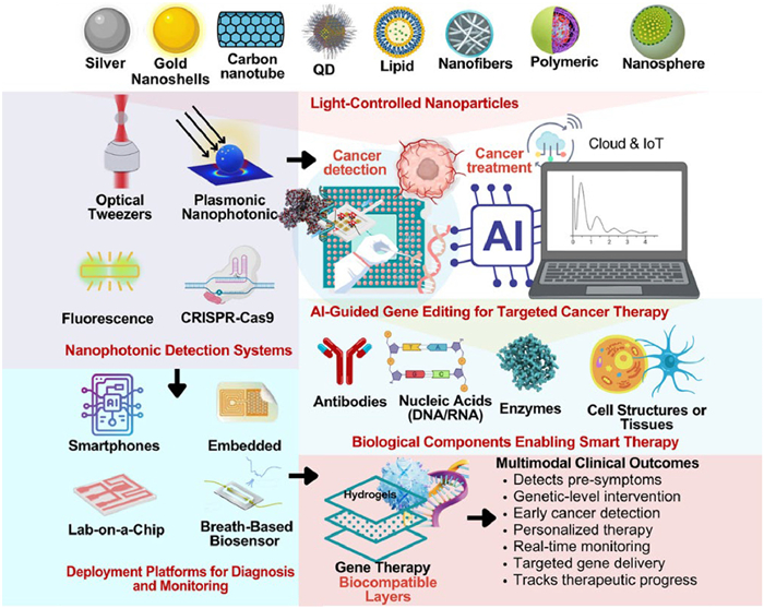
Figure 1.
A multimodal platform that combines light-responsive nanophotonics for early cancer diagnosis with AI-guided CRISPR gene editing for targeted therapy and real-time monitoring.
Advancements in nanophotonics and smart nanomaterials integrated with artificial intelligence-driven gene editing: A paradigm shift in cancer diagnosis and therapeutic
Bakr Ahmed Taha , Ali J. Addie , Luai Farhan Zghair Kolie , Saba Talib Wahhab , Sinan Adnan Abdulateef , Adawiya J. Haider , Khalid Ibnaouf , Norhana Arsad

Modern medicine's gene therapy represents a revolutionary approach to treating and even curing genetic disorders, including cancer. Genome therapy was first tested in 1990 to treat severe combined immunodeficiency (SCID), which emerged in the latter half of the 20th century. The combination of gene therapy with nanophotonic and nanomaterial-based techniques is a huge step forward in the fight against cancer. Tumor heterogeneity, medication resistance, and systemic toxicity provide significant treatment challenges for cancer, which continues to be a top cause of death globally. The targeting capabilities and multifunctionality of nanoscale delivery devices have made them attractive vehicles for addressing these issues [1–3]. Molecular biology and biotechnology developments have enabled gene therapy to move from an experimental stage to a clinical setting. In gene therapy, functional genes are delivered into a patient's cells through viral or non-viral vectors, either to replace mutated genes, silence malfunctioning genes, or to introduce new genes to treat diseases [4–8]. In oncology, this approach is important because cancer is a genetic disease characterized by uncontrolled cell growth caused by mutations. Many strategies are available for gene therapy for cancer, including gene replacement therapy, suicide gene therapy, RNA interference therapy (RNAi), and immune-based gene therapy. As an alternative to suicide gene therapy, alternative therapy uses a functional gene to replace a dysfunctional gene. The suicide gene therapy injects prodrugs into cancer cells and kills them [9]. Oncogenes are targeted and silenced in RNAi-based therapies, so cancer-promoting proteins cannot be generated. Genetic engineering allows a person's immune system to recognize and destroy cancer cells more effectively. These therapies have shown promise in leukemia, melanoma, and glioblastoma [10,11]. Applying gene therapy in treating PROM1-related retinal degeneration highlights the precision required in treating genetic disorders. Findings emphasize the importance of targeting genetic mutations at the molecular level, an approach that is directly relevant to cancer treatment [12].
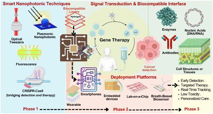
Moreover, investigations into patient perspectives on gene therapy for lysosomal storage disorders shed light on the critical role of patient acceptance and understanding factors that are vital in oncology [13,14]. Nanotechnology has improved genomic therapy, especially in cancer treatment. Therapeutics delivered at the nanoscale (1–100 nm) have fewer side effects. A nanoparticle (NP) differs from a viral vector because it does not cause immune rejection or toxicity. Photonic nanomaterials used to manipulate biological processes enable nanophotonics to be a unique application of gene therapy. Microscopes have pioneered optogenetics, which allows excessive-precision control of gene expression with excessive spatial and temporal accuracy. Genes control the use of mild-sensitive proteins, resulting in focused healing procedures that do much less damage to organs close to. It has been verified that numerous styles of nanomaterials are powerful at handing over genes, which include lipid NPs (LNPs), gold NPs (AuNPs), polymeric NPs (PNPs), and graphene-based nanocarriers. LNPs are one of the maximum widely used nanocarriers for encapsulating nucleic acids, which include mRNA and small interfering RNAs (siRNAs), seeing that they shape stable LNPs around them [15,16]. This perspective paper aims to analyze the effect of new practices of nanophotonics and nanomaterials on cancer treatment and even early detection about gene therapy. The connection between cancer therapies and nanotechnology used in gene therapy has led to the formulation of effective and targeted less invasive therapies, which solve many issues faced in traditional chemotherapy and radiation. Using nanotechnology inventions, we aim to increase gene transfer efficacy, enhance therapeutic effects, and decrease unwanted effects. Artificial intelligence (AI) will be discussed in relation to the control of gene therapy techniques (Fig. 1).
QD lipid is a quantum dot-conjugated lipid probe that improves fluorescence detection with strong signal intensity and strong photostability. Using protocols like those used in thorough in vivo and in vitro validations, physicochemical properties, including bio-compatibility and consistent emission, have been confirmed. Furthermore, this is evident in studies where experimental parameters were meticulously controlled and corrected [17]. Techniques like machine learning and deep learning (DL) are increasingly being used to mine large genomic datasets, establish the prediction of results of the gene therapy, and the construction of optimized delivery mechanisms. The combination of AI with nanotechnology and gene therapy is a new frontier in precision medicine for cancer patients, where real-time reactions to therapies given to them can be monitored, and personalized treatment measures can be taken.
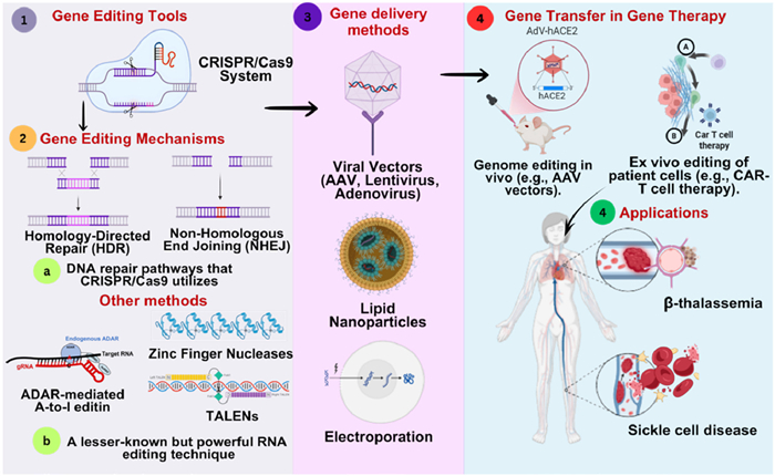
The mechanism of gene therapy works as follows: it alters the genetic information in the target cell so that essential proteins work again in the body. Tissues and organs in the body are made of sophisticated molecules of proteins and perform various functions. Multiple alleles or different variants of one gene may interfere with the protein synthesis or activity that is important for proper functioning of the body. If the genetic change meant for a specific disease is repaired or compensated for, then these proteins can recover their lost activities and allow the body to function properly [18]. Technological advances have become remarkably helpful for the genetic therapy clinic, especially the surgical methods of genes, by allowing new curricula to adjust the genetic fabric of the organism. Clustered regularly interspaced short palindromic repeats/CRISPR-associated protein 9 (CRISPR/Cas9) is the most popular technology because of its effectiveness and simplicity. Using this technique, RNA is collected manually using a molecule called guide RNA (GRNA). As a result, GRNA leads Cas9 to the place where discounts are calculated for the behavior of the genetic changes [19]. When compared to prior technologies such as zinc finger nucleases (ZFNs) or transcription activator-like effector nucleases (TALENs), CRISPR/Cas9 is becoming greater adaptable, efficient, and low priced. As a result, it is the maximum commonplace alternative for gene enhancement [20].
Besides CRISPR/Cas9, different gene enhancing techniques consisting of RNA enhancing and epigenetic modifications are gaining traction. RNA enhancement, in particular (A-to-I) modification mediated by enzymes such as adenosine deaminase acting on RNA (ADAR), plays a critical position in regulating gene expression and has implications for sicknesses, which include cancer and autoimmune issues [21–23]. Furthermore, epigenetic equipment, such as those defined by Pillai et al., permits the change of gene expression without altering the underlying DNA series, supplying a complementary technique to standard gene modification [24]. The delivery mechanisms for the gene-modifying equipment are essential for their healing programs. Various strategies have been advanced to beautify the transport of CRISPR components, inclusive of viral vectors, liposomes, and electroporation strategies. Adeno-associated virus (AAV) vectors have capacity to deliver CRISPR/Cas9 components in vivo, although challenges remain concerning their efficiency and specificity. Recent advancements in NP generation and electroporation strategies have shown promise in improving shipping ribonucleoprotein complexes, facilitating higher modifying efficiencies in goal cells [25–27]. Further, applications of gene editing are in specific diseases, such as amyotrophic lethal sclerosis (ALS) and heart disease, and complex genetic disorders [28,29], to these techniques to address the capacity of these techniques shed light. The gene therapy targeted to SOD1 genes in ALS has shown the possibility of using CRISPR/Cas9 for medical intervention [30]. Likewise, research outlines the use of gene editing for haemoglobinopathies, such as β-thalassemia and sickle cell disease (SCD), and the clinical relevance of these technologies [30,31]. To treat genetic illnesses like as β-thalassemia and SCD, gene therapy involves a series of steps, beginning with gene editing using tools like CRISPR/Cas9 and continuing with delivery via viral vectors and NPs, and finally concluding in gene transfer. It draws attention to critical processes, such as DNA repair routes, delivery strategies, and gene therapy therapeutic applications (Fig. 2).
These breakthroughs, together with advancements in delivery systems, continue to increase the accuracy, efficiency, and clinical application of gene treatments, providing fresh hope for patients with previously untreatable illnesses. Table 1 provides a summary of a comparison of three major gene editing technologies such as CRISPR-Cas9, ZFNs, and TALENs.
 DownLoad:
CSV
DownLoad:
CSV
| Target | CRISPR-Cas9 | ZFN | TALEN |
| Mechanism | GRNA directs Cas9 to DNA | Protein-based zinc finger motifs bind DNA | Protein-based TALE motifs recognize DNA sequences |
| DNA Recognition | RNA-guided | Protein-guided | Protein-guided |
| Editing precision | High (but can have off-target effects) | Moderate | High (more specific than ZFNs) |
| Ease of design | Simple (RNA customization) | Complex (protein engineering required) | Complex (protein engineering required) |
| Cost | Low | High | High |
| Efficiency | High | Moderate | High |
| Off-Target effects | Moderate (can be reduced with modifications) | Low | Low |
| Repair mechanisms | Non-homologous end joining (NHEJ) or homology-directed repair (HDR) | NHEJ or HDR | NHEJ or HDR |
| Delivery methods | Plasmids, viral vectors, NPs | Viral vectors, electroporation | Viral vectors, electroporation |
| Main application areas | Biotechnology in agriculture, and precision medicine | Synthetic biology, tailored gene therapy, and industrial biotechnology | Therapeutic development, functional genomics, and translational medicine |
| Experimental uses | Gene knockout, disease research, therapeutics | Genome editing, gene therapy | Gene therapy, disease modeling |
Plasmonic NPs have shown to be a powerful tool in cancer diagnosis and medicine through their ability to increase surface plasmon resonance (SPR) effects. It is widely used to detect photothermal therapy (PTT), molecular imaging, and biomarkers [32]. Research from Latfulin and George highlights Molecular targeting enables precise thermal therapy using plasmonic NPs in selective nanophotothermolysis [33]. Furthermore, surface-enhanced Raman scattering (SERS)-based plasmonic biosensors can detect microRNA biomarkers without labeling, facilitating early-stage cancer detection [34]. Besides improving imaging and PTT, studies have demonstrated that plasmonic gold nanostars also reduce systemic toxicity and enhance specificity [35]. It has been possible to manipulate cancer cells at the molecular level with optical tweezers, allowing precise drug delivery and real-time analysis of cellular responses. Optical tweezers facilitate tracking cancer cell behavior and interactions, allowing deeper insights into cancer pathogenesis and therapy. Drug delivery nanosystems have also been manipulated using these techniques, enhancing cancer treatment [36]. Cancer imaging and therapy have been significantly enhanced by fluorescence-based nanophotonics (Fig. 3).
Li et al. demonstrated that core-shell plasmonic nanostructures enhance fluorescence signals for targeted cancer cell imaging and PTT [37]. Fluorescence and plasmonic biosensors have been developed for detecting cancer biomarkers, enabling early and accurate diagnosis [38]. The CRISPR/Cas9 system, an RNA-oriented gene editing tool, has significant benefits to older prospects such as ZFN and TALENs. It is more cost-effective, adaptable and user-friendly, and it is a popular tool for genome editing. While obstacles exist, CRISPR/Cas9 is one of the most profitable gene editing equipment known, especially in effective distribution and security problems. Given its ability to edit genomes properly and cure deviations, it provides tremendous ability as a medical method for genetic diseases, especially in cancer therapy. This article addresses CRISPR-Cas-based gene editing, its classification, mechanisms of action and many medical applications, where genetic changes with emphasis on cancer therapy, its functions in targeting existing distribution paths and its advantages and boundaries are included [39].
Nanotechnology has improved RNAi by improving stability and targeting siRNAs. According to Abosalha et al., RNAi NPs based on NPs can improve gene silencing and therapeutic efficacy [40].
Furthermore, RNA-conjugated nanomaterials have become effective tools for cancer diagnosis and treatment, offering high specificity in gene targeting [41]. Combined optical tweezers and fluorescent techniques also advance cancer diagnosis. These methods allow for the manipulation and analysis of single cells and biomolecules, providing insight into cellular mechanisms and interactions [42]. Fluorescent biosensors, especially those that detect specific RNA sequences, can identify highly specific cancer biomarkers. A silicon-photonic biosensor that detects real-time microRNA has further improved the ability to diagnose microRNA disorders properly [43].
The CRISPR era has revolutionized gene editing and holds widespread capability for targeting most cancer healing procedures. By utilizing CRISPR-based systems, researchers can precisely edit genes associated with most cancer progression, offering new avenues for treatment. RNAi strategies have been hired to silence oncogenes, correctly reducing tumor growth and improving the efficacy of existing treatment options [44,45]. The mixture of CRISPR and RNAi with nanophotonic techniques can cause extra effective and targeted techniques in most cancer therapy, as these methods permit specific manipulation of gene expression and cellular responses. RNA enhancement biosensors have enabled new ways to analyze cancer using nanomaterials. These biosensors increase the signals from RNA goals and increase detection sensitivity [46]. The combination of plasmonic nanostructures with RNA biosensors is displayed to improve the detection of unique RNA sequences associated with cancer, allowing the rapid diagnosis of the disease and progression of the disease to be better monitored [47]. In a label-free approach, the ability to identify RNA biomarkers significantly simplifies the clinical process, leading to further cost-effectiveness [48].
Nanomaterials have revolutionized gene therapy by improving delivery, targeting, and controlling genetic payloads. Different nanomaterials, such as liposomes, dendrimers, AuNPs, and PNPs, have been extensively studied for cancer treatment. Because of liposomes, their biocompatibility and their ability to face nucleic acids, genes increase transfusion efficiency and reduce systemic poisoning [49]. Dendrimers provide accurate control over surface functionalities, making them effective in targeted gene therapy [50]. Gold NPs serve as a skilled gene carrier with the advantage of photos that can help with treating cancer [51]. Those designed for PNPs, especially biodegradable and non-toxicity, have achieved traction in clinical surroundings for their ability to provide therapeutic genes to tumor sites [52]. Nanomaterials, such as LNPs and AuNPs, show different toxicity and biodistribution characteristics depending on their size, content, and surface changes, according to existing in vivo toxicity studies. The liver and spleen are the most common sites for the accumulation of bigger AuNPs and those not engineered for biodegradation, according to a plethora of research. AuNPs conjugated with antibodies were shown to be quickly eliminated from circulation but to accumulate significantly in the liver and spleen for extended periods. Further, this has led to worries regarding possible toxicity caused by this persistent buildup [53].
A key advantage of nanomaterials in gene therapy is their ability to deliver therapeutic genes specifically to cancer cells, which minimizes off-target effects and enhances treatment precision. It has been demonstrated that tumor-targeting nanocarriers, including PNPs and liposomal formulations, can accumulate genetic information in malignant cells while sparing healthy tissues [54]. The enhanced permeability and retention (EPR) effect enables NPs to accumulate in tumor tissues because of leaky vasculature. In contrast, active targeting strategies involve ligand-mediated interactions with cancer-specific receptors [55]. The delivery of therapeutic genes and chemotherapy agents by NPs can also enhance the efficacy of combination therapy [56], nanomaterials have shown promise in improving the sensitivity and specificity of biomarker detection beyond cancer treatment. It has been shown that functionalized nanomaterials, such as aptamer-functionalized NPs and quantum dots, enhance the detection of cancer by binding to tumor biomarkers [57]. Imaging engineered NPs in real-time can enable early detection of cancer and the precise location of tumors. Furthermore, nanomaterial-based biosensors can perform noninvasive liquid biopsies, so traditional biopsy procedures are no longer required [58].
A biodegradability and long-term safety assessment of nanomaterials is required before humans may consume them. In addition, the development of multifunctional nanomaterials capable of diagnosing and treating cancer simultaneously could revolutionize the field of personalized medicine. Biomedical applications and cancer diagnosis rely heavily on nanomaterials, which are classified according to their respective functions. Inorganic NPs (2–1000 nm), such as AuNPs, iron oxide NPs (IONPs), and quantum dots, are widely used in imaging and biosensing for diagnosis. NPs (2–1000 nm) and self-assembling proteins have been applied to several biomedical applications, including protein cages for the delivery of nanocarrier drugs, de novo peptides for targeted therapy, bacteriophages to treat bacterial infections, and virus-like particles (VLPs) to develop vaccines (Fig. 4).
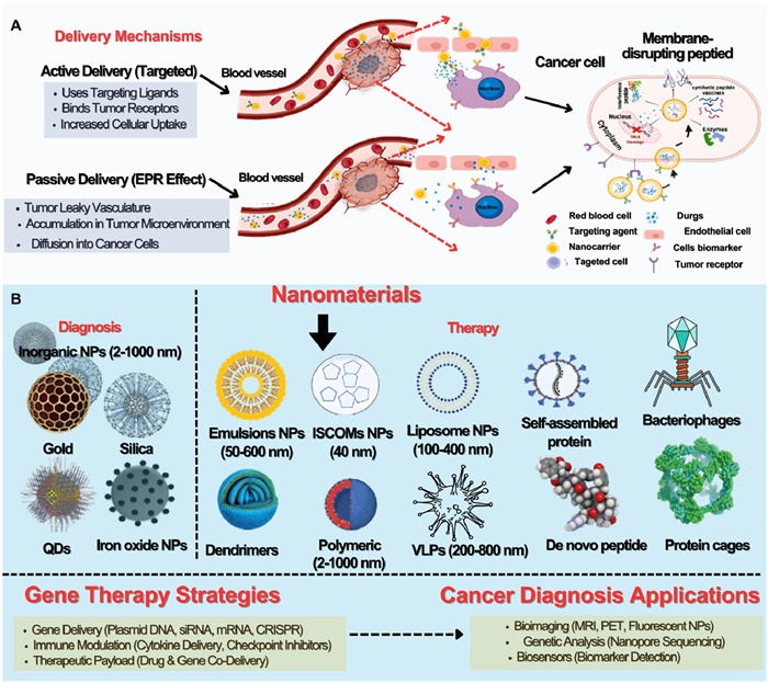
It is possible to enhance drug delivery and treatment effectiveness by using different NP systems in therapy, including emulsion NPs (50–600 nm), which increase drug solubility; immune stimulating complex (ISCOM) NPs (40 nm), which help vaccines work better; and LNPs (100–400 nm), which can encapsulate and deliver hydrophilic and hydrophobic drugs. Modern medicine has demonstrated the potential of these nanomaterials for targeting cancer treatment, precision medicine and immunotherapy, leading to more effective and personalized healthcare solutions. Gene delivery, targeting, and efficiency have all been enhanced by nanomaterials, leading to improvements in gene therapy for cancer treatment and diagnostics. In diagnostics, these compounds are used to increase the accuracy and early diagnosis of cancer, while in targeted treatments, they are used to minimize side effects and maximize therapeutic advantages. Further research must focus on identifying translational hurdles to close the gap between laboratory findings and clinical trials. Due to their surface modifiability, adaptive physicochemical properties, and ability to enhance genetic cargo targeting, nanomaterials are being investigated as a potential alternative gene delivery platform. Nanostructure surface manipulation offers a potential solution for improved tissue targeting while avoiding nonspecific interactions with the human immune system.
Researchers have demonstrated that nonviral nanomaterials exhibit lower pre-existing immunogenicity compared to viral vectors, which is a significant advantage when aiming to minimize unwanted immune responses. Despite these benefits, nanomaterials are still not very good at transfecting genes into living things. It typically requires either new methods or novel combinations of existing ones to achieve therapeutic effectiveness comparable to viral systems (Table 2) [59,60]. Alternatively, AAVs and other viral vectors have shown promising results in both laboratory and clinical settings when it comes to gene delivery and transfection efficiency [61]. They are a powerful tool for gene therapy due to their inherent capacity to directly penetrate cells and promote long-term expression. One major problem with AAVs is that they can trigger immunological responses, both innate and adaptive. Moreover, this can shorten the time that AAVs can be used therapeutically and make attempts to modulate the immune system necessary [62]. Furthermore, there are obstacles to their clinical translation, especially for systemic applications, due to difficulties in producing vectors on a large scale and rising prices [63].
DownLoad:
CSV
| Category | Nanomaterial | Viral vector (AAV) |
| Efficacy | Effectiveness in vivo is moderate; surface engineering has the potential to improve targeting | Longer expression and high transfection rates in healthcare situations |
| Immunogenicity | Lack of immunogenicity; surfaces that can be adjusted to lower immune activation | Capable of inducing both peripheral and systemic immunological responses |
| Scalability | Synthesis that is both easy and inexpensive, with excellent repeatability | Manufacturing is a challenging and costly process with ongoing challenges with scalability |
| Clinical translation | Insufficient data on long-term safety; fewer clinical trials | Clinical translation is impeded by immune reactions and the difficulty of manufacturing |
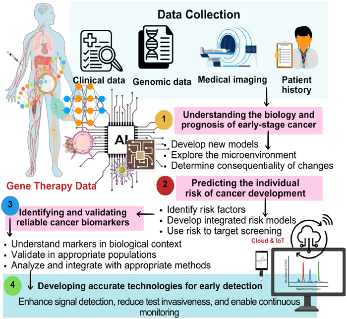
Nanophotonics and nanomaterials have made tremendous strides in gene remedy. Gene therapy efficacy and accuracy may be enhanced via nanocarrier optimization and prediction fashions for drug shipping systems [64–66]. A key application of AI in this field is to enhance the optimization of nanocarriers for gene delivery. Nanomaterials can enhance the pharmacokinetics and biodistribution of therapeutic agents [67], AI is used in the production of pharmacokinetic models to maximize therapeutic results for cancers. Combinations of medications and their delivery time are optimized through the models. It is crucial to broaden predictive models of nanobio interactions, which can be constructed with the help of synthetic intelligence, to ensure that nanomedicines are safe and effective [68]. Lin et al. Tested that machine learning techniques can provide predictive electricity for nanocarrier distribution to tumors, demonstrating that AI can also assist in improving nanocarrier targeting [69]. AI is also used to investigate the protein corona that forms around NPs, therefore affecting their interaction with biological systems. By anticipating the composition of the protein corona, AI-based high-throughput computational methods help to create more efficient gene therapy nanocarriers [70–73]. A protein corona can affect the biological identity of NPs, influencing their therapeutic effectiveness and safety. Nanocarriers may benefit from AI not only in terms of their performance but also in terms of the design and execution of complete gene therapy programs. Combining nanotechnology and AI makes it possible to tailor medicine to the needs of individual patients. AI may close the gap generated using pattern analysis and classification algorithms by intratumor and interpatient heterogeneities, hence improving diagnosis and treatment accuracy, by Adir et al. [74]. Gene therapy is particularly crucial in tumor heterogeneity, where this can significantly compromise treatment efficacy.
Furthermore, new medical treatments can be developed through the integration of AI and nanotechnology. El-atty et al. reported AI-enabled nanotherapeutics that respond to specific biological stimuli result in better gene therapy outcomes [75]. Qi et al. describe how AI can revolutionize data analysis and therapy customization, thus enhancing the integration of omics data into cancer nanomedicine [76]. Through AI-implemented screening strategies, drug selection, dosage, and nanomaterial properties can be analyzed to improve medical results. In addition, nano-information provides AI and nanotechnology, more individual gene therapy by improving patient data analysis and nanomaterial design [77]. One of the most significant advances in nanomedicine has been the incorporation of AI into the creation of nanocarriers and personalized cancer therapies. However, algorithms, datasets, and validation techniques must be thoroughly evaluated before being extensively used in clinical settings. A strong data platform is crucial for generating successful machine learning insights, as the performance of AI applications in this industry is substantially dependent on the quality and reliability of the raw data [78]. According to Bhange and Telange, AI is crucial in the field of nanocarrier design and individualized therapy, as it enables the creation of targeted drug delivery systems. However, to ensure AI works effectively and is safe for use in this industry, specific algorithms, datasets, and validation measures are necessary [79].
Nanocarrier property prediction and design optimization are common use cases for machine learning methods, such as random forest and support vector machines (SVMs). Accordingly, SVM excel in pattern recognition and data classification in nanotheranostics [80]. At the same time, other ML techniques may optimize IoT carrier network communication parameters by modelling the relationship between system settings and performance [81]. Convolutional neural networks (CNNs) and transformer-based models are examples of DL approaches that build upon these capacities by enhancing diagnosis accuracy through the analysis of complicated medical data. To identify hidden patterns that indicate risks such as cardiovascular disease, researchers can combine demographic, clinical, and lifestyle data [82,83]. In healthcare, generative AI enhances personalized treatment recommendations, generates realistic patient data while protecting privacy, facilitates secure data exchange, and creates synthetic datasets for data augmentation. Dataset quality is a crucial factor in determining the effectiveness of these AI methods. The datasets can include information from a variety of sources, such as clinical data, genomic profiles, electronic health records, nanocarrier synthesis, drug release, and cellular uptake data from animal studies, as well as multi-omics datasets that facilitate precision medicine by combining genomic, proteomic, and transcriptomic data [84–87]. Similarly important are image-based databases, such as those used to analyze open-access MRI tumor pictures or to track the release of nanocarrier drugs into cancer cells [88]. The accuracy, precision, recall, F1-score, ROC-AUC, overlap, sensitivity, and specificity of AI models are some of the validation metrics used. Other outcome metrics include the negative predictive value and the Matthews correlation coefficient, while test-based metrics include sensitivity and specificity [89]. The integration of diverse data sources, guidance of precise therapy selection, improvement of risk prediction models, and, finally, support for physicians in designing individualized treatment plans across a wide range of health disorders are all ways in which these AI-driven approaches enable more accurate and personalized healthcare [90,91]. Further, AI can predict therapeutic outcomes in areas such as cancer therapy and gene editing. CRISPR applications can be improved using AI-based methods to minimize off-target effects and enhance the overall efficacy of gene editing procedures. Machine learning algorithms integrate biological data from large datasets and aid in identifying ideal gene targets [92]. As a result of DL's success in predicting therapeutic intervention outcomes from high-dimensional clinical datasets, Huang et al. and Wang et al. developed more personalized medicine approaches based on patient genetic profiles [93,94]. Protein corona prediction models, aided by AI, are essential for comprehending the biological systems' interactions with nanocarriers. Nanocarrier design optimization for enhanced biocompatibility and medicinal efficiency is possible with accurate protein corona predictions [95]. Nanocarriers can be more reliably utilized in clinical settings with the aid of machine learning, which can simulate these interactions and predict their impact on drug delivery [96].
Gene therapy has also been revolutionized by AI-powered DNA nanobots that integrate CRISPR/Cas9 for precise genetic modifications, reducing off-target effects [77]. The CRISPR/Cas9 applications are also adapted to AI-driven protein installation, resulting in more accurate and effective gene editing [97]. Gene therapy based on nanophotonics, and nanomaterials will be more efficient, accurate and accessible in the future because of progress in AI. Using AI in nanophotonic and nanomaterial-based gene therapy not only improves nanocarrier design and optimization but also allows for the development of individualized and successful treatment techniques. AI's capacity to evaluate complicated biological data and anticipate interactions at the nanoscale is changing the face of gene therapy, making it a critical component of current biomedical research (Fig. 5).
Although AI is discussed in several applications in the literature, many descriptions are not clear enough. There is a lack of information on the algorithms utilized and thorough validation metrics like accuracy, specificity, and sensitivity. Explicit algorithmic details are frequently lacking in other sectors where AI is utilized, even if research in financial analysis shows that algorithms such as the random forest algorithm outperform others when it comes to predicting stock market fluctuations [98]. The absence of precise performance measures might impair the reliability and repeatability of model results, which is especially problematic in domains like biodistribution models and associated clinical predictions [99]. Strong metrics for validation are essential in healthcare settings. Sarao et al. discuss on the topic of geographic atrophy identification offers accurate performance metrics, which strengthen the scientific rationale for more adoption and provide credence to clinical practices. When moving from proofs of concept to real-world uses of AI in critical healthcare contexts, this degree of specificity is crucial [100].
Drug discovery, clinical diagnostics, and healthcare administration are all being affected by the ways in which AI is changing biomaterials and biomedicine. From this perspective, we can see the potential, the difficulties, and the necessity of multidisciplinary cooperation in AI integration in biomedicine [101]. Biotechnology, powered by AI, is enhancing healthcare with innovative methods that utilize molecular docking and in vitro testing to validate efficiency. Zhang et al. employ DL and a BERT-based framework to discover antioxidant compounds from traditional herbal treatments [102]. By utilizing liposomal delivery methods, this approach aims to counteract oxidative stress and aging, while also enhancing bioavailability. Using DL to allow gesture detection and overcome issues in conductivity, mechanical performance, and self-healing capabilities, Liu et al. present lignin-reinforced eutectogels for wearable strain sensors [103]. There is considerable hope that new AI technologies, such as AlphaFold 3, can improve medication discovery and accelerate medical advancements in treating diseases [104]. AI and structure-based approaches, such as molecular docking and dynamics, can be used to select aptamers, like monoclonal antibodies. Predicting the binding characteristics of aptamers to their targets is now easier and faster with AI-driven pipelines, such as machine learning and DL [105].
Cui et al. present recent developments in chemical imaging, with an emphasis on systems that utilize AI to enhance sensitivity, contrast, and resolution. With an emphasis on enhancing imaging tools for improved disease knowledge and treatment, it illustrates how these advances are transforming biomedical research and diagnosis, despite the hurdles [106]. Huang et al. introduce an intelligent approach for real-time monitoring of copper absorption in algae. This method enhances our understanding of metal ion dynamics in ecological systems by predicting bioavailability, speciation, and uptake processes of copper at the interface between algae and water [107]. A framework for chemical reaction prediction based on DL is introduced by Xue et al., called bidirectional chemical intelligent net (BiCINet). Its potential to assist in drug discovery, molecular research, and expanding our understanding of chemical transformations is evident in organic synthesis, enzyme-mediated reactions, and retrosynthetic planning [108].
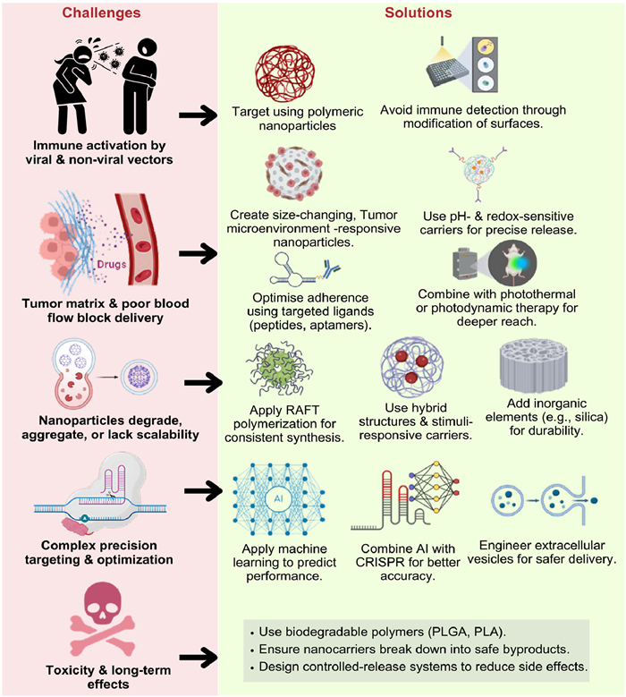
The integration of nanophotonic and nanomaterial-based approaches in gene therapy represents a transformative way to treat a variety of genetic disorders and diseases. However, the clinical application of these novel therapies is hindered by several challenges, including immunogenicity [109,110], off-target effects, efficient targeting and penetration into the tumor microenvironment (TME) [111,112], and the stability and scalability of nanomaterials [113,114]. To overcome these obstacles, the development of innovative and advanced solutions is required.
A major hurdle in gene therapy is the immune response triggered by both viral and non-viral vectors. Although viral vectors are efficient in gene delivery, they can elicit strong immune responses that can compromise therapeutic efficacy and cause adverse effects [115]. Non-viral vectors, including polymeric nanomaterials and LNPs, can also induce inflammatory responses, raising the question of their biocompatibility [116]. Recent developments have focused on the development of delivery systems that reduce immunogenicity and increase specificity to address these issues. The non-viral delivery system based on PNPs seems to be an interesting way forward [117]. PNPs can be engineered to reduce off-target effects by tailoring them to specific cell types and carrying more genetic payloads than conventional viral vectors [118]. In addition, PNPs can be administered repeatedly without eliciting significant immune responses [119]. This solves the problem of immunogenicity and enables a continuous therapeutic effect. PNPs are a compelling substitute for conventional vectors, as their adaptability in handling different genetic materials and their surface modification ability help to avoid immune recognition [120].
The delivery of genes to solid tumors is hindered by the TME, which features a dense extracellular matrix (ECM), disordered vasculature, and hypoxia. The dense ECM along with high interstitial pressure forms a physical barrier against NPs while irregular vascularity leads to flawed blood flow [121–123]. In response to this, researchers have been working on resizable TME-responsive NPs that modify themselves in response to acidic or hypoxic conditions to promote intertumoral penetration and binding [121,124]. Also, active modulation of TME NPs contributes toward overcoming their immunosuppressive background, with a consequent increase of therapeutic efficacy [125,126].
Multifunctional stimuli-responsive nanocarriers driven by specific triggers in the TME, such as pH changes, redox variations, and enzymatic activity, are presently the focal point of strategies in enhancing the gene delivery to solid tumors. These nanocarriers enable one to accomplish direct, controlled release of genetic payloads directly at the tumor [127,128]. For instance, the engineered pH-sensitive NPs use the acidic environment inside a tumor as a trigger for the release of genes near that tumor, thereby enhancing localization and reducing systemic side effects [128,129]. Introducing redox-sensitive linkages into NP design allows higher release in conditions resembling the high endogenous glutathione levels found in the TME [130]. The targeted approach will not only increase the therapeutic index of gene therapies but also address challenges imposed by dense ECM and abnormal vasculature present in a hostile TME, thus improving treatment outcomes [127,131]. This innovative type of idea would help significantly improve the safety and efficacy of gene therapies against cancer.
Conjugation of targeted ligands such as peptides, antibodies or aptamers to nanocarrier surfaces enhances selective binding to tumor-specific antigens or receptors, which improves the accumulation of therapeutics in the tumor and enables deeper penetration into the tissue. The use of prostate-specific membrane antigens (PSMA) as targeting ligands on polymeric nanocarriers shows improved tumor localization and increased therapeutic efficacy in prostate cancer models [132,133]. The combination of these targeting strategies with physical methods such as PTT or photodynamic therapy (PDT) increases the permeability of the tumor tissue by disrupting the vasculature or altering the TME, thus improving the accessibility of the nanocarrier to the tumor cells [134,135]. Dual-targeting ligands offer an ingenious way to target multiple tumor markers to improve therapeutic outcomes [136,137]. One potential avenue for improving gene delivery in oncology is the strategic design of multifunctional nanocarriers containing targeted ligands in combination with synergistic treatments. Practical application of nanomaterial-based gene therapies faces challenges related to nanocarrier stability during storage, systemic circulation, and production scalability. NPs may aggregate, degrade prematurely, or release their cargo unintentionally, which compromises therapeutic efficacy and reproducibility [138,139]. Gold nanocarriers, for example, demonstrate rapid cellular uptake yet encounter stability issues that affect in-vivo distribution and effectiveness [138]. Microencapsulated graphene oxide shows promise for sustained siRNA delivery, but precise retention of the cargo is critical to avoid premature release [139]. High encapsulation stability is essential to maintain nanocarrier integrity until therapeutic agents reach target tissues, with cargo release kinetics significantly influencing outcomes [140]. Recent advancements in hybrid colloidal structures and stimuli-responsive nanocarriers address these stability challenges by enabling controlled release triggered by specific environmental conditions [141,142]. Enhancing the stability and scalability of nanocarrier production remains essential for translating nanomaterial-based gene therapies into clinical practice.
To address these problems, scientists are exploring reversible addition-fragmentation chain transfer (RAFT) polymerization and other advanced polymerization techniques. The effective control of NP size, shape, and surface features, made possible by RAFT polymerization, is crucial for preserving stability and function during storage and systemic circulation [143]. This regulated synthesis enhances their utility for therapeutic usage by making them more scalable and repeatable [143]. The quality of NPs produced by RAFT polymerization is a good indicator of their therapeutic efficiency, as it helps decrease batch-to-batch variability [144]. The durability of nanocarriers has been enhanced by enzyme-catalyzed RAFT polymerization under moderate conditions, which has yielded promising results in well-defined polymer nano-objects [145]. Surface changes facilitate effective gene transport by preventing aggregation and early degradation, thereby stabilizing PNPs [146]. Major strides have been made in overcoming the limits of nanomaterial-based gene therapy thanks to these advancements in polymerization processes.
In addition, the development of hybrid nanostructures that combine organic and inorganic components improves both the mechanical stability and the functional versatility of the nanocarriers. The incorporation of inorganic elements such as iron oxide into polymer matrices adds magnetic properties that allow external magnetic fields to direct and concentrate therapeutic agents to target sites, while offering the potential for real-time imaging and delivery monitoring [147]. Mesoporous silica NPs (MSN) are known for their high porosity and easy surface modification. They are mechanically, chemically and thermally stable and are excellent for controlled release by encapsulating various therapeutic agents while maintaining their integrity during circulation [148]. The development of these hybrid nanostructures, which respond to external stimuli such as magnetic fields or light, enables precise spatial and temporal control of drug release, thus increasing therapeutic efficacy [149,150]. This flexible method of nanocarrier design offers a good opportunity to increase the efficiency of drug delivery systems in cancer treatment and other medical applications.
The combination of nanotechnology with AI and genome editing appears to be very exciting for the next stage of gene therapy. Through their interaction with biological systems, AI-driven platforms are accelerating the discovery and optimization of nanocarriers and facilitate the development of safe and efficient delivery systems [151,152]. Machine learning techniques recognize trends and predict the performance of nanomaterials based on huge data sets. This enables customized treatments with improved efficacy and fewer side effects [78]. The integration of AI into DNA nanostructures optimizes delivery systems for CRISPR/Cas applications ensuring accurate targeting and improved uptake into cells [153,154]. Moreover, AI enhances the mechanical stability and functional adaptability of hybrid nanostructures including organic and inorganic elements [155]. This flexible approach not only improves the accuracy of therapeutic delivery and targeting but also provides real-time imaging and monitoring possibility, so advancing gene therapy [78]. In parallel, advanced nanocarriers integrated with genome editing technologies such as CRISPR/Cas systems provide a disruptive mode of repairing genetic defects with extreme targeting. Nanomaterials based delivery systems protect the genome editing machinery from inactivation, transport across cellular barriers, and ensure release at the desired intracellular site [156]. Targeted delivery is key to attaining high editing efficiencies while limiting off-target and immune responses. DNA-based nanomaterials are emerging as more promising per CT-DCRISPR/Cas delivery applications, enhancing stability and functionality in biological environments [156].
The TME poses significant challenges to efficient drug administration because of its aberrant stromal architecture and thick ECM [157]. Scientists have devised size-switchable NPs to overcome these obstacles; these particles may shrink in size as they approach tumours by taking advantage of environmental cues like matrix metalloproteinase-2 (MMP-2). According to Cun et al. and Duan et al., these NPs were originally made at around 100 nm to have an EPR effect and sustained circulation. However, once within the tumor, they shrink down to around 30 nm, which greatly improves ECM penetration. In addition, stromal-targeting medicines, like as hyaluronidase, can break down ECM components such as hyaluronic acid, which lowers interstitial pressure and increases matrix permeability. This method works in conjunction with size-switchable systems to improve drug distribution and therapeutic efficacy. Enzymatic remodeling of the TME allows the now-smaller NPs to penetrate deeper, leading to better drug distribution. To circumvent the TME's biochemical and physical obstacles, a potent multi-pronged approach is to combine size-switchable NPs with ECM-modulating enzymes like hyaluronidase. Thus, this combined method improves the penetration of NPs and the accumulation of drugs within tumors, particularly those with thick stromus components [158,159]. Engineered extracellular vesicles and ribonucleoprotein complexes further increase the delivery of CRISPR components, thereby facilitating higher-level genome editing with lower immunogenicity [160]. The application of these advanced nanocarrier systems provides an opportunity for the researchers to best use the therapeutic potential of CRISPR/Cas technologies in creating novel genetic disorder and other diseases treatments [113,161].
Development of biodegradable and biocompatible nanomaterials addresses concerns related to long-term toxicity and environmental impact. Polymers like poly(lactic-co-glycolic acid) (PLGA) and polylactic acid (PLA) exhibit favorable degradation profiles, making them ideal for designing nanocarriers that safely deliver genetic material [162,163]. Engineered to break down into non-toxic byproducts, these materials help minimize adverse effects on human health and the environment [164]. PLGA NPs, for example, effectively encapsulate therapeutic agents while ensuring controlled release at target sites a critical factor for enhancing therapeutic efficacy [165]. Incorporating biodegradable components not only improves the safety profile of nanocarriers but also enables gradual release of the therapeutic payload, reducing toxicity risks associated with accumulation in the body [166]. This strategy proves particularly beneficial in cancer therapy, where targeted delivery and controlled release maximize treatment effectiveness and minimize side effects [164,167].
Development and implementation of NPs for drug administration can be somewhat challenging, however there are creative alternatives being investigated to overcome these hurdles. Improved medication delivery with fewer side effects is possible with the use of PNPs, which can be fine-tuned to target only the cells or tissues of interest. Surface modification, such as PEGylation, can be used to tailor NPs to escape immune detection. This shortens their circulation length and improves therapeutic efficacy, further enhancing targeting. Making NPs that can change size in response to the TME is another potential strategy. To improve their targeting of cancer cells, these NPs may enlarge or contract in response to changes in tumor pH or temperature.
NPs can be fine-tuned for targeted adhesion using aptamers or peptides, which enhances the specificity of drug delivery by binding to specific cells and tissues. Uniform NP synthesis is possible with RAFT polymerization, which makes it an excellent choice for large-scale manufacturing as it guarantees uniformity and scalability in NP production. More personalized and efficient medication delivery options are possible thanks to machine learning, which also plays a crucial role in predicting how NPs will behave in various settings. In gene therapy applications, particularly when merging AI with CRISPR, precise alterations to NPs can be enabled, thereby increasing their efficacy and reducing the error rate, resulting in enhanced precision. Incorporating inorganic materials, such as silica, into NP designs enhances their stability and endurance, thereby increasing their resistance to degradation within the body.
It concludes by outlining potential solutions to the primary problems with nanocarrier-mediated cancer therapy. (ⅰ) On the one hand, there is the issue of immune activation caused by both viral and non-viral vectors; (ⅱ) On the other hand, there are concerns about toxicity and long-term side effects; (ⅲ) NP instability leads to degradation, aggregation, or poor scalability; (ⅳ) Precise targeting and optimization are difficult to achieve; and (ⅴ) concerns about drug delivery are highlighted on the left side. Finally, a promising direction is emerging in the research and development of extracellular vesicles for targeted drug delivery. Medicinal compounds can be delivered more efficiently and with reduced toxicity when these naturally occurring structures are engineered, leading to improved patient outcomes (Fig. 6).
Numerous clinical trials are currently investigating the possibilities of gene editing and smart NPs. Gene therapy heavily utilizes gene editing technologies, such as ZFN, TALEN, and CRISPR, according to a comprehensive analysis of clinical studies. Several protocols have demonstrated both safety and therapeutic efficacy, and over 30 genome editing therapy research studies are active as of November 2022 [168–170]. Clinical experiments using CRISPR/Cas9 technology have demonstrated remarkable advancements, particularly in the treatment of genetic diseases, including SCD and transfusion-dependent beta-thalassemia (TDT) [171]. The first proof of concept for ex vivo gene-edited cell treatments was demonstrated using ZFN to target the CCR5 gene in autologous CD4⁺ T cells, resulting in populations of human immunodeficiency virus (HIV) -resistant cells that could be reinfused. Later developments with TALENs and CRISPR have broadened the editing horizons, with therapeutic uses in immuno-oncology and haematological diseases being more often documented. Allogeneic, commercially available platforms are now the focus of research into multiplex editing methods [172]. Improved adoptive cell treatment for cancer has been made possible by ex vivo editing of T cells, T-cell receptor T-cell therapy (TCR-T), and chimeric antigen receptor T-cell therapy (CAR-T). Consequently, this has enabled the production of universal allogeneic CAR-T cells and the elimination of checkpoint inhibitors. The use of editing has expanded beyond cancer to other fields, such as cervical carcinoma, HIV (to create immune cells resistant to the virus), and foetal haemoglobin in SCD and β-thalassemia (by reprogramming hematopoietic stem cells). Additional uses of in vivo techniques include ocular gene repair in Leber's congenital amaurosis and AAV-mediated liver-targeted editing for systemic protein synthesis. Clinical translation relies on thorough safety evaluations and education of stakeholders; however, these trials demonstrate that gene editing has great therapeutic potential [173]. Clinical trial recruiting is one area where AI is having a noticeable effect on the healthcare industry. AI may automate the process of participant identification in cancer trials by connecting with electronic health records (EHRs) and applying machine learning algorithms. It would improve the trials' speed, accuracy, and efficiency. Encryption and differential privacy can help enhance data privacy and security, while standardized EHR formats can address the issue of interoperability [174].
Combining gene therapy with nanophotonic technology has led to significant therapeutic advances in cancer treatment. Versatile platforms that overcome the limits of conventional cancer treatments are multifunctional NPs, which enhance diagnostic accuracy and enable more tailored therapy methods with decreased systemic toxicity. When used as contrast agents in magnetic resonance imaging (MRI), optical imaging (OI), and photoacoustic imaging (PI), these nanocarriers enhance diagnostic accuracy and enable more effective monitoring of therapy responses. Their ability to accumulate preferentially within tumor tissues via passive or active targeting mechanisms is a major advantage, as it increases the local concentration of the medication and prevents the early destruction of encapsulated therapies. In addition, chemo-PT and other combination techniques utilize synergistic effects to increase treatment efficacy by delivering multiple therapeutic agents simultaneously using nanocarriers [175–178].
The CRISPR/Cas system is a highly adaptable and accurate gene-editing technique that has recently been developed for use in bioimaging, diagnostics, and medicines. A detailed investigation was conducted on its background, principles of design, and methods of distribution, with a focus on vectors derived from DNA nanomaterials [179]. Delivery and spatiotemporal control are two challenges that limit the potential of CRISPR-therapeutic Cas9. To facilitate the targeted delivery of CRISPR/Cas9 and on-demand release, a tailored nanocapsule containing upconversion DNA was developed using near-infrared (NIR) light. The system's ability to inhibit tumor growth in both laboratory and living organism settings suggests it may be useful for deep-tissue treatments and remote-controlled gene editing [180]. Displacement of strings, as in DNA circuits, possesses great potential for molecular computation; nonetheless, it encounters difficulties related to complexity and contamination. Amplification and decision-making capacities are improved, contamination hazards are reduced, and design is simplified with CRISPR-Cas-based DNA circuits. CRISPR-Cas functional circuits can be built more efficiently and flexible by focusing on current tactics, constraints, and future possibilities [181]. Endocrine therapy is the main method of treatment for oestrogen receptor (ER)-positive breast cancer, which is the most prevalent subtype. To combat resistance and maximize effectiveness, new studies emphasize combination medicines that target ER in conjunction with pathways, including cyclin-dependent kinases 4/6, phosphatidylinositol 3-kinase, and Histone Deacetylase. Additionally, the study examines inhibitor structures, explores structure-activity relationships, and offers suggestions for future research into more effective treatment methods [182]. Nanozymes based on metal-organic frameworks (MOFs) integrate inorganic nanomaterials with biological systems, exhibiting a wide range of catalytic activities comparable to those of natural enzymes. As a result of heat, light, and ultrasound stimulation, various MOFs can adjust their activity. Due to their multifunctional enzyme-like properties and the challenges in preparing and functionalizing them, these MOFs hold significant promise for use in biological applications [183].
Integration of nanophotonic and nanomaterial-based strategies into gene therapy offers a promising approach to advance cancer treatment by enhancing precision, efficacy, and safety of oncology therapies. Preclinical studies indicate that NPs can act as radiosensitizers, systemically targeting tumor cells to boost the effects of X-ray radiotherapy against cancer cell lines [184]. Encapsulating radioactive molecules within a nanocrystalline matrix controls biohazards for healthy cells, thereby improving radiotherapy safety [184]. NPs also protect genome editing tools like CRISPR/Cas systems from degradation and promote their transport across cellular barriers, which is critical for high editing efficiency while reducing off-target effects and immune responses [185–187]. The multifunctionality of nanocarriers enables combination of therapeutic modalities, such as PTT and PDT, which further enhance treatment outcomes [188,189]. Continued exploration of these nanomaterial-based strategies in gene therapy promises to significantly improve cancer treatment paradigms.
Despite promising results in NP-based gene therapies, considerable hurdles persist in translating these innovations from laboratory research to clinical application. Limited targeting accuracy remains a major challenge, with studies reporting that only about 0.7% of administered NPs reach tumor sites [190]. Low accumulation emphasizes the need for more effective targeting strategies to ensure therapeutic efficacy. Engineered living materials such as modified bacteria or cells are being explored as alternatives to conventional NPs because they can attach more effectively to tumor sites [191,192]. Engineered bacteria, for example, demonstrate the ability to migrate into the tumor environment, thereby improving therapy outcomes [192]. Parallel efforts focus on inorganic NPs, particularly IONPs, which offer magnetic properties and improved biocompatibility. IONPs are being investigated for applications ranging from treating iron deficiency anemia to cancer diagnosis and therapy, with their capacity to induce pro-inflammatory macrophage polarization in tumor tissues highlighting their therapeutic potential [193,194]. Combining engineered living materials with advanced inorganic NPs represents a versatile approach to overcoming the challenges associated with NP-based gene therapies.
The path to clinical application of NP-based gene therapies demands rigorous evaluation through clinical trials. Despite promising efficacy in preclinical models, translation into clinical success remains limited, largely due to challenges in ensuring consistent manufacturing practices that maintain quality and safety [195,196]. Long-term biocompatibility is critical, as potential toxicity and adverse cellular reactions must be thoroughly addressed [197,198]. Complex regulatory frameworks further complicate the process, necessitating standardized protocols for NP characterization, safety evaluation, and efficacy assessment [199,200]. Collaboration among researchers, clinicians, and regulators is essential to develop guidelines that can streamline approval of these novel therapies. Advances in biodegradable and biocompatible nanomaterials such as those based on black phosphorus or mesoporous silicon promise to overcome these challenges by enhancing safety profiles and therapeutic efficacy [198,201]. Improving targeting methods, delivery systems, and our knowledge of NP tumor interactions are crucial for the future of nanophotonic and nanomaterial-based gene therapy for cancer. To conquer existing obstacles and fully realize the potential of these novel cancer treatments, interdisciplinary collaboration is necessary that combines nanotechnology, molecular biology, and clinical oncology.
Advances in gene therapy, such as CRISPR-Cas9, as well as integrated nanophotonic and nanomaterial-based methods, have the potential to transform precision oncology. Nanocarrier systems containing lipid, gold, and PNPs enable highly focused gene delivery, enhance biocompatibility, and reduce off-target effects compared to traditional approaches. In addition to improving molecular imaging and biomarker identification, nanophotonics now allows for early cancer diagnosis and real-time monitoring of therapy effectiveness. AI enhances the platform by optimizing nanocarrier design, predicting biodistribution, and customizing personalized treatment regimens based on multi-omics data, resulting in a coherent platform for targeted therapy, early diagnosis, and continuous monitoring. Despite these advancements, obstacles remain, such as vector immunogenicity, tumor penetration limitations, and NP manufacturing scalability. Future research should focus on enhancing nanocarrier stability and tumor penetration, developing multimodal theranostic systems that integrate diagnostic and therapeutic functions, investigating clinical translational pathways through phase studies of CRISPR-based nanocutters, and integrating multimodal diagnosis and treatment strategies for more precise interventions. AI-driven personalization can accelerate the implementation of scalable, patient-specific medicines while also optimizing clinical decision-making. Collaboration across clinical oncology, materials science, and molecular biology will be crucial for translating these findings into reality, ultimately enhancing precision medicine and improving patient outcomes through personalized, less invasive cancer therapies.
The authors declare that they have no known competing financial interests or personal relationships that could have appeared to influence the work reported in this paper.
Bakr Ahmed Taha: Writing – review & editing, Writing – original draft. Ali J. Addie: Writing – review & editing, Writing – original draft. Luai Farhan Zghair Kolie: Writing – review & editing. Saba Talib Wahhab: Writing – review & editing. Sinan Adnan Abdulateef: Writing – review & editing. Adawiya J. Haider: Writing – review & editing. Khalid Ibnaouf: Writing – review & editing. Norhana Arsad: Writing – review & editing.
This research was funded by Universiti Kebangsaan Malaysia through INISIATIF BELANJAWAN 2025: AI ENHANCEMENT, Malaysia (No. WARISAN-2025–023), as well as the University of Technology, Baghdad, Iraq.
D. Cross, J.K. Burmester, Clin. Med. Res. 4 (2006) 218–227. doi: 10.3121/cmr.4.3.218
B.A. Taha, A.J. Addie, A.J. Haider, A. Norhana, Bionanoscience 15 (2025) 268. doi: 10.1007/s12668-025-01876-9
Y. Li, X. Zhang, Z. Xiang, et al., JAMA Netw. Open 6 (2023) E2328352. doi: 10.1001/jamanetworkopen.2023.28352
S. Kerpel-Fronius, V. Baroutsou, S. Becker, R. Carlesi, et al., Front. Med. 7 (2020) 608249. doi: 10.3389/fmed.2020.608249
B.A. Taha, A.C. Kadhim, A.J. Addie, et al., ACS Chem. Neurosci. 16 (2025) 895–907. doi: 10.1021/acschemneuro.4c00809
B.A. Taha, Z.M. Abdulrahm, A.J. Addie, et al., Talanta 287 (2025) 127693. doi: 10.1016/j.talanta.2025.127693
B.A. Taha, A.J. Addie, E.M. Abbas, et al., J. Photochem. Photobiol. C: Photochem. Rev. 60 (2024) 100678.
B.A. Taha, A.J. Addie, S. Chahal, et al., J. Biotechnol. 400 (2025) 29–47. doi: 10.1016/j.jbiotec.2025.02.005
N.D. Germain, W.K. Chung, P.D. Sarmiere, Mol. Aspects Med. 91 (2023) 101148. doi: 10.1016/j.mam.2022.101148
J. Xue, K. Chen, H. Hu, S.C.B. Gopinath, Biotechnol. Appl. Biochem. 69 (2022) 1166–1175. doi: 10.1002/bab.2193
B. Cesur-Ergün, D. Demir-Dora, J. Gene Med. 25 (2023) e3550. doi: 10.1002/jgm.3550
J. Cehajic-Kapetanovic, J. Birtel, M.E. McClements, et al., JAMA Netw. Open 2 (2019) e195752. doi: 10.1001/jamanetworkopen.2019.5752
E.C.B. Eskes, C.R.L. Beishuizen, E.M. Corazolla, et al., Orphanet J. Rare Dis. 17 (2022) 383. doi: 10.1186/s13023-022-02543-y
B.A. Taha, A.J. Addie, A.J. Haider, et al., Langmuir 40 (2024) 23549–23561. doi: 10.1021/acs.langmuir.4c03513
A.A. Rizvanov, Pers. Psychiatry Neurol. 3 (2023) 3–6. doi: 10.52667/2712-9179-2023-3-1-3-6
O.L. Aiyegbusi, K. Macpherson, L. Elston, et al., Nat. Commun. 11 (2020) 6265. doi: 10.1038/s41467-020-20096-1
S.R. Weaver, E.L. Taylor, E.L. Zars, et al., J. Bone Miner. Res. 36 (2021) 2100–2101.
J.T. Bulcha, Y. Wang, H. Ma, P.W.L. Tai, G. Gao, Signal Transduct. Target. Ther. 6 (2021) 53. doi: 10.1038/s41392-021-00487-6
D. Desai, H. Panchal, S. Patel, K. Nayak, Int. J. Adv. Res. 8 (2020) 1127–1132. doi: 10.21474/ijar01/11943
I. Ates, T. Rathbone, C. Stuart, P. Hudson Bridges, R.N. Cottle, Genes 11 (2020) 1113. doi: 10.3390/genes11101113
T.W. Chan, T. Fu, J.H. Bahn, et al., Genome Biol. 21 (2020) 268. doi: 10.1186/s13059-020-02171-4
S. Wu, Q. Xue, X. Qin, et al., Genes 14 (2023) 919. doi: 10.3390/genes14040919
N.I. Vlachogiannis, A. Gatsiou, D.A. Silvestris, et al., J. Autoimmun. 106 (2020) 102329. doi: 10.1016/j.jaut.2019.102329
A. Pillai, V. Verma, S. Galande, BioEssays 47 (2024) 2400186.
X. Liu, J. Jiang, J. Liu, et al., Adv. Healthc. Mater. 13 (2024) 2400645. doi: 10.1002/adhm.202400645
D. Wang, F. Zhang, G. Gao, Cell 181 (2020) 136–150. doi: 10.1016/j.cell.2020.03.023
S. Zhang, J. Shen, D. Li, Y. Cheng, Theranostics 11 (2020) 614–648.
H. Wan, W. Qian, B. Wei, et al., Front. Neurosci. 18 (2024) 1499025. doi: 10.3389/fnins.2024.1499025
X. Wu, J. Yang, J. Zhang, Y. Song, MedComm 5 (2024) e639. doi: 10.1002/mco2.639
D.A. Amado, B.L. Davidson, Mol. Ther. 29 (2021) 3345–3358. doi: 10.1016/j.ymthe.2021.04.008
F. Locatelli, A.A. Thompson, J.L. Kwiatkowski, et al., N. Engl. J. Med. 386 (2022) 415–427. doi: 10.1056/nejmoa2113206
B.A. Taha, I.A. Al-Tahar, A.J. Addie, et al., Appl. Mater. Today 38 (2024) 102229. doi: 10.1016/j.apmt.2024.102229
R.R. Letfullin, T.F. George, Introduction to cancer therapy and detection by plasmonic nanoparticles, in: Computational Nanomedicine and Nanotechnology, Springer, Cham, 2016, pp. 133–182.
H.N. Wang, B.M. Crawford, S.J. Norton, T. Vo-Dinh, J. Phys. Chem. B 123 (2019) 10245–10251. doi: 10.1021/acs.jpcb.9b06804
R.A. Odion, Y. Liu, T. Vo-Dinh, Cancers 14 (2022) 5737. doi: 10.3390/cancers14235737
E. Spyratou, Front. Phys. 10 (2022) 812192. doi: 10.3389/fphy.2022.812192
L. Fu, C. Te Lin, H. Karimi-Maleh, F. Chen, S. Zhao, Biosensors 13 (2023) 977. doi: 10.3390/bios13110977
D.N. Moorthy, D. Dhinasekaran, P.N.B. Rebecca, A.R. Rajendran, J. Biophotonics 17 (2024) e202400243. doi: 10.1002/jbio.202400243
A. Karimian, K. Azizian, H. Parsian, et al., J. Cell. Physiol. 234 (2019) 12267–12277. doi: 10.1002/jcp.27972
A.K. Abosalha, P. Islam, J.L. Boyajian, et al., J. Cancer Biomol. Ther. 1 (2024) 29–37.
R. Arshad, I. Fatima, S. Sargazi, et al., Nanomaterials 11 (2021) 3330. doi: 10.3390/nano11123330
C. del Real Mata, O. Jeanne, M. Jalali, Y. Lu, S. Mahshid, Adv. Healthc. Mater. 12 (2023) 2202123. doi: 10.1002/adhm.202202123
O. Calvo-Lozano, P. García-Aparicio, L.Z. Raduly, et al., Anal. Chem. 94 (2022) 14659–14665. doi: 10.1021/acs.analchem.2c02895
H. Wu, S. Ou, H. Zhang, et al., Cancer Cell Int. 22 (2022) 220. doi: 10.1186/s12935-022-02640-9
B. Yin, W.K.H. Ho, X. Xia, et al., Small 19 (6) (2023) 2206762. doi: 10.1002/smll.202206762
X. Lu, C. Yao, L. Sun, Z. Li, Biosens. Bioelectron. 203 (2022) 114041. doi: 10.1016/j.bios.2022.114041
N. Sharma, A.D. Ankalgi, U. Thakur, M.S. Ashawat, N. Sharma, J. Drug Deliv. Ther. 12 (2022) 192–198.
E. Avci, H. Yilmaz, N. Sahiner, et al., Cancers 14 (20) (2022) 5021. doi: 10.3390/cancers14205021
H. Tiwari, N. Rai, S. Singh, et al., Bioengineering 10 (2023) 760. doi: 10.3390/bioengineering10070760
J. Kim, D.R. Wilson, C.G. Zamboni, J.J. Green, J. Drug Target. 23 (2015) 627–641. doi: 10.3109/1061186X.2015.1048519
J.A. Barreto, W. O’Malley, M. Kubeil, et al., Adv. Mater. 23 (2011) H18–H40.
P. Zhang, G. Ye, G. Xie, et al., Front. Bioeng. Biotechnol. 11 (2023) 1240529. doi: 10.3389/fbioe.2023.1240529
N. Daems, B. Verlinden, K. Van Hoecke, et al., J. Biomed. Nanotechnol. 16 (2020) 985–996. doi: 10.1166/jbn.2020.2928
P. Gao, W. Pan, N. Li, B. Tang, ACS Appl. Mater. Interfaces 11 (2019) 26529–26558. doi: 10.1021/acsami.9b01370
H. Kang, S. Hu, M.H. Cho, et al., Nano Today 23 (2018) 59–72. doi: 10.1016/j.nantod.2018.11.001
Y. Mei, R. Wang, W. Jiang, et al., Biomater. Sci. 7 (2019) 2640–2651. doi: 10.1039/c9bm00214f
R. Singh, S. Kumar, Nanomaterials 12 (2022) 2283. doi: 10.3390/nano12132283
N. Rashidi, M. Davidson, V. Apostolopoulos, K. Nurgali, J. Drug Deliv. Sci. Technol. 95 (2024) 105599. doi: 10.1016/j.jddst.2024.105599
S. Trigueros, Res. Med. Eng. Sci. 6 (2018) RMES.000633.2018.
D. Kasala, A.R. Yoon, J. Hong, S.W. Kim, C.O. Yun, Nanomedicine 11 (2016) 1689–1713. doi: 10.2217/nnm-2016-0060
P.E. Monahan, C. Négrier, M. Tarantino, L.A. Valentino, F. Mingozzi, J. Clin. Med. 10 (2021) 2471. doi: 10.3390/jcm10112471
F. Mingozzi, K.A. High, Blood 122 (2013) 23–36. doi: 10.1182/blood-2013-01-306647
D. Sharon, A. Kamen, Biotechnol. Bioeng. 115 (2018) 25–40. doi: 10.1002/bit.26461
V. Chaudhary, B.A. Taha, Lucky, et al., ACS Sens. 9 (2024) 4469–4494. doi: 10.1021/acssensors.4c01524
B.A. Taha, A.J. Addie, A.C. Kadhim, et al., Microchim. Acta 191 (2024) 250. doi: 10.1007/s00604-024-06314-3
R.M. Mat Yeh, B.A. Taha, N.N. Bachok, et al., Food Control 161 (2024) 110399. doi: 10.1016/j.foodcont.2024.110399
G. Lin, R.A. Revia, M. Zhang, Adv. Funct. Mater. 31 (2021) 2007096. doi: 10.1002/adfm.202007096
D. Fan, Y. Cao, M. Cao, et al., Signal Transduct. Target. Ther. 8 (2023) 293. doi: 10.1038/s41392-023-01536-y
Z. Lin, W.C. Chou, Y.H. Cheng, et al., Int. J. Nanomedicine 17 (2022) 1365–1379. doi: 10.2147/ijn.s344208
H. Lee, Pharmaceutics 16 (2024) 1419. doi: 10.3390/pharmaceutics16111419
B.A. Taha, V. Chaudhary, S. Rustagi, et al., ECS J. Solid State Sci. Technol. 13 (2024) 047004. doi: 10.1149/2162-8777/ad3d0a
B.A. Taha, N.M. Ahmed, R.K. Talreja, et al., ACS Synth. Biol. 13 (2024) 1600–1620. doi: 10.1021/acssynbio.4c00070
B.A. Taha, E.M. Abbas, A.C. Kadhim, et al., Microelectron. Eng. 292 (2024) 112228. doi: 10.1016/j.mee.2024.112228
O. Adir, M. Poley, G. Chen, et al., Adv. Mater. 32 (2020) 1901989. doi: 10.1002/adma.201901989
S.M. Abd El-Atty, N.A. Arafa, A. Abouelazm, et al., Comput. Model. Eng. Sci. 133 (2022) 111–131.
L. Qi, Z. Li, J. Liu, X. Chen, Adv. Mater. 36 (2024) e2409102. doi: 10.1002/adma.202409102
M. Soltani, F.M. Kashkooli, M. Souri, et al., Cancers 13 (2021) 2481. doi: 10.3390/cancers13102481
X.J. Gao, K. Ciura, Y. Ma, et al., Adv. Mater. 36 (2024) 2407793. doi: 10.1002/adma.202407793
M. Bhange, D. Telange, Discov. Oncol. 16 (2025) 77. doi: 10.1007/s12672-025-01821-y
M.S. Islam, M.M.T.G. Manik, M. Moniruzzaman, et al., J. Posthumanism 5 (2025) 4916–4934.
F. Jia, N. Feng, Q. Liu, Optimal design of carrier communication matching in internet of things based on machine learning algorithm, in: 2024 Asia-Pacific Conference on Software Engineering, Social Network Analysis and Intelligent Computing (SSAIC), New Delhi, India, 2024, pp. 819–824.
S. Anuyah, M.K. Singh, H. Nyavor, World J. Adv. Res. Rev. 24 (2024) 1–25. doi: 10.1145/3661725.3661733
S. Vaishali, T. Mohan, S. Navaneethan, AI-powered prediction models for cardiovascular disease risk assessment, in: 2024 7th International Conference on Contemporary Computing and Informatics (IC3I), Greater Noida, India, 2024, pp. 350–355.
R. Lin, AI-driven Personalized Healthcare: leveraging multimodal data for precision medicine, in: 2024 IEEE/WIC International Conference on Web Intelligence and Intelligent Agent Technology (WI-IAT), Bangkok, Thailand, 2024, pp. 693–697.
D. Ramakrishnan, L. Jekel, S. Chadha, et al., Sci. Data 11 (2024) 254. doi: 10.1038/s41597-024-03021-9
T. Henser-Brownhill, L. Martin, P. Samangouei, et al., Adv. Sci. 11 (2024) 2401935. doi: 10.1002/advs.202401935
A. Ortiz-Perez, D. van Tilborg, R. van der Meel, F. Grisoni, L. Albertazzi, Digit. Discov. 3 (2024) 1280–1291. doi: 10.1039/d4dd00104d
S. Goswami, K.D. Dhobale, R.D. Wavhale, B. Goswami, S.S. Banerjee, Appl. AI Lett. 3 (2022) e50. doi: 10.1002/ail2.50
M.E. Klontzas, K.B.W. Groot Lipman, T. Akinci D’ Antonoli, et al., Eur. Radiol. (2025), doi: 10.1007/s00330-025-11890-w.
H. Khude, P. Shende, Adv. Integr. Med. 12 (2025) 100529. doi: 10.1016/j.aimed.2025.100529
N. Latif, R. Babe, K. Raz, et al., Front. Heal. Informatics 13 (2024) 895–901.
J. Choi, I. Oh, S. Seo, J. Ahn, Sci. Rep. 8 (2018) 13729. doi: 10.1038/s41598-018-32180-0
B. Huang, S. Zheng, B. Ma, et al., BMC Pregnancy Childbirth 22 (2022) 36. doi: 10.1109/sdpc55702.2022.9915939
K. Wang, R. Yang, J. Li, et al., Front. Pharmacol. 16 (2025) 1591438. doi: 10.3389/fphar.2025.1591438
A. Khurana, P. Hartmann, World J. Gastroenterol. 31 (2025) 109105.
S.Z. Alshawwa, A.A. Kassem, R.M. Farid, et al., Pharmaceutics 14 (2022) 883. doi: 10.3390/pharmaceutics14040883
L. Lin, Z. Cui, S. Wang, et al., J. Theory Pract. Eng. Sci. 4 (2024) 183–190. doi: 10.53469/jtpes.2024.04(03).17
E.A. Avelar, R.V.D. Jordão, Manag. Decis. 63 (2025) 3533–3556. doi: 10.1108/MD-09-2023-1625
V.K. Venugopal, R. Takhar, S. Gupta, V. Mahajan, J. Med. Syst. 46 (2022) 10–15. doi: 10.1007/s10916-021-01777-w
V. Sarao, D. Veritti, A. De Nardin, M. Misciagna, G. Foresti, P. Lanzetta, BMJ Open Ophthalmol. 8 (2023) e001411. doi: 10.1136/bmjophth-2023-001411
S. Han, J. Wu, Smart Mater. Med. 5 (2024) 251–255.
X. Zhang, Z. Zheng, L. Xie, et al., Mater. Horiz. 12 (2025) 7416–7424. doi: 10.1039/d5mh00699f
D. Liu, S. Wang, H. Wang, et al., J. Mater. Chem. B 12 (2024) 6102–6116. doi: 10.1039/d4tb00809j
S. Zhang, Y. Hou, Y. Huang, MedComm Biomater. Appl. 3 (2024) 2–5. doi: 10.1117/12.3040968
A. Fallah, S.A. Havaei, H. Sedighian, et al., J. Mater. Chem. B 12 (2024) 8825–8842. doi: 10.1039/D4TB00909F
Y. Cui, Z. Zhang, Y. Shi, Y. Hu, J. Mater. Chem. B 13 (2025) 6916–6948. doi: 10.1039/d4tb02876g
Z. Huang, H. Li, J. Luo, et al., Chin. Chem. Lett. 36 (2025) 110209. doi: 10.1016/j.cclet.2024.110209
X. Xue, K. Chen, H. Sun, et al., Chin. Chem. Lett. 36 (2025) 110968. doi: 10.1016/j.cclet.2025.110968
S. Zhang, J. Zhang, X. Fan, et al., Int. J. Nanomedicine 17 (2022) 3497–3507. doi: 10.2147/ijn.s372947
E. Catanzaro, O. Feron, A.G. Skirtach, D.V. Krysko, Front. Immunol. 13 (2022) 925290. doi: 10.3389/fimmu.2022.925290
X. Chen, Y. Chen, H. Xin, T. Wan, Y. Ping, Proc. Natl. Acad. Sci. U. S. A. 117 (2020) 2395–2405. doi: 10.1073/pnas.1912220117
J. Zeng, X. Huang, Y. Yang, et al., ACS Appl. Mater. Interfaces 17 (2024) 701–710. doi: 10.3390/land13050701
Z. Xu, Q. Wang, H. Zhong, et al., Explorationvol 2 (2022) 20210081. doi: 10.1002/EXP.20210081
H.X. Wang, M. Li, C.M. Lee, et al., Chem. Rev. 117 (2017) 9874–9906. doi: 10.1021/acs.chemrev.6b00799
H. Zu, D. Gao, AAPS J. 23 (2021) 78. doi: 10.1208/s12248-021-00608-7
B. Tafech, F. Mohabatpour, S. Hedtrich, J. Gene Med. 26 (2024) e3642. doi: 10.1002/jgm.3642
S. Gillella, M. Divyanjali, S. Rishitha, et al., J. Innov. Appl. Pharm. Sci. 9 (2024) 25–31.
M.L. Seelam, S.R. Yarraguntla, V.K.K. Paravastu, et al., GSC Biol. Pharm. Sci. 20 (2022) 44–55. doi: 10.30574/gscbps.2022.20.1.0259
A. Piperno, M.T. Sciortino, E. Giusto, et al., Int. J. Nanomedicine 16 (2021) 5981–6002. doi: 10.2147/ijn.s321329
A.H. Umam, T. Alam, I. Hussain, Int. Res. J. Mod. Eng. Technol. Sci. 5 (2023) irjmets38886.
F. Xu, X. Huang, Y. Wang, S. Zhou, Adv. Mater. 32 (2020) 1906745. doi: 10.1002/adma.201906745
G. Lin, S. Chen, P. Mi, J. Biomed. Nanotechnol. 14 (2018) 1189–1207. doi: 10.1166/jbn.2018.2546
Q.V. Le, J. Suh, Y.K. Oh, AAPS J. 21 (2019) 64. doi: 10.1208/s12248-019-0333-y
F. Pi, X. Deng, Q. Xue, et al., J. Nanobiotechn. 21 (2023) 90. doi: 10.1186/s12951-023-01850-1
J. Wu, J. Chen, Y. Feng, et al., J. Gene Med. 21 (2019) e3088. doi: 10.1002/jgm.3088
Y. Zhou, X. Ren, Z. Hou, et al., Nanoscale Horizons. 6 (2021) 120–131. doi: 10.1039/d0nh00480d
J.Z. Du, H.J. Li, J. Wang, Acc. Chem. Res. 51 (2018) 2848–2856. doi: 10.1021/acs.accounts.8b00195
J. Yang, H.L. Lee, J.J. Yun, et al., Materials 15 (2022) 3795. doi: 10.3390/ma15113795
Q. Leng, Z. Imtiyaz, M.C. Woodle, A.J. Mixson, Pharmaceutics 15 (2023) 1482. doi: 10.3390/pharmaceutics15051482
T. Ma, W. Li, J. Ye, et al., Nanoscale 15 (2023) 16947–16958. doi: 10.1039/d3nr03881e
Y. Cao, J. Zhu, J. Kou, et al., J. Chem. Theory Comput. 20 (2024) 4045–4053. doi: 10.1021/acs.jctc.4c00231
N. Meher, G.W. Ashley, K.N. Bobba, et al., Adv. Healthc. Mater. 13 (2024) 2304618. doi: 10.1002/adhm.202304618
N. Meher, G.W. Ashley, A.P. Bidkar, et al., ACS Appl. Mater. Interfaces 14 (2022) 50569–50582. doi: 10.1021/acsami.2c15095
T. Cheng, Y. Zhang, J. Liu, et al., ACS Appl. Mater. Interfaces 10 (2018) 5296–5304. doi: 10.1021/acsami.7b18137
P. Mi, H. Cabral, K. Kataoka, Adv. Mater. 32 (2020) 1902604. doi: 10.1002/adma.201902604
Y. Liu, Y. Hui, R. Ran, et al., Adv. Healthc. Mater. 7 (2018) 1800106. doi: 10.1002/adhm.201800106
F. Luo, T. Zhong, Y. Chen, et al., Pharmaceutics 15 (2023) 2014. doi: 10.3390/pharmaceutics15072014
N. Fatima, U. Akcan, M. Kaya, et al., Nanomedicine 16 (2021) 709–720. doi: 10.2217/nnm-2020-0469
R. Imani, S. Prakash, H. Vali, et al., Biomed. Res. Int. 2022 (2022) 5866361. doi: 10.1155/2022/5866361
L. Palanikumar, E.S. Choi, J.Y. Oh, et al., Biomacromolecules 19 (2018) 3030–3039. doi: 10.1021/acs.biomac.8b00589
M. Tang, Y. Li, X. Zhang, et al., ACS Appl. Nano Mater. 5 (2022) 13854–13861. doi: 10.1021/acsanm.2c01612
W. Mao, X. Yang, D. Ma, ChemNanoMat 6 (2020) 118–123. doi: 10.1002/cnma.201900577
J. Cao, Y. Li, Y. Tan, et al., Polym. Chem. 15 (2023) 106–117.
F. Khodabakhsh, M. Bourbour, M.T. Yaraki, et al., Molecules 27 (2022) 5418. doi: 10.3390/molecules27175418
J. Tan, Q. Xu, X. Li, et al., Macromol. Rapid Commun. 39 (2018) 1700871. doi: 10.1002/marc.201700871
Y. Dai, X. Chen, X. Zhang, Macromol. Rapid Commun. 40 (2019) 1800541. doi: 10.1002/marc.201800541
A.A. Ali, W.H. Abuwatfa, M.H. Al-Sayah, G.A. Husseini, Nanomaterials 12 (2022) 3706. doi: 10.3390/nano12203706
F. Adnane, S.M.A. Soliman, E. ElZayat, et al., Lasers Med. Sci. 339 (2024) 45.
L.N. Besada, P. Peruzzo, A.M. Cortizo, M.S. Cortizo, J. Nanoparticle Res. 20 (2018) 67. doi: 10.1007/s11051-018-4169-7
Q. Zhou, L. Zhang, T.H. Yang, H. Wu, Int. J. Nanomedicine 13 (2018) 2921–2942. doi: 10.2147/ijn.s158696
M. Kokudeva, M. Vichev, E. Naseva, D.G. Miteva, T. Velikova, World J. Exp. Med. 14 (2024) 96042.
B.A. Taha, A.C. Kadhim, A.J. Addie, et al., Microchem. J. 205 (2024) 111307. doi: 10.1016/j.microc.2024.111307
W. Tang, J. Liu, B. Ding, Interdiscip. Med. 1 (2023) e20220014. doi: 10.1002/INMD.20220014
R. Shahbazi, G. Sghia-Hughes, J.L. Reid, et al., Nat. Mater. 18 (2019) 1124–1132. doi: 10.1038/s41563-019-0385-5
A. Dag, P.S.O. Ozgen, B.Z. Kurt, Z. Durmus, Turkish J. Chem. 46 (2022) 404–414. doi: 10.55730/1300-0527.3316
Z. Lv, Z. Li, P. Li, et al., Small Struct. 4 (2023) 2300086. doi: 10.1002/sstr.202300086
P. Singh, J. Ravishankar Univ. 37 (2024) 268–287. doi: 10.52228/jrub.2024-37-2-19
X. Cun, M. Li, S. Wang, et al., Nanoscale 10 (2018) 9935–9948. doi: 10.1039/c8nr00640g
S. Duan, F. Sun, P. Qiao, et al., ACS Biomater. Sci. Eng. 9 (2023) 1437–1449. doi: 10.1021/acsbiomaterials.2c01179
X. Yao, P. Lyu, K. Yoo, et al., J. Extracell. Vesicles 10 (2021) e12076. doi: 10.1002/jev2.12076
Y. Yang, J. Xu, S. Ge, L. Lai, Front. Med. 8 (2021) 649896. doi: 10.3389/fmed.2021.649896
B.K. Lee, Y. Yun, K. Park, Adv. Drug Deliv. Rev. 107 (2016) 176–191. doi: 10.1016/j.addr.2016.05.020
S. Su, P.M. Kang, Nanomaterials 10 (2020) 656. doi: 10.3390/nano10040656
B.Q.G. Le, T.L.H. Doan, Wiley Interdiscip. Rev. Nanomed. Nanobiotechnol. 15 (2023) e1874. doi: 10.1002/wnan.1874
S. Wu, A.J. Fowler, C.B. Garmon, et al., BMC Cancer 18 (2018) 457. doi: 10.1007/s40242-018-7398-5
D. Cao, L. Chen, Z. Zhang, et al., J. Mater. Chem. B 11 (2023) 1829–1848. doi: 10.1039/d2tb02591d
M. Tekade, P. Pingale, R. Gupta, et al., Pharmaceutics 15 (2023) 735. doi: 10.3390/pharmaceutics15030735
M. Raigani, Z. Eftekhari, A. Adeli, F. Kazemi-Lomedasht, Mol. Ther. Nucleic Acids 36 (2025) 102666. doi: 10.1016/j.omtn.2025.102666
K. Mitani, Transl. Regul. Sci. 2 (2020) 100–106. doi: 10.33611/trs.2020-007
B.A. Taha, A.J. Addie, M.A. Al-Rawi, et al., J. Drug Deliv. Sci. Technol. 114 (2025) 107488. doi: 10.1016/j.jddst.2025.107488
B. Cetin, F. Erendor, Y.E. Eksi, et al., Expert Rev. Mol. Med. 27 (2025) e16. doi: 10.1017/erm.2025.10
C. Ashmore-Harris, G.O. Fruhwirth, Clin. Transl. Med. 9 (2020) 15. doi: 10.1186/s40169-020-0268-z
M.P.T. Ernst, M. Broeders, P. Herrero-Hernandez, et al., Mol. Ther. Methods Clin. Dev. 18 (2020) 532–557. doi: 10.1016/j.omtm.2020.06.022
A.J. Nashwan, S.B. Hani, Contemp. Clin. Trials Commun. 36 (2023) 101223. doi: 10.1016/j.conctc.2023.101223
B. Carrese, G. Sanità, A. Lamberti, Cancers 14 (2022) 30–35. doi: 10.1201/9781846198403-7
B.A. Taha, S.A. Abdulateef, A.J. Addie, et al., Mater. Res. Bull. 190 (2025) 113511. doi: 10.1016/j.materresbull.2025.113511
A.C. Kadhim, A.S. Azzahrani, B.A. Taha, 3D phase profile for biological particle characterization using digital holographic microscopy, Frontiers in Optics + Laser Science 2024 (FiO, LS), Technical Digest Series, Optica Publishing Group, 2024 JW5A.40.
B.A. Taha, M.A. Al-Rawi, A.J. Addie, et al., Microchim. Acta 192 (2025) 547. doi: 10.1007/s00604-025-07415-3
N. Song, S. Li, Z. Lv, et al., Chin. Chem. Lett. 34 (2023) 108134. doi: 10.1016/j.cclet.2023.108134
Y. Liu, S. Zhang, K. Jiang, et al., Chin. Chem. Lett. 36 (2025) 110282. doi: 10.1016/j.cclet.2024.110282
X. Li, C. Li, C. Shi, et al., Chin. Chem. Lett. 36 (2025) 110507. doi: 10.1016/j.cclet.2024.110507
L. Liu, Z. Zhao, F. Zou, et al., Chin. Chem. Lett. 36 (2025) 111451. doi: 10.1016/j.cclet.2025.111451
M.K. Sarangi, L. Patel, G. Rath, et al., Chin. Chem. Lett. 35 (2024) 109381. doi: 10.1016/j.cclet.2023.109381
L. Zhao, M. Li, C. Shen, et al., Research 7 (2024) 0429. doi: 10.34133/research.0429
W. Sun, Z. Gu, Sci. China Mater. 60 (2017) 511–515.
F. Hejabi, M.S. Abbaszadeh, S. Taji, et al., Front. Chem. 10 (2022) 957572. doi: 10.3389/fchem.2022.957572
T. Li, Y. Yang, H. Qi, et al., Signal Transduct. Target. Ther. 8 (2023) 36. doi: 10.1117/12.2686793
W. Cheng, J. Nie, N. Gao, et al., Adv. Funct. Mater. 27 (2017) 1704135. doi: 10.1002/adfm.201704135
P. He, G. Yang, D. Zhu, et al., J. Nanobiotechnology 20 (2022) 483. doi: 10.1186/s12951-022-01691-4
L. Gandarias, D. Faivre, ChemPlusChem 89 (2024) e202400090. doi: 10.1002/cplu.202400090
S. Zhou, C. Gravekamp, D. Bermudes, K. Liu, Nat. Rev. Cancer 18 (2018) 727–743. doi: 10.1038/s41568-018-0070-z
R. Omer, M.Z. Mohsin, A. Mohsin, et al., Front. Bioeng. Biotechnol. 10 (2022) 870675. doi: 10.3389/fbioe.2022.870675
S. Zanganeh, G. Hutter, R. Spitler, et al., Nat. Nanotechnol. 11 (2016) 986–994. doi: 10.1038/nnano.2016.168
J. An, Research on the advances in the application of superparamagnetic iron oxide nanoparticles in targeted diagnosis and treatment in pancreatic cancer, in: Proceedings of the 2021 International Conference on Social Development and Media Communication, 2022, doi:
W. Liu, A. Dong, B. Wang, H. Zhang, Adv. Sci. 8 (2021) 2003033. doi: 10.1002/advs.202003033
W. Xu, K. Tamarov, L. Fan, et al., ACS Appl. Mater. Interfaces 10 (2018) 23529–23538. doi: 10.1021/acsami.8b04557
F. Yin, K. Hu, Y. Chen, et al., Theranostics 7 (2017) 1133–1148. doi: 10.7150/thno.17841
C.F. He, S.H. Wang, Y.J. Yu, et al., Cancer Biol. Med. 13 (2016) 299–312. doi: 10.20892/j.issn.2095-3941.2016.0052
L. Li, S. Fu, C. Chen, et al., ACS Nano 10 (2016) 7094–7105. doi: 10.1021/acsnano.6b03238
C. Xu, Y. Xu, M. Yang, et al., Adv. Funct. Mater. 30 (2020) 2000177. doi: 10.1002/adfm.202000177
J. Xu, W. Han, P. Yang, et al., Adv. Funct. Mater. 28 (2018) 1803804. doi: 10.1002/adfm.201803804

Figure 1 A multimodal platform that combines light-responsive nanophotonics for early cancer diagnosis with AI-guided CRISPR gene editing for targeted therapy and real-time monitoring.

Figure 2 Gene therapy overview, showing key tools, delivery methods, gene transfer techniques, and clinical applications.

Figure 3 A three-phase smart biosensing system for early cancer diagnosis and therapeutic assistance that employs nanophotonic approaches, transduction layers, and portable monitoring platforms.

Figure 4 (A) Passive and active drug delivery through nanocarriers to cancer cells. Active targeting uses receptor-ligand interactions rather than EPR. Nanocarriers deliver peptide vaccines, enzymes, drugs, and interference peptides. Reproduce with permission [58]. Copyright 2024, The Authors. (B) Comparison of NP sizes and applications, with an emphasis on immunotherapy.

Figure 5 Describes how AI-driven platforms can integrate diverse datasets (clinical, genomic, imaging, and patient history) to address key challenges in cancer diagnosis and therapy. By leveraging machine learning and predictive analytics, AI can help (a) understand early cancer biology, (b) determine individual risk profiles, (c) validate detection biomarkers, and (d) develop accurate, non-invasive monitoring technologies for early intervention.

Figure 6 Challenges and potential solutions related to nanocarrier-based cancer treatments include methods for avoiding immune activation, unstable delivery, complicated targeting, and long-term toxicity. Polymeric and stimuli-responsive NPs, optimization using AI, CRISPR integration, and biodegradable carriers are some of the approaches that improve therapeutic efficacy, precision, and safety.
Table 1. A comparison of gene editing technologies CRISPR-Cas9, ZFNs, and TALENs.
| Target | CRISPR-Cas9 | ZFN | TALEN |
| Mechanism | GRNA directs Cas9 to DNA | Protein-based zinc finger motifs bind DNA | Protein-based TALE motifs recognize DNA sequences |
| DNA Recognition | RNA-guided | Protein-guided | Protein-guided |
| Editing precision | High (but can have off-target effects) | Moderate | High (more specific than ZFNs) |
| Ease of design | Simple (RNA customization) | Complex (protein engineering required) | Complex (protein engineering required) |
| Cost | Low | High | High |
| Efficiency | High | Moderate | High |
| Off-Target effects | Moderate (can be reduced with modifications) | Low | Low |
| Repair mechanisms | Non-homologous end joining (NHEJ) or homology-directed repair (HDR) | NHEJ or HDR | NHEJ or HDR |
| Delivery methods | Plasmids, viral vectors, NPs | Viral vectors, electroporation | Viral vectors, electroporation |
| Main application areas | Biotechnology in agriculture, and precision medicine | Synthetic biology, tailored gene therapy, and industrial biotechnology | Therapeutic development, functional genomics, and translational medicine |
| Experimental uses | Gene knockout, disease research, therapeutics | Genome editing, gene therapy | Gene therapy, disease modeling |
 下载: 导出CSV
Table 2. Discusses in brief the relative merits of AAV-based viral vectors and nanomaterials in terms of immunogenicity, scalability, and the difficulties inherent in clinical translation.
| Category | Nanomaterial | Viral vector (AAV) |
| Efficacy | Effectiveness in vivo is moderate; surface engineering has the potential to improve targeting | Longer expression and high transfection rates in healthcare situations |
| Immunogenicity | Lack of immunogenicity; surfaces that can be adjusted to lower immune activation | Capable of inducing both peripheral and systemic immunological responses |
| Scalability | Synthesis that is both easy and inexpensive, with excellent repeatability | Manufacturing is a challenging and costly process with ongoing challenges with scalability |
| Clinical translation | Insufficient data on long-term safety; fewer clinical trials | Clinical translation is impeded by immune reactions and the difficulty of manufacturing |
下载: 导出CSV

 扫一扫看文章

扫一扫关注我们
 下载:
下载: