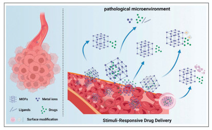
Figure 1.
Schematic illustration of MOF-based stimuli-responsive system for drug delivery. Image created with BioRender.com, with permission.
Engineered stimuli-responsive MOFs: Toward intelligent drug delivery systems for precision biomedicine
Wenting Wu , Zhao Chen , Man Zhe , Peiyun Yu , Sujan Shakya , Fei Xing , Ulrike Ritz

Stimuli-responsive systems, as an emerging drug delivery technology, effectively address the limitations of traditional drug components that rely on direct release [1-3]. These systems demonstrate multiple advantages, including high bioavailability, targeted distribution, controlled release, reduced dosing frequency, and minimized systemic side effects [4-6]. Within such systems, drugs remain stably encapsulated during transportation and achieve localized release at target sites upon accumulation or environmental stimuli activation [7-9]. The development of diverse stimuli-responsive carriers enables adaptability to the specific requirements of different administration routes and release environments [10,11]. Given these advantages, stimuli-responsive systems have garnered significant attention in the context of novel delivery systems and innovative drug development [12-15].
Metal-organic frameworks (MOFs), crystalline porous materials formed by coordination bonds between metal ions/clusters and organic ligands [16,17], exhibit extraordinary application potential across diverse fields. These including rechargeable batteries [18-20], gas and liquid separation [21-23], electrochemical sensors [24,25], catalysis [26,27], pollutant removal [28-30], and biomedicine [31-33]. Such versatility stems from their ultra-high specific surface area, tunable nanostructures, excellent biocompatibility, and uniformly distributed catalytic sites.
Recently, MOFs have gained prominence in stimuli-responsive drug delivery [34-37], due to their advantages manifested in several aspects: (a) Ultra-high porosity enabling high efficiency drug loading [38,39]. (b) Diverse pore architectures and surface properties that ensure compatibility with various drug molecules [40]. (c) Biodegradability to ensure delivery safety while minimizing carrier accumulation toxicity [41,42]. (d) Designable compositions and structures that respond to environmental stimuli via coordination bond cleavage, shell disintegration, or structural phase transitions [43,44].
Given rapid advances in stimuli-this review aims to (1) summarize recent progress, (2) identify emerging trends, and (3) provide design guidelines for clinically translatable delivery systems. This review systematically summarizes the primary synthesis methods of MOFs, classifies stimuli-responsive MOFs according to stimulus types, and elaborates on their drug release mechanisms and advancements in biomedical applications. Lastly, addressing the limitations identified in current research, it explores the challenges and future directions for stimuli-responsive MOFs in drug delivery (Fig. 1).
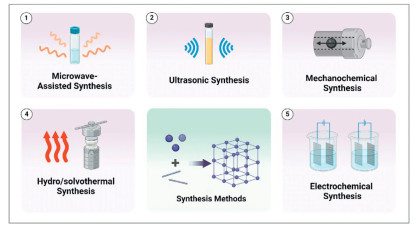
MOFs can be synthesized via diverse methodologies. Since slight variations in synthesis methods or reaction conditions significantly influence final product characteristics, careful optimization of these parameters is essential. We will outline several common MOFs synthesis techniques and their application scenarios (see Supporting information for details), thereby offering guidance for researchers to identify optimal synthetic strategies (Fig. 2).
Stimuli-responsive MOFs are intrinsically linked to precision medicine, serving critical functions in disease diagnosis, therapeutic intervention, and targeted drug delivery. We will discuss the specific applications of MOFs in precision medicine from two aspects: disease diagnosis and disease therapy, with detailed content provided in Supporting information.
In the context of drug delivery systems (DDS), diverse materials have been explored as drug carriers, each demonstrating distinct advantages and limitations in practical applications. In recent years, MOFs have garnered increasing attention in drug delivery due to their excellent physicochemical properties, functional versatility, and advanced encapsulation capabilities. We will detail the specific advantages of MOFs in the field of drug delivery, with specific content available in Supporting information.
Stimuli-responsive MOFs represent a class of intelligent materials that integrate the stimulus-responsive groups into their frameworks. These groups can specifically respond to environmental changes, triggering molecular-level structural transformations such as conformational rearrangements, hydrolytic cleavage, or protonation. Such dynamic responses enable precise control over the release of guest drug molecules [45]. This mechanism acts as the core regulatory unit of intelligent DDS, ensuring targeted drug release in specific organs. This section systematically elaborates on the response mechanisms of stimuli-responsive MOFs, focusing on ten key stimuli: redox reactions, enzyme, hydrogen sulfide (H2S), adenosine triphosphate (ATP), pH, temperature, magnetic fields, ions, humidity, and multi-stimuli conditions. Among them, the introduction to ions-, humidity-, and multi-stimuli-responsive mechanisms can be found in Supporting information.
Based on a comprehensive understanding of the drug delivery mechanisms of stimuli-responsive MOFs, this review aims to guide researchers in selecting stimuli that match the pathophysiological microenvironment of diseases, thereby facilitating the design of optimized intelligent DDS.
Redox homeostasis serves as a fundamental mechanism governing physiological activities in the human body. Disruption of redox equilibrium during these processes can lead to homeostasis breakdown, which may initiate or exacerbate pathological conditions, including cancer [46]. Given the marked redox concentration gradients between pathological and normal tissues, redox-responsive MOFs have emerged as promising platforms for achieving tissue-specific targeting and spatiotemporally controlled release of therapeutic agents, consequently attracting substantial research attention. Currently, redox-responsive MOF systems are primarily categorized into two types: GSH-responsive MOFs and glucose-responsive MOFs. This section will systematically elucidate the distinct activation mechanisms underlying each subtype.
GSH, a critical reducing tripeptide in biological systems, regulates cellular redox homeostasis through thiol-mediated interactions with oxidation-sensitive functional groups. In physiological processes, GSH modulates key signaling pathways governing cell proliferation, differentiation, and apoptosis via thiol modification of proteins [47]. Pathological conditions induce significant alterations in microenvironmental GSH levels. For example, the metabolic reprogramming of tumor cells induces reductive stress, elevating tumor microenvironment GSH concentrations far beyond those in normal tissues [48]. Capitalizing on this tumor-specific GSH overexpression, the engineering of GSH-responsive MOFs has emerged as a cornerstone strategy for precision tumor-targeted therapy.
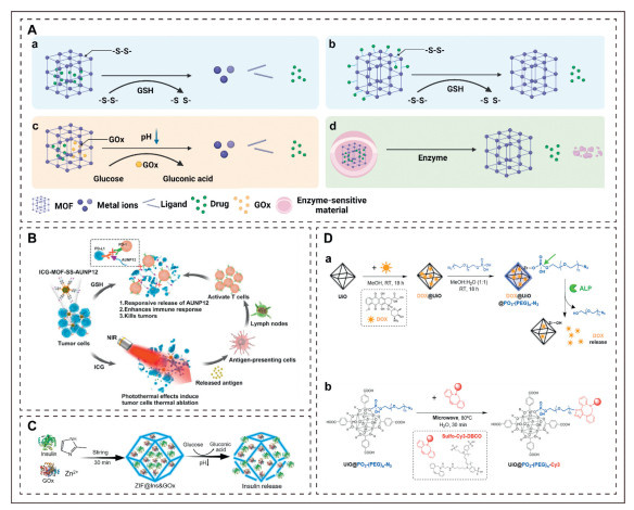
Central to this approach is the integration of disulfide bonds (-S-S-) as GSH-responsive motifs within MOF architectures. Upon entering high-GSH environments, disulfide bonds undergo reductive cleavage, triggering structural dissociation of MOFs and subsequent drug release. Lei et al. synthesized MOF-M(DTBA) (M = Fe, Al, Zr) carriers using 4,4′-dithiodibenzoic acid (4,4′-DTBA) as the ligand [49]. Experimental validation confirmed that disulfide bond cleavage under tumor-mimetic GSH conditions induced framework disintegration, enabling controlled release of the anticancer drug curcumin. In vitro studies demonstrated significantly enhanced antitumor efficacy compared to free curcumin. In contrast to conventional designs, Hao et al. engineered MOFs with dual therapeutic modules by anchoring AUNP12 peptides on the surface (Fig. 3) [50]. The first module responds to elevated tumor GSH levels, cleaving disulfide bonds to release AUNP12 peptides for programmed cell death protein-1 (PD-1)/programmed cell death ligand 1 (PD-L1) immune checkpoint inhibition. The second module uses co-encapsulated indocyanine green (ICG), that generates localized hyperthermia under NIR irradiation, enabling spatiotemporal controlled photothermal-immunotherapy.
The aforementioned studies demonstrate that GSH-responsive MOFs achieve spatiotemporally precise drug release and synergistic effects by leveraging microenvironment-specific disulfide cleavage mechanisms. Such advancements signify the evolution of redox-responsive DDS into intelligent platforms capable of multimodal therapeutic interventions.
Initially, glucose-responsive systems have garnered significant research attention in insulin delivery due to their unique properties. Studies demonstrate that glucose oxidase (GOx), functioning as a glucose-specific responsive component, catalyzes the conversion of glucose to gluconic acid in glucose-enriched microenvironments. This reaction induces localized acidification [53]. To enable glucose-triggered drug release, researchers utilized acid-labile MOFs such as MIL-100 [54], and ZIF-8 [51], which degrade in acidic environments. When insulin is co-encapsulated with GOx within these MOFs, the system achieves glucose-responsive insulin release (Fig. 3) [51]. GOx has emerged as key component in glucose-responsive systems.
Beyond insulin delivery, such materials demonstrate unique advantages in infected wound therapy. Given that inflammatory processes in infected wounds often coincide with glucose accumulation, researchers have developed GOx-based glucose-responsive MOFs for wound treatment. For instance, Wang et al. engineered a synergistic antibacterial platform by integrating GOx with gold nanozymes into ZIF-8 [55]. Upon glucose exposure, GOx and gold nanozymes initiate a cascade catalytic system that sequentially converts glucose to gluconic acid/H2O2 and generates hydroxyl radicals (•OH). Concurrently, gluconic acid lowers the microenvironmental pH, triggering ZIF-8 degradation and Zn2+ release, ultimately achieving synergistic antibacterial effects via OH and Zn2+.
Collectively, glucose-responsive MOFs exhibit versatile applicability across biomedical fields. Notably, unlike conventional pH-responsive materials, the acid-triggered dissociation here arises from enzymatic substrate conversion, enabling biomarker-specific responsiveness.
Enzyme-responsive strategies have been extensively employed in MOF-based DDS. For instance, in tumor tissues, dysregulated metabolism alters the balance between catabolism and anabolism, leading to significant overexpression of enzymes in the tumor microenvironment. Enzyme-responsive MOF systems achieve targeted drug release through recognition of these specifically overexpressed enzymes, thereby accomplishing precision therapeutics. For instance, capitalizing on the marked overexpression of alkaline phosphatase (ALP) in gastric cancer, pancreatic carcinoma, and osteosarcoma [56], the Carrion research team constructed an ALP-responsive intelligent DDS based on a Zr-based MOF (UiO-66) (Fig. 3) [52]. Using phosphate-Zr coordination interactions, the team anchored N3–(PEG)n–PO3 ligands onto the MOF surface, successfully creating both a stable PEG coating and clickable reactive sites. In the tumor microenvironment, the overexpressed ALP can specifically hydrolyze phosphate ester bonds, leading to the destabilization of the PEG coating and subsequent liberation of encapsulated doxorubicin. Furthermore, strategic exploitation of terminal azide moieties on the ligands enabled covalent conjugation of the fluorescent probe (Sulfo-Cy3-DBCO) to UiO@PO3–PEG–N3 particle surfaces via click chemistry, thereby establishing an integrated theranostic platform with precise drug-controlled release and real-time monitoring capabilities.
In the realm of tumor bone metastasis therapy, enzyme-responsive strategies demonstrate distinctive advantages. To address the critical challenge of inefficient drug delivery caused by the persistent adhesion of bone-targeting nanoparticles (NPs) to bone matrix in tumor bone metastasis therapy, Yang et al. designed a cascade delivery system with detachable targeting functionality [57]. This nanocarrier was composed of MMP enzyme-sensitive bone-targeting polypeptide ligand D8-M3, CD44-targeted hyaluronic acid (HA), and proteasome inhibitor bortezomib (Bortezomib)-loaded ZIF-8 NPs. Experiments demonstrated that the D8-M3-HA-ZIF8@BTZ NPs targeted bone tissue via D8-M3 peptides and detached from the bone matrix in response to tumor MMP enzymes. Concurrently, HA-mediated targeting enhanced endocytosis and cytotoxicity toward CD44-overexpressing tumor cells.
In summary, enzyme-responsive MOFs leverage enzymatic hydrolysis to specifically target abnormally expressed tumor enzymes, enabling precise drug delivery.
H2S, an endogenous gaseous signaling molecule, plays critical roles in physiological processes such as tumor immunotherapy [58], and protection against ischemia/reperfusion injury [59]. In specific pathological microenvironments, endogenous H2S concentrations are markedly higher than in normal tissues, positioning it as a potential target for stimuli-responsive therapeutic systems.
Leveraging these pathological features, Ma et al. proposed a novel MOF-based photosensitizer for colorectal cancer therapy. This system enables targeted photodynamic therapy (PDT) via a H2S-activated controllable singlet oxygen release mechanism within the tumor microenvironment [60]. The researchers constructed a fluorescent MOF system using a Zn-metalated porphyrin ligand (ZnTcpp) coordinated with Cu2+. In this system, Cu2+ completely quenched ligand fluorescence and suppressed ROS generation. H2S-mediated removal of Cu2+ nodes restores the photosensitive ligand's activity, allowing simultaneous fluorescence imaging and controlled therapy. Experimental results demonstrated that this system effectively responds to tumor microenvironmental signals and exerts antitumor effects.
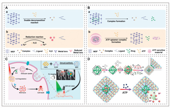
In a parallel study, Li et al. developed an H2S-responsive MOF-based photosensitive system through one-pot synthesis using porphyrin photosensitizer TCPP and Fe3+, enabling precision therapy for colorectal cancer (Fig. 4) [61]. Under high H2S concentration, Fe3+ reduction triggers MOF dissociation and TCPP release. This process simultaneously activates both fluorescence imaging and photodynamic therapy functions.
In the aforementioned studies, H2S-responsive MOFs activated photosensitizer via dynamic metal node modulation, either Cu2+-specific removal or Fe3+ reductive dissociation. This strategy overcomes the targeting limitations of conventional photosensitizers. Their synergistic "fluorescence imaging-PDT" mechanism provides a novel strategy for precision theranostics in colorectal cancer and related diseases.
As a pivotal nucleotide, ATP powers physiological processes via hydrolysis of its phosphoanhydride bonds. Studies demonstrate that ATP level fluctuations are closely linked to various pathologies. This correlation establishes ATP as a critical biomarker to differentiate pathological cells from normal counterparts [63]. ATP's nitrogen atoms possess abundant lone electron pairs, granting this metastable high-energy compound strong coordination capabilities. When nitrogen-containing groups, such as benzene rings, imidazole rings, and amino groups, expose lone electron pairs to metal ions in solution, coordination reactions would be induced [64]. ATP initiates framework dissociation and drug liberation via competitive coordination with metal sites in MOFs.
Building upon this mechanism, Chen et al. developed RhI-DOX@ZIF-90, an ATP-responsive NIR fluorescent nanoprobe. By co-loading the fluorophore RhI and doxorubicin into ZIF-90, they achieved tumor microenvironment-specific responsiveness and theranostic integration [65]. Upon detecting abnormally elevated ATP levels in tumor tissues, ATP competitively coordinates with Zn2+−2-imidazolecarboxaldehyde (2-ICA) linkages, triggering framework disassembly. This process simultaneously activates RhI's NIR fluorescence and release doxorubicin. Experimental validation demonstrated the nanoprobe's mitochondria-targeting capability, precise ATP level monitoring via fluorescence imaging, concurrent induction of tumor cell apoptosis, and effective in vivo antitumor therapy.
Beyond the aforementioned innovations, researchers have also explored designing ATP-responsive coatings on MOFs surfaces for controlled drug release. Chen et al., for example, encapsulated doxorubicin into MOFs coated with nucleic acid-based polyacrylamide hydrogels, achieving ATP-responsive tumor-specific drug delivery (Fig. 4) [62]. This system utilized hybridization chain reactions to synthesize duplex-bridged hydrogels embedded with caged ATP aptamers. Elevated ATP levels in tumor cells induce ATP-aptamer complex formation, leading to hydrogel disintegration and subsequent doxorubicin release. Experimental data revealed selective cytotoxicity against tumor cells.
These ATP-responsive strategies provide a novel pathway for intelligent drug delivery with broad therapeutic potential. However, further research is needed to translate these systems into clinical applications.
pH-responsive MOFs typically incorporate pH-sensitive functional groups that degrade in acidic environments, enabling controlled release of guest molecules [66]. Four main mechanisms drive drug delivery in pH-responsive MOFs. The dominant mechanism, protonation-induced coordination breaking, involves protonation of ionizable chemical groups in MOFs under acidic conditions. This causes charge reversal that disrupts metal-ligand coordination bonds. This structural disintegration leads to the release of guest molecules in acidic environments such as tumor sites [67]. Major acid-sensitive MOFs include carboxyl-rich MIL-n series [68,69], imidazolyl-based ZIF-n series [70,71]. Taking ZIF-8 as an example, under alkaline or neutral conditions, the nitrogen lone pairs in imidazolyl ligands demonstrate the capacity to coordinate with Zn2+ to form a stable tetrahedral framework. Under acidic conditions (pH < pKa1 = 7.85), protonation of 2-methylimidazole occupies the nitrogen lone pairs, preventing Zn2+ coordination and destabilizing the framework. This mechanistic alteration substantially accelerates the structural dissociation of ZIF-8.
Zhang et al. engineered a tumor-targeted MIL-100 platform coated with hyaluronic acid-polydopamine (HA-PDA), encapsulating GOx [54]. Upon exposure to acidic tumor microenvironments, the MIL-100 framework disintegrates to release GOx, which catalyzes glucose conversion into gluconic acid, thereby elevating local acidity and H2O2 levels. The MIL-100 then initiates Fenton-like reactions to convert H2O2 into cytotoxic •OH. Concurrently, the generated O2 augments GOx enzymatic activity, establishing a self-amplifying positive feedback loop. This cascade amplifies intracellular H+/H2O2/O2 accumulation gradients, synergistically suppressing tumors through oxidative stress and metabolic disruption. To improve siRNA stability and chemo-gene therapy synergy, Cai et al. designed PEG-CPP33@NPs, a pH-responsive MOF NPs modified with cell-penetrating peptides [72]. This system used ZIF-90 as a carrier to co-deliver the anticancer drug oridonin (ORI) and survivin siRNA, achieving lysosomal escape through the pH sensitivity of ZIF-90, and precisely releasing the drugs in the tumor microenvironment. Experiments confirmed that this system had good tumor selectivity and pH sensitivity, could be effectively internalized into the cytoplasm, and exhibited excellent gene silencing efficiency and antitumor effects. Yang et al. developed miR-34a-m@ZIF-8, a pH-responsive ZIF-8-based nano-delivery system [73]. This system utilized the pH-responsive properties of ZIF-8 to rapidly release miR-34a-m and Zn2+ in the lysosomes of tumor cells. Among them, Zn2+ induced tumor cell apoptosis by triggering the production of ROS. Simultaneously, the released miR-34a achieved a synergistic effect of gene therapy and chemodynamic therapy (CDT) by silencing the Bcl-2 gene. In another study, researchers developed ZGGO@ZIF-8, a novel core–shell nanoplatform combining chromium-doped Zn gallogermanate NPs (ZGGO PLNPs) with pH-responsive ZIF-8. This system enables dual imaging and drug delivery functions [74]. Leveraging the pH-responsive characteristics of ZIF-8, this platform exhibited accelerated drug release kinetics in the weakly acidic tumor microenvironment. Furthermore, the synergistic integration of imaging materials with MOF properties provides a novel strategy for precise tumor theranostics.
Another strategy uses acid-labile covalent bonds (e.g., hydrazine [75], epoxy bond [76], amide [77], and ether [78]) between drugs and MOFs for pH-responsive release. These covalent bonds generally maintain structural integrity under physiological pH conditions while undergoing hydrolytic cleavage in acidic microenvironments. As demonstrated in Zhang et al.'s study, the amino-rich chemotherapeutic doxorubicin forms amide bonds with surface acyl groups on ZIF-90. By exploiting the pH-dependent instability of the amide bonds, the DDS achieved sustained release kinetics at pH 5.5, attaining > 95% cumulative drug release within a 16-h period [79].
The construction of pH-sensitive coatings on MOFs surfaces represents another validated strategy [80], with commonly employed coating materials including chitosan [81], poly-histidine [82], glucose [83], gelatin polymer [84], and carboxymethylcellulose [85]. In a recent study addressing therapeutic challenges in triple-negative breast cancer (TNBC), Du et al. developed a multifunctional core/shell nanoreactor based on MOFs (Fig. 5) [80]. This system integrates a pH-responsive polymeric shell (PEG–CDM–PEI) with a ROS-sensitive degradable MOF core, enabling co-loading of glycolysis inhibitor glucose oxidase (GOD) and glutamine metabolism inhibitor BPTES for synergistic dual-metabolic blockade. In acidic tumor microenvironments, the polymeric shell degrades, releasing GOD and BPTES. These inhibitors synergistically enhance TNBC cytotoxicity by blocking glycolysis and glutamine metabolism. This work establishes a novel intelligent delivery strategy for TNBC therapy. In another study, Hang et al. proposed an innovative strategy for constructing an intelligent theranostic platform based on MOFs (Fig. 5) [83]. Using Gd3+-coordinated 5-boronobenzene-1,3-dicarboxylic acid (BBDC), this approach functionalized MOFs with glucose via diol-borate condensation. It simultaneously enhanced biocompatibility, tumor targeting, and controlled release. Experimental results demonstrated that the glucose coating not only improved tumor targeting via GLUT1 protein interaction but also served as a pH-responsive gatekeeper to prevent premature drug leakage. Concurrently, the Gd3+ ions endowed the system with MR imaging capabilities, successfully realizing MR imaging-guided precision chemotherapy.
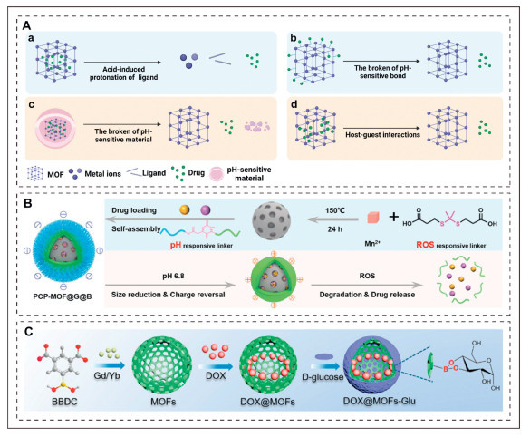
In addition to the aforementioned mechanisms, pH-responsive drug release can also be achieved through transformations in host-guest interactions within MOFs. For instance, Yang et al. developed a nanoscale cationic porous drug carrier, ZJU-101, via solvothermal synthesis, which demonstrates efficient loading of the anionic drug diclofenac sodium through dual mechanisms of ion exchange and permeation [86]. Experimental results revealed that ZJU-101 exhibits accelerated pH-responsive release kinetics in inflammatory microenvironments (pH 5.4) compared to physiological conditions (pH 7.4), mediated by competitive anion exchange between phosphate-buffered saline (PBS) anions and diclofenac anions.
In summary, pH-responsive MOFs enable precise drug release in acidic conditions via four key mechanisms: protonation-induced coordination bond cleavage, acid-labile covalent bonds, pH-sensitive coatings, and host-guest interactions. These systems show great promise for tumor and inflammation therapy, especially in targeted delivery and combination therapies, advancing the development of intelligent drug delivery platforms.
Temperature-responsive MOFs can dynamically sense temperature changes and regulate drug release behavior via structural phase transitions or modified intermolecular interactions. Poly(N-isopropyl acrylamide) (PNIPAM), a well-characterized temperature-sensitive polymer, exhibits a lower critical solution temperature (Tc) of 31 ℃. Below this threshold, PNIPAM is hydrophilic and water-soluble; above this threshold, it transitions to a hydrophobic aggregated state [87]. This behavior enables switchable PNIPAM-modified MOFs (e.g., UiO-66-PNIPAM) for temperature-responsive drug delivery (Fig. 6) [88]. Researchers loaded UiO-66-PNIPAM with resorufin, caffeine, and procainamide. The system remained stable at 40 ℃ but released drugs rapidly at 25 ℃, thus confirming its temperature-responsive release kinetics. Nagata et al. engineered a dual-responsive (temperature/pH) DDS by grafting P(NIPAM-AA) onto UiO-66 via post-synthetic modification (Fig. 6) [89]. At > 40 ℃ or pH < 4.95, the polymer collapses into a compact globular conformation, inhibiting procainamide diffusion. Conversely, at < 25 ℃ or pH > 6.86, the polymer extends into a coil conformation, enabling rapid drug release. In another study, Jiang et al. developed a Zr cluster-based MOF (ZJU-801) as a high-efficiency temperature-responsive drug delivery platform [90]. Naphthalene moieties in the organic ligands enhanced π-π interactions with diclofenac sodium. Experimental data demonstrated negligible drug release at ambient temperatures and significantly accelerated release kinetics at elevated temperatures. Structural analysis showed that naphthalene functionalization improved framework stability and created π-π stacking sites for drug anchoring. Moreover, thermal stimulation triggered release through disruption of these host-guest interactions.

Furthermore, Lin et al. constructed two Zn-based MOFs (ZJU-64 and ZJU-64-CH3) using zinc ions, adenine, and carboxylate ligands [91]. These isostructural MOFs exhibit temperature-responsive behavior with methotrexate (Methotrexate) loading capacities of 13.45 wt% (ZJU-64) and 10.63 wt% (ZJU-64-CH3). Drug release at 37 ℃ required 72 h, but at 60 ℃, equivalent release was achieved in 1.5 h (ZJU-64) and 6 h (ZJU-64-CH3). The researchers attributed this differential behavior to thermally enhanced disruption of host-guest interactions (e.g., hydrogen bonding and π-π stacking between aromatic moieties). Notably, methyl functionalization in ZJU-64-CH3 induced channel contraction compared to the parent ZJU-64, resulting in reduced cumulative drug release and attenuated release kinetics.
In summary, temperature-responsive MOFs regulate drug release via structural phase transitions or modified interactions, enabling temperature-dependent kinetics, and advancing intelligent DDS. However, the underlying temperature-responsive mechanisms require further investigation.
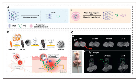
Magnetic-responsive drug delivery utilizes external magnetic fields to guide drug-loaded carriers to target sites and trigger stimuli-dependent drug release [92]. Owing to their material properties, magnetic-responsive systems offer advantages in magnetic hyperthermia, targeted delivery, separation, and MR imaging [93]. Fe3O4 is commonly used as the magnetic core in MOFs to enable magnetic responsiveness. For instance, a magnetic MOF-based drug carrier was fabricated by combining Fe3O4 nanorods with HKUST-1 (Cu3(BTC)2), demonstrating targeted delivery capabilities [94]. This system exhibits magnetic properties and high porosity, encapsulating nimesulide for targeted delivery and magnetic separation. Sharma et al. designed an iron carboxylate nanoMOF (M-NMOF) for co-delivering doxorubicin and methylene blue. This system combines magnetic guidance with light-activated therapy [95]. The aforementioned research findings indicate that magnetic-responsive MOFs have potential advantages in targeted cancer therapy. Chung et al. developed MagNORM, a composite of NO donor [Fe(μ-S-thioglycerol)(NO)2]2, MOF-derived Fe3O4@C, and poly(lactic-co-glycolic acid) (PLGA) microspheres. The temperature-responsive PLGA encapsulation enables controlled NO release (Fig. 7) [96]. Under alternating magnetic field (AMF), Fe3O4 generates heat, triggering PLGA phase transition and high-concentration NO release for broad-spectrum antibacterial effects. By modulating AMF frequency, MagNORM achieved dual-phase controlled NO release, providing an innovative solution for precise treatment of infected wounds. Moreover, due to their ability to simultaneously enable drug delivery and MR imaging, magnetic-responsive MOFs demonstrate unique application value in tumor treatment and tracking. Wang et al. created Fe3O4@C@MIL-100(Fe) (FCM), a nanoplatform with a Fe3O4@C core and MIL-100(Fe) shell, for dual imaging and therapy (Fig. 7) [97]. The system integrates carbon quantum dots for two-photon fluorescence imaging (TPFI) and hydrophobic MIL-100(Fe) pores to load DHA and Fe(Ⅲ) ions. In acidic tumor microenvironments, MIL-100(Fe) degradation enabled pH-triggered co-release of DHA and Fe(Ⅲ), while Fe3O4-mediated magnetic targeting enhanced drug accumulation at tumor sites. Combined MR/TPFI imaging allowed real-time therapeutic monitoring. Zhao et al. developed a Fe3O4@UiO-66 core-shell composite via by growing UiO-66 on carboxylate-terminated Fe3O4 cores. This dual-functional platform serves as a drug carrier and MR contrast agent, showing promise for cancer theranostics [98].
In summary, magnetic-responsive MOFs enable targeted drug delivery via external magnetic fields. Incorporating magnetic materials enhances capabilities in hyperthermia, targeted delivery, and MR imaging. These systems show broad potential in cancer therapy, antibacterial action, and real-time monitoring, advancing precision medicine strategies.
MOFs, emerging as advanced nanoscale drug carriers, exhibit unique potential for intelligent drug delivery owing to their exceptional biocompatibility, ultrahigh drug loading capacity, facile functionalization, and precisely tunable architectures. However, their clinical translation remains hindered by multifaceted challenges. First, current mechanistic studies remain largely theoretical, lacking in situ molecular characterization and dynamic tracking of key pathways in stimuli-responsive release. Second, validation systems over-rely on in vitro models. However, in vivo physiological complexity exceeds simulated conditions, making full replication impossible. Third, the dynamic behavior of MOF systems in pathophysiological microenvironments is poorly understood. Despite their apparent biocompatibility, systematic evaluation of long-term biosafety risks remains lacking. To address these bottlenecks, future research should integrate material characterization, physicochemical analysis, and release kinetics modeling to clarify microenvironment-triggered release mechanisms. Concurrently, there is a need to: (1) Develop experimental animal models replicating authentic pathological features, and (2) establish evaluation systems covering pharmacokinetics, spatiotemporal distribution, metabolic clearance, and long-term biosafety to assess biological effects. By integrating experimental testing, computational design, and stabilization strategies, we can develop MOFs with both high stability and multi-stimulus responsiveness. This approach ensures durable, reliable drug release with precise spatiotemporal control, accelerating clinical translation.
Numerous stimuli-responsive MOF-based DDS have been developed to date, demonstrating precise drug release triggered by specific stimuli. These systems exhibit significant potential for improving therapeutic outcomes in tumors and inflammatory diseases, underscoring MOFs' advantages in precision medicine. MOFs enable targeted drug delivery activated only under disease-specific conditions, maximizing therapeutic efficacy and minimizing side effects. As promising biomedical candidate materials, MOFs require standardized in vitro and in vivo studies to rigorously assess their long-term biosafety and efficacy. Developing MOFs that combine enhanced stability with multi-stimuli responsiveness is a critical research focus. Stable MOFs maintain structural integrity in biological environments long-term, ensuring durable and reliable drug release. Such systems can precisely control drug release by synergistically responding to multiple stimuli, enhancing treatment accuracy and accelerating clinical translation.
The authors declare that they have no known competing financial interests or personal relationships that could have appeared to influence the work reported in this paper.
Wenting Wu: Writing – review & editing, Writing – original draft. Zhao Chen: Writing – review & editing, Writing – original draft, Conceptualization. Man Zhe: Writing – review & editing, Visualization. Peiyun Yu: Visualization, Conceptualization. Sujan Shakya: Supervision, Conceptualization. Fei Xing: Writing – review & editing, Writing – original draft, Supervision, Conceptualization. Ulrike Ritz: Writing – review & editing, Writing – original draft, Supervision.
This work was supported by the National Natural Science Foundation of China (Nos. 32371420, 82202705), China Postdoctoral Science Foundation (No. 2023M742485), Clinical Research Incubation project of West China Hospital of Sichuan University (No. 2019HXFH041), Postdoctoral research and Development Foundation of West China Hospital of Sichuan University (No. 2024HXBH153). Graphical abstract was created with BioRender.com.
Supplementary material associated with this article can be found, in the online version, at doi:
Y. Song, H. Jing, L.B. Vong, et al., Chin. Chem. Lett. 33 (2022) 1705–1717. doi: 10.1016/j.cclet.2021.10.055
X. Ji, Y. Meng, Q. Wang, et al., ACS Nano 17 (2023) 5421–5434. doi: 10.1021/acsnano.2c10042
Y. Wu, Z. Wang, Y. Ge, et al., J. Control. Release 370 (2024) 747–762. doi: 10.3390/cryst14080747
S. Adepu, S. Ramakrishna, Molecules 26 (2021) 5905. doi: 10.3390/molecules26195905
Y. Wang, J. Yan, N. Wen, et al., Biomaterials 230 (2020) 119619. doi: 10.1016/j.biomaterials.2019.119619
Y. Wu, Y. Ge, Z. Wang, et al., J. Nanobiotechnol. 22 (2024) 188. doi: 10.1186/s12951-024-02465-w
Q. Zhou, L. Zhang, T. Yang, et al., Int. J. Nanomed. 13 (2018) 2921–2942. doi: 10.2147/ijn.s158696
D. Wei, Y. Sun, H. Zhu, et al., ACS Nano 17 (2023) 23223–23261. doi: 10.1021/acsnano.3c06019
T. Nie, H. Liu, Z. Fang, et al., ACS Nano 17 (2023) 10925–10937. doi: 10.1021/acsnano.3c02803
B. Gowda, M.G. Ahmed, A. Sahebkar, et al., Biomacromolecules 23 (2022) 1519–1544. doi: 10.1021/acs.biomac.1c01691
L. Mao, P. Ma, X. Luo, et al., ACS Nano 17 (2023) 9826–9849. doi: 10.1021/acsnano.3c02273
N. Deirram, C. Zhang, S.S. Kermaniyan, et al., Macromol. Rapid Commun. 40 (2019) e1800917. doi: 10.1002/marc.201800917
S. Lee, S.J. Song, J. Lee, et al., Pharmaceutics 12 (2020) 876. doi: 10.3390/pharmaceutics12090876
T. Wang, C. Wu, Y. Hu, et al., RSC Adv. 13 (2023) 16488–16511. doi: 10.1039/d3ra00866e
J. Liu, X. You, L. Wang, et al., Small 20 (2024) e2401438.
M.T. Khulood, U.S. Jijith, P.P. Naseef, et al., Int. J. Pharm. 673 (2025) 125380. doi: 10.1016/j.ijpharm.2025.125380
H. Chu, G. Liu, F. Wang, et al., Chin. Chem. Lett. 36 (2025) 110745. doi: 10.1016/j.cclet.2024.110745
X. Zhang, J. Qiao, Y. Jiang, et al., Nano-Micro Lett. 13 (2021) 135. doi: 10.1007/s40820-021-00658-8
Q. Zhang, S. Jiang, T. Lv, et al., Adv. Mater. 35 (2023) e2305532. doi: 10.1002/adma.202305532
Z. Liu, X. Zhang, J. Luo, et al., Chin. Chem. Lett. 35 (2024) 109500. doi: 10.1016/j.cclet.2024.109500
J. Li, P.M. Bhatt, J. Li, et al., Adv. Mater. 32 (2020) e2002563. doi: 10.1002/adma.202002563
J. Li, J.X. Wu, T. Liu, et al., Angew. Chem. Int. Ed. 64 (2025) e202411150. doi: 10.1002/anie.202411150
Y. Ma, W. Shi, G. Wang, et al., Chin. Chem. Lett. 36 (2025) 109729. doi: 10.1016/j.cclet.2024.109729
D.B. Gorle, S. Ponnada, M.S. Kiai, et al., J. Mat. Chem. B 9 (2021) 7927–7954. doi: 10.1039/d1tb01403j
Y. Liu, M. Liu, S. Shang, et al., ACS Appl. Mater. Interfaces 15 (2023) 16991–16998. doi: 10.1021/acsami.3c00005
Y. Liu, X. Zhou, W. Zhu, et al., Inorg. Chem. 62 (2023) 15022–15030. doi: 10.1021/acs.inorgchem.3c01874
L. Ma, F. Jiang, X. Fan, et al., Adv. Mater. 32 (2020) e2003065. doi: 10.1002/adma.202003065
I. Salahshoori, A. Vaziri, R. Jahanmardi, et al., ACS Appl. Mater. Interfaces 16 (2024) 26685–26712. doi: 10.1021/acsami.4c01365
A. Almahri, N.M. El-Metwaly, Int. J. Biol. Macromol. 276 (2024) 133909. doi: 10.1016/j.ijbiomac.2024.133909
P. Qin, S. Zhu, M. Mu, et al., Chin. Chem. Lett. 34 (2023) 108620. doi: 10.1016/j.cclet.2023.108620
K. Xiang, H. Wu, Y. Liu, et al., Theranostics 13 (2023) 2721–2733. doi: 10.7150/thno.83543
X. Ge, R. Wong, A. Anisa, et al., Biomaterials 281 (2022) 121322. doi: 10.1016/j.biomaterials.2021.121322
M.K. Sarangi, L.D. Patel, G. Rath, et al., Chin. Chem. Lett. 35 (2024) 109381. doi: 10.1016/j.cclet.2023.109381
Z. Guo, Y. Xiao, W. Wu, et al., J. Nanobiotechnol. 23 (2025) 157. doi: 10.7575/aiac.ijels.v.13n.2p.157
Y. Zeng, C. Zhang, Du D., et al., Acta Biomater. 145 (2022) 43–51. doi: 10.1016/j.actbio.2022.04.003
W. Cai, J. Wang, C. Chu, et al., Adv. Sci. 6 (2019) 1801526. doi: 10.1002/advs.201801526
L. Yang, S. Zhu, Z. He, et al., Chin. Chem. Lett. 33 (2022) 314–319. doi: 10.1016/j.cclet.2021.07.007
A. Hamedi, A. Anceschi, A. Patrucco, et al., J. Drug Target. 30 (2022) 381–393. doi: 10.1080/1061186x.2021.2012683
V. ClaudiaáCanale, D. MariaáCuscela, J. Mat. Chem. B 12 (2024) 3807–3839. doi: 10.1039/D4TB00312H
M. Eddaoudi, J. Kim, N. Rosi, et al., Science 295 (2002) 469–472. doi: 10.1126/science.1067208
F. Xing, J. Xu, Y. Zhou, et al., Nanoscale 16 (2024) 4434–4483. doi: 10.1039/d3nr05776c
D.S. Khafaga, M.T. El-Morsy, H. Faried, et al., RSC Adv. 14 (2024) 30201–30229. doi: 10.1039/d4ra04441j
H. Liu, F. Xing, P. Yu, et al., J. Mat. Chem. B 12 (2024) 8235–8266. doi: 10.1039/d4tb00768a
N. Srivastava, H. Chaurasia, A. Pandey, et al., Metal-organic frameworks (MOFs) for drug delivery: part B, in: B. Jain, D.K. Verma, A.K. Singh, et al. (Eds.), Metal Organic Frameworks, Elsevier, 2024, pp. 289–310.
S. Mura, J. Nicolas, P. Couvreur, Nat. Mater. 12 (2013) 991–1003. doi: 10.1038/nmat3776
Y. Ren, R. Wang, S. Weng, et al., Mol. Cancer 22 (2023) 130. doi: 10.1007/978-981-99-1639-9_11
R. Njälsson, S. Norgren, Acta Paediatr. 94 (2005) 132–137. doi: 10.1111/j.1651-2227.2005.tb01878.x
J. Li, M. Huo, J. Wang, et al., Biomaterials 33 (2012) 2310–2320. doi: 10.1016/j.biomaterials.2011.11.022
B. Lei, M. Wang, Z. Jiang, et al., ACS Appl. Mater. Interfaces 10 (2018) 16698–16706. doi: 10.1021/acsami.7b19693
Y. Hao, T. Liu, H. Zhou, et al., Front. Bioeng. Biotechnol. 11 (2023) 1294074. doi: 10.3389/fbioe.2023.1294074
C. Zhang, S. Hong, M. Liu, et al., J. Control. Release 320 (2020) 159–167. doi: 10.1016/j.jconrel.2020.01.038
C. Carrillo-Carrión, V. Comaills, A.M. Visiga, et al., ACS Appl. Mater. Interfaces 15 (2023) 27600–27611. doi: 10.1021/acsami.3c03230
Y. Duan, F. Ye, Y. Huang, et al., Chem. Commun. 54 (2018) 5377–5380. doi: 10.1039/c8cc02708k
Y. Zhang, L. Lin, L. Liu, et al., Biomaterials 216 (2019) 119255. doi: 10.1016/j.biomaterials.2019.119255
M. Wang, X. Zhou, Y. Li, et al., Bioact. Mater. 17 (2022) 289–299.
S.R. Rao, A.E. Snaith, D. Marino, et al., Br. J. Cancer 116 (2017) 227–236. doi: 10.1038/bjc.2016.402
H. Yang, Z. Yu, S. Ji, et al., RSC Adv. 12 (2022) 14707–14715. doi: 10.1039/d2ra00051b
T. Yue, J. Li, J. Zhu, et al., Cancer Res. 83 (2023) 595–612. doi: 10.1158/0008-5472.can-22-1837
S. Scheid, M. Goeller, W. Baar, et al., Int. J. Mol. Sci. 22 (2021) 10099. doi: 10.3390/ijms221810099
Y. Ma, X. Li, A. Li, et al., Angew. Chem. Int. Ed. 56 (2017) 13752–13756. doi: 10.1002/anie.201708005
H. Li, M. Huang, Z. Wei, et al., Front. Bioeng. Biotechnol. 10 (2022) 1032571. doi: 10.3389/fbioe.2022.1032571
W.H. Chen, W.C. Liao, Y.S. Sohn, et al., Adv. Funct. Mater. 28 (2018) 1705137. doi: 10.1002/adfm.201705137
G.G. Yegutkin, D. Boison, Pharmacol. Rev. 74 (2022) 799–824. doi: 10.1124/pharmrev.121.000528
X. Yang, Q. Tang, Y. Jiang, et al., J. Am. Chem. Soc. 141 (2019) 3782–3786. doi: 10.1021/jacs.8b11996
X. Chen, M. Hou, G. Mao, et al., Microchim. Acta 188 (2021) 287. doi: 10.1080/10864415.2021.1943170
M. Kanamala, W.R. Wilson, M. Yang, et al., Biomaterials 85 (2016) 152–167.
A.B. Lago, A. Pino-Cuevas, R. Carballo, et al., Dalton Trans. 45 (2016) 1614–1621. doi: 10.1039/C5DT04031K
J.J. Shen, S.J. Xue, Z.H. Mei, et al., Heliyon 10 (2024) e28066. doi: 10.1016/j.heliyon.2024.e28066
Z.J. Zhang, Y.K. Hou, M.W. Chen, et al., J. Nanobiotechnol. 21 (2023) 18.
Y. Ge, K. Wang, J. Liu, et al., Colloid Surf. B: Biointerfaces 212 (2022) 112354.
X. Meng, J. Guan, S. Lai, et al., RSC Adv. 12 (2022) 10005–10013. doi: 10.1039/d1ra09450e
M. Cai, Y. Yao, D. Yin, et al., J. Colloid Interface Sci. 646 (2023) 370–380. doi: 10.1016/j.jcis.2023.04.126
H. Zhao, T. Li, C. Yao, et al., ACS Appl. Mater. Interfaces 13 (2021) 6034–6042. doi: 10.1021/acsami.0c21006
Y. Lv, D. Ding, Y. Zhuang, et al., ACS Appl. Mater. Interfaces 11 (2018) 1907–1916.
Y. Liu, C.S. Gong, Y. Dai, et al., Biomaterials 218 (2019) 119365.
H. Chen, Z. Chen, Y. Kuang, et al., Colloids Surfaces B: Biointerfaces 167 (2018) 407–414.
H. Zhang, Q. Li, R. Liu, et al., Adv. Funct. Mater. 28 (2018) 1802830.
A. Cabrera-García, E. Checa-Chavarria, E. Rivero-Buceta, et al., J. Colloid Interface Sci. 541 (2019) 163–174.
F. Zhang, H. Dong, X. Zhang, et al., ACS Appl. Mater. Interfaces 9 (2017) 27332–27337. doi: 10.1021/acsami.7b08451
H. Du, S. Meng, M. Geng, et al., Small 19 (2023) 2303253.
V. Nejadshafiee, H. Naeimi, B. Goliaei, et al., Mater. Sci. Eng. C 99 (2019) 805–815.
Y. Yang, L. Xu, W. Zhu, et al., Biomaterials 156 (2018) 121–133.
H. Zhang, Y. Shang, Y. Li, et al., ACS Appl. Mater. Interfaces 11 (2018) 1886–1895. doi: 10.1109/jsyst.2016.2600582
A. Karakeçili, B. Topuz, S. Korpayev, et al., Mater. Sci. Eng. C 105 (2019) 110098.
S. Javanbakht, M. Pooresmaeil, H. Hashemi, et al., Int. J. Biol. Macromol. 119 (2018) 588–596.
Y. Yang, Q. Hu, Q. Zhang, et al., Mol. Pharm. 13 (2016) 2782–2786. doi: 10.1021/acs.molpharmaceut.6b00374
D. Roy, W.L. Brooks, B.S. Sumerlin, Chem. Soc. Rev. 42 (2013) 7214–7243. doi: 10.1039/c3cs35499g
S. Nagata, K. Kokado, K. Sada, Chem. Commun. 51 (2015) 8614–8617.
S. Nagata, K. Kokado, K. Sada, CrystEngComm 22 (2020) 1106–1111. doi: 10.1039/c9ce01731c
K. Jiang, L. Zhang, Q. Hu, et al., Chemistry 23 (2017) 10215–10221. doi: 10.1002/chem.201701904
W. Lin, Q. Hu, J. Yu, et al., ChemPlusChem 81 (2016) 804–810. doi: 10.1002/cplu.201600142
C.S. Kumar, F. Mohammad, Adv. Drug Deliv. Rev. 63 (2011) 789–808.
R. Hergt, S. Dutz, R. Müller, et al., J. Phys.: Condens. Matter 18 (2006) S2919.
F. Ke, Y. Yuan, L. Qiu, et al., J. Mater. Chem. 21 (2011) 3843–3848. doi: 10.1039/c0jm01770a
S. Sharma, K. Sethi, I. Roy, New J. Chem. 41 (2017) 11860–11866.
C. Chung, B. Liao, S. Huang, et al., ACS Appl. Mater. Interfaces 14 (2022) 6343–6357. doi: 10.1021/acsami.1c20802
D. Wang, J. Zhou, R. Chen, et al., Biomaterials 107 (2016) 88–101.
H. Zhao, Q. Zou, S. Sun, et al., Chem. Sci. 7 (2016) 5294–5301.

Figure 1 Schematic illustration of MOF-based stimuli-responsive system for drug delivery. Image created with BioRender.com, with permission.

Figure 2 Schematic illustration of MOFs synthesis techniques. Image created with BioRender.com, with permission.

Figure 3 (A) Mechanisms of MOF-based redox-responsive (a–c) and enzyme-responsive (d) DDS: (a, b) GSH-responsive; (c) glucose-responsive; (d) enzyme-responsive. (B) Schematic representation of the anti-tumor activity of ICG-MOF-SS-AUNP12 NPs through synergistic photothermal and immunotherapy. Copied with permission [50]. Copyright 2023, The Authors. (C) Synthesis of ZIF@Ins & GOx and its glucose-responsive insulin release mechanism. Copied with permission [51]. Copyright 2020, Elsevier Ltd. (D) (a) Synthesis route of UiO@PO3–PEG–N3 and its enzyme-responsive release mechanism; (b) schematic diagram of the modification of Sulfo-Cy3-DBCO on the surface of UiO@PO3–PEG–N3. Copied with permission [52]. Copyright 2023, American Chemical Society.

Figure 4 (A) Mechanisms of MOF-based H2S-responsive DDS: (a) Double decomposition reaction; (b) reduction rection. (B) Mechanisms of MOF-based ATP-responsive DDS: (a) Complex formation; (b) ATP-aptamer complex formation. (C) Synthetic route of MOF and its role in fluorescence imaging-guided PDT. Copied with permission [61]. Copyright 2022, The Authors. (D) Synthesis of ATP-responsive DNA/polyacrylamide-hydrogel-coated NMOFs loaded with a dye or doxorubicin, and its release performance via the formation of ATP–aptamer complexes. Copied with permission [62]. Copyright 2018, Wiley-VCH GmbH.

Figure 5 (A) Mechanisms of MOF-based pH-responsive DDS: (a) Protonation-induced bond breakage; (b) pH-cleavable bonds; (c) pH-induced shell disintegration and (d) host-guest interactions. (B) Synthetic route of PCP-MOF@G@B and its pH-responsive release mechanism. Copied with permission [80]. Copyright 2023, Wiley-VCH GmbH. (C) Synthetic route of DOX@MOFs-glucose nanocarrier. Copied with permission [83]. Copyright 2018, American Chemical Society.

Figure 6 (A) Mechanisms of MOF-based temperature-responsive DDS: (a) Alterations of intermolecular interactions; (b) structural phase transitions. (B) Temperature-responsive behavior of UiO-66-PNIPAM. Copied with permission [88]. Copyright 2015, The Royal Society of Chemistry. (C) Synthetic route of UiO-66–P(NIPAM–AA) and its temperature-responsive release mechanism. Copied with permission [89]. Copyright 2020, The Royal Society of Chemistry.

Figure 7 (A) Mechanisms of MOF-based magnetic-responsive DDS: (a) Magnetic targeting; (b) magnetic hyperthermia. (B) Schematic illustration of the preparation of MagNORM and its mechanism of action in infected wounds. Copied with permission [96]. Copyright 2022, American Chemical Society. (C) Transverse (a) and coronal (c) MR images of a HeLa-xenografted tumor in vivo before and after intravenous injection of FCM. Copied with permission [97]. Copyright 2016, Elsevier Ltd.

 扫一扫看文章

扫一扫关注我们
 DownLoad:
 下载:
下载:
 下载:
下载: