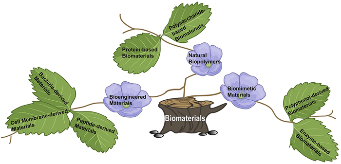
Figure 1.
The classification of novel biomaterials for DFUs regulation.
Biomaterials for regulating the micro-environment of diabetic foot ulcers
Qun Zhang , Zhengkun Liu , Keke Wu , Zhipeng Zhang , Weisheng Guo , Xiaoying Guan

Diabetic foot ulcers (DFUs) represent a highly prevalent and therapeutically challenging complication in diabetic patients, arising from the complex interplay between peripheral neuropathy and peripheral artery disease, which is further exacerbated by chronic hyperglycemia. Pathophysiological mechanisms include hyperglycemia-driven glycosylation of hemoglobin, leading to vasoconstriction and altered erythrocyte membrane properties, ultimately resulting in tissue hypoxia [1]. The advanced glycation end products (AGEs) can also contribute to oxidative damage, alter the extracellular matrix (ECM), and trigger the release of pro-inflammatory cytokines [2]. These processes are compounded by persistent inflammation at the wound site, creating a vicious cycle of tissue damage. Furthermore, the accumulation of endogenous reactive oxygen species (ROS) impairs immune surveillance and cellular homeostasis. Conventional treatment strategies, which primarily rely on surgical interventions (encompassing debridement, offloading, antibiotic therapy, glycemic control, and advanced wound care), often demonstrate limited efficacy in concurrently addressing these multifaceted pathological processes [3-5]. Of particular concern, repeated surgical debridement and the emergence of antibiotic-resistant pathogens escalate healthcare costs and contribute to a high recurrence rate (80%) following initial wound closure, significantly complicating diabetes management [6].
Contemporary advancements in biomaterial science have introduced a spectrum of innovative wound care solutions to augment clinical management strategies for DFUs. Collagen (COL)-based matrices are frequently employed to enhance cellular motility and tissue reconstitution, leveraging their inherent biocompatibility and structural similarity to native extracellular components. Hydrogel formulations are designed to maintain a physiologically moist microenvironment, facilitating autolytic debridement and mitigating pain, while silver-incorporated biomaterials provide potent antimicrobial activity to diminish microbial burden. Despite these advancements, commercially available biomaterials exhibit inherent limitations in their capacity to recapitulate the dynamic complexity of the native ECM within the DFUs microenvironment [7]. These shortcomings highlight the imperative for next-generation therapeutic approaches. The present review integrates a comprehensive analysis of distinct diabetic wound microenvironments with corresponding biomaterial-based therapeutic strategies, with the objective of equipping clinicians and researchers with actionable insights to guide the development and deployment of context-specific, efficacious interventions.
The pathophysiology of DFUs is multifaceted, involving intricate interactions between intrinsic and extrinsic factors. The internal wound microenvironment is marked by a constellation of pathological features, including angiogenesis impairment, persistent inflammation, ECM remodeling, and neurovascular dysregulation. Hyperglycemia-induced autonomic neuropathy compromises skin integrity and enhances susceptibility to microbial colonization [8]. Concurrent reductions in tissue perfusion and oxygen tension, coupled with microvascular dysfunction and elevated pro-inflammatory cytokine profiles, synergistically disrupt reparative processes. Aberrant ECM architecture impedes cellular motility, proliferation, and differentiation, thereby further impeding wound closure [9]. Externally, the wound microenvironment is subjected to diverse mechanical, thermal, and chemical stressors. Elevated plantar pressures, shear forces, and frictional forces exacerbate tissue injury and provoke inflammatory responses, fostering ulcer progression. This interplay of internal and external factors establishes a complex pathophysiological milieu that significantly challenges effective DFUs management. Comprehensive elucidation of these intricate mechanisms is essential to guide the rational design of targeted therapeutic interventions that address the specific microenvironmental challenges inherent to DFUs healing.
Hyperglycemia exerts a profound inhibitory effect on the critical transition from the inflammatory to proliferative phase of wound healing. It compromises fibroblast functionality and collagen (COL) biosynthesis while perturbing intracellular redox homeostasis [10]. Concurrently, the hyperglycemic state fosters bacterial biofilm formation and stabilization in the wound bed, contributing to the persistence of open wounds for durations of 4–6 weeks. During this protracted period, the subcutaneous tissues of chronic, non-healing ulcers remain vulnerable to environmental exposures, significantly increasing the risk of complications such as osteomyelitis, hemorrhagic episodes, and potentially fatal outcomes in severe instances [11]. Moreover, elevated glucose levels disrupt the stabilization and functional capacity of hypoxia-inducible factor-1 alpha (HIF-1α), a pivotal transcriptional regulator of hypoxic cellular responses. This dysregulation not only exacerbates impairments in neovascularization but also contributes to the broader spectrum of diabetic complications [9,12].
DPN, a symmetrical, length-dependent sensorimotor polyneuropathy, arises from metabolic and microvascular alterations secondary to chronic hyperglycemia and concomitant cardiovascular risk factors. While not directly contributing to DFUs pathophysiology, DPN exerts a profound indirect influence on ulcer development and progression, warranting its careful consideration in DFUs management strategies. Sensory deficits in the lower extremities predispose patients to unnoticed traumatic injuries and repetitive mechanical stress, facilitating skin breakdown and ulcer initiation [8,13,14]. Sensory symptoms, including anesthesia and neuropathic pain, often progress to severe morbidity, including limb amputation [15]. The economic burden associated with DFUs treatment and amputation is substantial, while DPN-related impairments in postural control and affective regulation contribute to psychological distress [16]. Furthermore, DPN-mediated alterations in foot biomechanics and gait increase ulceration risk, while neuroinflammatory processes within affected nerves precipitate pain hypersensitivity, impaired wound healing, and heightened susceptibility to infection [17,18]. Insulin resistance and metabolic perturbations in neuronal tissues exacerbate these pathological sequelae.
In the context of DFUs, hypoxia represents a critical microenvironmental feature that synergizes with persistent inflammation to further compromise circulatory impairment [19]. The resultant oxygen deprivation disrupts the functional capacity of key wound-healing cellular populations, including fibroblasts, endothelial cells, and immune effectors [20,21]. This physiological deficit decelerates reparative processes and establishes a permissive niche for bacterial colonization and progressive tissue destruction [22]. The hypoxic milieu thus constitutes a substantial therapeutic challenge in DFUs management, as it amplifies oxidative stress while impeding the biosynthesis of essential regenerative proteins and enzymes [23].
HIF-1α serves as a pivotal transcriptional regulator in the cellular adaptive response to hypoxic stress, exerting profound effects on DFUs pathophysiology. This oxygen-sensitive factor orchestrates the expression of genes encoding erythropoietin (EPO) and vascular endothelial growth factor (VEGF), thereby stimulating erythropoiesis and promoting angiogenesis, respectively [12]. Beyond these systemic effects, HIF-1α directly modulates critical wound-healing processes, including granulation tissue formation, wound contraction, and re-epithelialization. Under normoxic conditions, HIF-1α is subject to proline hydroxylation by prolyl hydroxylase domain enzymes (PHDs), targeting it for proteasomal degradation. However, in hypoxic environments, diminished PHD activity stabilizes HIF-1α, enabling its accumulation and the activation of adaptive gene programs that enhance tissue tolerance to oxygen deprivation [24]. Recognizing the dual imperative of optimizing oxygen delivery while harnessing endogenous HIF-1α-mediated adaptive mechanisms underscores the complexity of therapeutic strategies aimed at accelerating DFUs resolution.
Dysregulated pH homeostasis represents a hallmark of DFU, significantly impacting multiple facets of wound biology, including angiogenesis, hemodynamic regulation, infectious susceptibility, inflammatory dynamics, and ECM remodeling. Temporal fluctuations in wound exudate pH and metabolic profiles emerge as critical biomarkers of healing progression. Notably, acute wounds and healthy skin maintain an acidic microenvironment (pH 4–6), whereas chronic and infected ulcers exhibit an alkaline shift (pH 7.3–9), which fosters a microbial-permissive niche [24]. Elevated pH not only enhances the proliferation of bacterial pathogens and opportunistic microorganisms [25] but also skews macrophage polarization toward the pro-inflammatory M1 phenotype. This phenotypic switch is accompanied by the secretion of cytokines such as interleukin-1β (IL-1β) and monocyte chemoattractant protein-1 (MCP-1), which collectively impede wound resolution [26]. Furthermore, the disruption of M1-to-M2 macrophage transition under alkaline conditions compromises the reparative functions of VEGF, particularly its capacity to promote M2 macrophage differentiation [27]. Elucidating the pH-dependent modulation of macrophage polarization provides mechanistic insights into DFUs pathogenesis and informs the development of targeted therapeutic interventions.
Chronic wounds in diabetic patients are characterized by a protracted inflammatory phase, frequently compounded by dysbiotic microbial colonization. The skin microbiota plays a pivotal role in maintaining epidermal homeostasis and orchestrating wound repair processes [28]. In diabetic ulcers, the alkaline wound microenvironment facilitates the overgrowth of diverse microbial communities, encompassing Staphylococcus, Streptococcus, Pseudomonas, Corynebacterium species, as well as anaerobic organisms, culminating in recalcitrant infections [15,29]. The formation of biofilms further complicates therapeutic interventions by conferring protection against both host immune responses and antimicrobial therapies. The accumulation of necrotic tissue and cellular debris provides a fertile substrate for microbial proliferation. Notably, anaerobic bacteria are more prevalent in deeper wound strata, correlating with clinical indicators such as pyrexia, malodorous exudate, and ulcer severity [15]. This intricate interplay between microbial dysbiosis, biofilm formation, and impaired host defenses underscores the complexity of infection management in diabetic chronic wounds.
At the wound interface, infiltrating neutrophils and monocytes evoke a pro-inflammatory cascade through the secretion of the tumor necrosis factor-α (TNF-α), augmenting tissue injury and inflammation. Infectious processes and hyperglycemia synergistically trigger the release of neutrophil extracellular traps (NETs), which contribute to delayed wound closure. Elevated NET concentrations correlate with impaired wound healing and diminished expression of growth factors and pro-angiogenic proteins, such as platelet-derived growth factors (PDGFs) and VEGF. Additionally, certain commensal bacterial species elicit a Th17-mediated immune response, further escalating inflammation and hindering repair mechanisms [30]. Fig. S1 (Supporting information) illustrates the principal pathogenic factors in diabetes and their roles in DFUs progression. Notwithstanding these challenges, certain skin-resident microorganisms, such as members of the actinomycetes’ order, synthesize antibacterial metabolites that promote wound resolution. The maintenance of a balanced microbial ecosystem by these organisms is essential for optimal healing outcomes. Emerging therapeutic strategies leveraging probiotics and microbiota modulation are under investigation as potential adjuvants to enhance DFUs management [22].
Immune effector cells, including neutrophils and classically activated (M1) macrophages, augment the expression of matrix metalloproteinase-9 (MMP-9), which facilitates the transition from acute to chronic wound states by degrading ECM components and growth factors critical for tissue repair [31,32]. Aberrant serine protease activity and dysregulation of cysteine proteases further exacerbate tissue proteolysis, thereby suppressing growth factor bioavailability and impeding healing progression [33]. Pro-inflammatory cytokines, such as TNF-α and IL-1, amplify MMP-9 production, establishing a self-perpetuating cycle of inflammation and matrix destruction [34]. Overexpression of MMP-9 additionally compromises angiogenic responses and re-epithelialization processes [35].
In the context of DFUs, aberrant COL and fibronectin deposition can result in pathological fibrosis and hypertrophic scarring [36]. Ceramides, in concert with cholesterol and fatty acids, constitute essential extracellular lipid layers that preserve skin barrier function [37]. Infection by Pseudomonas aeruginosa, among other pathogens, induces ceramidase (CDase) expression, which not only enhances bacterial colonization and invasive capabilities but also disrupts host tissue integrity. Bacterial pathogens themselves secrete CDase-like enzymes that are integral to their growth, persistence, and virulence mechanisms [38]. Notably, the development of DFUs involves complex interactions between diverse proteases and bioactive molecules, giving rise to distinct proteolytic signatures among affected individuals.
Contemporary multifunctional biomaterials, integrating antibacterial, anti-inflammatory, and hemostatic functionalities, are progressively supplanting conventional materials limited to physical barrier functions and isolation [39]. Within the realm of tissue engineering, these advanced biomaterials function as dynamic scaffolds for cellular growth and expansion, simultaneously serving as controlled delivery systems for therapeutic agents and bioactive molecules. Under precisely regulated conditions, they enable the de novo synthesis of functional tissues or organs to restore compromised physiological structures [40]. The escalating utilization of natural biopolymers in DFUs management reflects their inherent advantages, particularly their biocompatibility and biodegradable nature, which synergistically promote wound healing. Furthermore, bio-mimetic biomaterials have revolutionized wound care paradigms by recapitulating the structural and functional complexities of the native ECM, thereby augmenting cellular motility, proliferation, and facilitating spatially controlled drug release.
Protein-polysaccharide bio-composites, a class of natural biopolymers, provide an absorbable and biocompatible scaffolding that effectively manages wound exudate. Their intrinsic antibacterial activity and biodegradable characteristics collectively contribute to accelerated wound healing. The comprehensive analysis presented in Table S1 (Supporting information) elucidates both the advantageous properties and the current challenges associated with the clinical translation of these biopolymer-based wound care strategies.
COL, a versatile biopolymer available in diverse configurations, can be strategically integrated with growth factors, pharmacological agents, or antimicrobial compounds to augment wound repair processes [36,41]. A notable advancement in this domain is the development of epidermal growth factor (EGF)-loaded polydopamine-modified COL-hyaluronic acid (HA) scaffolds (EGF-loaded CHS-PDA) by Wang et al., engineered for the repair of diabetic cutaneous lesions. These scaffolds demonstrate exceptional mechanical resilience, biocompatibility, and structural coherence, while enabling sustained EGF release and promoting cellular proliferation, coagulation, and ROS detoxification [42]. Although supplementary incorporation of VEGF, stromal cell-derived factor 1 (SDF-1), and PDGF represent viable strategies, COL-based biomaterials may encounter limitations in effectively addressing tissue fluid exudation, inflammatory responses, and infection risks. To circumvent these challenges, the incorporation of rare-earth elements such as praseodymium-cobaltite into COL matrices has shown promise in enhancing angiogenic potential and guiding mesenchymal stem cell (MSC) differentiation towards endothelial lineages [43]. Parallel innovations in COL crosslinking methodologies have further advanced the field, as exemplified by the research of Zhe et al [44]. This work emphasizes the optimization of COL casing crosslinking with glutaraldehyde (GTA) through precise pH modulation to induce the formation of conjugated structural motifs. Under mild alkaline conditions (pH 8–10), GTA undergoes accelerated polymerization, resulting in the generation of conjugated structures such as -C = C—C = O and -N = C—C = C-. These structural modifications not only elevate crosslinking efficiency but also confer enhanced mechanical robustness, thermal stability, and a reduced swelling ratio to COL films. Of clinical relevance, the minimized swelling ratio of such crosslinked COL under the specific pH environment of DFUs enables superior management of wound exudates, thereby maintaining an optimal moisture balance conducive to healing. Li et al. [45] engineered an injectable COL/chitosan (CTS)/genipin hydrogel system, further augmented with phycocyanin nanoparticles and ND-336, tailored for the specific demands of diabetic wound management. This hydrogel formulation was designed to overcome several inherent limitations associated with COL, namely its suboptimal mechanical robustness, rapid biodegradation, and insufficient antimicrobial efficacy. The strategic incorporation of genipin, a naturally derived crosslinker, not only mitigated cytotoxicity concerns but also enhanced the overall biocompatibility of the hydrogel matrix. Preclinical evaluations of this hydrogel demonstrated significant potential in facilitating fibroblast migration, suppressing the expression of MMP-9, a key mediator of ECM degradation, and ultimately accelerating wound closure rates. These findings collectively underscore the hydrogel’s promise as a multifaceted therapeutic platform addressing critical challenges in diabetic wound care.
Platelet-rich plasma (PRP) is characterized by a high concentration of platelets and a rich array of growth factors, including VEGF, PDGF, and fibroblast growth factor (FGF). These bioactive components exhibit multifaceted therapeutic effects, such as chemoattraction of endogenous progenitor cells, induction of M2 macrophage polarization, attenuation of inflammatory responses, and stimulation of cellular migration and proliferation [28]. Despite its therapeutic potential, PRP is hindered by inherent limitations, including a brief bioactive half-life, rapid biodegradation, and non-injectable formulation, which complicates its clinical translation. To address these challenges, recent studies have explored the use of functionalized polyethylene glycol (PEG) to crosslink CTS, creating a sophisticated hydrogel delivery system [46]. This engineered hydrogel facilitates sustained release of PRP components, enhances resistance to enzymatic degradation, and fosters angiogenesis. Furthermore, the degradation products of the hydrogel contribute to a microenvironment conducive to cellular proliferation, inflammation resolution, and COL biosynthesis, collectively advancing tissue repair and regeneration.
Sericin (Ser), a hydrophilic protein derived from silkworm cocoons, has garnered significant attention in the realm of hydrogel-based wound dressing development. Its biocompatibility, natural degradability, and multifaceted biological activities, encompassing anti-aging, anticoagulant, and anti-tyrosinase properties, render it particularly attractive for biomedical applications. Of particular interest is Ser’s demonstrated proficiency in stimulating the proliferation and growth of fibroblasts and keratinocytes, underscoring its therapeutic potential in wound healing paradigms [47]. Through chemical modification with methacrylic anhydride (MA), Ser is transformed into methacrylic anhydride Ser (SerMA), a derivative that exhibits enhanced functional attributes. SerMA not only retains the inherent bioactivity of native Ser but also acquires injectability, adhesive characteristics, and superior biocompatibility, thereby expanding its utility in advanced biomaterial designs. These attributes collectively position SerMA as a promising candidate for next generation wound care strategies.
Alginate (Alg), a biocompatible and gel-forming polysaccharide extracted from brown algae, has emerged as a versatile biomaterial in the development of medical dressings and drug delivery systems [48]. Its unique capability to crosslink with divalent cations, such as barium [49] and calcium [50], enables the formation of hydrogels with enhanced mechanical properties, including improved mechanical strength, elasticity, thermal stability, and reduced swelling [51]. These attributes make Alg-based hydrogels particularly suitable for managing exudative wounds [52]. In a recent study, a three-dimensional scaffold (PANB) was engineered by cross-linking Alg with Ca2+ using multi-vinyl-modified gold nanorods. This innovative approach not only augmented the hydrogel’s mechanical integrity and thermal stability but also facilitated effective near-infrared (NIR) photothermal therapy for diabetic mice with oral mucosa ulcers and skin lesions. The treatment exhibited remarkable antibacterial activity and promoted angiogenesis, highlighting its potential in advanced wound care strategies [53]. Notably, Alg’s therapeutic efficacy extends beyond NIR-assisted applications, as evidenced by its ability to manage DFUs independently of photothermal intervention. Another study demonstrated the functionalization of Alg with poly-imidazolium-maleimide to create a novel hydrogel (Alg-PPN). This hydrogel accelerated wound healing, potentiated bactericidal effects, and mitigated pro-inflammatory markers, including matrix MMP-9 and ROS, in diabetic mouse models infected with antibiotic-resistant bacteria [54]. Collectively, these findings underscore the multifaceted therapeutic potential of Alg-based biomaterials in addressing complex wound care challenges.
CTS, a biopolymer derived from the deacetylation of chitin, exhibits procoagulant and thrombogenic properties attributable to its cationic nature [55]. The presence of multiple reactive amino groups in CTS confers significant advantages in promoting diabetic wound healing; however, its inherent solubility limitations have impeded its widespread application in antibacterial wound dressings [56,57]. To address this challenge, recent studies have explored chemical modifications of CTS, such as conjugation with lipoic acid to enable UV-induced gelation or introduction of quaternary ammonium moieties to enhance aqueous solubility and broaden the spectrum of antibacterial activity [58]. An injectable polysaccharide hydrogel dressing (QOP), composed of quaternized chitosan (QCS) and oxidized β-glucan, has been developed. This hydrogel system incorporates polydopamine nanoparticles (PDA NPs) and demonstrates remarkable photothermal bactericidal efficacy against Staphylococcus aureus (S. aureus) and Escherichia coli (E. coli) upon exposure to NIR laser irradiation. These advancements highlight the potential of CTS-based biomaterials in innovative wound care solutions.
A critical determinant of platelet regulation is the surface charge characteristic of biomaterials. Exposure to positively charged surfaces triggers the rapid accumulation of blood cells, a phenomenon attributable to the relatively large size, expansive surface area, and morphological alterations exhibited by activated platelets. These interactions precipitate the swift formation of fibrin-dependent blood clots [59]. Capitalizing on this principle, the amino, hydroxyl, and other polar functional groups inherent to CTS may hold substantial promise for hemostatic applications. By exploiting the propensity of positive charges to induce blood cell aggregation and efficient platelet activation, CTS-based biomaterials could augment coagulation mechanisms, thereby presenting novel avenues for wound care and hemorrhage control.
HA, a non-sulfated glycosaminoglycan component of the ECM, plays a pivotal role in angiogenesis, tissue repair, and wound healing. However, its inherent limitations, including poor protein affinity, rapid enzymatic degradation, and absence of intrinsic antibacterial activity, have restricted its full therapeutic potential [60]. To address these shortcomings, chemical modification strategies have been employed, such as the sulfation process developed by Zhou, utilizing sulfur trioxide-pyridine complex and ethanol precipitation, which effectively enhances HA’s functional properties [61]. The resulting sulfated hyaluronic acid (SHA) was integrated with COL to fabricate SHA-COL composites and SHA/HA/COL hybrid nanofibers. These biomaterial constructs were further reinforced with polyurethane (PU) to impart improved mechanical strength and flexibility. These advanced scaffolds demonstrated remarkable efficacy in accelerating diabetic wound healing and skin remodeling processes, significantly enhancing tissue regeneration and repair in DFUs.
The advancement of therapeutics is significantly propelled by the systematic classification of biomaterials originating from polyphenols and enzymes within this domain. For a comprehensive understanding of the advantages and current limitations associated with bio-mimetic biomaterials, Table S2 (Supporting information) demonstrates a detailed analysis of these aspects.
The unique enzyme-like characteristics, coupled with their facile synthesis and biocompatibility, have rendered nanozymes a valuable asset in the realm of material design [62]. Nevertheless, the alkaline microenvironment typically associated with chronic wounds, coupled with the scarcity of hydrogen peroxide (H2O2), imposes significant constraints on the efficacy of nanozymes. Furthermore, the non-selective nature of certain nanozymes may inadvertently augment the risk of bacterial infections and compromise the viability of healthy cells. To surmount these limitations, Chen et al. engineered an activatable nanozyme system, designated as APGH. This innovative system entails the encapsulation of aptamer-functionalized platinum nanozymes (Apt-PtNZ) and glucose oxidase (GOX) within a HA shell [63]. Building upon this understanding, Shang et al. developed a multi-enzyme-like nanozyme hydrogel spray, which manifests five distinct enzyme-like activities. This multifaceted approach effectively counteracts the detrimental effects of ROS within the DFUs microenvironment [64]. Concurrently, efforts are underway to devise nitric oxide synthase (NOS) analogues capable of modulating inflammatory cytokines. These analogues hold promises in disrupting bacterial biofilms, mitigating bacterial adhesion, and promoting extracellular polymeric substance (EPS) formation, ultimately culminating in bacterial eradication. Additionally, the emergence of Janus liposozymes, such as the therapeutic targeting redox and immune homeostasis in infected wounds, demonstrates considerable potential in restoring equilibrium within the DFUs milieu. Wei et al. fabricated a Janus liposozyme (TSeL) loaded with the photosensitizer TDTM. This system exhibits the ability to regulate ROS levels by generating light-induced ROS under controlled irradiation, while simultaneously attenuating abrupt ROS surges to effectively eliminate bacterial infections. Notably, TSeL subjected to white light irradiation demonstrated nearly complete inhibition against methicillin-resistant S. aureus (MRSA), Pseudomonas aeruginosa, and S. aureus [65].
Puerarin (PUE), a bioactive compound extracted from the root of the kudzu vine (Pueraria lobata), has demonstrated cardioprotective, neuroprotective, and blood glucose-regulatory properties in the context of diabetes mellitus [66]. To address the inherent challenges associated with PUE’s poor hydrophilicity, limited bioavailability, and suboptimal tissue penetration, Chen et al. employed a novel encapsulation strategy. The researchers incorporated PUE within a CTS matrix, yielding a composite nanofiber hydrogel designated as C@P. This encapsulation process involved mechanical grinding and pH adjustment to 6.15, which facilitated the self-assembly of the hydrogel. The resultant C@P hydrogel exhibits injectable and self-healing properties, while effectively reducing shear forces. In vivo investigations revealed that this hydrogel system attenuated the presence of pro-inflammatory M1 macrophages and augmented the expression of VEGF along with COL types Ⅰ and Ⅲ in diabetic mouse models of cutaneous wounds, thereby promoting wound healing [67].
Tannic acid (TA), a polyphenolic compound naturally occurring in various plant tissues including tree bark, leaves, wood, fruit, and herbs, is recognized for its astringent and antioxidant attributes [68]. The molecular structure of TA is characterized by multiple hydroxyls and galloyl groups, which facilitate the formation of robust hydrogen bonds with metal ions. This unique property enables the fabrication of dark-hued, photothermal biomaterials with tailored functionalities [69]. Drawing inspiration from the adhesive proteins found in marine mussels, Fu et al. harnessed the potential of TA in the development of TA-engineered catechol-based bioadhesives (TE-CMBAs). These bioadhesives were formulated using citric acid as a base component, with TA serving as a cross-linking agent. Under NIR light irradiation, these TE-CMBAs demonstrated remarkable shape adaptability, robust tissue adhesion, rapid self-healing capabilities, pH-responsive release profiles, biocompatibility, and exceptional photothermal antibacterial efficacy [68].
A nanoplatform has been recently developed by Zeng et al. [70] featuring a biomimetic architecture that encapsulates the neddylation inhibitor MLN4924 (Pevonedistat) within macrophage membrane-coated poly(lactic-co-glycolic acid) (PLGA) nanoparticles. This innovative platform operates at low therapeutic doses to effectively inhibit the neddylation process, a critical post-translational modification implicated in inflammatory responses. By doing so, it not only suppresses the activation of pro-inflammatory M1 macrophages but also facilitates their phenotypic transition towards anti-inflammatory M2 macrophages. Additionally, the biomimetic design of M-NPs/MLN4924 allows for specific binding to key protein receptors, namely TNF receptor 1 and IL-6 receptors, which are abundantly expressed on macrophage membranes. This targeted interaction significantly reduces the secretion of inflammatory cytokines such as TNF-α and IL-6. Consequently, the platform is capable of precisely targeting the inflammatory microenvironment characteristic of diabetic wounds, ensuring efficient delivery of the therapeutic cargo to the site of injury.
Bacterial cellulose (BC) is distinguished by its unique nanofibrillar architecture, inherent biocompatibility, superior moisture permeability, and exceptional mechanical resilience. These distinctive attributes render BC an optimal candidate for advanced wound healing dressings. The glucose subunits of BC are richly adorned with hydroxyl groups, conferring exceptional hydrophilicity and chemical reactivity, which further enhances its potential as a scaffold for the incorporation of bioactive agents. Zhao et al. [71] capitalized on these advantageous properties by employing BC as a foundational scaffold and zeolitic imidazolate framework-8 (ZIF-8) as a carrier for naringin (Nar), a bioactive compound known for its antioxidant and pro-angiogenic properties. This synergistic combination facilitates efficient absorption of exudates from DFUs, while concurrently exerting antimicrobial, anti-inflammatory, antioxidant, and pro-angiogenic effects. Moreover, the implementation of ethanol treatment followed by freeze-drying significantly augmented the mechanical robustness of BC, elevating its tensile strength to 2.28 MPa, thereby fulfilling the stringent requirements for clinical-grade wound dressing applications.
Mukharya et al. [72] conducted a comprehensive review examining the therapeutic potential of integrating probiotics with antimicrobial peptides (AMPs), including human β-defensins (hBDs) and LL-37, as a multi-modal anti-infective strategy. AMPs typically exhibit a net positive charge, facilitating their electrostatic interaction with negatively charged bacterial cell membranes. Beyond membrane disruption, these peptides impede bacterial DNA replication and interfere with RNA and protein synthesis, while concurrently promoting the generation of ROS, culminating in proteolytic degradation. Specifically, probiotics can competitively inhibit the colonization of drug-resistant pathogens, while AMPs, characterized by their broad-spectrum antimicrobial activity, directly engage and compromise bacterial membrane integrity, thereby effectively eradicating the microbial threat [73]. This integrated strategy holds promise in impeding the emergence of resistance mechanisms. Furthermore, the employment of advanced drug delivery systems, such as nanoparticles and nanoemulsions, enables the targeted and controlled release of AMPs and probiotics at the wound site. This precision delivery approach not only enhances the therapeutic efficacy but also minimizes off-target effects, thereby optimizing the overall treatment outcomes.
Traditional clinical interventions for DFUs commonly encompass vascular surgical procedures, advanced wound care dressings, systemic antibiotic therapies, and surgical debridement [74]. In recent years, biomaterials have emerged as promising therapeutic modality, owing to their exceptional biocompatibility, tunable biodegradability, and inherent capacity to stimulate angiogenesis. Additionally, these materials often exhibit intrinsic antimicrobial properties and the ability to maintain a physiologically optimal, moist microenvironment at the wound interface. Such characteristics facilitate seamless integration with host tissues, promote neovascularization, regulate the local immune response, and effectively mitigate infection risks, collectively orchestrating a conducive milieu for accelerated DFUs healing.
A detailed overview of innovative biomaterial-based strategies is schematically presented in Fig. 1.
Biological skin substitutes have gained widespread clinical adoption, with biomaterials such as CTS and growth factors demonstrating remarkable efficacy in augmenting granulation tissue formation. These substitutes are often employed in conjunction with conventional wound dressings, including hydrogels, films, foams, hydrocolloids, and Algs. According to Gordon’s 2019 meta-analysis, biological skin substitutes exhibit a 1.67-fold increased likelihood of achieving DFUs healing within a 12-week period, relative to standard care protocols [75]. The implementation of various biologic skin substitutes as the interventional arm yielded significant improvements compared to standard care, with a reduced recurrence rate of 11.21% versus 15.15%, respectively. Conversely, advanced topical dressings (ATDs) are typically applied on a daily or weekly basis and are notably more cost-effective than biological skin substitutes. A clinical trial involving 8 patients revealed substantial granulation of DFUs following the application of a 2% CTS gel and film [76]. Furthermore, in a clinical investigation with 167 DFUs patients, topical recombinant human epidermal growth factor (rhEGF) treatment resulted in a noTable 73.2% healing rate, significantly surpassing the 50.6% healing rate observed in the placebo group [77].
Biomaterials have the potential to significantly mitigate the discomfort and inconvenience associated with DFUs management through the incorporation of tunable, sustained-release, and glucose-responsive mechanisms. Volpatti et al. [78] encapsulated glucose-responsive dextran nanoparticles in porous Alg micro-gels, facilitating durable insulin release over 28 days through CAT, GOx, and insulin. As shown in Wang et al.’s work (Figs. S2A–D in Supporting information) [79], which focuses on a novel Janus dendrimer (JD) system, incorporating two distinct dendrons: one with superior protein-binding properties and the other with protein-repelling properties. This dual functionality allows for efficient protein encapsulation while minimizing premature release. Another advancement is the development of Nanosugar, a phytoglycogen derivative functionalized with phenylboronic acid (PBA) and amine groups, which facilitates rapid and sustained insulin release in dynamic response to fluctuations in blood glucose levels [80]. Dong et al. [81] developed a Bi-directional drug delivery system (BDRS) using insulin-loaded black phosphorus nanosheets (BPNs) and glucose-loaded pressure-responsive nanovesicles (PRNV) to control blood glucose in DFUs patients, employing NIR irradiation and external pressure for insulin and glucose release. Tai et al.’s [82] amphiphilic Janus dendrimer (JD) nano-carriers, decorated with arginine and heptadecanoic acid, effectively regulate blood sugar in BALB/c mice through insulin binding.
While autograft remains the gold standard for the management of DPN, there is a growing interest in exploring alternative therapeutic strategies. Nerve conduits represent a specialized category of devices engineered to facilitate nerve repair. Functioning as a "biological bridge", these conduits are designed to physically connect the severed ends of damaged nerves, thereby providing directional guidance and structural support for regenerating nerve fibers while simultaneously shielding them from invasive surrounding tissues [83]. Notably, conduits fabricated from biomaterials such as polysaccharides or COL have demonstrated promise, as they not only minimize secondary injury but also exhibit superior biocompatibility and controlled biodegradability. This technology has recently garnered significant attention in the field of peripheral nerve injury repair, highlighting its potential to enable more precise and individualized treatment paradigms. In addition to the aforementioned strategies, there is a critical need to prioritize the preservation of nerve fibers and the prevention of neuronal apoptosis [84]. Emerging evidence underscores the therapeutic potential of electrical conductivity in mitigating neuropathy and fostering nerve regeneration, as it effectively alleviates pain, augments nerve function, and upregulates critical growth factors such as VEGF/NGF. Notably, Anamika et al.’s research has demonstrated that polypyrrole-based biomaterials can significantly enhance neurite outgrowth, offering a promising avenue for nerve repair. Furthermore, exosomes derived from bone marrow mesenchymal stem cells (BMSCs) have shown considerable promise in preclinical models of DPN, exhibiting both regenerative and neuroprotective properties. A particularly innovative approach is the designer conducting exosomal system (DCES), which integrates liposomes, exosomes, and polypyrrole nanoparticles. This system, when combined with electrical stimulation (ES), has been shown to substantially improve motor nerve conduction velocity (MNCV) and compound muscle action potential (CMAP), achieving values that closely approximate those observed in healthy subjects [85]. Additionally, Nanivadekar et al. has introduced lumbosacral cord stimulation (SCS), a technique that employs electrode-mediated activation to evoke somatosensory responses. This method holds potential for improving standing ability, gait balance, and alleviating phantom limb pain (PLP) [86].
Hypoxia control is crucial for diabetic wound healing, especially in DFUs [87] and severe limb ischemia [88]. Zhao et al. [89] engineered a catalase-like nanozyme, termed MnCoO@PDA/CPH hydrogel, for targeted oxygen delivery. This nanozyme was synthesized by coating Mn3[Co(CN)6]2 metal-organic frameworks with mesoporous silica, followed by annealing to form MnCoO nanoparticles. The surface of these nanoparticles was further treated with polydopamine (PDA) to enhance biostability and biocompatibility. Subsequent incorporation of polyvinyl alcohol (PVA), HA, acrylonitrile (AN), and acrylic acid (ABA) resulted in a nanozyme-decorated hydrogel (Fig. S3A in Supporting information), which exhibits promising oxygen delivery capabilities. Gao et al. [90] introduced a living Chlorella-loaded polyionic liquid-based membrane (PILMN—Chl), fabricated via photo-crosslinking. This innovative material provides a sustained carbon source, enabling continuous oxygen production through photosynthetic activity. The green, translucent PILMN—Chl is particularly suited for diabetic wound dressings, as it not only ensures an adequate oxygen supply but also facilitates easy monitoring of healing progress (Fig. S3B in Supporting information).
While Fe2+ and Cu2+ ions have been shown to individually promote COL regeneration and angiogenesis via mechanisms such as enzyme cofactor activity, VEGF secretion, and enhancement of endothelial cell functions, the investigation into their synergistic effects in the context of wound tissue regeneration remains understudied. To bridge this knowledge gap and explore the combined potential of metal ions and electrical potential stimulation in wound healing, a novel triboelectric nanogenerator (TENG)-based patch was developed [91]. This advanced patch is engineered with a dual-layer polydimethylsiloxane (PDMS) membrane, between which a conductive hydrogel (GelNCs) is sandwiched. The hydrogel is embedded with Eggshell@CuFe₂O₄ nanocomposites, creating a system capable of generating electrical potentials. These potentials serve as directional cues, effectively attracting and guiding specific cell types, including epithelial cells, fibroblasts, and vascular endothelial cells, to migrate along the electric field lines and accumulate at the site of injury.
By harnessing the power of such electrical stimulation, the patch is designed to compensate for the diminished endogenous electric field often observed in diabetic wounds, which is a consequence of impaired wound-healing capacity. In doing so, it aims to augment or restore the physiological electric field necessary to support and accelerate tissue regeneration processes.
The maintenance of physiological pH levels at DFUs sites is a critical determinant of wound healing progression and bacterial activity [29,92]. To address the challenges associated with drug stability and efficacy in the alkaline microenvironment characteristic of DFUs, researchers have engineered sophisticated pH-responsive drug delivery systems. A notable advancement in this area was reported by Yang et al. [93], who developed Cur/MH/HKUST-1@Gel, an innovative formulation integrating a copper-based metal-organic framework (MOF) with curcumin and metformin hydrochloride (HCl), embedded within a thermosensitive hydrogel matrix. This formulation was specifically designed to overcome the inherent solubility limitations of curcumin and metformin HCl, while achieving pH-dependent controlled release profiles. Under physiological conditions (pH 7.4), the system demonstrated a release efficiency of 56.13% for curcumin and 91.43% for metformin HCl. The hydrogel matrix was observed to modulate the release kinetics, with curcumin exhibiting a sustained release profile and metformin HCl showing an initial rapid release phase followed by stabilization. Beyond its drug delivery capabilities, the Cur/MH/HKUST-1@Gel system exhibited multifaceted therapeutic benefits, including potent antimicrobial activity, stimulation of angiogenesis, preservation of COL integrity, and mitigation of copper-induced toxicity.
The bacterial cell wall, a critical determinant of bacterial structural integrity and pathogenicity, is fundamentally composed of peptidoglycan. This polymeric network is further differentiated by the presence of unique surface components: Gram-positive bacteria are distinguished by their teichoic acid-rich layers, whereas Gram-negative bacteria are characterized by their outer membrane containing lipopolysaccharide (LPS). These structural features not only define bacterial taxonomy but also serve as important targets for therapeutic intervention strategies aimed at disrupting bacterial viability. Gao et al. [94] introduced artificial peroxisomes with Co-Ru centers (APCR), synthesized via a hydrothermal method, as a novel antimicrobial agent. In vitro characterization revealed a remarkable pH-dependent catalytic duality: under acidic conditions, APCR functions as a peroxidase (POD), generating ROS to induce oxidative stress, while in neutral to alkaline environments, it acts as a catalase (CAT), effectively decomposing H2O2. This dual functionality allows APCR to modulate redox homeostasis in a manner that is detrimental to bacterial survival. Mechanistically, APCR disrupts bacterial energy metabolism by inhibiting ATP production, compromises membrane integrity, and accelerates bacterial cell death. Beyond its direct antimicrobial effects, APCR also demonstrated pro-angiogenic properties, as evidenced by its ability to promote vascular regeneration in human umbilical vein endothelial cells (HUVECs). In a preclinical model of MRSA-infected diabetic rabbit wounds, APCR treatment significantly reduced inflammatory responses and increased the presence of pro-regenerative macrophages, further supporting its multi-faceted therapeutic potential. Collectively, these findings underscore APCR’s efficacy as an antibacterial, anti-inflammatory, and pro-angiogenic agent, as schematically represented in Fig. S4 (Supporting information).
Advanced antimicrobial materials are engineered with multifaceted strategies to combat bacterial infections, including controlled release of antimicrobial agents and surface modifications that impede bacterial adhesion and biofilm development [95]. Emerging research has elucidated the mechanism of action of carbon dots (CDs), which exhibit potent antibacterial activity through interference with bacterial protein synthesis machinery. Specifically, CDs disrupt the assembly and function of critical structural proteins, encompassing ribosomal subunits, cytoskeletal elements, secreted extracellular proteins, and metabolic enzymes. By obstructing transcription and translation processes, CDs effectively mimic the mode of action of traditional antibiotics but with a distinct molecular mechanism. Furthermore, CDs induce conformational changes in extracellular proteins, thereby compromising bacterial virulence and survival strategies. Notably, the synthesis of CDs can be achieved using biologically derived materials such as CTS and other natural polymers. This approach integrates the inherent biocompatibility and environmental sustainability of natural resources, thereby enhancing the translational potential of CDs in biomedical applications and antibacterial therapies [96]. Such bio-inspired nanomaterials represent a promising paradigm shift towards sustainable and effective antimicrobial solutions.
Conventional hydrogel systems are contingent upon bacterial ingress into their porous matrices to exert antimicrobial activity, a process that is inherently slow and may paradoxically exacerbate inflammatory responses, thereby impeding wound repair processes. This limitation underscores the critical need for strategies that enable rapid bacterial sequestration and neutralization to enhance antimicrobial efficacy. The strategic deployment of these cell wall-targeting agents could significantly improve clinical outcomes by synergistically enhancing bacterial clearance and promoting tissue repair. Future investigations into the optimization of delivery systems and combinatorial therapies are warranted to fully realize the therapeutic potential of these targeted antimicrobial interventions in diabetic wound management [97].
Chronic inflammation is frequently associated with dysregulation in the production of key cytokines, including IL-6, TNF-α, and IL-10. Restoring the equilibrium of these inflammatory mediators is essential for mitigating inflammatory responses and promoting effective wound healing. Chen et al. [98] developed a novel Bacillus chlorella (Bac-Chl) gel formulation capable of sustained hydrogen gas production over a 60-h period. This innovative gel demonstrated remarkable anti-inflammatory properties by enhancing the secretion of IL-10, a potent anti-inflammatory cytokine, while concurrently suppressing IL-6 expression. The hydrogen molecules generated by the Bac-Chl gel were shown to induce a favorable shift in the cytokine balance, fostering an environment conducive to angiogenesis and tissue regeneration. Notably, the gel exhibited exceptional biocompatibility and minimal cytotoxicity, underscoring its potential for therapeutic applications. Furthermore, the study highlighted the role of tumor necrosis factor-alpha-induced protein 6 (TNFAIP6), which plays a critical role in cellular motility and ECM stabilization. TNFAIP6 was found to exert anti-inflammatory effects across various experimental models, contributing to the attenuation of inflammation within the DFUs microenvironment [21]. These findings collectively suggest that targeted modulation of cytokine networks and stabilization of the ECM represent promising strategies for the management of chronic inflammation and promotion of wound healing in complex pathological settings.
The pathogenesis of DFUs is closely linked to immune cell dysregulation, particularly the overabundance of TNF-α and pro-inflammatory T lymphocytes, which perpetuate inflammatory responses within the wound microenvironment [99]. M1 macrophages, characterized by their secretion of TNF-α and IL-1β, exacerbate inflammatory processes, whereas M2 macrophages contribute to tissue repair by producing IL-10, which stimulates angiogenesis and facilitates tissue regeneration. Additionally, reduced levels of monocyte chemoattractant protein-1 (MCP-1) impair immune surveillance and disrupt interactions between keratinocytes and immune cells, resulting in diminished dendritic cell migration to the wound site and impaired healing in diabetic conditions [100].
Biomaterial-based strategies offer a promising avenue for modulating immune responses through the controlled delivery of immunoregulatory molecules. For instance, TGF-β has been shown to promote fibroblast proliferation while inhibiting TNF-α production, thereby contributing to a more favorable wound healing environment. Hauck et al. [101] developed a COL/hyaluronan hydrogel incorporating SHA, which demonstrated the ability to regulate macrophage polarization, favoring a shift from pro-inflammatory M1 to reparative M2 phenotypes. Emerging research is focusing on the use of RNA-based therapies to influence macrophage polarization. Recognizing the potential of RNA molecules to modulate immune responses, investigators are designing biomaterials capable of delivering RNA payloads to wound sites. Li et al. [102] conducted a comprehensive analysis of gene expression profiles in skin tissues from DFUs patients, revealing distinct differences in immune-related gene expression patterns between pro-inflammatory M1 macrophages and DFUs skin. Notably, a specific circular RNA (circRNA) was found to be highly abundant in DFUs wounds but scarcely present in acute wounds. This circRNA was shown to promote keratinocyte proliferation while inhibiting their migratory capacity, suggesting a complex role in wound healing dynamics.
The encapsulation of RNA lipoplexes within natural polymer hydrogels, such as CTS or Alg, represents a promising strategy for achieving controlled release and enhancing the therapeutic efficacy of RNA-based immunomodulatory agents in the context of DFUs repair. Such approaches hold significant potential for advancing the development of targeted therapies that can effectively modulate immune responses and promote healing in chronic wounds.
Over the past two decades, both natural and engineered biomaterials have emerged as valuable tools for delivering personalized therapeutic interventions that cater to the heterogeneous microenvironments’ characteristic of DFUs. These biomaterials exhibit a range of beneficial properties, including blood glucose-lowering effects, antimicrobial activity, anti-inflammatory action, and immunomodulatory capabilities. Concurrently, chemically modified biomaterials, such as liposomes, stimuli-responsive hydrogels, and nanofibers, have been integrated with smart monitoring devices (e.g., mobile phone-based platforms) to enable non-invasive, continuous, and real-time monitoring of critical physiological parameters, such as blood glucose levels and exudate pH. This technological advancement facilitates early detection of complications, timely intervention, and improved management of DFUs, ultimately reducing the risk of infections and amputations and enhancing the overall care of this patient population.
Despite these remarkable progressions, the field of biomaterials continues to grapple with the challenge of achieving precise customization to meet the highly individualized needs of DFUs patients. This is largely due to the significant inter-patient variability in wound microenvironments. In response, the current focus in biomaterials design is shifting towards the development of degradable materials that can be tailored to align with the specific healing timeline of individual patients. This approach involves the customization of critical mechanical properties, including strength, flexibility, and durability, to precisely match the unique requirements of the patient’s tissue. By leveraging patient-specific clinical data, it is possible to engineer biomaterials that are not only tailored to the distinct genetic profile and health status of each patient but also capable of adapting to the dynamic changes occurring during the healing process. Furthermore, the active involvement of healthcare professionals and patients in the biomaterials design phase can ensure that the resulting therapies are not only effective in improving clinical outcomes but also aligned with the patient’s preferences and lifestyle, thereby enhancing their overall quality of life.
The authors declare that they have no known competing financial interests or personal relationships that could have appeared to influence the work reported in this paper.
Qun Zhang: Writing – original draft. Zhengkun Liu: Data curation. Keke Wu: Formal analysis. Zhipeng Zhang: Supervision. Weisheng Guo: Resources, Investigation. Xiaoying Guan: Writing – review & editing.
This work was supported by the National Nature Science Foundation of China (No. 82372063).
Supplementary material associated with this article can be found, in the online version, at doi:
L.R. Kalan, M.B. Brennan, Ann. N.Y. Acad. Sci. 1435 (2019) 79–92. doi: 10.1111/nyas.13926
S. Chen, Y. Zhu, Q. Xu, et al., Nat. Commun. 13 (2022) 5684. doi: 10.1038/s41467-022-33475-7
F. Huang, X. Lu, Y. Yang, et al., Adv. Sci. 10 (2023) e2203308. doi: 10.1002/advs.202203308
J.R. Bardill, M.R. Laughter, M. Stager, et al., Acta Biomater. 138 (2022) 73–91. doi: 10.1016/j.actbio.2021.10.045
Y. Xu, L. Ma, Y. Wang, C. Shi, Chin. Chem. Lett. 36 (2025) 109766. doi: 10.1016/j.cclet.2024.109766
G. Wang, Z. Lin, Y. Li, et al., Adv. Drug Deliv. Rev. 194 (2023) 114727. doi: 10.1016/j.addr.2023.114727
L. SoldevilaBoixader, A.P. Fernandez, J.M. Laguna, I. Uckay, Antibiotics 12 (2023) 124. doi: 10.3390/antibiotics12010124
G. Sloan, D. Selvarajah, S. Tesfaye, Nat. Rev. Endocrinol. 17 (2021) 400–420. doi: 10.1038/s41574-021-00496-z
X. Qi, E. Cai, Y. Xiang, et al., Adv. Mater. 35 (2023) 2306632. doi: 10.1002/adma.202306632
X. Qi, Y. Xiang, E. Cai, et al., Chem. Eng. J. 439 (2022) 135691. doi: 10.1016/j.cej.2022.135691
S.H. Hamed, E.A. Azooz, E.A.J. AlMulla, Nano Biomed. Eng. 15 (2023) 425–435. doi: 10.26599/nbe.2023.9290039
G. Li, C. Ko, D. Li, et al., Nat. Commun. 12 (2021) 3363. doi: 10.1038/s41467-021-23448-7
M.K. Handzlik, J.M. Gengatharan, K.E. Frizzi, et al., Nature 614 (2023) 118–124. doi: 10.1038/s41586-022-05637-6
S.A. Eid, A.E. Rumora, B. Beirowski, et al., Neuron 111 (2023) 2623–2641. doi: 10.1016/j.neuron.2023.05.003
F. Villa, H. Marchandin, J. Lavigne, et al., Clin. Microbiol. Rev. 37 (2024) e0014323. doi: 10.1128/cmr.00143-23
B. Zaino, R. Goel, S. Devaragudi, et al., Dis. Mon. 69 (2023) 101582. doi: 10.1016/j.disamonth.2023.101582
N.T. Fiore, S.R. Debs, J.P. Hayes, S.S. Duffy, G. MoalemTaylor, Nat. Rev. Neurol. 19 (2023) 199–220.
A.J. Macpherson, V. Pachnis, M. Prinz, Immunity 56 (2023) 1712–1726. doi: 10.1016/j.immuni.2023.07.011
H. Chen, F. Fu, Q. Chen, Y. Zhang, X. Zhang, Nano Lett. 23 (2023) 5595–5602. doi: 10.1021/acs.nanolett.3c01023
Z. Zhu, L. Wang, Y. Peng, et al., Adv. Funct. Mater. 32 (2022) 2201875. doi: 10.1002/adfm.202201875
G. Theocharidis, B.E. Thomas, D. Sarkar, et al., Nat. Commun. 13 (2022) 181. doi: 10.1038/s41467-021-27801-8
C. Liu, A.J. Ponsero, D.G. Armstrong, B.A. Lipsky, B.L. Hurwitz, BMC Med. 18 (2020) 358. doi: 10.1186/s12916-020-01820-6
X. Qi, X. Ge, X. Chen, et al., Adv. Funct. Mater. 34 (2024) 2400489. doi: 10.1002/adfm.202400489
Y. Zhu, J. Zhang, J. Song, et al., Adv. Funct. Mater. 30 (2020) 1905493. doi: 10.1002/adfm.201905493
W. Liu, S. Liu, M. Sun, et al., Acta Biomater. 181 (2024) 161–175. doi: 10.1016/j.actbio.2024.04.035
V. Vijayan, S. Sreekumar, F. Singh, et al., ACS Appl. Bio Mater. 2 (2019) 3458–3472. doi: 10.1021/acsabm.9b00405
S. Matoori, A. Veves, D.J. Mooney, Sci. Transl. Med. 13 (2021) eabe4839. doi: 10.1126/scitranslmed.abe4839
J. Zhang, Q. Luo, Q. Hu, et al., Acta Pharm. Sin. B 13 (2023) 4318–4336. doi: 10.1016/j.apsb.2022.11.006
B. Qin, S. Wu, H. Dong, et al., ACS Appl. Mater. Interfaces 15 (2023) 33207–33222. doi: 10.1021/acsami.2c22650
C. Hurabielle, V.M. Link, N. Bouladoux, et al., Proc. Natl. Acad. Sci. U. S. A. 117 (2020) 16465–16474. doi: 10.1073/pnas.2003022117
T. Fu, P. Stupnitskaia, S. Matoori, ACS Meas. Sci. Au 2 (2022) 377–384. doi: 10.1021/acsmeasuresciau.2c00023
L. Wu, Y. Chen, G. Zeng, et al., Chem. Eng. J. 457 (2023) 141244. doi: 10.1016/j.cej.2022.141244
Z. Shao, T. Yin, J. Jiang, et al., Bioact. Mater. 20 (2023) 561–573.
J.J. Hu, X.Z. Yu, S.Q. Zhang, et al., iScience 26 (2023) 106775. doi: 10.1016/j.isci.2023.106775
A. PageMcCaw, A.J. Ewald, Z. Werb, Nat. Rev. Mol. Cell Biol. 8 (2007) 221–233. doi: 10.1038/nrm2125
E. Rezvani Ghomi, N. Nourbakhsh, M. Akbari Kenari, M. Zare, S. Ramakrishna, J. Biomed. Mater. Res. B 109 (2021) 1986–1999. doi: 10.1002/jbm.b.34881
J.X. Zou, W. Chua, Z. Ser, et al., Angew. Chem. Int. Ed. 62 (2023) e202307553. doi: 10.1002/anie.202307553
C.M. Kusminski, P.E. Scherer, Science 365 (2019) 319–320. doi: 10.1126/science.aax6594
Z. Bei, J. Zheng, MedComm Biomater. Appl. 3 (2024) e101.
X. Zhu, Y. Li, N. Gu, Nano Biomed. Eng. 15 (2023) 342–353. doi: 10.26599/nbe.2023.9290035
M. Abedi, M. Shafiee, F. Afshari, H. Mohammadi, Y. Ghasemi, Appl. Biochem. Biotechnol. 196 (2023) 5563–5603.
Y. Wang, L. Chen, D.Y. Ren, et al., Mater. Today Bio 15 (2022) 100320. doi: 10.1016/j.mtbio.2022.100320
S. Jana, P. Datta, H. Das, et al., ACS Biomater. Sci. Eng. 8 (2022) 734–752. doi: 10.1021/acsbiomaterials.1c01098
Z. Yu, J. Wu, T. Zhang, et al., Collag. Leather 6 (2024) 29. doi: 10.1186/s42825-024-00172-8
Z. Li, C. Qian, X. Zheng, et al., Int. J. Biol. Macromol. 266 (2024) 131220. doi: 10.1016/j.ijbiomac.2024.131220
Z. Qian, H. Wang, Y. Bai, et al., ACS Appl. Mater. Interfaces 12 (2020) 55659–55674. doi: 10.1021/acsami.0c17142
J. Wang, X. Ge, Y. Xiang, et al., Chin. Chem. Lett. 36 (2025) 109819. doi: 10.1016/j.cclet.2024.109819
P. Pecchini, D.A. Aguilera, A. Soccio, et al., ACS Catal. 14 (2024) 8096–8104. doi: 10.1021/acscatal.3c06036
T. Huang, Z. Sun, D.E. Heath, N. O’BrienSimpson, A.J. O’Connor, Chem. Eng. J. 492 (2024) 152117. doi: 10.1016/j.cej.2024.152117
H.M. Nguyen, T.T. Ngoc Le, A.T. Nguyen, H.N. Thien Le, T.T. Pham, RSC Adv. 13 (2023) 5509–5528. doi: 10.1039/d2ra07673j
W. Li, S. Wu, L. Ren, et al., ACS Nano 17 (2023) 22106–22120. doi: 10.1021/acsnano.3c09220
K. Sackheim, T.S. De Araujo, R.S. Kirsner, Dermatol. Ther. 19 (2006) 338–347. doi: 10.1111/j.1529-8019.2006.00092.x
X. Li, J.P. Gong, Nat. Rev. Mater. 9 (2024) 380–398. doi: 10.1038/s41578-024-00672-3
D. Pranantyo, C.K. Yeo, Y. Wu, et al., Nat. Commun. 15 (2024) 954. doi: 10.1038/s41467-024-44968-y
M.S. Mirbagheri, S. AkhavanMahdavi, A. Hasan, M.S. Kharazmi, S.M. Jafari, Carbohydr. Polym. 313 (2023) 120512. doi: 10.1016/j.carbpol.2022.120512
Y. Wang, K. Liu, W. Wei, H. Dai, Adv. Funct. Mater. 34 (2024) 2402531. doi: 10.1002/adfm.202402531
F. Mottaghitalab, M.K. Yazdi, M.R. Saeb, T. B ˛ aczek, M. Farokhi, Chem. Eng. J. 492 (2024) 152288. doi: 10.1016/j.cej.2024.152288
K. Liu, Y. Kang, X. Dong, et al., Chem. Eng. J. 470 (2023) 143987. doi: 10.1016/j.cej.2023.143987
R. Li, K. Liu, X. Huang, et al., Adv. Sci. 9 (2022) 2105152. doi: 10.1002/advs.202105152
Y. Xiong, Q. Feng, L. Lu, et al., Adv. Funct. Mater. 33 (2023) 2213066. doi: 10.1002/adfm.202213066
Y. Zhou, W. Jia, J. Bi, et al., Carbohydr. Polym. 334 (2024) 122025. doi: 10.1016/j.carbpol.2024.122025
Q. Wang, Z. Luo, Y. Wu, Z. Li, Adv. NanoBiomed Res. 3 (2023) 2200110. doi: 10.1002/anbr.202200110
L. Chen, S. Xing, Y. Lei, et al., Angew. Chem. Int. Ed. 133 (2021) 23726–23731. doi: 10.1002/ange.202107712
L. Shang, Y. Yu, Y. Jiang, et al., ACS Nano 17 (2023) 15962–15977. doi: 10.1021/acsnano.3c04134
T. Wei, T. Pan, X. Peng, et al., Nat. Nanotechnol. 19 (2024) 1178–1189. doi: 10.1038/s41565-024-01660-y
B. Chen, H. Zhang, J. Qiu, et al., Small 18 (2022) e2201766. doi: 10.1002/smll.202201766
X. Zeng, B. Chen, L. Wang, et al., Bioact. Mater. 19 (2023) 653–665.
M. Fu, Y. Zhao, Y. Wang, et al., Small 19 (2023) 2205489. doi: 10.1002/smll.202205489
Y. Wang, C. Wang, K. Li, et al., J. Control. Release 330 (2021) 618–640. doi: 10.1016/j.jconrel.2021.01.002
R. Zeng, B. Lv, Z. Lin, et al., Bioact. Mater. 34 (2024) 366–380.
Z. Wei, M. Robertson, J. Qian, Z. Qiang, J. Ren, ACS Appl. Mater. Interfaces (2025).
A. Mukharya, R. Pokale, A.A. Roy, et al., J. Drug Deliv. Sci. Technol. 105 (2024) 106590.
A. Herman, A.P. Herman, Adv. Wound Care. 14 (2025) 619–634. doi: 10.1089/wound.2023.0011
V. Falanga, R.R. Isseroff, A.M. Soulika, et al., Nat. Rev. Dis. Primers 8 (2022) 50. doi: 10.1038/s41572-022-00377-3
A.J. Gordon, A.R. Alfonso, J. Nicholson, E.S. Chiu, Ann. Plast. Surg. 83 (2019) S31–S44. doi: 10.1097/sap.0000000000002096
A.C. Chen, Y. Lu, C. Hsieh, et al., Adv. Wound Care. 13 (2024) 97–113. doi: 10.1089/wound.2023.0024
K.H. Park, S.H. Han, J.P. Hong, et al., Diabetes Res. Clin. Pract. 142 (2018) 335–344. doi: 10.1016/j.diabres.2018.06.002
L.R. Volpatti, A.L. Facklam, A.B. Cortinas, et al., Biomaterials 267 (2021) 120458. doi: 10.1016/j.biomaterials.2020.120458
L. Wang, C. Shi, X. Wang, et al., Biomaterials 215 (2019) 119233. doi: 10.1016/j.biomaterials.2019.119233
R. Xu, S.K. Bhangu, K.C. Sourris, et al., Adv. Mater. 35 (2023) e2210392. doi: 10.1002/adma.202210392
L. Dong, Y. Ren, W. Zhang, et al., Biomater. Sci. 10 (2022) 5318–5325. doi: 10.1039/d2bm00920j
Y. Tai, L. Zheng, J. Liao, Z. Wang, L. Zhang, Heliyon 9 (2023) e18651. doi: 10.1016/j.heliyon.2023.e18651
S. Kumar, R. Malviya, S. Sundram, MedComm Biomater. Appl. 3 (2024) e72.
M.G. Monaghan, R. Borah, C. Thomsen, S. Browne, Adv. Drug Deliv. Rev. 203 (2023) 115120. doi: 10.1016/j.addr.2023.115120
A. Singh, A. Raghav, P.A. Shiekh, A. Kumar, Bioact. Mater. 6 (2021) 2231–2249.
A.C. Nanivadekar, R. Bose, B.A. Petersen, et al., Nat. Biomed. Eng. 8 (2024) 992–1003.
M. Dubský, J. Husáková, R. Bem, et al., Front. Endocrinol. 13 (2022) 888809. doi: 10.3389/fendo.2022.888809
Q. Xuan, F. Jiang, H. Dong, et al., Adv. Funct. Mater. 31 (2021) 2106705. doi: 10.1002/adfm.202106705
Y. Zhao, D. Wang, T. Qian, et al., ACS Nano 17 (2023) 16854–16869. doi: 10.1021/acsnano.3c03761
S. Gao, Y. Rao, X. Wang, et al., Adv. Mater. 36 (2024) 2307585. doi: 10.1002/adma.202307585
Y. Zhang, C. Xue, Y. Zhang, et al., Chin. Chem. Lett. 35 (2024) 109196. doi: 10.1016/j.cclet.2023.109196
B. Prasad Mahindrakar, A.G. Goswami, F. Huda, M. Naithani, S. Basu, Int. J. Low. Extrem. Wounds 24 (2025) 1129–1139. doi: 10.1177/15347346231156962
L. Yang, F. Liang, X. Zhang, et al., Chem. Eng. J. 427 (2022) 131506. doi: 10.1016/j.cej.2021.131506
Y. Gao, Y. Deng, W. Geng, et al., Adv. Mater. 36 (2024) 2408787. doi: 10.1002/adma.202408787
J. Cai, R. Liu, Biomater. Sci. 8 (2020) 6812–6813. doi: 10.1039/d0bm90100h
S. Wang, D. Wang, G. Wang, et al., Mater. Today Bio. (2024) 101383.
X. Wang, M. Zhang, T. Zhu, et al., Adv. Sci. 10 (2023) 2206154. doi: 10.1002/advs.202206154
H. Chen, Y. Guo, Z. Zhang, et al., Nano Lett. 22 (2022) 229–237. doi: 10.1021/acs.nanolett.1c03693
J. Moura, P. Madureira, E. Leal, A. Fonseca, E. Carvalho, Clin. Immunol. 200 (2019) 43–54. doi: 10.1016/j.clim.2019.02.002
J. Song, L. Hu, B. Liu, et al., J. Inflamm. Res. 15 (2022) 4119–4138. doi: 10.2147/jir.s371939
S. Hauck, P. Zager, N. Halfter, et al., Bioact. Mater. 6 (2021) 4342–4359.
Y. Li, X. Li, S. Ju, et al., Front. Pharmacol. 13 (2022) 1098041.

 扫一扫看文章

扫一扫关注我们
 DownLoad:

 下载:
下载:
 下载:
下载: