
Figure 1.
Molecular design of the photo-responsive fungicides. NV-PHE and DEACM-PHE could undergo the release of PHE by UV light (365 nm) and blue light (470 nm), respectively.
Design, synthesis and bioassay of the emerging photo-responsive fungicides
Wen Fu , Xinyue Hu , Qinglong Yuan , Zhiping Xu , Jiagao Cheng , Zhong Li , Xusheng Shao

Pesticides could enhance 22%−53% of crop production [1,2], contributing to reduce the population in hunger. However, the high-efficient and “smart” strategies of pesticide delivery are in demand due to the low efficiency of agrochemicals [3]. Chemical pesticides persisted in the environment and eventually posed a threat to human health [4]. Additionally, the traditional formulations, as mixtures of active ingredients and adjuvants, usually cause heavy environmental pollution [5]. Researchers are committed to developing different controlled release systems (CRSs) and designed new materials available in agriculture [6]. Many stimuli, such as light, pH, temperature, microbes, could finish controlled-release of pesticides [7–12]. Light offers unparalleled advantages among other environmental responsive factors, because light could realize the precise control over the pesticide release without limitation of space and time.
The emerging photoremovable protecting groups (PRPGs) have drawn attentions since they are found to utilize light to achieve the precise spatial and temporal control over the release of drugs. PPRGs were discovered in the organic synthesis [13–15] and applied into biology and medicine as the presentation of “cage” [16]. Some agricultural fluorescent probes have been developed based on the similar structure and optical properties to PRPG molecules [17,18]. However, caging bioactive molecules by various PRPGs, such as NP-IPTC [19], DNB-SS-NAC [20], HCK [21], CD-MSN [22], and the design and bioassay evaluations were conducted to explain the realizable for precise and spatiotemporal release of active chemicals to regulate biological function. Covalently linking PPRGs with plant regulators [23,24], herbicides [25] and insecticides [26,27] elucidated the feasibility in spatiotemporal modulations on plant/weed growth and pest control. While no reference was reported to study the photo release of agricultural fungicides using PRPGs. In addition, improper application of phenamacril (PHE) posed a potential threat to the ecosystem [28,29]. Among the most reported PPRGs, such as o-nitrobenzyls [30] and coumarins [31], we incorporated 4,5-dimethoxy-o-nitrobenzyl (NV) and 7-diethylaminocoumarin (DEACM) into PHE and synthesized novel PPRGs-PHE. We expected the new photo-responsive fungicides would take advantages of PPRGs, and realize precise delivery of PHE.
PHE provides an amine part which could allow the installation of NV and DEACM. Therefore, NV-PHE and DEACM-PHE were designed as shown in Fig. 1. The synthetic routes were shown on Scheme S1 (Supporting information). NV-PHE was obtained via a two-step synthesis including esterification and ester-amide exchange reactions. DEACM-PHE was generated via oxidation, reduction, esterification and ester-amide exchange reactions. All compounds were charactered with 1H NMR, 13C NMR and HRMS.
Photophysicochemical properties of NV-PHE and DEACM-PHE clearly illustrated the feasibility for optical control release of active molecule PHE. NV-PHE was sensitive to UV light while DEACM-PHE answered to blue light rapidly. UV absorption spectra of PHE, NV-PHE and DEACM-PHE could be found in Fig. S1 (Supporting information). The maximum absorption wavelength of 288 nm referred to PHE while the 295 nm corresponded to NV-PHE and DEACM-PHE. The 295 nm absorption decrease and shifting-to-288 nm both indicated the release of PHE. The UV absorption spectra variations could not be changed after 150-min UV light and 30-min blue light irradiation on NV-PHE and DEACM-PHE, respectively. The UV–vis spectrometer analysis suggested the cleavages of NV-PHE and DEACM-PHE and the generation of PHE.
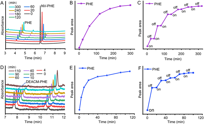
In order to quantitively validate whether PHE could be released from NV-PHE and DEACM-PHE by light, UPLC was used to analyze the photolysis of NV-PHE and DEACM-PHE. The peak area changes of NV-PHE and DEACM-PHE could be clearly observed in Fig. 2. The peak area of PHE increased while that of NV-PHE and DEACM-PHE decreased, interpreting that PHE could be generated under the illumination of NV-PHE or DEACM-PHE. As shown in Fig. 2 and Table S1 (Supporting information), 44% and 98% PHE could be photo-released from NV-PHE and DEACM-PHE after 300-min and 110-min irradiation, respectively. Remarkably, about 50% PHE would be released from DEACM-PHE if exposing to blue light for 20 min. We speculated that the releasing rates might have a positive correlation with the following bioactivity. The photolysis of NV-PHE and DEACM-PHE belonged to first-order kinetics, and the photolysis half-lives of the two compounds were 34.6 min and 18.7 min, respectively. Moreover, the light might be the only responsive factor for releasing PHE from NV-PHE and DEACM-PHE. Because no hydrolysis was detected when the NV-PHE and DEACM-PHE were respectively stored in darkness for 300 min and 110 min, and the release could be paused if taking light off. The precise delivery of PHE could be realized, and the releasing amount of PHE depended on the duration and intensity of light.
NV-PHE and DEACM-PHE were further evaluated with the in-vitro and in-vivo fungicidal activities against Fusarium graminearum. As our previously reported [32,33], mycelial growth rates measure and leaf-dipped method were used in our biological assay. The details could be found in Supporting information. The results were depicted in Figs. 3 and 4, Fig. S2 and Tables S2 and S3 (Supporting information). The in-vitro assay was firstly carried out by method of mycelial growth rates. The non-irradiated NV-PHE and DEACM-PHE either exhibited extremely low fungicidal activity. In Table S2, NV-PHE at 44 µmol/L and DEACM-PHE at 40 µmol/L showed inhibitory rates of 9.33% and 11.11% against F. graminearum, respectively. After illumination with UV light or blue light, NV-PHE and DEACM-PHE displayed EC50 of 4.4 µmol/L and 0.96 µmol/L against F. graminearum, respectively. In addition, no fungicidal activity was observed after dosing with PRPGs including NV and DEACM. In comparison to the direct use of PHE (EC50 = 1.4 µmol/L), the optical control-release manner really worked and retained high fungicidal activities. As shown in Fig. 3, the inhibition patterns of mycelium growth exhibited great difference before and after photo-releasing. The irradiated DEACM-PHE showed higher activity than the irradiated NV-PHE, corresponding to our previous speculation in the correlation of photo-releasing rates and bioactivity. Thus, DEACM would be a more effective PRPG than NV in optical controlled release of fungicides.

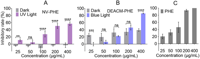
Next the in-vivo fungicidal assay was performed by leaf-dipped method. Fig. 4 gave the inhibition bar graph with five gradient concentrations of the target compounds. As for every concentration of the photo-responsive fungicides both in dark and light, difference analysis was conducted using the GraphPad Prism software and most of them showed significant difference at P<0.0001 level. Table S3 presented the results of EC50 and EC90 against F. graminearum. It seems to be similar for the control effect of the irradiated DEACM-PHE (EC50 = 0.38 mmol/L, EC90 = 1.1 mmol/L) and PHE (EC50 = 0.33 mmol/L, EC90 = 1.0 mmol/L). Besides, Fig. S2 visibly revealed the inhibition effect of the irradiated DEACM-PHE against F. graminearum. The results indicated that the optical controlled release of PHE from DEACM-PHE worked well. Because the irradiated DEACM-PHE retained the similar fungicidal activity of PHE, and DEACM-PHE in dark and light exhibited significant difference in control of F. graminearum.
In conclusion, we synthesized two types of photo-responsive fungicides by introducing PRPGs into PHE. The caging PHE could be optical released, and inhibited the mycelial growth of F. graminearum in vitro and in vivo. In dark, photolysis of NV-PHE and DEACM-PHE could not be occurred, resulting in very poor or no fungicidal activity. Interestingly, DEACM-PHE showed more potent photo-releasing efficiency than NV-PHE. For example, the higher photo-releasing rate of PHE, the more excellent in-vitro or in-vivo fungicidal activity, and the significant activity differences between treatments in dark and light. Therefore, the optical controlled releases of PHE from NV-PHE and DEACM-PHE were achieved at space and time precision. The results provided a new direction in development of CRSs. Thereinto, DEACM might be a more effective photo cage for further explorations on other chemical pesticides and tend to be attempted in agricultural applications.
The authors declare that they have no known competing financial interests or personal relationships that could have appeared to influence the work reported in this paper.
This work was financially supported by the Natural Science Foundation of China (Nos. 21877039, 32072441), National Key Research and Development Program of China (No. 2018YFD0200100), and Innovation Program of Shanghai Municipal Education Commission (No. 2017–01–07–00–02-E00037).
Supplementary material associated with this article can be found, in the online version, at
E.C. Oerke, J. Agric. Sci. 144 (2005) 31–43.
P. Maienfisch, T.M. Stevenson, Modern Agribusiness - Markets, Companies, Benefits and Challenges, American Chemical Society, Washington, DC, 2015.
N. Li, C. Sun, J. Jiang, et al., J. Agric. Food Chem. 69 (2021) 12579–12597. doi: 10.1021/acs.jafc.0c05431
D. Adam, Nature 408 (2000) 125.
X. Pan, F. Dong, X. Wu, et al., J. Integr. Agr. 18 (2019) 840–853. doi: 10.1016/S2095-3119(18)61929-X
A. Singh, N. Dhiman, A.K. Kar, et al., J. Hazard. Mater. 385 (2020) 121525. doi: 10.1016/j.jhazmat.2019.121525
C. Gao, Q. Huang, Q. Lan, et al., Nat. Commun. 9 (2018) 1–13. doi: 10.1038/s41467-017-02088-w
A.E. Kaziem, Y. Gao, S. He, J. Li, J. Agric. Food Chem. 65 (2017) 7854–7864. doi: 10.1021/acs.jafc.7b02560
S. Kumar, N. Chauhan, M. Gopal, R. Kumar, N. Dilbaghi, Int. J. Biol. Macromol. 81 (2015) 631–637. doi: 10.1016/j.ijbiomac.2015.08.062
Y. Liu, Y. Sun, G. Ding, et al., J. Agric. Food Chem. 63 (2015) 4263–4268. doi: 10.1021/jf5055062
C. Xu, L. Cao, P. Zhao, et al., Int. J. Mol. Sci. 19 (2018) 854. doi: 10.3390/ijms19030854
Z. Yi, H.I. Hussain, C. Feng, et al., ACS Appl. Mater. Interfaces 7 (2015) 9937–9946. doi: 10.1021/acsami.5b02131
A. Patchornik, B. Amit, R.B. Woodward, J. Am. Chem. Soc. 92 (1970) 6333–6335. doi: 10.1021/ja00724a041
D.H.R. Barton, Y.L. Chow, A. Cox, et al., Tetrahedron Lett. (1962) 1055–1057. doi: 10.1016/S0040-4039(00)70957-9
J.A. Barltrop, P. Schofield, Tetrahedron Lett. (1962) 697–699. doi: 10.1016/S0040-4039(00)70935-X
J.H. Kaplan, B. Forbush, Ⅲ, J.F. Hoffman, Biochemistry 17 (1978) 1929–1935. doi: 10.1021/bi00603a020
J. Li, C. Zhang, S. Yang, et al., Anal. Chem. 86 (2014) 3037–3042. doi: 10.1021/ac403885n
X. Zeng, Y. Huang, J. Dong, et al., Adv. Agrochem. 1 (2022) 73–84. doi: 10.1016/j.aac.2022.08.001
D.D. Young, A. Deiters, Angew. Chem. Int. Ed. 46 (2007) 4290–4292. doi: 10.1002/anie.200700057
A. Chaudhuri, Y. Venkatesh, J. Das, et al., J. Org. Chem. 84 (2019) 11441–11449. doi: 10.1021/acs.joc.9b01224
W. Zhou, C.P. Hankinson, A. Deiters, ChemBioChem 21 (2020) 1832–1836. doi: 10.1002/cbic.201900757
Q. Lin, Q. Huang, C. Li, et al., J. Am. Chem. Soc. 132 (2010) 10645–10647. doi: 10.1021/ja103415t
N. Kaewchangwat, E. Thanayupong, S. Jarussophon, et al., J. Agric. Food Chem. 68 (2020) 6268–6279. doi: 10.1021/acs.jafc.0c00138
C.A. Griffiths, R. Sagar, Y. Geng, et al., Nature 540 (2016) 574–578. doi: 10.1038/nature20591
S. Atta, A. Jana, R. Ananthakirshnan, et al., J. Agric. Food Chem. 58 (2010) 11844–11851. doi: 10.1021/jf1027763
Z. Gao, P. Yuan, D. Wang, et al., Bioorg. Med. Chem. Lett. 27 (2017) 2528–2535. doi: 10.1016/j.bmcl.2017.03.091
Z. Xu, Z. Gao, X. Shao, Chin. Chem. Lett. 29 (2018) 1648–1650. doi: 10.1016/j.cclet.2018.01.025
S.S. Donau, U.E. Bollmann, R. Wimmer, et al., Chemosphere 233 (2019) 873–878. doi: 10.1016/j.chemosphere.2019.06.015
J. Xu, D. Kong, N. Song, X. Kong, Z. Shan, J. Agro. Environ. Sci. 32 (2013) 2005–2011.
P. Wang, Asian J. Org. Chem. 2 (2013) 452–464. doi: 10.1002/ajoc.201200197
L. Josa-Cullere, A. Llebaria, ChemPhotoChem 5 (2021) 298–316.
W. Fu, X. Hu, Q. Yuan, et al., J. Agric. Food Chem. 69 (2021) 13448–13459. doi: 10.1021/acs.jafc.1c05551
W. Fu, Z. Shao, X. Sun, et al., J. Agric. Food Chem. 70 (2022) 4279–4290. doi: 10.1021/acs.jafc.1c08198

Figure 1 Molecular design of the photo-responsive fungicides. NV-PHE and DEACM-PHE could undergo the release of PHE by UV light (365 nm) and blue light (470 nm), respectively.

Figure 2 UPLC traces and peak area changes of NV-PHE and DEACM-PHE in aqueous methanol (4 × 10⁻⁵ mol/L) after exposure to UV or blue light. (A) Overlaid UPLC chromatograms of NV-PHE at several intervals of irradiation with 365 nm light (the dark line marked ‘0 min’ refers to the fresh prepared NV-PHE, and the light blue line marked ‘300 min’ represents the final released state). (B) Peak area changes of PHE released from NV-PHE under continuous 365 nm light. (C) Peak area changes of PHE released from NV-PHE under discontinuous 365 nm light. (D) Overlaid UPLC chromatograms of DEACM-PHE at several intervals of irradiation with 470 nm light (the dark line marked ‘0 min’ refers to the fresh prepared DEACM-PHE, and the brown line marked ‘110 min’ represents the final released state). (E) Peak area changes of PHE released from DEACM-PHE under continuous 470 nm light. (F) Peak area changes of PHE released from DEACM-PHE under discontinuous 470 nm light.

Figure 3 The in-vitro fungicidal activity of NV-PHE and DEACM-PHE against Fusarium graminearum before and after irradiation. The inhibitory rates were 58% for (A-Light) at 5.5 µmol/L, 9.33% for (A-Dark) at 44 µmol/L, 64% for (B-Light) at 1.3 µmol/L, 11.11% for (B-Dark) at 40 µmol/L, respectively.

Figure 4 The in-vivo fungicidal activity of NV-PHE, DEACM-PHE and PHE against Fusarium graminearum before and after exposing to light. (A) The inhibitory rates of NV-PHE against Fusarium graminearum under dark (gray bar) and UV light (violet bar). (B) The inhibitory rates of DEACM-PHE against Fusarium graminearum under dark (gray bar) and blue light (blue bar). (C) The inhibitory rates of PHE against Fusarium graminearum. Each concentration was repeated with three replicates. **P < 0.01; ***P <0.001; ****P < 0.0001; ns, *P > 0.5.

 扫一扫看文章

扫一扫关注我们
 DownLoad:
 下载:
下载:
 下载:
下载: