Citation:
Sida Huang, Shihai Yin, Aparna Kushwaha, Abhinav Kumar, Yu Deng, Yanqiong Peng, Ying Pan, Jianqiang Liu, Yong Huang. MOFs-based nanomaterials for bone repair and regeneration: Current status and future perspectives[J]. Chinese Chemical Letters,
2026, 37(8): 111211.
doi:
10.1016/j.cclet.2025.111211
MOFs-based nanomaterials for bone repair and regeneration: Current status and future perspectives
English
MOFs-based nanomaterials for bone repair and regeneration: Current status and future perspectives
Received Date:
13 January 2025 Accepted Date:
15 April 2025 Revised Date:
08 April 2025 Available Online:
15 August 2026
Abstract:
Bone abnormalities induced due to inflammation, trauma, or diabetes have emerged as a serious issue during treatment and post-treatment. Currently, the prevalent treatments for bone abnormalities are surgery and antibiotics. However, they are progressively struggling to meet clinical needs. Metal-organic frameworks (MOFs) are acquiring attention among many bone repair and bone regeneration materials due to their excellent role in this area of research. MOFs -based nanomaterials are characterized by long-term stability, large specific surface area, and can be easily modifiable. Furthermore, their porous structure and increased mechanical capabilities are similar to alternative tissues, making them useful for bone repair and regeneration. This review article outlines the application of MOFs in bone repair and bone regeneration therapy. First, the mechanics of bone repair and regeneration are discussed, followed by a summary of the most frequent synthetic methods of MOFs for treating bone defects. Secondly, the strategies of different types of MOFs in bone repair and bone regeneration therapies are reviewed. Eventually, the challenges and future of MOFs in bone repairs and bone regeneration therapies are described, introducing a novel approach to treating bone abnormalities.
Bone tissue is composed of living cells and bioactive minerals. With the aging of the population, bone-related diseases such as fractures and bone abnormalities have increasingly become a problem that cannot be ignored [1]. Reports show that there was a total of 178 million cases of fractures worldwide in 2019 [2]. Therefore, how to reconstruct bone defects caused by trauma, congenital diseases, and tumor resection has become a major challenge in recent years [3,4]. Currently, bone grafting and drug therapy are the most commonly used methods to address bone abnormalities caused by osteoporosis, osteogenesis imperfecta, and surgical injuries [5]. Bone grafting can be classified into autologous bone grafting and allogeneic bone grafting. However, apart from bringing additional trauma and pain to patients, autologous bone grafting may lead to complications in the bone donor site, such as infection, bleeding, nerve injury. Most importantly, the source of autologous bone is limited, and it is often difficult to meet the requirements for large-area bone defects. However, allogeneic bone grafting has even more limitations such as rejection reactions, limited application scope, and transplantation risks [6,7]. As for drug therapy, it usually has better effects in the early stage. However, since it is impossible to efficiently concentrate the drugs on the bone defect area, long-term medication not only leads to a decrease in curative effect but also easily causes adverse reactions. In order to overcome these challenges, scholars are exploring bone materials suitable for clinical implantation that can reduce wound infections, have high mechanical stability, match in terms of comfort, and possess good biocompatibility and biodegradability [8–10].
Nowadays, nanomaterials research can be regarded as the most explored field in medicinal sciences. This is because the physico-chemical properties of materials alter on moving from macro- to nano-dimension due to enhancement in specific surface area and reactivity. Amongst varied classes of nanomaterials, that derived from metal-organic frameworks (MOFs) are up-surging interest of biomaterial scientists as they are a new assorted class of synthetic materials that emerged over the past 30 years and had gained immense attention in varied application areas [11–16]. In addition, MOF nanostructures have currently attracted widespread interest in both academia [11–13] and industry [14–16] due to their unique contributions in the fields of medicine, remote sensing, wastewater treatment, air purification, and renewable energy. MOFs may be innately bioactive or acquire bioactivity arising because of their constituent metal ions, ligands, or their synergistic contribution. It can be employed as an encapsulating agent or combined with pharmaceutical agents such as medications and hydrogels can uplift the performance of others. In the aspect of bone repair and regeneration, MOFs possess great potential [17–19].
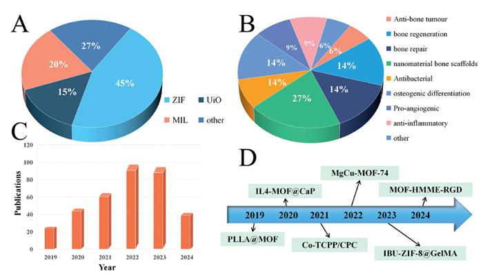
As research and technology advance, bioengineering technology becomes more commonly used in the therapeutics of bone abnormalities. Tissue-engineered bone technology is a new treatment method. Due to their outstanding physicochemical characteristics, nanomaterials are likely to be an essential part of the treatment of bone defects, such as promoting the differentiation and proliferation of bone cells and accelerating the bone healing process. At present, the research related to nanomaterials for bone repair and regeneration mainly focuses on the bionic design of nanomaterials and the development of multifunctional nanocomposites. However, not all nanomaterials are suitable for bone repair and regeneration. For example, there are problems such as potential toxicity, lack of targeted drug delivery ability, unsatisfactory mechanical properties, and poor biocompatibility. MOFs have evinced to be beneficial bone materials (Fig. 1). Among them, MOFs stand out among numerous nanomaterials due to their unique advantages. MOFs show similar behavior as natural tissues as they provide the physical stability essential for the bone, toughness, and mechanical strength to restore the wounded limb's load-bearing capacity. In the field of bone tissue treatment and repair, the nanoscale porous network structure of MOFs is highly similar to the trabecular structure of natural bone. The pore size range (1–50 nm) can be adjusted, providing three-dimensional spatial support for cell adhesion, proliferation, and vascularization. In addition, MOFs can carry a variety of metal ions that are beneficial to bone tissue and cell therapy. For example, Zn2+ can promote the activity of alkaline phosphatase and accelerate the formation of mineralized nodules; Mg2+ can upregulate the expression of osteogenesis-related genes (such as Runx2 and OCN); Sr2+ can inhibit the activity of osteoclasts and balance bone metabolism. Finally, the efficient drug loading capacity and the ability to fabricate personalized MOFs composite scaffolds through controllable 3D printing make the strategies of MOFs in bone treatment and repair flexible and diverse. Compared with traditional materials, the advantages of MOFs in bone repair and regeneration are reflected in their high porosity, large specific surface area, good biocompatibility, controllability, and easy functionalization.
Figure 1
Figure 1.
(A) Proportion of MOFs used to treat bone defects. (B) MOF-based nanomaterials for therapeutic modalities in bone defects. (C) Research advances in MOF-based treatment of bone defects in past 5 years (Source: Web of Science, 8 July 2024). (D) Study of MOF in Bone Defects, 2019–2024.
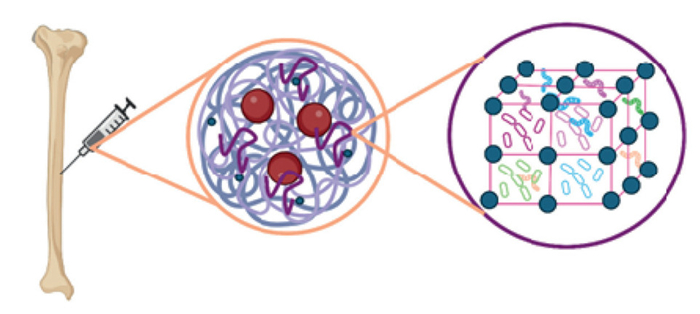
In summary, this review presents the concept of constructing bioactive bone tissue using cells, growth factors, and scaffold materials based on tissue-engineered bone technology to promote bone repair and regeneration [20–22]. MOFs possess the capability to release metal ions, which in turn create an excellent ion microenvironment once the encapsulated metal ions penetrate the bone tissue environment. Simultaneously, they can also serve as drug delivery vehicles. The two can play a synergistic role, offering ideas and methods for bone repair and regeneration therapy. The benefit of this procedure is that it reduces treatment time, improves the standards of life of patients, and avoids some shortcomings of traditional transplantation methods (Fig. 2).
Figure 2
Figure 2.
A simplified diagram depicting MOFs utilized as drug delivery devices for bone-related disorders.
MOF nanocomposites for bone repair and regeneration are continuously being researched and developed. Currently, MOFs are widely studied in the fields of bone tissue engineering scaffolds, drug delivery carriers, ion release for promoting bone regeneration, and 3D printed personalized bone scaffold materials. In addition, MOFs are also expected to be further explored in photothermal therapy for osteosarcoma, immunomodulation for promoting bone healing, as well as gene delivery and gene therapy. Until recently, the potential synergistic effects of adding hydrogels to MOFs to acquire extended retention and sustained therapeutic action were mainly unknown [23]. Hence, the current highlight focuses on the implementation of MOFs as a biomaterial component in biomaterial for bone repair and regeneration.
2.
Mechanisms of bone repair and regeneration
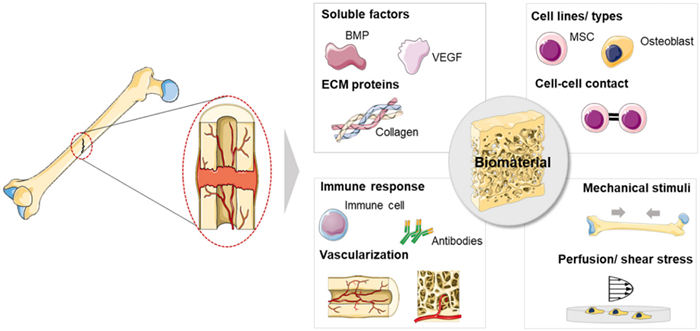
Bone regeneration and repair are the biological mechanisms that allow bone to restore its structure and function following injury (Fig. 3) [24]. This process usually involves a sequence of complex biological events, such as cell proliferation, differentiation, matrix synthesis, and mineralization. The mechanism of bone regeneration and repair is involved at various levels, including molecular, cellular, and tissue levels. At the molecular level, Bone morphogenetic proteins (BMPs), transforming growth factor beta (TGF-β), and fibroblast growth factors (FGFs) are crucial for bone production and repair [25]. These growth factors promote biological processes after injury by activating specific receptors. At the cellular level, bone marrow stromal stem cells (BMSCs) and osteoblasts are the prime cell types for bone regeneration and repair. BMSCs can differentiate into osteoblasts, which further secrete collagen and other organic matrix, and finally mineralize to form new bone [26]. In addition, immune cells, endothelial cells, and fibroblasts also contribute during bone repair [27], supporting bone formation by secreting extracellular matrix and regulating the inflammatory response. At the tissue level, bone repair also involves angiogenesis and bone remodeling. Angiogenesis provides nutrients and oxygen to the newly formed bone while removing metabolic waste. Bone remodeling is to adjust the shape and size of bone to adapt towards changes in mechanical load by maintaining pivotal balance between bone resorption and bone formation.
Figure 3
Figure 3.
Physiological substances involved in the repair of bone damage. Reprinted with permission [24]. Copyright 2018, Elsevier Ltd.
BMPs serve an important part in the regeneration of bone. They are a class of proteins that can promote bone formation and regeneration and are members of the transforming growth factor beta (TGF-β) family. BMPs often activate the Smad signaling pathway by binding to their receptors and then encourage the propagation and differentiation of osteoblasts, thereby leading to bone formation [28]. During bone regeneration, BMPs not only directly stimulate bone formation, rather promote bone tissue reconstruction by attracting blood vessels and other types of cells [29]. Moreover, BMPs can also promote the formation of cartilage and provide a template for the deposition of new bone. BMPs are also widely employed in bone tissue engineering. The recombinant human BMP-2 (rhBMP-2) has been used in clinical settings to improve fracture healing and spinal fusion surgery [30]. In addition, BMPs have also been used to make artificial bone materials to replace lost bone tissue [31].
Pluripotent stem cells, also called mesenchymal stem cells (MSCs) can develop varied cell types like osteoblasts, chondrocytes, and adipocytes [32]. The process of differentiating into osteoblasts involves a series of complex molecular signals and intracellular changes [33]. At the early stage of differentiation, MSCs are first induced to enter the precursor state of osteoblasts. Growth factors and cytokines including BMPs, TGF-β, and FGFs typically govern this process. As differentiation progresses, MSCs begin to produce additional osteoblast markers, including alkaline phosphatase (ALP) and osteocalcin.
At the same time, the interior structure of the cell also changes, forming a bone-like mineralized area, which is an important sign of osteoblast maturation. At the late stage of differentiation, osteoblasts further mature and eventually form intact bone tissue. This stage involves the deposition of extracellular matrix and cell-cell interactions. Mature osteoblasts secrete collagen and other proteins to form a solid bone matrix. Subsequently, through the mineralization process, calcium salt gradually deposits on the matrix, and finally forms bone.
Angiogenesis is essential for the process of bone mending [34]. First, neovascularization can provide the damaged area with the necessary nutrients and oxygen, which are necessary for bone regeneration and repair. Second, angiogenesis aids in delivering stem cells and growth factors to the bone defect site, that are essential for promoting new bone generation and tissue reconstruction. In addition, neovascularization can also help remove dead cells and metabolic waste, maintain the cleanliness of the bone repair surroundings and thus promote the formation of healthy bone tissue.
In recent years, investigators have been exploring new approaches for bone repair and regeneration using bioactive materials such as scaffolds that can carry growth factors and stem cells to encourage bone formation [35]. Gene therapy and cell therapy also showed potential application value [36]. Among them, MOFs are particularly effective in bone regeneration and bone repair [37]. These new technologies are predicted to uplift the efficiency and progress rate of bone repair, particularly for refractory fractures and bone deformities.
3.
Properties and preparation of MOFs
3.1
Properties of MOFs
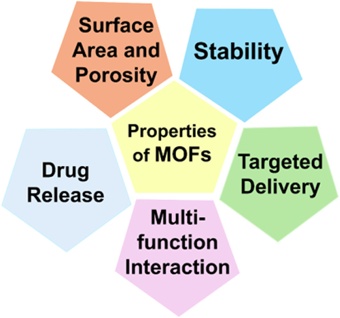
The host's reaction to implanted biomaterials, specifically their ability to steer the neighboring tissues of bone defects in a repairable pathway, is critical to successful bone healing. Implanted biomaterials with strong biocompatibility, osteoconductivity osteoinductivity, osseointegration, and osteogenesis, can increase the differentiation and proliferation of bone mesenchymal stem cells while also promoting mineralization and fresh bone production, resulting in effective bone healing [38]. MOF materials have distinct physicochemical features, including cargo transport capacity, releasing ability (ions and ligands) and formability, setting them apart from biomaterials functionalized techniques [39]. The physico-chemical and medicinal properties of materials alter on moving from macro- to nano-dimension due to enhancement in specific surface area and reactivity (Fig. 4). Compared with other nanomaterials, MOFs possess the following characteristics that facilitate the application of MOF-based nanomaterials in bone repair and regeneration.
Figure 4
Figure 4.
Properties of MOFs with biomedical applications.
To achieve bio-adhesive and targeting properties, the nanoscale-metal organic framework (nMOF) system needs to be designed appropriately. The biophysical characteristics of nMOFs, like surface hydrophilicity, the ligand characteristics play a crucial role when nMOFs interact with proteins, lipids, and ions in biological medium. Altering the exterior surface of nMOFs can enhance their stability and movement in the blood, facilitating purposeful distribution. Post-synthetic alteration which adds a functional layer to the surface, is the most common method for modifying the surface properties of nMOFs.
The apt nMOF surface modifiers include organic polymers, lipid bilayers, and silica shells. Amongst these, silica acting as enveloping material improves water dispersibility and reduces nMOF decomposition. In addition, the size of MOFs has a significant impact on their bio-distribution, circulatory lifespan, and targeting capacities and studies reveal that the ideal nMOF size is < 200 nm [40]. MOFs due to their large specific surface area and tunable pore structure are capable of carrying huge amounts of drug molecules and attain precise regulation of drug release rate by controlling the pore size. The particle size of nMOF affects its mobility, diffusion, and susceptibility to being absorbed in tumour cells or removed by macrophages or the renal system to prevent severe side effects [41].
For example, in bone repair and regeneration, by loading growth factors that promote bone growth (such as bone morphogenetic proteins) into the pores of MOFs and then implanting them into bone defect sites, slow and sustained release of growth factors can be achieved. This provides long-term stimulation for the proliferation and differentiation of bone cells and the formation of new bone, thereby promoting bone repair.
3.1.2
Good biocompatibility and stability
Biocompatibility is an essential need for the clinical translation of biomaterials. Many MOFs materials show good biocompatibility, which is crucial for drug carriers because they need to deliver drugs safely in vivo. MOFs comprise different organic linkers and metal ions and the stability of their structure poses a potential issue for biomedical applications. Many parameters influence the structural stability of MOFs in aqueous medium that includes M-L bond stability, oxidation state of Mn+, donor ability of ligand, and its basicity and framework dimensionality [42]. Although short-term cell/animal models in vitro have shown negligible toxicity and side effects, a small number or high concentration of MOFs may produce toxicity. MOF constituting metals ions, such as Al3+, Cr3+, Mn2+, Fe2+/Fe3+, Cu2+, Zn2+, Zr4+ and Mg2+, exerts varying toxic effects. The Zr4+-, Cr3+- and Mg2+-based MOFs are least hazardous, whereas Cu2+- and Mn2+-MOFs are relatively toxic. Oxidative stress is a contributing factor to MOF-induced cytotoxicity. Excessive ROS can lead to the activation of inflammatory signaling, resulting in protein, mitochondrial membrane, and DNA damage and apoptosis. Furthermore, in antibacterial applications, MOFs may impair cell membrane integrity while destroying bacterial membranes, limiting bacterial growth, and causing changes in membrane permeability and cell homeostasis [43]. In addition, most MOFs, such as ZIF-8 and Mg-UiO-66, exhibit good biocompatibility and do not cause significant immune responses or cytotoxicity. This allows MOFs to coexist harmoniously with bone tissue, providing a safe microenvironment for bone repair. Additionally, good biocompatibility also facilitates the adhesion, proliferation, and differentiation of cells on or within MOF materials, promoting the growth and repair of bone tissue. In addition, some MOFs also exhibit excellent chemical and thermal stability, enabling them to maintain structural integrity under harsh physiological conditions.
3.1.3
Targeted delivery ability
Due to poor aqueous or lipid solubility, stability issues, toxicity, and restricted biodistribution, formulation scientists have struggled with drug delivery. However, the evolution of MOFs in drug delivery systems has acknowledged that MOFs can help alleviate these difficulties [44]. MOFs can be designed as targeted drug carriers for specific cells or tissues by surface modification or binding to specific ligands. Conjugating molecules containing bone tissue-specific ligands, such as those with the arginine-glycine-aspartic acid (RGD) sequence, to the surface of MOFs enables specific binding to integrin receptors on bone cell surfaces, achieving targeted drug delivery to bone tissue. MOFs have a wide surface area and structural divergence, making it easier to load pharmaceuticals on the outside or inside of pores utilizing a variety of loading methods. There are two commonly utilized drug-loading strategies: one-step and two-step techniques [45]. In the first method, therapeutic actives are immediately merged into MOFs during production in one step. This approach uses uniform distribution and has a high drug-loading capability. However, managing the size of the particle, shape, and physiochemical properties of current MOFs is difficult. Additionally, extra steps must be taken to guarantee that medicine is not destroyed throughout the synthesis process. The second approach of drug loading encapsulates pharmaceuticals inside the structure of previously synthesized nMOFs to preserve particle shape. When medications have smaller molecular dimensions than the pore diameter of nMOFs, they are frequently expected to be trapped inside the scaffolds via hydrogen bonding or several other host-guest interactions. Large-sized medicine molecules with opposite charges are expected to be absorbed by nMOFs via electrostatic interactions [46]. This targeting may boost treatment efficacy while minimizing negative effects on normal tissues.
3.1.4
Controllable drug release mechanism
The drug release by the drug delivery system is crucial as it affects the efficacy of the dosage form. MOFs with good porosity are employed for medication delivery; hence their formulations are assessed to monitor the patterns of drug-release. MOFs can control drug release in a variety of ways, including pH response, temperature sensitivity, light triggering. These intelligent response mechanisms allow the controlled release of drugs when reaching the lesion site, thereby enhancing treatment efficiency and reducing side effects. Horcajada et al. created nano-porous MIL-53 (Fe, Cr) for IBU delivery. The transport of IBU was explained by employing simulated body fluid (SBF) at 37 ℃ and analyzing SBF through HPLC which revealed 21 days of delayed delivery that demonstrated the involvement of two procedures [47]. Maryam et al. demonstrated Fe3O4@PAA@ZIF-8 aided ciprofloxacin (CIP) transportation and release against S. aureus and E. coli at pH 5 and 7.4. This nanocomposite was examined using multiple kinetic models revealing that Higuchi and Peppas models fitted best with the kinetic data [48]. In bone repair and regeneration, controlled release of drugs from MOFs in bone tissue can be achieved via pH and enzyme-responsive mechanisms.
3.1.5
Multi-function integration
Multifunctional integration serves as the main strategy for current nanomaterials used in bone repair and regeneration. In this regard, MOFs have three features that act simultaneously: crystalline nature, porosity, and the presence of strong metal-ligand interactions. MOFs are a unique category of materials due to their peculiar characteristics. Their low density, high specific surface area, structural flexibility, and configurable pore functionality make them ideal for a numerous application such as supercapacitors, gas storage and delivery, medication delivery, catalysis, rechargeable batteries, separation membranes, and sensing [49]. Biomedicine is among the fields in which MOFs have been used most recently. MOFs have proved to be employed in drug delivery for bio-agents because of their chemical characteristics, drug-MOF interaction, pore size distribution, heat stability and release kinetics, size dispersion, loading capacity, and other factors [50]. MOFs cannot only act as drug carriers, but also integrate other functions such as diagnostic markers, imaging agents, to form a "treatment diagnosis integration" platform, which provides new possibilities for personalized medicine [51]. Based on these applications, numerous researchers have developed combined therapeutic strategies such as "drug release + metal ion release" [52], "antioxidant + anti-inflammatory" [53], or 'antioxidant + antibacterial' [54] for bone tissue treatment.
In summary, large specific surface area and porosity; good biocompatibility and stability; targeted delivery ability; controllable drug release mechanism and multi-function integration make MOFs an interesting class of materials finding therapeutic applications to treat bone defects.
3.2
Preparation methods to obtain targeted MOFs
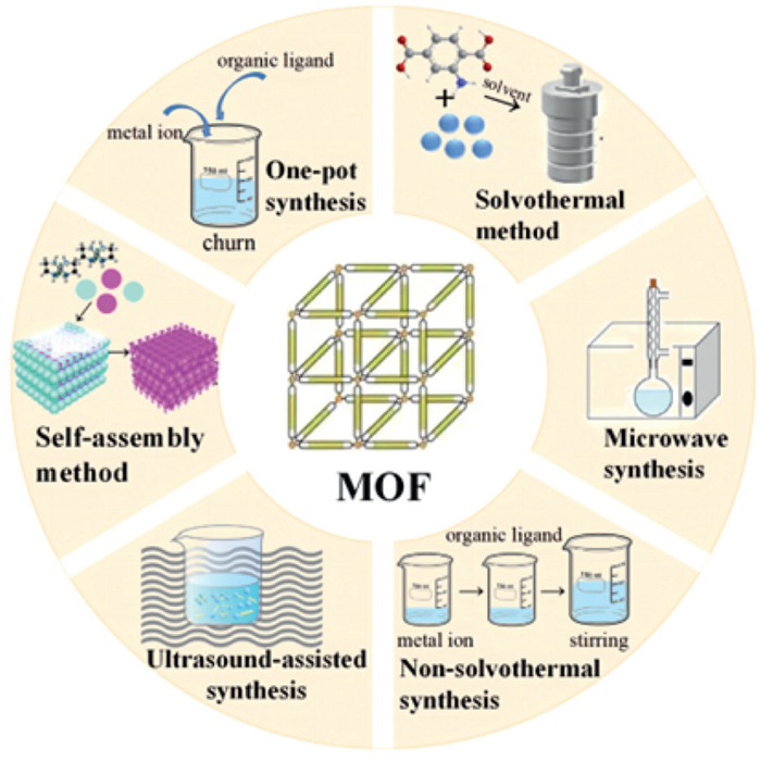
In recent years, researchers have committed to developing new MOFs with specific functions, such as MOFs nanomaterials with core-shell structure, which show prospects in the fields of catalysis, energy conversion, and biomedicine [55]. At the same time, the preparation methods of MOFs are also constantly improving, including solvothermal method, microwave-assisted synthesis, electrochemical method, etc., which helps to improve the synthesis efficiency and purity of MOFs. Selecting the appropriate synthetic technology can certify the syntheses of MOFs with the properties required for bone repair and regeneration therapeutic applications. Here, the commonly used MOF synthetic techniques in the treatment of bone injury are summarized, mainly including one-pot, self-loading syntheses, ultrasonic chemical syntheses, solvothermal and microwave-assisted methods (Fig. 5).
Figure 5
Figure 5.
Scheme for preparation methods for MOFs for bone repair and regeneration.
One-pot synthesis is a simple and effective chemical synthesis strategy, which allows multiple reactants to be converted into target molecules or materials by heating or other conditions in a single reaction vessel. It is particularly useful in the synthesis of MOFs because it can simplify the synthesis steps, reduce the purification process, and sometimes increase the yield and control the crystal quality. For the preparation of MOFs using the one-pot synthesis strategy, it is crucial to select the appropriate metal ion, organic linker, and appropriate solvent and temperature conditions. In the synthesis process, metal ions and organic linkers copolymerized in solution to form a framework structure and then the final MOF material can be obtained by evaporation of the solvent, crystallization, and other steps. As the most widely synthesized MOF, ZIF-8 can be synthesized by one-pot synthesis in addition to non-solvothermal synthesis. Researchers could improve bone regeneration by loading ZIF-8 with bone-treating medications if they were able to produce it with a greater encapsulation efficiency using a single-pot strategy [56].
3.2.2
Self-assembly method
Another typical MOF preparation approach is self-assembly synthesis, which relies on interactions between organic ligands and metal ions such as dative and non-covalent interactions to render complexes with 3D frameworks. This method does not require the intervention of external forces but relies on the interaction of molecules or nanoparticles to achieve self-assembly. In the self-assembly method, metal salts are usually dissolved in the solvent first, and then organic ligands are added to control the formation process of MOFs by adjusting the reaction conditions, viz. pressure, temperature, concentration, solvent. This method can engender MOFs with tuned pore sizes and volumes with functionalized surfaces, which show excellent performance in varied application areas. MOFs synthesized by the self-assembling method include ZIF (zinc imidazolate framework). MOFs self-assembled by zinc ions along with imidazole ligands and with high porosity and adjustable pore size have been employed to carry drugs for bone injury treatment [57]. HKUST-1 MOFs, self-assembled from copper ions and benzoic acid, have a special three-dimensional pore structure.
3.2.3
Solvothermal method
The solvothermal technique is a standard and well-established strategy for the synthesis of MOFs using metal salts and organic linkers and dissolving them in appropriate solvents and the reaction is performed at high temperature and high pressure. This method can lead to a self-assembly reaction to engender targeted MOFs [58]. This method can control the crystallinity and pore building of the product. Temperature plays a key role in the solvothermal method. The reaction temperature ranges from 80 ℃ to 260 ℃ and the reaction time is usually 48–96 h or longer. In addition to temperature, the morphology of MOFs synthesized by the solvothermal method is also affected by cation, surfactant, polymer, and other regulators, which can compete for ligand complexation. The solvothermal method can produce crystalline MOFs with better yield. The MOFs synthesized by the solvothermal method include Zr-MOF obtained by the reacting 2,2′-bipyridine-5,5′-dicarboxylic acid (H2L) with Zr4+ source possess excellent water stability and photoluminescence properties [59]. MIL-101-(Fe) is usually prepared by in situ hydrothermal method involving FeCl3·6H2O as iron source and terephthalic acid (H2BDC) as polycarboxylate ligand [60]. Different adsorbents can be prepared by changing the drying temperature. In UiO-66, MOF materials with specific structures can be generated by the reaction of metal salts (such as zinc salt) and organic linkers (such as benzene tetracarboxylate) at high temperatures and high pressures [61]. In addition, MIL-100 and MOF-74 can also be synthesized by solvothermal method [62,63].
3.2.4
Ultrasonic chemical synthesis
Ultrasonic chemical synthesis is a method that uses ultrasonic energy (20–1000 kHz) to promote chemical reactions. This method has been extensively employed in the preparation of nanomaterials. Through the cavitation effect of ultrasound, tiny bubbles can be generated in the liquid. In the process of oscillation, growth, contraction, and collapse, these bubbles will produce extreme temperature and pressure, which will cause physical and chemical changes and help to synthesize nanomaterials. The advantages of this method for MOF synthesis include rapid synthesis, scalability, high efficiency, and green process. This method can be carried out at a relatively low temperature and produces consistent nano-sized MOF. The MOFs synthesized by the sonochemical method include Zr-based porphyrin MOFs, with high purity and homogeneous size MOF-525 and MOF-545 produced by the sonochemical process in 2.5 and 0.5 h, respectively [64]. Compared with the conventionally prepared MOFs, the samples synthesized by this method have more defect sites and improved tissue properties. Also, the previously mentioned MIL-101-(Fe) and UiO-66 MOFs can also be synthesized by the sonochemical method.
3.2.5
Microwave-assisted syntheses
Microwave-assisted syntheses utilize microwave radiation to accelerate the chemical reaction process. Thus, it also shortens the synthesis time and improves the quality of products. Compared with traditional synthesis methods, microwave-assisted synthesis has many advantages including faster reaction rate, higher yield and better crystal quality. In addition, due to the uniformity of microwave heating, this method can also reduce the formation of by-products with high product purity. In this method, the selection of solvent and suitable reaction vessel is the key limiting factors. This method can shorten the time and improve the purity and crystallinity of the product [65].
3.2.6
Non-solvothermal (Neat) method
Non-solvothermal (Neat) method is a method used to synthesize these materials that differs from traditional solvent thermal methods, which typically involve chemical reactions in liquid solvents. Non-solvent thermal methods involve heat treatment in solvent-free or non-traditional solvent systems to promote chemical reactions and material formation. Non-solvent thermally synthesized MOFs may lead to stronger crystal structures and have fewer lattice defects due to their formation under milder conditions, thus improving the thermal stability of the material. Lower synthetic temperature parameters help to reduce the movement of atoms in the lattice and reduce thermally induced phase transitions or structural collapse, allowing the material to remain stable at higher temperatures. In addition, non-solvent thermal synthesis can sometimes promote tighter molecular packing, further enhancing the thermal stability of the material.
Each of the above MOFs synthesis methods has advantages and disadvantages, and the optimum synthesis strategy is determined in light of the application for which targeted MOF is being synthesized. To achieve this, optimization of experimental conditions is crucial. With the continuous advancement of synthetic technologies, new synthetic methods and improvement in existing methods are being explored to meet diverse application requirements. For the syntheses of MOFs with desirable bone applications, the synthetic goals must aim on the purity, porosity, physical characteristics and biocompatibility of MOFs.
In summary, appropriate MOFs and rational synthesis methods can be selected based on the therapeutic requirements for bone defects, such as the need for specific metal ions, drug loading efficiency, feasibility of functionalization, and mechanical requirements. For MOF-based nanomaterials used in bone repair and regeneration, more attention is paid to controlling particle size during synthesis, drug-loading capacity, biocompatibility, and subsequent functionalization requirements. Although both MOFs and MOF-nanomaterials are based on the coordination self-assembly of metal ions and organic ligands, achieving structural control through the regulation of reaction conditions (such as metal/ligand ratio, solvent type, temperature). However, in terms of synthesis methods, MOFs rely more on traditional methods such as solvothermal and one-pot approaches, whereas MOF-nanomaterials may utilize advanced techniques like microfluidics, templating, and 3D printing.
4.
Application of MOFs-based nanomaterials in bone repair and regeneration
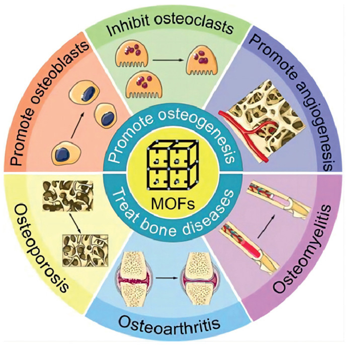
MOFs can play vital role as antibacterial, anti-inflammatory and drug carriers to enhance bone repair and regeneration (Fig. 6) [66]. MOFs can be used as scaffold materials for bone tissue engineering to stimulate bone cell adherence, differentiation and proliferation. Furthermore, the pore structure of MOFs can be used for loading growth factors, medicines or other bioactive chemicals that can be delivered at precise times and locations to stimulate bone repair and regeneration. Compared with traditional materials, MOFs have excellent structure and better biocompatibility, appropriate for cell growth and differentiation, revealing their better osteogenesis [67]. In addition, studies have shown that MOF with Zn, Zr and Ca as metal ions promote osteogenesis through different metal ions, which affect different proteins [68]. This section mainly discusses the utility of MOFs in bone defect treatment and bone tissue engineering from the types of MOFs and the combination with other materials (Table 1) [69–88].
Figure 6
Figure 6.
Scheme for application of MOFs in bone repair and regeneration. Reprinted with permission [66]. Copyright 2023, Elsevier B.V.
4.1
Application of ZIFs-based nanomaterials in bone repair and regeneration
Zeolites imidazole framework materials (ZIFs) are prepared by crosslinking transition metal ions (Zn) with organic imidazole esters [89]. ZIFs have different applications in various fields, especially in medical-related fields [90]. They are often used in biomedical engineering and drug delivery systems as they can exhibit good biocompatible and non-toxic nature [91,92].
Based on the characteristics that the continuous release of Zn2+ is essential in osteogenesis and antibacterial activity. Liu et al. prepared ZIF-8 MOF for bone regeneration and antibacterial activity [69]. The skeleton can gradually release Zn2+ ions, successfully promoting bone regeneration by elevating osteogenic-related genes or proteins expression like Runx2 and OCN. At the same time, it also showed strong antibacterial activity and both of them are used for alveolar bone regeneration.
To solve the problem of alendronate (AlN) targeting and slow controlled release, Baadani et al. [70] prepared pg/aln-ZIF-8 electrospinning membrane by loading AlN into the pores of ZIF-8 and studied its osteogenic properties. The skeleton can manipulate and slowly release AlN and its anti-osteoporosis properties were confirmed by significantly enhancing ALP activity, osteogenic mineralization ability, scaled regulation of osteogenic related genes and inhibiting bone resorption.
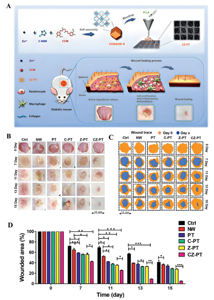
The slow release of metal ions from ZIF or the slow release of loaded drugs is usually not a single step process but exhibits a dual release of the metal ions as well as the loaded drugs, exhibiting synergistic effects. Since both curcumin (CCM) and Zn2+ can produce anti-inflammatory and antioxidant effects, Wang et al. [71] reported the synthesis of (CCM@ ZIF-8) by adopting the eutectic method to coordinate CCM with Zn2+ in combination with PLLA scaffolds of PLA (Fig. 7). This nano scaffold possesses the ability to degrade and slowly release curcumin and Zn2+. The in vitro and in vivo experiments revealed that it could synergistically reduce reactive oxygen species levels and alleviate inflammatory reactions by inducing anti-inflammatory and antioxidant effects. Further, curcumin and Zn2+ were released slowly through the nano scaffold that promoted cell proliferation. Qiao et al. [72] reported a ZIF-8 scaffold loaded with simvastatin (SIM), which showed good biocompatibility and can continuously release drugs, SIM and Zn2+. The in vitro analysis confirmed that the skeleton could uplift the osteogenic differentiation ability of BMSCs. Furthermore, it possessed bone induction characteristics and in vivo tests confirmed that the skeleton can enhance the Runx2 expression suggesting excellent bone repair ability.
Figure 7
Figure 7.
(A) The fabrication of porous patterned PLLA composite electrospun scaffolds incorporated with CCM@ZIF-8 MOFs, and their promotion for diabetic wound healing process. (B) Research progress on the size change of back skin resection in diabetic mice at different periods. (C) Simulation of wound bed closure trace in each group in vivo. Light brown area represented the wounds area at day 0 and the blue area represented the wounds area at day x (x = 7, 11, 13 and 15). (D) The wound area of the six groups was statistically analyzed to show the healing effect. Reprinted with permission [71]. Copyright 2020, Elsevier B.V.
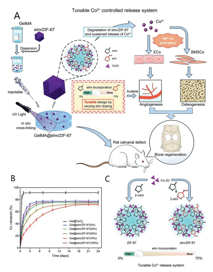
ZIFs can usually be used to further prepare the ZIF composite skeleton, which is modified with various molecules to functionalize the ZIF skeleton to enhance its properties. Sun et al. by regulating the release of Co2+ ion from ZIF-67, modified it with 2-ethylimidazole (EIM) and combined it with hydrogel [73], prepared GelMA@eIm/ZIF-67 hydrogel and studied its effect on bone formation (Fig. 8). The composite hydrogel can control the sustained and slow discharge of Co2+ ions for 21 days (Fig. 8), that promoted angiogenesis and confirmed its therapeutic potential in bone repair. Liu et al. [74] confirmed the bone regeneration effect of ZIF-8 by catechol chitosan modification in vivo. The multifunctional hydrogel promoted bone regeneration and osteogenic differentiation by promoting the generation of ALP and OCN. Shu et al. developed Cu-modified ZIF-8 and binding it to polycaprolactone (PCL) and polylactic acid (PLA) [75]. The resulting MOFs possess osteogenic properties and are capable of guiding bone regeneration.
Figure 8
Figure 8.
(A) Scheme for GelMA@eIm/ZIF-67 nanocomposite hydrogel formation. (B) GelMA@CoCl2 and GelMA@eIm/ZIF-67 release patterns in FBS. (C) Scheme of reaction between a linker solution comprising eIm and mIm and Co(Ⅱ). Reprinted with permission [73]. Copyright 2021, American Chemical Society.
ZIF can be widely applicable in drug delivery due to its pH responsiveness. As a pH sensitive MOF, ZIF can collapse and decompose at acidic pH and release metal ions, ligands or loaded drugs, thus playing a certain role. In addition, some scholars have constructed a drug delivery system loaded with anti-tumor drugs based on the acidic pH response of ZIF-8 [93]. Tao et al. [76] modified the surface of ZIF-8 loaded with drugs with hydroxyapatite (HA) and zoledronic acid (ZA) and constructed a MOF double targeted delivery system which effectively improved the bioavailability of drugs and reduced drug toxicity. At the same time, ZA also inhibited osteoclasts which have potential in the cure of rheumatoid arthritis.
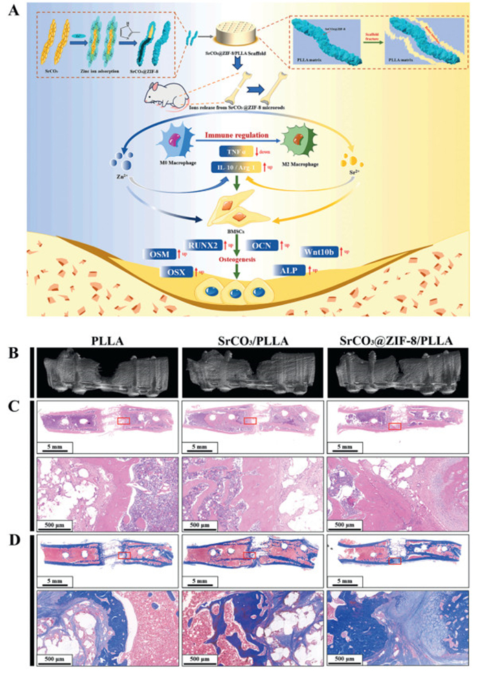
ZIF can also be combined with bioceramics. Bioceramics are extensively employed to enhance the osteogenic action of polymers due to their outstanding biological activity and osteogenic ability [94,95]. However, the mechanical characteristics of ceramics are impacted by the absence of interfacial interaction between them and polymers. Between various ceramic particles, strontium carbonate (SrCO3) is deliberated to be an encouraging bioceramic. SrCO3 promotes osteogenesis and inhibits osteoclast activity by releasing SR ions [96–98]. However, due to the apparent difference in physical and chemical properties between SrCO3 and polymer, the poor interface bond will deteriorate its mechanical properties. Thus, enhancing the interfacial compatibility of polymers and ceramics is a critical issue that must be solved. Qian et al. [77] reported core-shell SrCO3@ZIF-8 synthesized using in situ-grown ZIF-8 on SrCO3. Then it was added to polylactic acid and printed by selective sintering technology to engender SrCO3@ZIF-8/PLLA composite stent (Fig. 9). In raw cells, PLLA, SrCO3/PLLA, and SrCO3@ZIF-8, after co-culture with PLLA scaffold folding solution for 3 days, SrCO3/PLLA group had the largest number of cells and this material was proved to have good biocompatibility. In the mouse model experiment, the SrCO3@ZIF-8/PLLA group considerably increased the osteogenic development of mouse mesenchymal stem cells and accelerated the bone regeneration efficiency of rat femoral defect (Fig. 9).
Figure 9
Figure 9.
(A) Schematic illustration presenting syntheses of SrCO3@ZIF-8/PLLA moieties for regulating bone immune environment and enhancement in osteogenic differentiation. (B) Images of 3D reconstruction, (C) H&E, (D) and Masson trichrome staining of the femoral defects after implanting with PLLA, SrCO3/PLLA, and SrCO3@ZIF-8/PLLA scaffolds. Reprinted with permission [77]. Copyright 2023, Elsevier B.V.
In summary, ZIF has high porosity, good biocompatibility, and tunable drug release properties, which make it show potential application in bone regeneration and repair. ZIF can be used as a scaffolding material to furnish a convenient microenvironment to promote osteoblast adhesion and proliferation. The high porosity of ZIF can be used to encapsulate drugs and enable slow release of drugs as required for the therapeutic target. This controlled release property is critical for maintaining local therapeutic concentrations, reducing systemic side effects, and promoting bone repair. ZIF can release Zn ions to help induce vascular neovascularisation in the bone repair region, thereby accelerating the bone healing process. In addition, combining ZIF with other biomaterials can further enhance its potential for application in bone tissue regeneration. For example, ZIF-8 can be combined with chitosan to form a composite material that not only retains the excellent physicochemical properties of ZIF-8, but also mimics the mechanical strength and biocompatibility of natural bone tissue.
4.2
Application of UiO-based nanomaterials in bone repair and regeneration
UiO-66 is one of the most famous MOFs based on zirconium clusters. In the framework of UiO-66, the [Zr6O4(OH)4]12+ cluster is connected by organic dicarboxylic acid linker to form a face splitting cubic structure with FM-3m symmetry. The strong Zr-O bond is responsible for its unique thermodynamic stability [99]. Due to its good biocompatible, non-toxic nature and having good mechanical, chemical, thermal, and water stability, UiO-66 has been widely applicable as a drug carrier by most researchers [100–102]. The Zr center in the structure of UiO-66 can stimulate the bonding interaction, proliferation, and differentiation of osteoblasts to make osteogenesis, up-regulate the related osteogenic genes, and enhance the mineralization process [103]. The surface amino (-NH2) groups make this MOF conducive towards the osteogenic differentiation of mesenchymal stem cells [104]. Hence, many researchers use -NH2 modified materials to promote the regulation of osteoblast adhesion and differentiation to enhance the osteogenic potential [105]. These unique advantages make UiO-66 to gain rapid attention in the field of bone therapy.
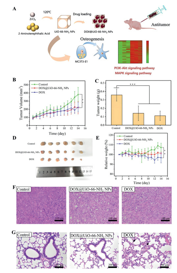
Based on these characteristics, Karakeili et al. prepared the chitosan scaffolds of UiO-66 nanocrystals containing fosfomycin. This framework not only possesses the ability of osteogenesis but also has the ability of anti-infection. It is used not only for bone regeneration but also for osteomyelitis. The up-regulation of the bone gene and the increase of ALP activity were also confirmed in the gene aspect. It was indicated that UiO-66 MOF nanocrystals possess high potential to accelerate bone regeneration without growth factors [106,107]. Yuan et al. [78] explored and designed a new type of bone marrow stromal cells along with dual anti-tumor and bone regeneration properties DOX@UiO-66-NH2 NPs (Fig. 10). The size of the hydrated DOX@UiO-66-NH2 NPs was found to be ~192.5 nm. However, the loading rate of DOX was about 12.6%. The DOX@UiO-66-NH2 NPs displayed good antitumor efficacy in vitro and in vivo. Also, compared with free DOX in vivo, DOX@UiO-66-NH2 NPs significantly turn down lung injury and the employment of UiO-66-NH2 NPs significantly encourages the osteogenic differentiation of osteoblasts (Fig. 10).
Figure 10
Figure 10.
(A) Scheme for UiO-66-NH2 NPs for the treating bone tumors and promote osteogenesis. (B) Tumor volume change profiles during treatment (P < 0.01 (**), n = 6). (C) Tumor weight after 14 days of treatment (P < 0.001 (***), n = 6). (D) Tumor images of mice in each group after 14 days of treatment. (E) Weight change profiles of mice during treatment cycles (P < 0.01 (**), n = 6). (F) H&E staining of nude mice tumor sections. (G) H&E staining of local lung section of nude mice. Reprinted with permission [78]. Copyright 2023, American Chemical Society.
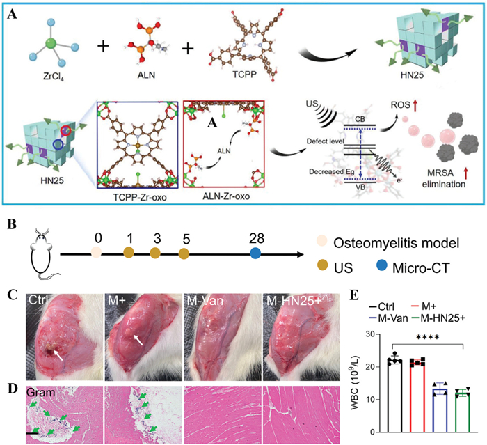
The main therapeutic therapies for infectious bone disorders are surgical debridement and antibiotic treatment; however, excessive use of either might exacerbate the bone defect. Antibiotics carry the potential of major adverse effects. Without surgery and antibiotic therapy, quick anti-infection and tissue restoration are hard to achieve. As for the anti-infection treatment of infectious bone defects, the current research was mainly focused on the slow-discharge of antibiotics and metal ions viz. Ag+ and Cu2+ [108–111]. Perhaps, these methods are uncontrollable in time and space with low antibacterial effect. Once the antibacterial substances are discharged completely, the anti-infection ability will vanish. To overcome this problem, Ma et al. used alendronate sodium (ALN) to construct a MOF-based sound sensitive agent (HN25) based on defect engineering porphyrin which can effectually eliminate methicillin-resistant Staphylococcus aureus (MRSA) infection and stimulate osteogenic differentiation under differential ultrasound irradiation (Fig. 11) [79]. Under 1.0 MHz and 1.5 W/cm2 irradiation, the antibacterial efficiency of HN25 was 98.97%. In the rat osteomyelitis model, compared with the control group HN25, no abnormal liver and kidney function was found and other major organs after 28 days of administration, which proved that it had good biocompatibility. Compared with the control group, the pyogenic mice caused by MRSA infection in the HN25 group had been repaired with the smallest bone defect.
Figure 11
Figure 11.
(A) Scheme for synthesis of ALN-mediated defective MOF for targeting infectious bone tissue repair. (B) The osteomyelitis experimentation process. (C) four-week-old wound photos of infected locations. (D) Gram staining. Scale bar = 100 µm. (E) WBC of different samples. n = 5, ****P < 0.0001. Reprinted with permission [79]. Copyright 2023, Wiley‐VCH GmbH.
In clinical practice, bone transplantation is limited by the long time-period, limited source, and bleeding of autologous bone tissue transplantation [112–114]. Sadek et al. found that UiO-66 nanomaterials have good blood and cell compatibility [80], and the treated bone defects have signs of healing. Therefore, the implantation of UiO-66 nanomaterials stimulated bone defect and could be used as a bone defect scaffold for the reconstruction of the rabbit femoral model.
The application of UiO-MOF in treating bone ailments is mainly based on its ability to mimic the microstructure of natural bone and provide an acceptable environment for cell attachment and growth. By adjusting its porosity and chemical composition, UiO-MOF can increase new bone formation by stimulating osteoblast proliferation and differentiation. UiO-MOF has a high porosity, which can be used as a drug carrier to achieve local slow-release therapy and reduce systemic side effects. In addition, the surface of UiO-66 can be further modified, such as surface-modified acoustic sensitizer as mentioned above, to exert therapeutic effects in response to ultrasound.
4.3
Application of MIL-based nanomaterials in bone repair and regeneration
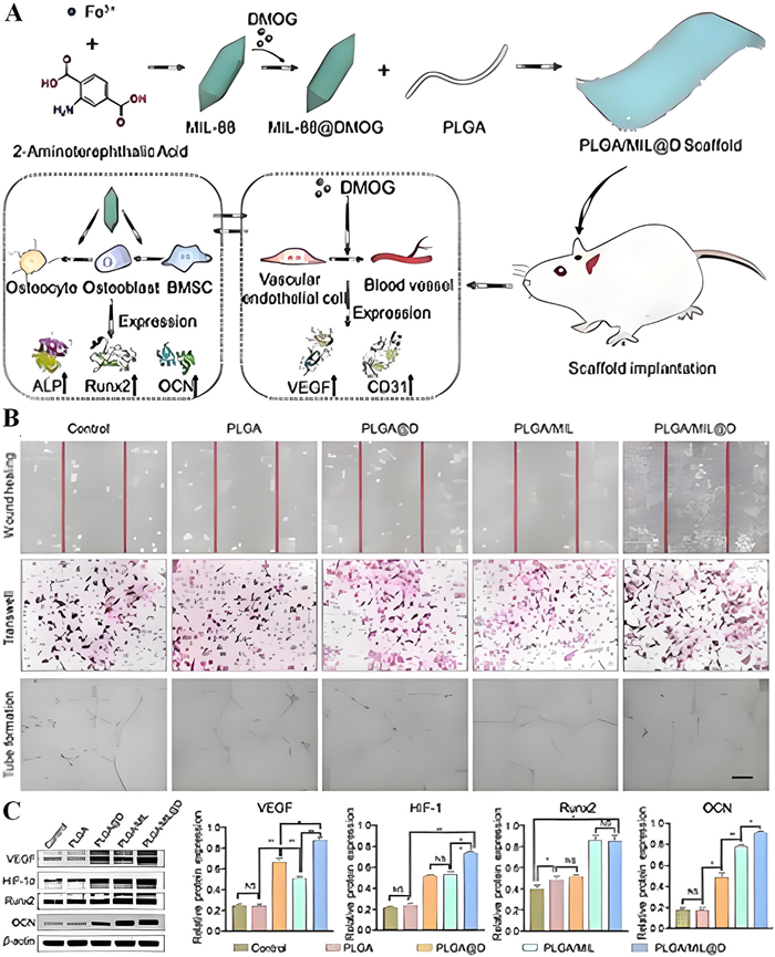
As one of the most popular MOFs materials of Lavassey Institute Materials (MIL) have good biocompatibility, unique skeleton flexibility, high specific surface area, and high stability. Consequently, they exhibit vast application prospects medicine sector [115], sensing [116], catalysis [117,118], etc. The traditional scaffolds used in bone regeneration in the past mainly affect bone repair through free diffusion and mass exchange, which poses a challenge to forming an operational vascular network [119]. The bone repair and regeneration phenomenon is closely related to the vascular network [120–124], which contributes significantly to the oxygen and nourishment required for bone repair and regeneration [125,126]. Therefore, there is a pressing requirement to develop a scaffold that can combine angiogenesis and osteogenesis to achieve this repair. As a bioactive ion, iron possesses antibacterial, osteogenic, and angiogenic effects. Iron-based MIL-based MOFs (such as MIL-88) also play a principal role in the field of biomaterials [127–129]. Based on this, Xu et al. [81] loaded the angiogenesis small molecule drug (dimethyloxalallyl glycine, dmog) into the iron-based MOF (MIL-88), and combined with PLGA nanofiber scaffold in which the PLGA nanofiber scaffold was prepared by electrospinning technology MIL@D scaffold and its related performance in bone repair. The scaffold can significantly strengthen vascularization and bone regeneration by promoting angiogenesis and mineralization at the bone defect site in vivo (Fig. 12). The scaffold can significantly enhance vascularization and bone formation by stimulating endothelial cell migration and tube formation, activating hypoxia inducible factor-1 and vascular endothelial growth factor signaling routes thereby promoting osteoblast related protein expression in vitro (Fig. 12).
Figure 12
Figure 12.
(A) Scheme for synthesis of DMOG-loaded MIL-88. (B) The corresponding images of assays presenting wound curing, transwell, and tube formation process (scale bar: 100 µm). (C) The VEGF, HIF-1α, Runx2 and OCN expression inspected with Western blot. NS, no consequence associated to regulator (n = 3), P < 0.05, **P < 0.01. Reprinted with permission [81]. Copyright 2023, Elsevier B.V.
Magnesium deficiency may lead to osteoporosis [130], because the lack of magnesium ion in bone will directly escalate the crystal size of hydroxyapatite in bone and decline the proportion of osteoblasts, which will lead to the reduction of bone hardness [131,132]. In addition, magnesium deficiency can also down regulate parathyroid hormone and change vitamin D levels, thus indirectly affecting bone formation [133–135]. Yu et al. [82] also incorporated magnesium ions into the cage of mil-100(FE) and to improve the loading rate of magnesium ions, polyacrylic acid (PAA) was modified on its surface to produce Mg@MIL-100(Fe)-PAA scaffolds and their related properties in bone repair were studied. The reports stated that the nano-scaffold offered good biocompatibility. By encouraging the proliferation and improving the ALP activity of MG-63 cells, it effectively promoted cell differentiation and accelerated the process of bone healing.
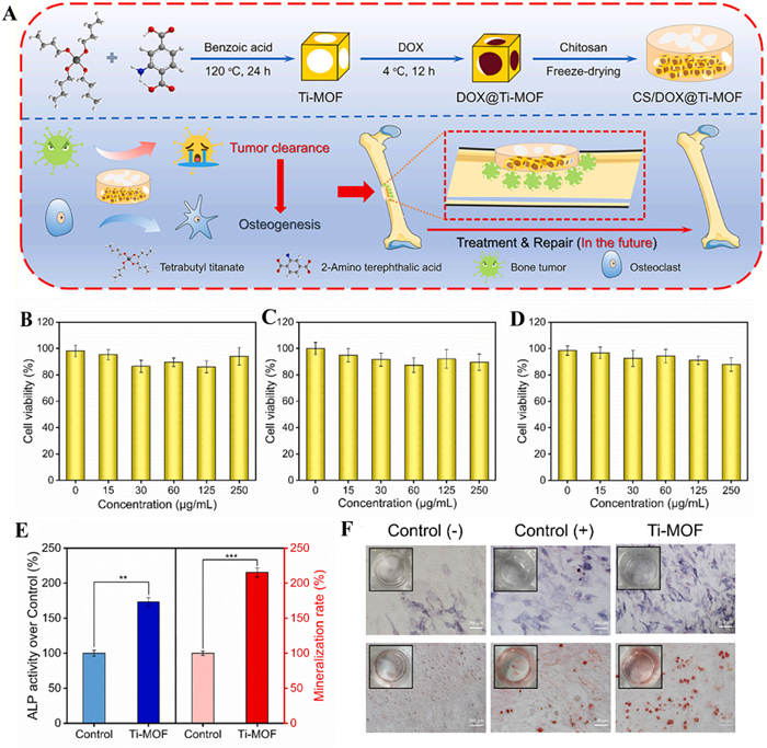
The recurrence and metastasis of remaining tumors continue to aggravate bone repair following bone tumour surgery. Because of the existence of wound, there are great requirements for the biocompatibility and degradability of the material. Ti-based MOF is distinguished by its exceptional composition of 2-aminoterephthalic acid and Ti8O8 ring clusters. Due to the non-toxicity of Ti4+ and organic ligands, they exhibit excellent biocompatibility and biodegradability compared with other MOFs [136,137]. Zeng et al. (Fig. 13) [83], designed a chitosan composite framework (CS/DOX@TiMOF) for tumor treatment and bone repair. The DOX@Ti-MOF exhibited a hydrodynamic dimension of 306 ± 20 nm. In this study, 250 µg/mL Ti-Mo was added to the culture dish of MC3T3-E1 cells, RAW264.7 cells, and HUVEC cells (Fig. 13). After incubating for 24 h, the relative cell activity was more than 80%, which proved good biocompatibility. The outcomes of alizarin red staining by measuring ALP activity and mineralized nodule generation showed that Ti-MOF could efficiently promote the osteogenic differentiation of MC3T3-E1 cells. After modification with chitosan, DOX@Ti-MOF showed an ideal degradation rate under the action of lysozyme. In animal models, CS/DOX@TiMOF was able to remove residual tumor cells in chronological order and then carry out osteogenic differentiation.
Figure 13
Figure 13.
(A) Scheme presenting CS/DOX@Ti-MOF synthesis and characteristics of biocompatibility and osteogenic activity. Comparative viability of cells of (B) MC3T3-E1, (C) RAW264.7 cells, (D) and HUVEC cells after incubation with variable concentrations of Ti-MOF for 01 day. (E) ALP activity in MC3T3-E1 cells treated differently after 01 week and quantitative study of alizarin red staining at 02 weeks. (F) Pictures of MC3T3-E1 cells stained with alizarin red after seven days and after various treatments. Reprinted with permission [83]. Copyright 2024, Elsevier B.V.
MOF organic ligands are classed as either exogenous or endogenous. In terms of biocompatibility, endogenous ligands have better safety performance in vivo compared to exogenous ligands that do not naturally exist in the human body [138]. Therefore, bio-metallic organic frameworks (BioMOFs) composed of biocompatible metal cations as nodes and common biomolecules as connectors have a variety of applications in biological environments. Jafari et al. [84] used chitosan nanofibers as a template, coordinated calcium ion with alendronate sodium, and added alginate and chitosan particle solution to obtain biomil-4/chitosan/alginate-based bone tissue scaffold and studied its bone regeneration potential. The composite scaffold offered good performance as the chitosan nanofibers provide a good biodegradation rate, alginate and chitosan particle solution provide a good stability, mechanical strength and biocompatibility and calcium ion and alendronate sodium released during the degradation of biomil-4 contribute to the process of bone regeneration.
In summary, apart from drug loading, MIL can enhance osteoblast adhesion, proliferation, and differentiation, thereby speeding up bone regeneration and repair. Its porous nature also facilitates the penetration and vascularisation of new bone tissue, which is particularly important for the treatment of bone defects. MIL can be used as a bone grafting material for the filling and restoration of fractures, bone defects, or osteochondromas, etc. MIL also carries and releases ions of calcium, magnesium, etc., which have a positive outcome on bone mineralization and metabolism. In addition, MIL's good degradability makes it useful in bone trauma.
4.4
Application of polyphenol-based MOF nanomaterials in bone repair and regeneration
Gallic acid (GA) also named as 3,4,5-trihydroxybenzoic acid, widely exists in grape berries as a plant polyphenol [139]. GA contains significant free radical scavenging and antioxidant properties, which can help the body's natural antioxidant defense system and prevent lipid peroxidation. GA can form good coordination complexes with metal ions and different metal ions will influence the physiological function of the resulting complexes [140]. Cooper et al. reported a MOF material of porous Mg-GA derivatives, which exhibited good biocompatibility and can be slowly released in physiological body fluid to regulate cell growth, cell metabolism, and proliferation [141].
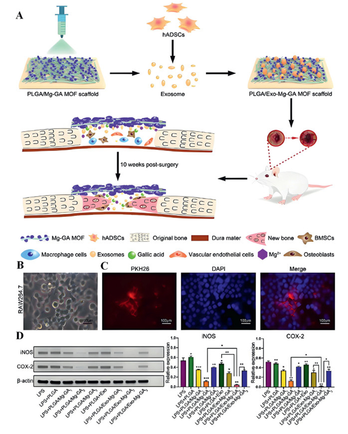
The MOF scaffolds formed by the coordination of GA and Mg2+, enhanced osteogenic differentiation, anti-inflammatory ability and accelerated bone regeneration for the human body. Kang et al. combined polylactic acid with polyglycolic acid (PLGA), to generate PLGA/Mg-GA MOF scaffolds by electrospinning technology for bone regeneration (Fig. 14) [85]. The nano scaffold structure can regulate the steady release of Mg2+ and GA, effectively enhance bone generation and angiogenesis in vivo, and enhance anti-inflammatory ability by affecting the osteogenic ability of hBMSCs, the angiogenesis ability of HUVECs and the anti-inflammatory effect of RAW264.7 cells (Fig. 14).
Figure 14
Figure 14.
(A) Scheme for PLGA/Mg-GA MOF for bone regeneration scaffolds. (B) Corresponding images of RAW264.7. (C) Cellular uptake assays displaying PKH26-labeled exosomes. (D) Western blot measurements of iNOS and COX-2 expression in RAW264.7 cells. Reprinted with permission [85]. Copyright 2022, The Authors.
Polyphenolic compounds usually have good biocompatibility and antioxidant properties, while MOF materials are known for their huge specific surface area, tunable pore structure along with functionalization ability. These traits make polyphenolic-MOF materials apt materials for bone tissue engineering, which apart from regulating cell growth, cell metabolism, and proliferation, also exert anti-inflammatory effects.
4.5
Application of MOF combined with gel in bone repair and regeneration
Gel is a specific concentration of a polymer solution or sol. The viscosity eventually loses its fluidity as it steadily rises under the correct circumstances. The entire system takes on the characteristics of an elastic semi-solid with a consistent look and shape. Gels can be prepared from a variety of materials including synthetic polymer materials and natural polymer materials and may be categorized into several categories based on their composition and properties. Hydrogels have attracted extensive attention and are a special type of high molecular polymer material, which has a three-dimensional network structure and can absorb a huge amount of water without disintegrating. Compared with other types of gels, hydrogels exhibit high water content, softness similar to natural tissues, biocompatibility, degradability, and stimulus-response [142]. In recent studies, the combination of gel and MOFs has become a research hotspot. The composite material of gel and MOFs combines the tunable pore structure, large specific surface area, superior adsorption performance of MOFs, as well as the good biocompatibility and mechanical flexibility of hydrogel, showing excellent therapeutic effects in bone therapy. For example, Li et al. [86] reported a convenient synthesis of organic/inorganic nanocomposite gel scaffold by combining the chemical and physical crosslinking of conjugated magnesium MOFs (Mg-MOF) and bone targeted alendronate sodium with biocompatible gelatin scaffold. This nanocomposite gel scaffold displayed good biocompatibility, sustained release of Mg2+ and alendronate sodium, strong bone affinity, and bone regeneration ability.
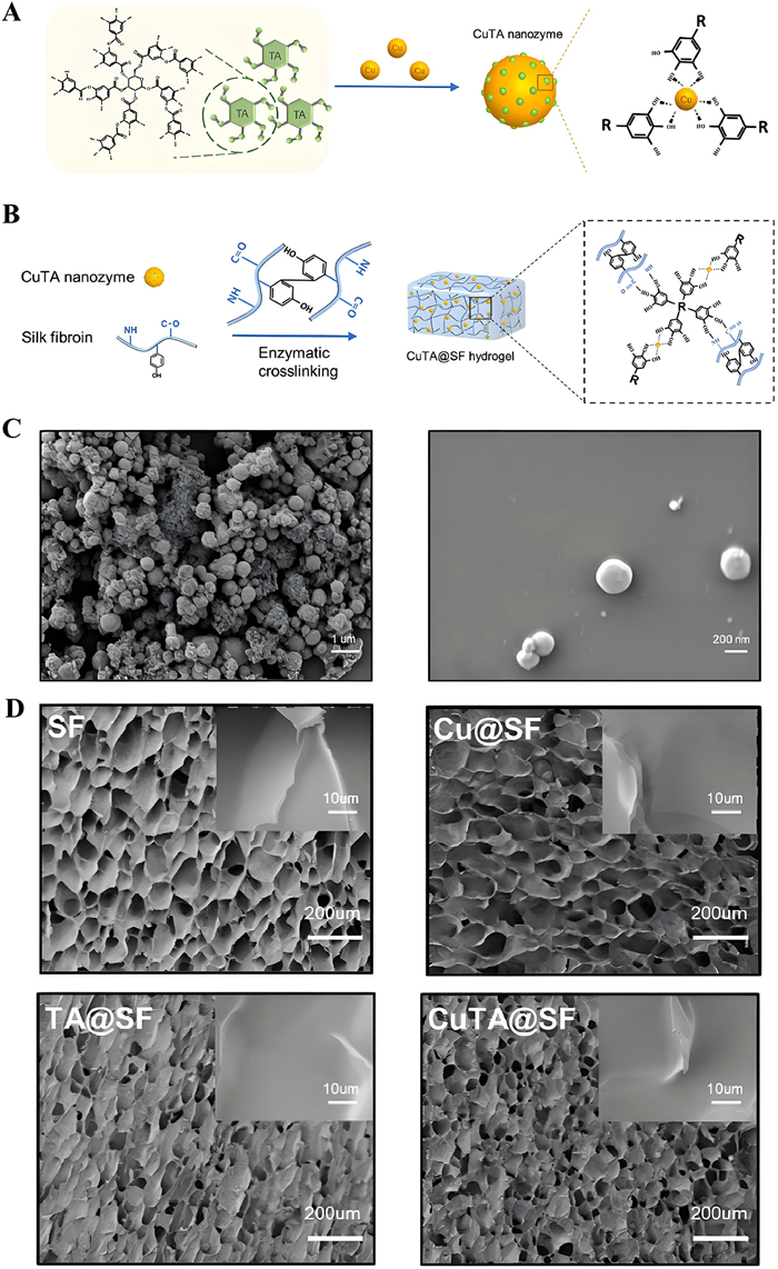
Hydrogels are usually amorphous and have a uniform microstructure and high hydrophilicity. Cao et al. [87] reported the synthesis of a polyfunctional silk-based hydrogel by combining MOFs nanoenzyme (CuTA@SF) (Fig. 15). The addition of CuTA nanoenzyme engendered SF hydrogel with homogenous microstructure and high hydrophilicity. The average diameter of the CuTA nanoenzyme was 470 ± 140 nm and the pore morphology changed after adding it into the hydrogel (Fig. 15). The CuTA@SF hydrogel showcased a long decaying time with a degradation rate of 87.9% after 70 days. Its delayed breakdown left room for the development of new tissues and this hydrogel promoted the growth, viability, antioxidant, and antibacterial activities of mesenchymal stem cells (MSCs) and chondrocytes grown in vitro. Under the inflammatory environment stimulated by interleukin-1 beta, CuTA@SF hydrogel still induced the osteogenesis of mesenchymal stem cells and the deposition of cartilage-specific extracellular matrix (ECM). In the full thickness osteochondral defects (OCD) model of rabbits, CuTA@SF hydrogel confirmed the favorable regeneration of in situ OCD by micro-CT, histology, and immunohistochemistry.
Figure 15
Figure 15.
(A) Scheme for the preparation of CuTA nanozyme. (B) Enzymatically crosslinked CuTA@SF hydrogel and its intermolecular integration inside the CuTA@SF hydrogel. (C) SEM image for CuTA nanozyme. (D) SEM images of lyophilized SF, Cu@SF, TA@SF, CuTA@SF hydrogels. Reprinted with permission [87]. Copyright 2022, The Authors.
Diabetic bone defects are a common diabetic consequence that has significant repercussions on patient quality of life. Because of the pathological milieu of hyperglycemia, oxidative stress, and inflammation in the diabetic bone defect site, repairing diabetic bone defects is one of the most difficult issues confronted in the clinic. The current tissue-engineered bone is capable of good bone regeneration and functional restoration. The ability to cure a diabetic bone defect by treating a single pathogenic cause remains suboptimal. To rectify the complicated pathological milieu of bone regeneration in diabetes, it is critical to synthesize a multi-functional drug delivery system that can achieve hypoglycemia, antioxidant, anti-inflammatory, angiogenesis, and osteogenesis simultaneously. Wang et al. [88] reported a multifunctional GelMA/HAMA microgel encapsulated Mg2+/emodin (MgEm) nano-rod release system towards the microenvironment of the diabetes pathway. In the microgel sustained-release system, emodin possesses hypoglycemic, antioxidant, and anti-inflammatory functions. Mg2+ ion promoted angiogenesis and bone regeneration. Then, the decalcified bone matrix and microgel loaded with BMSCs were prepared (BMSCs@MEGH-D) to construct tissue-engineered bone. Animal experiments show that the BMSCs@MEGH-D can effectively correct the pathological microenvironment of diabetes and promote vascularized bone regrowth in diabetic rabbit skull models.
In summary, when MOFs are combined with hydrogels, a synergistic phenomenon can enhance the bone repair and regeneration potential of the material. Such combinations typically aim to utilize the high specific surface area and tunable chemistry of MOFs to nurture cell attachment and growth, while utilizing the biocompatibility and moisture retention power of hydrogels to imitate the environment of natural bone tissue. And these composites combine the advantages of the versatility of MOFs with the biocompatibility and injectability of hydrogels. The metal-organic framework-based hydrogel composites are not only capable of repairing soft tissue injuries, but can also be effectively applied to the repair of bone defects. The design of this composite takes into account the special requirements of hard tissue repair, including mechanical strength, bioactivity, and integration ability with bone tissue. In addition, the multifunctionality of MOFs was utilized to improve the properties of the hydrogel.
5.
Summary and outlook
In summary, MOF-based composite materials demonstrate significant potential in bone repair and regeneration. MOFs stand out among various materials due to their high porosity, easy modification, tailorable mechanical properties, and excellent biocompatibility. Compared to other nanomaterials in bone repair and regeneration, MOFs offer the following advantages as drug carriers: Sustained local drug concentration over time, reducing systemic side effects; Promotion of osteocyte growth and differentiation; Antibacterial and anti-inflammatory effects. Additionally, when used as bio-scaffolds, MOF composites provide: Superior biocompatibility; Customizable mechanical properties; Minimal damage to surrounding tissues; Optimal degradation rates. Importantly, functionalized MOF nanomaterials can achieve multi-modal therapeutic effects for bone repair and regeneration. These characteristics position MOFs as a promising material platform for bone defect treatment.
In the field of MOF nanomaterials currently used for bone repair and regeneration, researchers have developed strategies such as "MOFs + hydrogel composites" and "MOFs + bioceramic hybrids" to enhance the mechanical properties or biocompatibility of nanocomposites through MOFs. Additionally, extensive research and discussions have been conducted on the use of MOFs jointly with novel therapies (e.g., photothermal therapy) for treating bone tissue defects or infections.
The application of MOFs in bone regeneration materials is still in the research stage, but their therapeutic efficacy is beyond doubt. Future studies should focus on exploring the effects of different types of MOFs in promoting bone healing, including their ability to release anti-inflammatory or osteoinductive factors as drug carriers. Additionally, optimizing the stability and functionality of MOF composites can expand their application scope in bone repair. Meanwhile, integrating advanced manufacturing technologies such as 3D printing allows customization of personalized MOF-based scaffolds to adapt to complex bone defect shapes and patient-specific treatment needs. Finally, it is necessary to clarify their metabolic mechanisms to avoid long-term damage to the body. In the future, with improvements in synthetic methods and deeper understanding of MOFs' biological effects, these materials are expected to be translated into clinically effective bone repair and regeneration solutions.
Declaration of competing interest
The authors declare that they have no known competing financial interests or personal relationships that could have appeared to influence the work reported in this paper.
The authors acknowledge funding from the Key Scientific Research Project of Colleges and Universities of Education Department of Guangdong Province (Nos. 2021ZDZX2052, 2022ZDZX2022), Dongguan Social Development Science and Technology Project (No. 20231800936222), Guangdong Medical University Research Project (Nos. 4SG24267G, GDMUZ2023001), the open research fund of Songshan Lake Materials Laboratory (No. 2023SLABFN30), Guangdong Basic and Applied Basic Research Foundation (No. 2023A1515011536), and Featured Innovation Project of Guangdong Province (No. 2022KTSCX045).
[1]
Y. Liu, S. Wang, C. Quan, et al., J. Mater. Chem. B 12 (2024) 637–649. doi: 10.1039/d3tb02620e
Figure 1
(A) Proportion of MOFs used to treat bone defects. (B) MOF-based nanomaterials for therapeutic modalities in bone defects. (C) Research advances in MOF-based treatment of bone defects in past 5 years (Source: Web of Science, 8 July 2024). (D) Study of MOF in Bone Defects, 2019–2024.
Figure 7
(A) The fabrication of porous patterned PLLA composite electrospun scaffolds incorporated with CCM@ZIF-8 MOFs, and their promotion for diabetic wound healing process. (B) Research progress on the size change of back skin resection in diabetic mice at different periods. (C) Simulation of wound bed closure trace in each group in vivo. Light brown area represented the wounds area at day 0 and the blue area represented the wounds area at day x (x = 7, 11, 13 and 15). (D) The wound area of the six groups was statistically analyzed to show the healing effect. Reprinted with permission [71]. Copyright 2020, Elsevier B.V.
Figure 8
(A) Scheme for GelMA@eIm/ZIF-67 nanocomposite hydrogel formation. (B) GelMA@CoCl2 and GelMA@eIm/ZIF-67 release patterns in FBS. (C) Scheme of reaction between a linker solution comprising eIm and mIm and Co(Ⅱ). Reprinted with permission [73]. Copyright 2021, American Chemical Society.
Figure 9
(A) Schematic illustration presenting syntheses of SrCO3@ZIF-8/PLLA moieties for regulating bone immune environment and enhancement in osteogenic differentiation. (B) Images of 3D reconstruction, (C) H&E, (D) and Masson trichrome staining of the femoral defects after implanting with PLLA, SrCO3/PLLA, and SrCO3@ZIF-8/PLLA scaffolds. Reprinted with permission [77]. Copyright 2023, Elsevier B.V.
Figure 10
(A) Scheme for UiO-66-NH2 NPs for the treating bone tumors and promote osteogenesis. (B) Tumor volume change profiles during treatment (P < 0.01 (**), n = 6). (C) Tumor weight after 14 days of treatment (P < 0.001 (***), n = 6). (D) Tumor images of mice in each group after 14 days of treatment. (E) Weight change profiles of mice during treatment cycles (P < 0.01 (**), n = 6). (F) H&E staining of nude mice tumor sections. (G) H&E staining of local lung section of nude mice. Reprinted with permission [78]. Copyright 2023, American Chemical Society.
Figure 11
(A) Scheme for synthesis of ALN-mediated defective MOF for targeting infectious bone tissue repair. (B) The osteomyelitis experimentation process. (C) four-week-old wound photos of infected locations. (D) Gram staining. Scale bar = 100 µm. (E) WBC of different samples. n = 5, ****P < 0.0001. Reprinted with permission [79]. Copyright 2023, Wiley‐VCH GmbH.
Figure 12
(A) Scheme for synthesis of DMOG-loaded MIL-88. (B) The corresponding images of assays presenting wound curing, transwell, and tube formation process (scale bar: 100 µm). (C) The VEGF, HIF-1α, Runx2 and OCN expression inspected with Western blot. NS, no consequence associated to regulator (n = 3), P < 0.05, **P < 0.01. Reprinted with permission [81]. Copyright 2023, Elsevier B.V.
Figure 13
(A) Scheme presenting CS/DOX@Ti-MOF synthesis and characteristics of biocompatibility and osteogenic activity. Comparative viability of cells of (B) MC3T3-E1, (C) RAW264.7 cells, (D) and HUVEC cells after incubation with variable concentrations of Ti-MOF for 01 day. (E) ALP activity in MC3T3-E1 cells treated differently after 01 week and quantitative study of alizarin red staining at 02 weeks. (F) Pictures of MC3T3-E1 cells stained with alizarin red after seven days and after various treatments. Reprinted with permission [83]. Copyright 2024, Elsevier B.V.
Figure 14
(A) Scheme for PLGA/Mg-GA MOF for bone regeneration scaffolds. (B) Corresponding images of RAW264.7. (C) Cellular uptake assays displaying PKH26-labeled exosomes. (D) Western blot measurements of iNOS and COX-2 expression in RAW264.7 cells. Reprinted with permission [85]. Copyright 2022, The Authors.
Figure 15
(A) Scheme for the preparation of CuTA nanozyme. (B) Enzymatically crosslinked CuTA@SF hydrogel and its intermolecular integration inside the CuTA@SF hydrogel. (C) SEM image for CuTA nanozyme. (D) SEM images of lyophilized SF, Cu@SF, TA@SF, CuTA@SF hydrogels. Reprinted with permission [87]. Copyright 2022, The Authors.


 DownLoad:


 下载:
下载:


 下载:
下载:

