chenjing6811@126.com (J. Chen). 1 These authors contributed equally to this work.
Received Date:
15 September 2025 Accepted Date:
01 December 2025 Revised Date:
29 November 2025 Available Online:
15 November 2026
Abstract:
Neural damage remains a serious clinical challenge. While electrical stimulation (ES) is a potent strategy to promote repair, traditional implantable electrodes face issues of invasiveness, rigidity, and finite longevity. In recent years, electroactive biomaterials have emerged as a revolutionary platform to overcome these hurdles by seamlessly integrating bio-mimetic conductivity with tunable biological and mechanical properties. This review outlines the types, advantages and disadvantages, preparation methods, and progress of conductive biomaterials commonly employed in nerve repair, and focus on pioneering non-invasive self-generating systems based on piezoelectric, magnetoelectric, and biofuel cell technologies. In addition, based on the relevant electrophysiological properties of electroactive materials, we synthesize a unified framework for their mechanisms of action, elucidating how electroactive interfaces orchestrate regeneration through tripartite regulation: Cellular behavior, signaling pathways, and microenvironment regulation. Finally, we discuss ongoing challenges and future perspectives, particularly regarding biodegradation, clinical translation, and the integration of smart feedback systems. This review aims to serve as a foundational reference for guiding the rational design of next-generation electroactive therapies for neural repair.
Nerve damage is a frequent type of clinical trauma that can result from accidents, diseases, or various traumatic events in everyday life and work. The World Health Organization reports that ~50 million individuals globally experience traumatic brain damage annually, whereas other studies indicate that up to 1 million individuals worldwide sustain peripheral nerve injuries (PNI) each year. In China, the number of patients with PNI is 30–500,000, accounting for 2.8% of trauma patients. Furthermore, the incidence of impairment following nerve injury is elevated, with the majority of cases resulting in sequelae, thus diminishing patients' quality of life and imposing a significant economic burden [1]. The central nervous system (CNS) largely lacks the ability to self-repair, whereas the peripheral nervous system (PNS) has inherent regenerative and repair capabilities. However, nerve injuries greater than 3 mm typically necessitate autologous nerve transplantation, which is regarded as the gold standard for nerve healing. Autologous nerve transplantation encounters challenges, including donor site morbidity, potential neuroma formation, diameter discrepancies; allogeneic nerve transplantation is hindered by immunological responses and rejection phenomena [2,3]. Consequently, the application of biomaterials for the repair of nerve injury is garnering increased interest. Owing to their distinctive physical and chemical characteristics, electroactive materials are considered optimal for nerve repair and regeneration.
Endogenous electric fields within the human body significantly influence nervous system [4]. Upon exposure to an electric field, the potential differential across the cell membrane generates "action potentials", which are electrical signals enabling neuronal communication and influencing cell migration, proliferation, differentiation, and other actions [5]. Furthermore, introducing electric currents alters neuronal excitability by causing membrane depolarization or hyperpolarization. To quantify the extent of this polarization, several studies have focused on measuring the impacts of electric field amplitude, frequency, direction, and other factors [6,7]. In this way, in the field of neural tissue engineering, the emergence of electroactive materials has opened up new possibilities for neural regeneration. Electroactive materials can facilitate the reconnection of damaged neurons. Crucially, these materials can mimic the electrophysiological environment of neurons. In addition, some electroactive materials can also release bioactive molecules, further promoting neural regeneration [8]. However, despite some progress in the utilization of electroactive materials in neural regeneration, many challenges remain. For example, how to precisely control the intensity and frequency of electrical stimulation (ES), and how to ensure the long-term stability and biocompatibility of electroactive materials, are issues that need further research [9].
Overall, the utilization of electroactive materials in neural regeneration is a promising research field. We aim to improve treatment techniques for neural regeneration and repair through further research and development, thereby improving the quality of life for individuals with nerve injuries. This article initially examines the mechanism of neural regeneration and the impact of ES on nerves, followed by an analysis of the performance, characteristics, functions, and applications of regularly utilized electroactive materials in nerve repair. It then introduces the preparation methods of various electroactive material implants for neural regeneration, traditional ES and new developments. Finally, to better explore the role of materials in the nervous system, this work depicts the specific mechanisms of these materials in neural regeneration from three perspectives: Regulation of cellular behavior, regulation of signaling pathways, and regulation of the microenvironment.
2.
Strategies and challenges in neural regeneration
2.1
Mechanisms of neural regeneration and repair
The PNS exhibits a relatively comprehensive regenerative mechanism. In contrast, the mechanisms underlying CNS regeneration remain insufficiently elucidated; however, modulation of key molecular pathways, immune cell functions, and external factors can, to some extent, facilitate the exploration of partial mechanisms for CNS regeneration and repair. For detailed specifications on neural regeneration and repair, please refer to Section S1 (Supporting information).
2.2
ES promotes neural regeneration
The nervous system is composed of electrically excitable tissues capable of conducting information. By electrically modulating neuronal membrane potential to induce artificial action potentials, physiological electric fields can be simulated. This principle forms the basis for the use of ES as a therapeutic approach to restore function in impaired neural tissues. Detailed content is provided in Section S2 (Supporting information).
2.3
Neural regeneration on the conductive material
Conductive materials enhance charge transport at the cell‒matrix interface interfaces. This capability reduces voltage loss caused by the surrounding medium, thereby improving ES, and enables conductive materials with ideal morphological, chemical, and mechanical properties to connect damaged nerves in scaffold form [10,11]. Research indicates that the application of ES to conductive materials markedly enhances the development, proliferation, and migration of neural stem cells (NSCs). Additionally, ES facilitates remyelination of NSCs [12]. Furthermore, Huang et al. reported that local ES enhances the migratory capacity of the NSCs into conductive conduits, paving the way for axon regeneration [13]. Consequently, ES employing conductive materials can offer a versatile framework for influencing cellular behavior and determining stem cell destiny in tissue engineering.
2.3.1
Design principles and requirements for conductive materials
Conductive materials for nerve restoration must have excellent electrical conductivity, electrochemical stability, favorable, biocompatibility, suitable flexibility, adequate porosity, and an appropriate degradation rate. Furthermore, its internal architecture and measurements must replicate those of an autograft, guaranteeing that there is no pressure on neural tissue. Detailed examples are provided in Section S3 (Supporting information).
2.3.2
Biocompatibility and challenges of conductive materials
The lack of adequate biocompatibility is a major limitation for the majority of conductive materials [14]. The primary biological toxicity of conductive polymers stems from low-molecular-weight hazardous impurities generated during synthesis processes as well as their degradation products [8]. The primary drawback of carbon-based materials lies in their poor water solubility and tendency to aggregate, which may induce various toxic effects. For instance, Singh et al. reported that CNTs could accumulate in multiple organs within 30 min post-intravenous injection, with particularly abundant presence observed in the blood, skin, muscle, and kidneys [15]. Therefore, a comprehensive evaluation of the blood clearance kinetics, circulation half-life, in vivo biodistribution, and tissue/organ accumulation of conductive materials is essential.
Conductive biomaterials can be engineered to promote neural regeneration and improve biocompatibility through the incorporation of biodegradable materials and various chemical modifications [16]. Detailed content is provided in Section S4 (Supporting information).
3.
Conductive biomaterials for neural regeneration
Various conductive substrates for neural repair include electroactive inorganic materials, conductive polymers, and metallic materials. Electroactive inorganic materials mostly pertain to carbon-based substances, including graphene and its derivatives, CNTs. Conductive polymers include mainly polypyrrole (PPy), polyaniline (PANI), polythiophene (PT), poly(3,4-ethylenedioxythiophene) (PEDOT) PT derivatives and poly(p-phenylvinyl) (PPV). Nano-metallic particles are significant conductive materials. The composite material, comprising nanometals and hydrogels, enhances the mechanical strength and electrical conductivity while regulating cell adherence and proliferation through the modulation of the metal concentration [17]. The inability of common nonbiodegradable metals (e.g., gold, platinum, iridium) and their alloys to degrade frequently induces substantial chronic inflammatory responses in surrounding tissues. The consumption of oxygen and by-products during metal corrosion can lead to necrosis of the surrounding tissue [8]. In addition, different organs, sexes and ages have different tolerances for various metals, so quantitative and systematic studies are needed. Various electroactive materials for neural regeneration are summarized in Table S1 (Supporting information)
3.1
Conductive polymers
Conductive polymers represent promising materials are highly regarded in the domain of neural regeneration. Conductive polymers have significant advantages as novel candidates for nerve repair therapy with metal electrodes, such as greater charge injection capacity, lower impedance, safer stimulation window, and better mechanical durability. However, the brittleness of pure conductive polymers limits their processability and applicability, while their suboptimal biocompatibility and lack of biodegradability further constrain utility. Consequently, doping or blending with supplementary materials becomes essential for property enhancement [18].
3.1.1
PPy
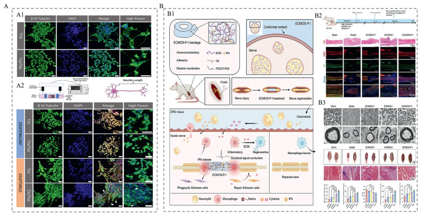
PPy, a prominent intrinsically conductive polymer (ICP) utilized in tissue engineering. While the pyrrole monomer is soluble in various solvents, polymerization renders it insoluble, resulting in limited processability [19]. Polypyrrole has high electrical conductivity (103 S/cm), facile synthesis, robust modification potential, excellent cytocompatibility and histocompatibility, and enhanced cell adhesion/tissue regeneration capacity [20], establishing significant potential for nerve repair applications. In vitro studies have consistently illustrated the impact of PPy as a conductive polymer on neuronal cells. PPy nanorods, produced through the oxidative chemical polymerization of pyrroles within alumina nanopores, have been shown to enhance cell differentiation, significantly promoting cell proliferation and expediting neuronal differentiation when electrically stimulated in PC12 cells [21]. Leahy et al. coated PPy on polycaprolactone (PCL) surfaces to create a three-dimensional (3D) conductive scaffold matching the corticospinal tract. The scaffold showed good conductivity (15 ± 5 S/m) and biocompatibility. ES transmitted through the scaffold resulted in longer neurite growth (Fig. 1A) [22]. The repair of peripheral nerves with PPy has been thoroughly investigated; for example, Song et al. fabricated a PPy-coated electrospun poly(L-lactic acid-ε-caprolactone)/silk fibroin (PLCL/SF) nerve guidance conduit for 15 mm sciatic nerve gap regeneration in rat models. The PPy coating on NGCs markedly enhanced SCs proliferation and demonstrated comparable performance to the autograft group at ES (100 mV, 1 h) [23]. By combining hyaluronic acid (HA), cystamine (Cys), and pyrrole-1-propionic acid (Py-COOH), Xuan et al. developed a hydrogel (HASPy) with self-repairing, injectable, biodegradable, and high conductivity (7.7 mS/cm) properties. Using the sciatic functional index (SFI) and the weight of the gastrocnemius muscle on the injured side and the control side of mice as indicators of neural function recovery, their experiment showed that HASPy significantly improved neural function recovery (SFI index increased to 87.5%, and the recovery rate of the gastrocnemius muscle was 75.49% ± 4.77%). In addition, they found that HASPy regulates SCs function through the IL-17RA-CD44 dual pathway (Fig. S1 in Supporting information) [24]. The long-term behavioural effects of SCI are generally evaluated via the Basso, Beattie, Bresnahan (BBB) motor rating scale [25]. A study used tubes rolled from a mixture of PPy particles and polylactic acid (PLA), implanted in rats with complete spinal cord transection injuries. The PPy material somewhat enhanced motor function recovery compared with that of the controls (BBB scores < 6 vs. 1–3 after 6 weeks) [26]. For 2 mm spinal cord hemisections in adult male mice, tannic acid-crosslinked PPy hydrogel patch implantation yielded superior motor recovery vs. controls (BBB ~14 vs. 7 in controls after 6 weeks). Regarding immune responses, implantation of the PPy-tannic acid hydrogel significantly decreases macrophage and microglial migration into injured areas [27].
Figure 1
Figure 1.
Design strategies and functional roles of conductive polymers in neural regeneration. (A) Modulation of neuronal behavior by PPy/PCL scaffolds. (A1) Neurons grow on PCL and PPy/PCL surfaces and neurite extension. (A2) Neurons and neurites grown on PCL and PPy/PCL after ES, analysis of neuronal metabolic activity (B-Ⅲ tubulin, 4′,6-diamidino-2-phenylindole (DAPI), merge (phalloidin, red)) and the morphology of growing cells and their neurites (high power). Scale bar: 50 µm. Copied with permission [22]. Copyright 2024, Elsevier. (B) Neural hydrogel bandage (ECMOS-P-Ⅰ) applied for nerve regeneration and functional recovery in rats. (B1) Schematic diagram of the ECMOS-P-Ⅰ component and its neural application functions. (B2) Assessment of functional and sensory recovery in rats. MBP (red) and NF (green). (B3) Transmission electron microscope (TEM) images of sciatic nerve regeneration and gross views with cross-sectional Masson’s trichrome staining of excised gastrocnemius muscle. Scale bar: 10, 2 µm (TEM images); 1 mm (gross images); 500 µm (cross-sectional views images). Reproduced with permission [28]. Copyright 2024, John Wiley and Sons.
The differentiation of human-induced pluripotent stem cell-derived neural progenitor cells (hNPCs) is crucial for advancing neurological disease models [29]. Positioning hNPCs within a PPy-based system and conducting ES on hNPCs in vitro (1 kHz, 1 h). Furthermore, implant a scaffold containing hNPCs stimulated with electricity into the brain of rats with induced stroke. These experiments indicated that ES could modify hNPCs gene expression within the vascular endothelial growth factor A (VEGF-A) pathway, which is believed to be essential for improved recovery [30]. Another study created a scaffold comprising electropolymerized 2D membrane and 3D tube PPy integrated into alginate hydrogel-fixed hNPCs. The electric field intensity of 40 mV/m increased the production of crucial neurotrophic factors linked to cell survival, synapse remodeling, and neural regeneration [29]. Furthermore, integrating hydrogel mechanics with material conductivity is being explored. Integration of SWCNTs and PPy into catechol-modified HA hydrogels substantially enhances hNSPC neuronal maturation [31].
In conclusion, PPy, the most common conductive polymer, has been preliminarily explored in various studies. Developing PPy hydrogel composites holds significant therapeutic potential. Nevertheless, these formulations require prefabrication because of the nonbiocompatible crosslinking conditions, such as the utilization of oxidative initiators such ferric(Ⅲ) chloride, which is inherently hazardous. With the ongoing development of this field, future studies should explore the preparation technology of PPy composites and application of ES in tandem with PPy, since matching of stimulation settings may be advantageous for neural regeneration. As a result, there is great room to pursue in vivo and clinical investigations connected to neurons and PPy.
3.1.2
PANI
Among conductive polymers for biomedical use, PANI holds secondary prevalence. The emeraldine-base (EM-B) form of PANI has excellent electrical conductivity (up to 2 × 10² S/cm), high ambient-temperature stability, and potential degradability through doping [32]. PANI offers advantages including straightforward synthesis, varied structural forms, affordability, electrical conductivity, notable environmental stability, in vitro cytocompatibility, and switchable conductivity via doping/dedoping [33]. In the field of nerve repair, PANI can be combined with scaffolds to enhance neuronal cell repair, encourage NSC proliferation and differentiation, and stimulate neurite outgrowth while responding to external ES (EES) to regenerate damaged peripheral nerve tissue. thus, it has value as a conductive polymer in nerve repair. Detailed content is provided in Section S5 (Supporting information).
3.1.3
PEDOT
PEDOT is a derivative of polythiophene [34]. PEDOT possesses moderate heat conductivity, strong electrical conductivity (up to 400 S/cm), and excellent chemical stability [5]. PEDOT is biocompatible with multiple cell types, including rat bone marrow-derived mesenchymal stem cells (rBMSCs) [35] and neurons [36]. The solubility of the monomers in water facilitates their integration with various materials in watery environments. This endows PEDOT with superior application for nerve repair compared with PANi and PPy [37]. Nonetheless, the inadequate processing, low solubility, and degradation of PEDOT, along with the ambiguous metabolic mechanisms in vivo, restrict its utility in nerve healing.
The use of PEDOT:polyethylene glycol (PEG) produced with ITO-coated glass substrates. PEDOT:PEG facilitates the differentiation of NSCs and pluripotent P19 embryonic cancer cells (P19 ECs) into neuronal phenotypes without surface modification or the addition of stimulating molecules [38]. Huang et al. demonstrated that incorporating PEDOT nanoparticles into chitin hydrogels enhanced Schwann cell (SC) adhesion and angiogenesis at the injury site and that the functionality of the gastrocnemius muscle and the thickness of myelinated axons were comparable to those of autografts in a 10 mm rat sciatic nerve defect [39]. Liu et al. amalgamated conductive PEDOT with biocompatible translucent bacterial cellulose (BC) and sulfonated nanofibers (SNFs). The proposed NGCs demonstrated exceptional biocompatibility, enhanced conductivity (up to 1.8 × 10⁻² S/cm), adequate mechanical strength, promoted the adhesion and proliferation of adipose-derived stem cells (ADSCs), and improved nerve restoration efficacy [40]. The advancement of integrated systems (ES, biomaterial integration, chemical signals, and physical attributes) aimed at enhancing neural repair may be a focal point for future research. Collazos-Castro et al. demonstrated that ES triggers basic fibroblast growth factor (bFGF) release from PEDOT-coated carbon microfibers. These structured microfibers further enhance proliferative cell alignment and drive glial progenitor differentiation into astrocytes [41].
PEDOT is doped with poly(4-styrene sulfonate) (PSS), resulting in the widely utilized colloidal suspension of PEDOT:PSS, which, when combined with other materials, has demonstrated beneficial effects for nerve restoration. Garrudo cross-linked PEDOT:PSS membranes utilizing 3-glycidyloxypropyltrimethoxytrimethoxysilane (GOPS) facilitated the targeted differentiation of NSCs into neurons and enhanced neurite elongation under 100 Hz pulsed DC ES [42]. Recently, Magaz et al. created conductive nanofibers by combining with dimethyl sulfoxide (DMSO)-treated PEDOT:PSS, which has demonstrated the ability to increase neurite outgrowth and improve neuronal cell metabolic activity and proliferation [43]. Superior solubility distinguishes PEDOT:PSS from other conductive polymers as its key advantage. This improvement has facilitated the creation of biocompatible PEDOT:PSS for use in the gelatinization process. Recently, Liu et al. utilized HA hydrogels doped with mecobalamin (MeCbl) and conductive PEDOT:PSS, combined with exogenous ES, for peripheral nerve regeneration (PNR) in rats. Their experiments showed that these hydrogels not only possess superior self-healing, injectability, adhesiveness, antioxidant, and degradation properties, but also promote nerve axon regeneration, myelination, and muscle function recovery through multiple synergistic effects, significantly enhancing peripheral nerve regeneration in rats [44]. Wang et al. have developed a nerve hydrogel bandage (ECMOS-P-Ⅰ) prepared and loaded with indole-3-propionic acid (IPA) by self-assembly of bioactive material extracellular matrix (ECM), oxidized polysaccharides and PEDOT:PSS, and Schiff base crosslinking. The nerve bandage hydrogel has good biocompatibility, conductivity and immune regulatory functions. Experiments show that the nerve bandage can effectively promote axon regeneration and myelin repair, while preventing muscle atrophy. In addition, motor function and electrophysiological signals have also been significantly improved (Fig. 1B) [28]. The suggested biomimetic, conductive, and injectable hydrogels have potential uses as minimally invasive systems for neurotherapy and the restoration of damaged brain tissue. In the future, neuroregenerative therapy may benefit from the utilization of bioprinted PEDOT:PSS hydrogels to enhance clinical translatability.
3.2
Graphene and its chemical derivatives
Graphene and its chemical derivatives used for manufacturing conductive bio-materials include but are not limited to graphene, graphene oxide (GO), reduced graphene oxide (rGO), which have been explored for neuroregeneration due to their high conductivity (103–104 S/cm) and extensive surface area facilitate biomolecule functionalization and cellular response modulation. This section examines graphene, GO, and rGO composites, detailing their use in conductive biomaterials for neural repair and analyzing their comparative advantages and limitations.
3.2.1
Graphene
Graphene is a 2D carbon nanomaterial structure composed of a hexagonal honeycomb lattice composed of sp2 hybrid graphite and the special structure of graphene is characterized by high strength, high flexibility, high thermal conductivity and bendability, superhydrophobicity, and superlipophilicity [45-47]. Simultaneously, graphene exhibits metallic or semimetallic characteristics resulting in excellent electrical conductivity [48]. Graphene has demonstrated biocompatibility, serves as a stable surface for nerve cell proliferation, and facilitates the connection between regenerating and preserved nerve cells [49]. Studies have indicated the potential benefits of graphene for neural regeneration. Detailed content is provided in Section S6 (Supporting information).
3.2.2
Go and rGo
GO is a form of graphene modified with oxidizing chemical groups [50]. GO derives its key properties (electrical, electrochemical, mechanical) from oxygen-containing groups. This chemical structure yields hydrophilicity and elevated biomolecule adsorption. This property is advantageous for integrating GO with other polymers. However, they unfortunately diminish conductivity [51,52]. rGO serves as a structural intermediary between graphene and GO and is characterized by a lower oxygen content and an increased specific surface area, which enhances its conductivity [53,54]. The abundant carboxy, hydroxyl, and epoxy groups on GO/rGO enable modifications like nitrogen doping, enhancing electrocatalytic activity for nerve cell attachment, proliferation, and differentiation [55]. The benefits of GO and rGO for neural repair and regeneration have been extensively studied. Detailed content is provided in Section S7 (Supporting information).
3.3
CNTs
CNTs are coiled and enveloped by graphene, resulting in cylindrical hollow structures characterized by a honeycomb lattice and a high aspect ratio. CNTs are categorized into single-walled CNTs (SWCNTs) and multiwalled CNTs (MWCNTs), which are 1–3 nm and 10–200 nm in size, respectively [56]. CNTs have exceptional mechanical, thermal, and electrical conductivity (up to 105 S/cm [57]), with favorable physical, optical, and chemical properties [58]. SWCNTs can be used as suitable carriers for loading drugs and biomolecules [59]. In biomedical applications, MWCNTs are superior to SWCNTs, because of SWCNTs are composed of a monolayer of hybrid carbon molecules, any chemical change significantly reduces their conductivity [60]. On the other hand, MWCNTs retain high conductivity post-chemical modification and composite integration, while their enhanced functionalization capacity improves hydrophilicity, dispersion, and biocompatibility [61,62].
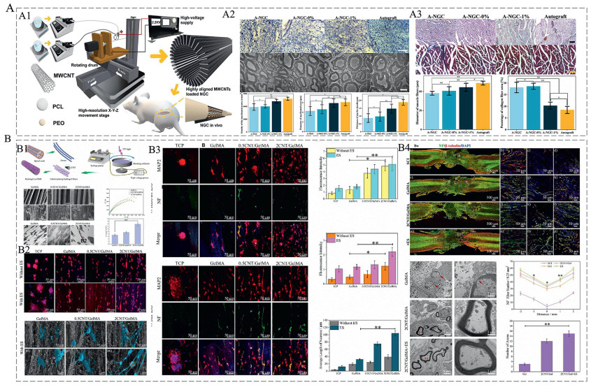
CNTs can create an ECM-mimetic milieu that regulates nerve cell adhesion, development, and differentiation [63]. As conductive materials, CNTs have also been shown to accelerate neuronal electrical activity, as nerve impulses are enhanced by charge recombination along the membrane surface [4]. Such as rope-like SWCNTs synthesized by chemical vapor depostion (CVD) technique induce neurite growth along their spiral morphology [64]. Overexpression of neuronal genes/proteins confirmed that 5 mV ES significantly enhances NSC differentiation into neurons when cultured on SWCNT ropes. CNTs are often mixed with other materials to promote neural regeneration. Combining carboxylated MWCNTs with a PCL-collagen mixture and applying 100 mV/cm ES for 5 days significantly promoted the proliferation of hippocampal neural progenitor cells (HT-22) in mice. Notably, MWCNTs endowed the cells with bipolarity, enabling directed extension of the cells [65]. Wang et al. fabricated a neural guidance scaffold (NGS) composed of MWCNTs/poly(lactic-co-glycolic acid) (PLGA) composite nanofibers, which were subsequently coated with poly-L-lysine to improve their hydrophilicity and cytocompatibility. The electrical activity of PC12 cells and DRG neurons was shown to enhance neurite length, whereas the combined influence of aligned nanofibers and ES amplify SC responses [66]. Recently, Sun et al. prepared core-shell fibers containing MWCNTs. The conduit has a shell composed of PCL and MWCNTs, with polyethylene oxide (PEO) as the inner layer, forming a directional fiber structure. The experiments showed that this NGC have good biocompatibility and mechanical properties. After 12 weeks of implantation, NGCs containing 1% MWCNTs showed recovery of muscle and sciatic nerve function and myelin regeneration similar to the gold standard autograft (Fig. 2A) [67]. Similarly, Yao et al. successfully prepared CNT/methacrylate acylated gelatin (GelMA) hydrogel fibers. This hydrogel promote NSC differentiation/axon outgrowth and restore motor function in T9-spinal cord injury models (Fig. 2B) [68].
Figure 2
Figure 2.
Carbon nanotube scaffolds enable multimodal neural regeneration via material optimization, amplified cellular behavior, and functional recovery in vitro/in vivo. (A) Core-shell fibers containing MWCNTs for neural regeneration and repair. (A1) Preparation of core-shell fibers containing MWCNTs. (A2) Staining analysis of gastrocnemius specimens at 12 weeks post-surgery, Scale bar: 100 µm, and statistical analysis of muscle fiber diameter and collagen fiber area. (A3) Analysis of nerve regeneration 12 weeks post-surgery: nerve endings, axons, myelin. Scale bar: 50 µm. Reproduced with permission [67]. Copyright 2024, John Wiley and Sons. (B) Multiscale functionality of CNT/GelMA hydrogel fibers in neural repair. (B1) Preparation and characterization of CNT/GelMA conductive hydrogel fibers. Red arrows indicate CNTs distribution. Scale bar: 5 µm, 500 nm, 200 nm (TEM). (B2) CNT/GelMA conductive hydrogel with ES promotes neuronal differentiation and neurite elongation in vitro. NF (green) and MAP-2 (red) immunofluorescence. Scale bar: 50, 10, 2 µm. (B3) CNT/GelMA conductive hydrogel promotes NSCs differentiation and adhesion. Red fluorescence (Rhodamine phalloidin) and blue fluorescence (DAPI) indicate cytoskeletons and nucleus, respectively. Scale bar: 50 µm. (B4) Representative images and quantification of post-surgical neural regeneration at 8 weeks. Axonal markers: NF (green), β-tubulin (red). Scale bar: 5, 1 µm (TEM). Reproduced with permission [68]. Copyright 2024, Elsevier.
While numerous reports have highlighted the adverse effects of CNTs on biological systems and cellular properties, studies investigating both pristine and functionalized multi-walled CNTs in SH-SY5Y cells have demonstrated neuroinflammatory and neurotoxic responses. Data indicate that multi-walled CNTs induce DNA damage and stimulate reactive oxygen species (ROS) production, leading to pro-inflammatory reactions. This is further confirmed by elevated concentrations of interleukin-6 (IL-6), tumor necrosis factor-alpha (TNF-α), IL-1β, and other cytokines, which substantiate the neuroinflammatory effects [69]. However, Ye et al. fabricated CNT-PEG hydrogel composites by embedding preformed CNT networks into PEG matrix. This material promotes neuronal differentiation via enhanced focal adhesion signaling from its microstructure, while hydrogel encapsulation maintains neural network excitability homeostasis in spinal cord injury by preventing direct CNT-cell contact (Fig. S3 in Supporting information) [70].
4.
Fabrication of electroactive biomaterial implants for neural regeneration
4.1
Conductive films (CFS)
CFS are attractive candidates for electroactive materials because of their high surface area, enhanced effective ion exchange with surrounding tissues, and better transmission of electrical stimuli [71]. However, CFS are very brittle, so the direct use of CFS as electroactive materials is difficult [72]. Therefore, in contrast to altering the structure and composition of the electrode independently, applying a conductive film on the material's surface to create a conductive coating is a more straightforward and economical approach. The coating material and its structure can be customized and modified according to user specifications [73].
There are various coating methods for different coating materials. (1) Chemical deposition. (2) Chemical vapor deposition (CVD). (3) Electron beam deposition (EBD). (4) Vapor phase polymerization (VPP). Detailed content is provided in Section S8 (Supporting information).
A key technological challenge in applying conductive coatings to tissue engineering scaffolds is achieving homogeneous, conformal coverage on complex 3D geometries [74]. Second, the degradation from repetitive stimuli and micromotions may lead to delamination of the nerve electrode coating. To enhance the mechanical stability of the coating on the nerve electrode, the method of including covalent bonds between the coating and the nerve electrode may be employed.
4.2
Conductive conduit
The role of the nerve guide conduit (NGCs) is to connect damaged nerves and provide structural and biochemical support [75]. ES has been proven to be an effective method of neuronal function and regeneration. Therefore, conductive conduit made via various methods have become popular electrically active material implants for neural regeneration in recent years.
At present, the manufacturing technology of conductive conduits for nerve repair can be divided into traditional and additive manufacturing (AM) methods [76]. Conventional technologies, including electrospinning, freeze-drying, injection molding, and thermally induced phase separation (TIPS), have demonstrated significant potential in the creation of conductive conduits. Li et al. reported one of the multichannel conductive conduits, which utilized injection molding techniques to produce conductive conduits with intraluminal channels. In vivo studies implanted this conduit with ES to repair a 10 mm transected sciatic nerve gap. We found that the nerve recovery was comparable to that of autologous transplantation one month after surgery [77]. Farzamfar et al. employed TIPS to create a multichannel conduit, successfully regenerating nerves in a rat sciatic nerve defect [78]. Electrospinning, a technique for producing nanofibers through the application of a high-voltage electric field to a polymer solution, has emerged as the predominant approach for fabricating conductive conduits because of its capacity to utilize uncomplicated apparatuses to assess shape, porosity, and composition [79]. Many studies on nerve conduit repair, including (Fig. S4A in Supporting information) [80] have been described. In addition, in vitro experiments have also demonstrated the repair effect of conductive conduits, such as Sadeghi et al. prepared a PCL/chitosan/PPy nanofiber composite conduits was prepared via an electrospinning which showed a 2.75-fold increase in PC12 cell proliferation [81].
These traditional techniques can generate scaffolds that exhibit good interconnectivity, porosity, and homogeneous pore size. Nevertheless, conventional approaches for constructing intricate structures with tunable micro and macro dimensions are challenging [83]. Consequently, AM technology has been developed. The obtained picture data or CAD models can be utilized to create intricate 3D constructions, which can be tailored to meet the specific requirements of each patient [84]. AM technologies is used for the preparation of conductive catheters normally include Novel electrohydrodynamic jet 3D printing (EHD-jet 3D printing) and fused electrowriting (MEW). EHD-jet 3D printing is suitable for conductive conduits. Its basic principle involves utilizing the strong electric field formed between the nozzle and the substrate to eject charged liquid from the nozzle which is then deposited on the substrate and layered to form a 3D structure. Sanjairaj et al. utilized EHD-jet 3D printing technology to manufacture PCL/PPy conductive conduits, observing increased glial heavy chain (NF-H) protein fluorescence in cells [85]. MEW can be combined with bionic technology. In the study, load placental mesenchymal stem cells (PDMSC) into bionic melt electrowriting (MEW) NGC to improve axon myelination, increase muscle mass, enhance the sciatic nerve functional index, and get a positive neural regeneration results [86].
Despite the advantages of conductive conduits in neural regeneration, but the significance and potential of these nonbiodegradable conductive conduits in clinical use are limited [87]. Therefore, determining the biodegradability of conductive conduits will be the focus of our research. In addition, combining patterning technology with bioprinting and conductive conduits to construct a multipath, multifunctional nerve conductive regeneration conduit to achieve better nerve repair, is expected to become a new direction of conduits development.
4.3
Electroconductive hydrogels (ECHs)
Owing to their similarity to natural neural tissue, hydrogels can effectively occupy damaged regions within neural tissue, making them highly suitable for neural repair [88]. Compared with traditional hydrogel materials, ECHs can enhance electrical coupling between normal and damaged tissues and provide additional ES to neural cells [89]. Therefore, ECHs have significant potential as electroactive material implants for neural repair.
ECHs can currently be synthesized through various techniques, such as physical crosslinking, chemical crosslinking, in-situ polymerization and 3D printing. Physical crosslinking primarily involves mixing electroactive components with nonconductive components, allowing them to self-assemble into ECHs [90]. Gan et al. manufactured a dual physically cross-linked double network (DN) hydrogel composite by embedding hydroxypropyl cellulose (HPC) biopolymer fibers within a poly(vinyl alcohol)-sodium alginate (PVA/SA) matrix. Subsequent immersion of the prestretched PVA-HPC/SA composite in CaCl2 solution yielded PVA-HPCT/SA-Ca DN hydrogels. They are noncytotoxic, and have high electrical conductivity and excellent tensile strength. PVA-HPCT/SA-Ca DN hydrogels exhibit potential for neural tissue replacement applications [91]. For in situ polymerization, the monomers of the conductive polymer diffuse into the nonconductive polymer hydrogel and polymerize within it. For example, Zhao et al. prepared a polyacrylamide/graphene oxide/pectin/sodium alginate (PAM/GO/Gel/SA) composite hydrogel through in situ polymerization by adding concentration of graphene oxide sodium alginate to a premade polyacrylamide solution, followed by the addition of gelatin and sodium alginate under the action of the initiator ammonium persulfate (AP) [92]. Their experiments revealed that this composite hydrogel could effectively support the adhesion and proliferation of SCs. Chemical crosslinking is the most commonly used method for synthesizing ECHs, where monomers form irreversible assemblies through covalent or ionic bonds, such as, Liu et al. assembled conductive hydrogels loaded with NSCs (ICH/NSC) using amino-modified gelatin (NH2-gelatin) and benzoate tetramer grafted oxidized HA (AT-OHA). When it was used for SCI repair, the results showed that this scaffold promotes direct NSC commitment toward neuronal lineages while inhibiting their transformation into astrocytes [93]. Compared with traditional ECHs, 3D printing represents an advanced manufacturing technology that allows for precise control over the geometry, anisotropy, pore size, and topography of hydrogels [94]. Various 3D printing methods for ECHs, including extrusion-based printing, stereolithography (SLA), and inkjet printing [94-97], have been explored. For example, Deo et al. recently reported a nanoscale printable 3D hydrogel ink made from sulfated gelatin and molybdenum disulfide. The experiment shows that hMSCs adhere and proliferate well on the hydrogel surface, with cell survival rates comparable to the control (GelMA) (Fig. S4B in Supporting information) [96]. Furthermore, Bioprinting denotes the direct incorporation of living cells into the printing process [98]. Song et al. developed an electrically conductive hydrogel scaffold loaded with NSCs using 3D bioprinting technology. The scaffold is centered around modified PEDOT and is improved for solubility and conductivity by incorporating chondroitin sulfate (CS) and tannic acid (TA). Gelatin methacryloyl (GelMA)/PEG diacrylate (PEGDA) hydrogel matrix is integrated. Experiments showed that the NSC survival rate in the scaffold exceeded 90%, and the polyphenolic structure of TA significantly induced differentiation into neurons while inhibiting astrocyte differentiation [99].
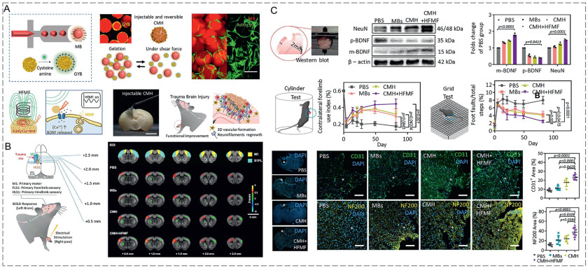
Notably, the mechanical properties of ECHs significantly weaken after repeated use; therefore, further efforts should be made to increase the mechanical stability of ECHs. Currently, multifunctional hydrogels are future novel therapeutic strategies. Recently, Hsu et al. synthesized a multifunctional conductive microporous hydrogel (CMH) using electromagnetized gold nanoyarn balls (GYBs)-coated injectable building blocks (microbeads, MBs) for neural repair after traumatic brain injury (TBI) (Fig. 3) [82]. This hydrogel combined with non-invasive high-frequency magnetic field (HFMF) stimulation promotes neural differentiation, neurite growth, and angiogenesis. In addition, after CMH implantation, the inflammatory response and glial scar thickness in TBI mice were significantly reduced, and Blood-oxygenation level-dependent functional magnetic resonance imaging (BOLD-fMRI) showed the recovery of functional connectivity in the motor cortex and sensory cortex.
Figure 3
Figure 3.
Summary of adaptive CMH for traumatic brain injury repair. (A) Schematic illustration of the fabrication and applications of adaptive CMH. (B) Blood oxygen level-dependent (BOLD) fMRI activation map during contralateral forcepaw stimulation. (C) Schematic of CMH application in neural repair related to traumatic brain injury (TBI). Cavity of tissue loss (white circle). Scale bar: 10 mm. Reproduced permission [82]. Copyright 2022, Springer Nature.
The preparation methods and characteristics of different electroactive material implants are summarized in Table S2 (Supporting information).
The efficacy of electroactive implants is profoundly influenced by the mode and parameters of ES applied. As detailed in previous sections, conductive films, conduits, and hydrogels serve not only as structural supports but also as efficient media for transmitting electrical cues to neural cells. The design of these implants, whether as flexible conductive films for enhanced surface contact, multi-channel conduits for guided axonal growth, or injectable hydrogels for minimally invasive delivery, must align with the intended ES strategy. For instance, the choice between conventional invasive ES and noninvasive self-generating systems dictates the material’s required conductivity, biodegradability, and mechanical compatibility. The following section will elaborate on how different ES modalities, from traditional implanted electrodes to advanced self-powering platforms, synergize with these electroactive constructs to optimize neural regeneration outcomes.
5.
Types of ES for neural regeneration
5.1
Conventional ES
ES has been employed in clinical practice, including cochlear implants [100], deep brain stimulators [101], epilepsy management [102]. Currently, the predominant method employed is traditional ES, which involves the implantation of electrodes or the integration of a power supply at the nerve injury site, the application of open ES during surgical procedures [103].
Epidural ES (EES), the most extensively researched type of conventional ES. Following total thoracic SCI in rats, this stimulation engages spinal cord motor networks, facilitating movement independent of descending input [104]. EES has demonstrated benefits including functional recovery of paralyzed extremities (arms, hands, trunk, legs), diaphragm activation, and partial restoration of bladder and bowel control [105]. In addition, EES further induces spontaneous movement in human subjects with neurologically complete impairment [106]. Nevertheless, implanted electrodes frequently result in interfacial reactions, following damage, inflammatory responses, challenges in degradation, and the necessity for further surgical resection [103]. Although transcutaneous and transcranial magnetic stimulation offer non-invasive alternatives, their therapeutic efficacy remains unestablished.
5.2
Noninvasive self-generating system
Conventional ES encounters obstacles, traditional electrodes tend to be rigid, cumbersome, and may incorporate hazardous compounds [107]. The finite longevity of the electrodes is a possible obstacle for postimplantation replacement [108]. Therefore, the development of a noninvasive self-generating system with self-power and physiological adaptation functions for neural regeneration is of interest [109]. The present development emphasis of Noninvasive self-generating system is on piezoelectric materials, magnetoelectric response, and biofuel cells (BFCs) (Fig. S5 in Supporting information). Detailed content is provided in Section S9 (Supporting information).
6.
Regulatory mechanism of electroactive materials in neural regeneration
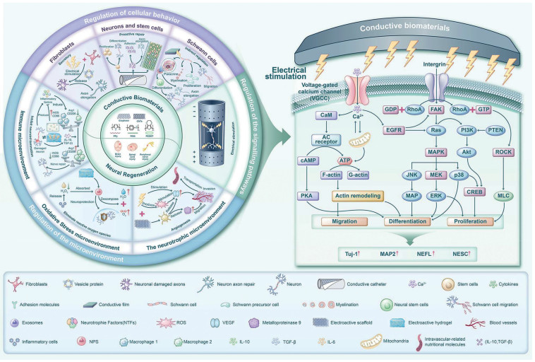
Electroactive materials can carry exogenous electron currents and convert them into ion currents used in signal transmission. This facilitates the regulation of intracellular metabolism and signal transduction, as well as the microenvironment [110]. Numerous studies have confirmed the significant role of electrically active materials in neural repair, but comprehensive exploration of the function and mechanism of electroactive materials in neural regeneration is lacking. The regulatory role of electroactive materials in neural regeneration is shown in Fig. 4. Mechanism of neural regeneration modulation by electroactive materials are summarized in Table S3 (Supporting information).
Figure 4
Figure 4.
Schematic illustration of the regulatory role and mechanisms of electrically active materials for materials in neural regeneration.
EES combined with conductive materials can trigger bioeffects within cells by altering the membrane's bioelectrophysiology, thus controlling the behavior of electrically active cells. As previously mentioned, extensive research has demonstrated that various electroactive materials have direct regulatory effects on the behavior of neural cells and related stem cells, such as cell adhesion, accelerating cell migration, cell proliferation, secretion and differentiation [111-113]. For instance, Chuan et al. demonstrated enhanced proliferation, neuronal differentiation, and axon elongation in NSCs cultured on conductive PLGA/GO composite membranes [114]. In another investigation, ES synergized with graphene-reinforced collagen conduits to drive neural differentiation in BMSCs, resulting in elevated microtubule-associated protein 2 (MAP-2) and β Ⅲ-tubulin protein levels [112]. Similarly, Wang et al. anchored laminin-modified graphene nanosheets on NSCs. These generated localized ES via electromagnetic induction, directing NSCs to differentiate into functional dopaminergic neurons (Figs. S6A–C in Supporting information) [115]. It is well-known that when exposed to an electric field, neural cells and axons align, a common indicator. Chen et al. reported that electric fields applied via bacterial cellulose (BC)/PEDOT microfibers modulate PC12 cell and neurite alignment [116]. However, Koppes et al. found that the morphological orientation provided by electroactive materials is the primary determinant for the alignment of DRG neuronal cells and axons [117]. Other groups have reported similar observations [42,118]. In conclusion, the morphological orientation of electroactive materials and the electric field they conduct can ensure specific cell and axon alignment and extension, thus promoting the correct regeneration of nerves. However, the base topography of the electroactive materials plays a decisive role.
Interestingly, beyond interacting with electrically responsive cells, electroactive materials exert functional effects on insulating tissue-derived cells such as SCs. SCs have important functions such as supporting and protecting axons, forming myelin sheaths, and nourishing nerve axons [119]. After Wallerian degeneration occurs distal to the injured nerve, SCs are involved in the removal of denatured axons and phagocytosis of myelin fragments. In addition, SCs also secrete active substances such as ECM, nerve growth factor(NGF), and cytokine [42]. Electroactive materials effectively mediate SC proliferation, migration, adhesion, and myelination [120]. Wu et al. demonstrated that SCs cultured on conductive flexible PGSAP-H membranes exhibit upregulated expression of peripheral myelin protein 22 (PMP22) and three key myelination-associated markers. Their findings further indicate that such conductive substrates promote PC12 neuronal differentiation by modulating SC paracrine signaling [121]. Conductive biomaterials can also promote cell directional extension. Li et al. prepared polydopamine (PDA) and carbon graphene oxide (CGO) composites of PPy films. The experiments showed that the grafted SCs exhibited good adhesion and neural protein expression, and were aligned along the flow direction [122]. In recent years, the role of SCs in vesicle metastasis has received increasing attention. Lopez-Leal et al. found that ES can stimulate the secretion of glutamate by SCs, which can increase SC-derived exosomes and intracellular Ca2+ concentrations, causing an increase in calcium-dependent proteins in exosomes, thereby enhancing the repair effect of exosomes on axons [123]. This important theory supports new treatment strategies.
During Wallerian degeneration, macrophages mediate phagocytic clearance of axonal and myelin degradation products [124]. Recent advances reveal macrophages actively drive neural regeneration through direct and indirect mechanisms, notably via secretion of growth factors, cytokines, and proteases [42]. Jia et al. placed macrophages between two parallel electrodes connected to a power source. They observed that the frequency of the applied electric field significantly influenced the polarization of macrophages from pro-inflammatory (M1) to inflammatory (M2) phenotypes [125]. It is noteworthy that, traditionally, fibroblasts invade the interior of the NGC to form a scar, which leading to adhesion between the newly regenerated nerves and surrounding tissues [126]. However, contemporary research establishes fibroblasts as critical mediators of peripheral nerve repair. Zhao et al. identified that fibroblast-derived extracellular vesicle protein TFAP2C enhances peripheral axon regeneration via the miR-132–5p/CAMKK1 pathway [127]. Therefore, the traditional concept of fibroblasts in the context of electroactive material repair of nerves must be reconsidered, and future research should explore this area.
6.2
The regulation of the signaling pathways
Neurons contain numerous ion channels. The ion exchange through these channels not only regulate the membrane potential but also control neurotransmitter release. As a special type of ion channel, The voltage-gated calcium channels (VGCCs) related mechanisms are one of the most well-known regulatory mechanisms in neuronal signaling pathways [128]. Electroactive materials induce transmembrane Ca2+ flux in neural cells via VGCCs, elevating cytoplasmic Ca2+ through controlled extracellular cation influx. Additionally, ES can induce the reorganization of membrane proteins through phospholipase C, leading to the release of Ca2+ from intracellular stores [129]. Wang and colleagues created a porous membrane composed of MoS2 nanosheets. Research confirms that Ca2+-bound calmodulin (CaM) initiates calcium-dependent kinase (CaMKII)-mediated phosphorylation of response element binding protein (CREB), inducing expression of genes critical for neuronal differentiation. Notably, cell adhesion activates the Rho GTPase/ROCK pathway to restructure the cytoskeleton, thereby promoting neurite extension and directed migration [130]. These experiments demonstrate the significant role of calcium ion channels in promoting the elucidation of ES effect mechanisms. Furthermore, Ca2+ is involved in other various signaling pathways. Wu et al. developed methyl methacrylate (MA)-modified hectopicon collagen-polypyrrole NP hybrid hydrogels. Their research demonstrated that the synergistic action of electrical conductivity and stimulation activates Ca2+-dependent PI3K/Akt and mitogen-activated protein kinase (MAPK)/Ras signaling pathways in BMSCs. These pathways modulate NSC migration while enhancing ATP and neurotransmitter secretion. Notably, the hydrogel scavenges ROS through water-generating redox reactions, protecting BMSCs from oxidative damage and thereby exerting anti-inflammatory effects (Figs. S7A–D in Supporting information) [131]. In vitro studies demonstrate that 1 h of ES (20 Hz, 3 V, 0.1 ms) downregulates phosphatase and tensin (PTEN), a critical growth-suppressing molecule. As a potent antagonist of the PI3K/Akt signaling pathway, PTEN suppression facilitates regulated cellular growth and differentiation [132]. ES-induced depolarization also promotes the tyrosine phosphorylation of the epidermal growth factor receptor (EGFR), activating MAPK, including ERK and p38 MAPK, which regulate cell differentiation, proliferation, and survival [133]. For example, Zhao et al. activated the MAPK pathway through electric stimulation transmitted by PPy/SF scaffolds to achieve bidirectional regulation, with ERK dominating growth signals and p38/JNK suppressing injury signals, thereby enhancing the proliferation and vitality of SCs while promoting the secretion of neurotrophic factors [134]. Simultaneously, ES activates the cAMP response element-binding protein (CREB) pathway via p38 MAPK signaling. In PC12 cells with NGF-induced axon growth deficits, ES application demonstrated that p38 MAPK induces CREB activation to promote axonal regeneration [135]. This indicates the related role of this pathway in promoting neuronal growth.
In addition, ES can regulate transmembrane receptors (such as integrin-β molecules), thereby influencing intracellular signaling pathways related to cell migration or differentiation through the effects of ligand-receptor binding [136]. ES can promote cellular metabolism and accelerate ATP depletion. Some reports indicate that direct current ES at 10–1000 µA promotes the synthesis of membrane-bound ATP, which is then consumed to convert monomeric G-actin into polymerized F-actin, facilitating the reorganization of the actin cytoskeleton [137]. Various signaling pathways involved in electric stimulation mediated by electroactive materials are summarized in Fig. 4.
While the precise molecular mechanisms through which electroactive materials mediate signal transduction and pathway activation require further investigation, evidence confirms that engineered neural scaffolds incorporating electroactive components directly or indirectly modulate intracellular signaling, regulate critical pathways, enhance neuronal survival, guide axonal growth, and ultimately promote superior neural regeneration outcomes.
6.3
Regulation of the microenvironment
When neural damage occurs, mitochondrial function is impaired, and a large amount of ROS and cytochrome P450 enzymes are produced, activating the hypoxia-inducible factor 1 (HIF-1) signaling pathway [138]. As previously mentioned, an appropriate oxidative stress environment can promote angiogenesis, delivering nutrients to the microenvironment for neural regeneration [139]. However, prolonged exposure to a chronic oxidative stress microenvironment can induce apoptosis of Schwann and neuronal cells. These studies lay the theoretical foundation for exploring the mechanisms of electroactive materials and oxidative stress, and pave the way for further development of oxidative stress-regulating electroactive materials.
6.3.1
The neurotrophic microenvironment
The nutrients required for neural regeneration are closely related to local blood supply, making the formation of microvessels essential for the recovery of neural nutrition [140]. The early vascularization of electrically active materials can provide neural cells with sufficient nutrition [141]. Detailed content is provided in Section S10 (Supporting information).
6.3.2
Oxidative stress microenvironment
When neural damage occurs, mitochondrial function is impaired, and a large amount of ROS and cytochrome P450 enzymes are produced, activating the HIF-1 signaling pathway [138]. As previously mentioned, an appropriate oxidative stress environment can promote angiogenesis, delivering nutrients to the microenvironment for neural regeneration [139]. However, prolonged exposure to a chronic oxidative stress microenvironment can induce apoptosis of Schwann and neuronal cells. Some studies lay the theoretical foundation for exploring the mechanisms of electroactive materials and oxidative stress, and pave the way for further development of oxidative stress-regulating electroactive materials. Detailed content is in Section S11 (Supporting information).
6.3.3
Immune microenvironment
In recent decades, the immune microenvironment has been studied, primarily regulated by immune cells and cytokines, gaining attention in the fields of tissue repair and regeneration due to its plasticity and uniqueness [142]. Generally, following neural injury, the recruited M1 subtype of macrophages act as cleaners, phagocytosing tissue debris and pathogens, and promoting the secretion of pro-inflammatory cytokines. Conversely, M2 macrophages release anti-inflammatory cytokines and play a role in tissue repair [143]. For instance, in the preclinical evidence of MCAO mice, macrophages infiltrate within hours after stroke damage, reaching a peak in three days. By the seventh day, M1 and M2 phenotypes of macrophages each account for half, but within the next two weeks, most macrophages exhibit an anti-inflammatory M2 phenotype [144]. Therefore, the balance between M1 and M2 macrophages adapts to different physiological and pathological conditions, playing a crucial role in the regulation of the immunomicroenvironment. Tu et al. developed graphene oxide-encapsulated hydrogels and demonstrated that conductive hydrogel substrates drive macrophage polarization from M1 to M2 phenotypes. The resulting M2 macrophages secrete abundant anti-inflammatory factors that promote angiogenesis, cellular proliferation, and regenerative microenvironment remodeling (Fig. S8A in Supporting information) [145]. Based on this theory, a multitude of electroactive materials have been studied for experiments. For example, Zhang et al. engineered a ECHs composed of biodegradable waterborne polyurethane (WPU) and PDA-reduced graphene oxide (pGO). The study found that this hydrogel exhibits immunosuppressive effects, with significantly elevated levels of the CD206 marker in M2-type macrophages, achieving a polarization shift from M1 to M2 type [146]. Notably, recently, Wang et al. designed a NGC loaded with regenerative silk fibroin (RSF), PEDOT:poly(styrene sulfonate) (P:P), and fumaric acid dimethyl ester (DMF), which provided a neuro-mechanically matched NGC that could gradually regulate the inflammatory microenvironment (IME) within the catheter through the release of DMF and pressure-resistance sensing monitoring of the sciatic nerve movement (Fig. S8B in Supporting information) [147]. The combination of NGC and electric stimulation can synergistically inhibit the apoptosis of SCs, reduce the release of inflammatory factors, polarize macrophages from the inflammatory M1 phenotype to the regenerative M2 phenotype, and lead to functional recovery of neurons. The DMF/RSF/P:P catheter provides a new multidimensional, adjustable method for regulating the immunomicroenvironment, offering new inspirations for future clinical treatments of neural repair.
These studies indicate that electroactive materials can effectively regulate the immunomicroenvironment by modulating the polarization of macrophages, thus establishing a favorable microenvironment for neural repair. The mechanisms may involve multiple levels, including the direct regulation of cellular behavior by electrical signals, synergy with material topological structure, and controlled release of immunomodulatory factors. Detailed content is provided in Section S12 (Supporting information).
7.
Conclusions and future perspectives
With the further improvement and clarification of the mechanism of neural regeneration, the further development of related research on ES-induced neural regeneration and repair, and conductive biomaterials, the new technology of ES-conductive biomaterials, by combining ES, biomaterial integration, chemical signals, and physical properties, Significant impact on the development of the field of neural repair. However, different injury sites may require personalized control strategies. Most conductive biomaterials have poor biodegradability, insufficient long-term stability, or potential neurotoxicity issues. Existing research on immune responses and material metabolism pathways is still insufficient. In addition, Noninvasive self-generating system enables physiologically adaptive stimulation. However, the mechanical energy conversion efficiency of piezoelectric materials is constrained by the tissue mechanical environment, and the electromagnetic wave attenuation of magnetoelectric materials remains unresolved. Therefore, future research directions can focus on: (1) Developing new biomimetic multifunctional composite conductive biomaterials. (2) Optimizing personalized control strategies for ES parameters is crucial. (3) Assessing immunotoxicity and enhancing biocompatibility are crucial for electroactive materials. (4) Balancing long-term stability and controlled degradation is essential for electroactive materials. Detailed content is provided in Section S13 (Supporting information).
Electroactive materials can effectively regulate the behavior of neural cells and other neuro-related cells through conduction or generation of electrical signals, especially in the regulation of macrophage polarization and the re-understanding of fibroblast function, further expanding their application value in the remodeling of the neural microenvironment. Currently, the research on the activation of related signaling pathways by electroactive materials is incomplete and mature, for example, the activation methods of specific signaling pathways by material surface morphology and electrical conductivity still need to be deeply analyzed. It is noteworthy that we emphasize the importance of the microenvironment for neural regeneration, and focusing on the research of angiogenesis, inflammatory balance, and immune regulation is promising.
This review makes the progress in research on the neural repair of conductive biomaterials and makes an initial exploration of future work. It is hoped that through the review, the understanding of the fundamental mechanisms and applications of electroactive materials in neural regeneration can be deepened, and ultimately, efficient and safe treatment strategies for neural injury repair can be provided through interdisciplinary cross-innovation.
The authors declare that they have no known competing financial interests or personal relationships that could have appeared to influence the work reported in this paper.
Acknowledgments
This work was supported by the National Natural Science Foundation of China (No. 82401629), the China Postdoctoral Science Foundation (Nos. 2024T170601, 2024M762228, GZC20231811), the Natural Science Foundation of Sichuan Province (No. 2024NSFSC1646).
Supplementary materials
Supplementary material associated with this article can be found, in the online version, at doi:10.1016/j.cclet.2025.112205.
[1]
R. Li, Z. Liu, Y. Pan, et al., Cell Biochem. Biophys. 68 (2014) 449–454. doi: 10.1007/s12013-013-9742-1
[2]
W. Dai, Y. Yang, Y. Yang, W. Liu, Nanotechnol. Rev. 10 (2021) 488–503. doi: 10.1515/ntrev-2021-0028
[3]
M. Kasper, M. Cydis, A. Afridi, et al., J. Mater. Chem. B 11 (2023) 7663–7674. doi: 10.1039/d2tb02766f
[4]
B.S. Eftekhari, M. Eskandari, P.A. Janmey, et al., Adv. Funct. Mater. 30 (2020) 1907792. doi: 10.1002/adfm.201907792
Z. Tu, M. Chen, M. Wang, et al., Adv. Funct. Mater. 31 (2021) 2100924. doi: 10.1002/adfm.202100924
[146]
Q. Zhang, J. Chen, Y. Feng, et al., J. Mater. Chem. B 11 (2023) 6308–6318. doi: 10.1039/d3tb00837a
[147]
J.Y. Wang, Y. Yuan, S.Y. Zhang, et al., Adv. Sci. 11 (2024) 2302988. doi: 10.1002/advs.202302988
Figure 1
Design strategies and functional roles of conductive polymers in neural regeneration. (A) Modulation of neuronal behavior by PPy/PCL scaffolds. (A1) Neurons grow on PCL and PPy/PCL surfaces and neurite extension. (A2) Neurons and neurites grown on PCL and PPy/PCL after ES, analysis of neuronal metabolic activity (B-Ⅲ tubulin, 4′,6-diamidino-2-phenylindole (DAPI), merge (phalloidin, red)) and the morphology of growing cells and their neurites (high power). Scale bar: 50 µm. Copied with permission [22]. Copyright 2024, Elsevier. (B) Neural hydrogel bandage (ECMOS-P-Ⅰ) applied for nerve regeneration and functional recovery in rats. (B1) Schematic diagram of the ECMOS-P-Ⅰ component and its neural application functions. (B2) Assessment of functional and sensory recovery in rats. MBP (red) and NF (green). (B3) Transmission electron microscope (TEM) images of sciatic nerve regeneration and gross views with cross-sectional Masson’s trichrome staining of excised gastrocnemius muscle. Scale bar: 10, 2 µm (TEM images); 1 mm (gross images); 500 µm (cross-sectional views images). Reproduced with permission [28]. Copyright 2024, John Wiley and Sons.
Figure 2
Carbon nanotube scaffolds enable multimodal neural regeneration via material optimization, amplified cellular behavior, and functional recovery in vitro/in vivo. (A) Core-shell fibers containing MWCNTs for neural regeneration and repair. (A1) Preparation of core-shell fibers containing MWCNTs. (A2) Staining analysis of gastrocnemius specimens at 12 weeks post-surgery, Scale bar: 100 µm, and statistical analysis of muscle fiber diameter and collagen fiber area. (A3) Analysis of nerve regeneration 12 weeks post-surgery: nerve endings, axons, myelin. Scale bar: 50 µm. Reproduced with permission [67]. Copyright 2024, John Wiley and Sons. (B) Multiscale functionality of CNT/GelMA hydrogel fibers in neural repair. (B1) Preparation and characterization of CNT/GelMA conductive hydrogel fibers. Red arrows indicate CNTs distribution. Scale bar: 5 µm, 500 nm, 200 nm (TEM). (B2) CNT/GelMA conductive hydrogel with ES promotes neuronal differentiation and neurite elongation in vitro. NF (green) and MAP-2 (red) immunofluorescence. Scale bar: 50, 10, 2 µm. (B3) CNT/GelMA conductive hydrogel promotes NSCs differentiation and adhesion. Red fluorescence (Rhodamine phalloidin) and blue fluorescence (DAPI) indicate cytoskeletons and nucleus, respectively. Scale bar: 50 µm. (B4) Representative images and quantification of post-surgical neural regeneration at 8 weeks. Axonal markers: NF (green), β-tubulin (red). Scale bar: 5, 1 µm (TEM). Reproduced with permission [68]. Copyright 2024, Elsevier.
Figure 3
Summary of adaptive CMH for traumatic brain injury repair. (A) Schematic illustration of the fabrication and applications of adaptive CMH. (B) Blood oxygen level-dependent (BOLD) fMRI activation map during contralateral forcepaw stimulation. (C) Schematic of CMH application in neural repair related to traumatic brain injury (TBI). Cavity of tissue loss (white circle). Scale bar: 10 mm. Reproduced permission [82]. Copyright 2022, Springer Nature.


 DownLoad:

 下载:


 下载:

