State Key Laboratory of Bioreactor Engineering, Shanghai Frontiers Science Center of Optogenetic Techniques for Cell Metabolism, Frontiers Science Center for Materiobiology and Dynamic Chemistry, Shanghai Key Laboratory of New Drug Design, School of Pharmacy, East China University of Science and Technology, Shanghai 200237, China
b.
College of Pharmacy, Dali University, Dali 671000, China
c.
Key Laboratory of Xinjiang Phytomedicine Resource and Utilization, Ministry of Education, School of Pharmacy, Shihezi University, Shihezi 832003, China
d.
Key Laboratory of Tropical Biological Resources of Ministry of Education, College of Pharmacy, Hainan University, Haikou 570228, China
e.
School of Chemical Engineering, Shandong Chemical Engineering & Vocational College, Weifang 261000, China
yixxu@ecust.edu.cn (Y. Xu). 1 These authors contributed equally to this work.
Received Date:
26 September 2025 Accepted Date:
15 December 2025 Revised Date:
12 December 2025 Available Online:
15 November 2026
Abstract:
Heart failure (HF) constitutes a global health burden with elevated mortality and rehospitalization, underscoring the urgent need for novel therapeutics to improve outcomes. Herein, we reported the discovery of a psoralen analog, xanthotoxin (XAT), which is a natural product isolated from Synotis nagensium with cardioprotective effects. To overcome XAT's phototoxicity and poor aqueous solubility, the rational structural optimization was performed to yield 26 psoralen derivatives. Among them, A22 emerged as the optimal derivative, showing abolished phototoxicity, improved aqueous solubility, and potent efficacy at 5 mg/kg in both isoproterenol (ISO)-induced and myocardial infarction (MI)-induced HF models. Mechanistic studies demonstrated that A22 attenuated oxidative stress and cardiomyocyte apoptosis via activation of the Kelch-like ECH-associated protein 1 (Keap1)–nuclear factor erythroid 2-related factor 2 (Nrf2)–antioxidant response element (ARE) pathway. Favorable safety and pharmacokinetic profiles further supported its translational potential. Together, these data supported A22 as a promising candidate for HF therapy and justified continued preclinical investigation toward clinical development.
Heart failure (HF) remains a significant global health burden, affecting over 64 million individuals worldwide and causing high mortality [1]. Despite therapeutic advances, the clinical outcomes remain suboptimal due to persistent disease progression, recurrent hospitalizations, and diminished quality of life [2]. Current therapeutic limitations, including partial efficacy, drug resistance, and adverse effects, underscore the urgent need for innovative therapeutic strategies [3]. Natural products remain a vital source for drug discovery, offering unique chemical scaffolds and novel mechanisms [4,5]. Historically, many HF therapeutics have been derived from natural sources. For instance, cardiac glycosides such as digoxin, was originally isolated from the foxglove and has been utilized for centuries to treat HF [6]. More recently, sodium-glucose co-transporter 2 (SGLT2) inhibitors, originally developed from the natural product phlorizin as antidiabetic agents [7], have emerged as a breakthrough therapy for HF [8]. The structural diversity, intrinsic safety, and broad bioactivity of natural products provide valuable leads for developing innovative HF therapeutics and uncovering novel molecular targets. In pursuit of innovative therapeutic strategies for HF, we sought to discover novel chemical entities from the rich repository of traditional Chinese medicinal herbs. Previously, an in-house screening campaign of 828 Chinese herbs was conducted in oxygen-glucose deprivation (OGD) induced cell injury model. Among these, the ethanol extracts of Veronica Anagallis-Aquatica L, Synotis nagensium, and others were identified as promising anti-HF botanical sources [9], and Synotis nagensium was extensively investigated in this study (Fig. S1 in Supporting information). Synotis nagensium (commonly known as Baiye Huocao, HLJ338) has long been used to treat chronic renal failure, asthma, tracheitis, and nephrogenic edema [10], yet its application in HF therapy remains unreported. Thus, the cardioprotective effects of HLJ338 have been systematically evaluated. The results showed that HLJ338 exhibited potent cardiomyocyte protective effect in OGD-induced cell injury model (Fig. S2 in Supporting information). In an isoproterenol (ISO)-induced HF mice model, HLJ338 treatment at doses of 0.1 and 0.3 g/kg significantly attenuated cardiac dysfunction and cardiac hypertrophy of HF mice, evidenced by increased ejection fraction (EF) and fractional shortening (FS), as well as reduced heart-to-body weight ratio, indicating its potential as a therapeutic agent for HF (Fig. S2).
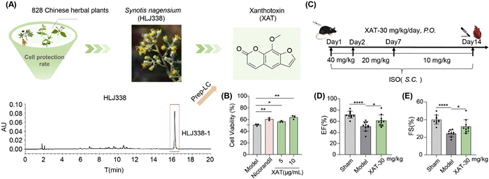
To identify the active constituents of HLJ338, phytochemical analysis was conducted via preparative liquid chromatography to isolate HLJ338–1 (Fig. 1A). The structural elucidation through multi-spectroscopic techniques (1H/13C NMR spectroscopy, high-resolution mass spectrometry (HRMS) coupled with HRMS-based spectral library matching identified HLJ338–1 as xanthotoxin (XAT) (Figs. S3–S6 in Supporting information). Subsequently, the cardioprotective effects of XAT were assessed in vitro and in vivo. The results showed that XAT protected H9c2 cells from OGD injury (Fig. 1B) and alleviated cardiac dysfunction induced by ISO in HF mice, as evidenced by elevated EF and FS (Figs. 1C–E).
Figure 1
Figure 1.
Xanthotoxin was identified as an active anti-HF constituent of Synotis nagensium. (A) The process of Synotis nagensium discovery and XAT isolation. (B) The cardiomyocyte protection of XAT on H9c2 cell lines (n = 3). Nicorandil, 100 µmol/L. (C) The schematic diagram of ISO-induced HF in C57BL/6J mice (n = 10). The cardiac function parameters of mice, EF for (D) and FS for (E). The data are shown as the mean ± SD. *P < 0.05, **P < 0.01, ****P < 0.0001 vs. the model group.
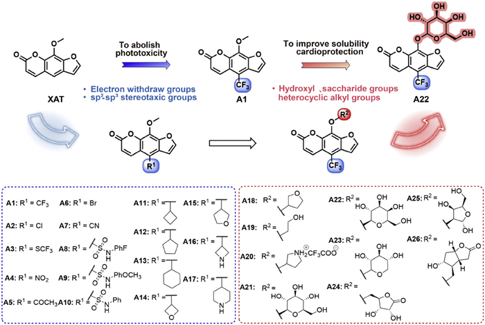
XAT is a clinically approved drug for vitiligo [11] and psoriasis [12]. Its therapeutic efficacy in psoriasis relies on long-wave ultraviolet irradiation, during which the psoralen scaffold undergoes [2 + 2] cycloaddition with pyrimidine bases to form photo-adducts, a process facilitated by its planar and electron-rich properties. Although this mechanism constitutes the pharmacological basis for its efficacy in psoriasis, it also accounts for the intrinsic phototoxicity associated with its clinical use [13]. Besides, psoralen-ultraviolet A (UVA) therapy has been reported to induce apoptosis in human keratinocytes cell line [14]. These limitations pose significant safety concerns for cardiovascular applications, potentially inducing adverse dermal reactions and triggering apoptosis in epidermal cells. Furthermore, XAT's poor aqueous solubility compromises formulation stability and oral bioavailability [15]. To address these challenges, structure optimization focusing on XAT remains a critical priority. Given that XAT's phototoxicity is associated with its electron-rich, planar psoralen scaffold [16,17], the electron-withdrawing substituents and C(sp2)–C(sp3) linkages were introduced at the C4 position, aiming to decrease electron density and disrupt planarity, yielding derivatives A1–A17 (Fig. 2). Among these, the C(sp2)–C(sp3) modifications were achieved via photo–nickel synergistic catalysis, a selective method widely used for C–H activation and C–C coupling [18,19]. After identifying the optimal substituent at the C4 position, hydrophilic moieties (hydroxyl, saccharides, heterocycles) were introduced at the C9 position to enhance aqueous solubility, affording derivatives A18–A26 (Fig. 2). Notably, our previous studies have demonstrated that the introduction of hydrophilic groups is favorable for cardiomyocyte protective effects [20,21]. Therefore, the modification strategy is expected to achieve dual optimization of both aqueous solubility and bioactivity. Collectively, 26 psoralen derivatives (A1–A26) were rationally designed and synthesized to mitigate phototoxicity, enhance cardiomyocyte protective effects, and improve aqueous solubility. The details of the synthetic procedures and structural characterization were described in Supporting information.
Figure 2
Figure 2.
The structural modification strategy and substituents of XAT-based derivatives.
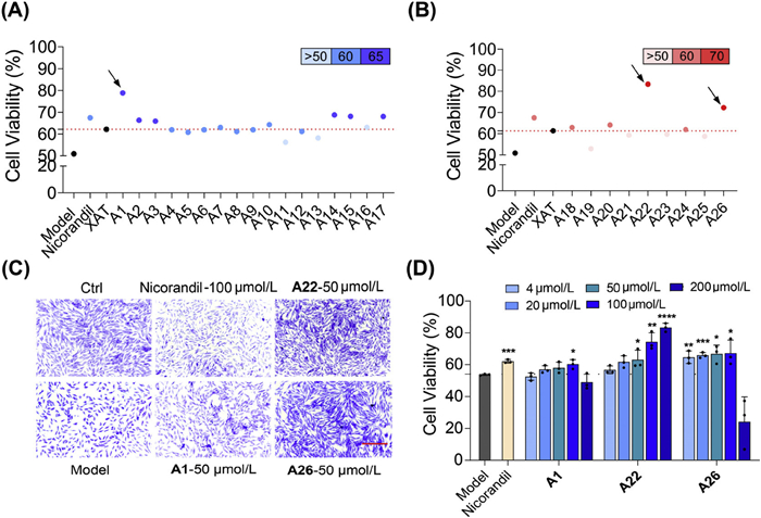
Herein, the cardiomyocyte protective effects of all derivatives in the OGD-induced H9c2 cell injury model were evaluated at concentrations of 4, 20, and 100 µmol/L, and the results were summarized in Tables S1 and S2 (Supporting information). Furthermore, to provide a more intuitive comparison of the activity of derivatives, the maximum cardiomyocyte survival rates at the tested concentrations were presented in scatter plots in Figs. 3A and B. Among the C4-position electron-withdrawing substituted derivatives (A1–A10), trifluoromethyl, chlorine, trifluoromethylthio (A1, A2, A3), and unsubstituted aromatic rings substitution (A10) were favorable for activity, which could be plausibly related to their electronegativity and steric hindrance. Among cyclic substituents (A11–A17), oxyheterocyclic alkane derivatives exhibited superior cardiomyocyte protective effects than azacycloalkyl and cycloalkyl analogs (A14vs.A11/A16, A15vs.A12). This may be due to the distinct electronic and steric characteristics of oxyheterocyclic alkane derivatives, which confer stronger hydrogen-bond accepting ability compared to azacycloalkyl and cycloalkyl analogs [22], ultimately leading to higher biological activity. Given that the trifluoromethyl-substituted derivative A1 exhibited the most potent cytoprotective activity (78.89% at 100 µmol/L), we retained the trifluoromethyl group at the C4 position and focused structural modifications on introducing hydrophilic groups at C9 position. Among the C9-position-substituted derivatives (A18–A26), the incorporation of galactose and corydaline lactone conferred pronounced cardiomyocyte protective effects (A22, A26), whereas other hydrophilic groups were not conducive to enhancing activity (especially for A22vs.A21/A23). The observed differences in activity may arise from variations in the saccharide ring configuration and the number of hydroxyl groups, which jointly modulate biological activity [23,24]. Notably, the aqueous solubility of A22 (183.3 µg/mL) and A26 (104.3 µg/mL) was distinctly improved compared with XAT (64.4 µg/mL) (Table 1).
Figure 3
Figure 3.
The cardiomyocyte protective effects evaluation of XAT-based derivatives in OGD-induced cell injury model. (A) The scatter plots of maximal cardiomyocyte protective effects of derivatives A1–A17. (B) The scatter plots of maximal cardiomyocyte protective effects of derivatives A18–A26. (C) Representative images of crystal violet-stained H9c2 cells subjected to OGD and treated with DMSO or derivatives. Scale bar: 100 µm. (D) The protective effects of derivatives A1, A22 and A26 on primary cardiomyocytes. The data are shown as the mean ± SD (n = 3). *P < 0.05, **P < 0.01, ***P < 0.001, ****P < 0.0001 vs. the model group.
a For cells with cytotoxicity both non-irradiation and irradiation: PIF < 2, no phototoxicity; 2 < PIF < 5, potential phototoxicity; PIF ≥ 5, phototoxicity. For cells without non-irradiation cytotoxicity: PIF > 1, phototoxicity. For cells with no cytotoxicity both non-irradiation and irradiation: PIF = 1*, no phototoxicity. b No cytotoxicity. c No test.
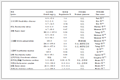
The photo-irradiation factor (PIF) is a key parameter for assessing phototoxicity. According to Organisation for Economic Co-operation and Development (OECD) guidelines, PIF is determined by measuring its cytotoxicity under irradiated vs. non-irradiated conditions on BALB/c 3T3 cells [25]. The representative derivatives that exhibited superior cardioprotective activity to XAT were selected for phototoxicity assessment (A1, A3, A14, A15, A17, A22, A26). As depicted in Table 1, XAT exhibited a PIF value exceeding 11.75, indicating strong phototoxicity [26,27]. In contrast, the derivatives A1, A3, A14, A15, A17, A22, and A26 showed markedly reduced PIF values.
According to the cardiomyocyte protective effects and PIF values, derivatives A1, A22 and A26 exhibited significant activity superior to 70% with no phototoxicity, thus were selected for further in vitro evaluation. Cell morphology and viability were assessed by crystal violet staining. OGD exposure caused approximately 50% cell loss, whereas treatment with A22 and A26 significantly improved cell density and preserved cellular morphology compared to A1 (Fig. 3C). To better mimic physiological conditions [28], the primary cardiomyocytes were isolated from heart of neonatal rats to evaluate protective effects of derivatives. A1 demonstrated moderate concentration-dependent protective effects compared to A22 and A26 (Fig. 3D). Notably, A22 increased the cell survival to 80% at 200 µmol/L. Taken together, A22 and A26 demonstrated superior protective effects in both primary cardiomyocytes and H9c2 cell lines compared to A1, justifying their selection for further in vivo studies.
Overall, after two series of structural modifications based on XAT, derivatives A22 and A26 exhibited significantly improved cardiomyocyte protective effects, mitigated phototoxicity, and improved aqueous solubility. These improvements effectively addressed the pharmacological limitations of XAT, underscoring the importance of rational structural optimization of the scaffold.
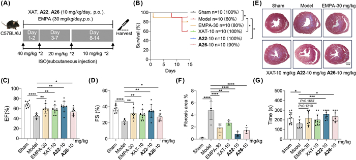
The cardioprotective effect of XAT and its optimized derivatives A22 and A26 were evaluated in ISO-induced HF mice (10 mg/kg, p.o.), with empagliflozin (EMPA) (30 mg/kg, p.o.) as a positive control (Fig. 4A). All animal care and experimental programs were in line with the "Guide of Laboratory Animals" (National Institutes of Health Publication, revised 1996, No. 86–23, Bethesda, MD), and approved by the Committee on the Ethics of Animal Experiments of Dali University, China (No. 2023-pz-277). ISO administration markedly decreased survival rate and impaired cardiac function indices, whereas treatment with XAT, A22, and A26 improved survival rate and cardiac contractility, as evidenced by the increased EF and FS (Figs. 4B–D). Additional cardiac function parameters, such as left ventricular internal diameter in systole (LVIDs) and left ventricular internal diameter in diastole (LVIDd) have been included in Fig. S7 (Supporting information). Masson's trichrome staining results revealed that XAT, A22 and A26 significantly ameliorated ISO-induced myocardial fibrosis, and A22-induced improvement was superior to that of XAT and A26 at the same dosage (Figs. 4E and F). Given that exercise intolerance is the manifestation of the poor prognosis of end-stage HF patients, we assessed exercise tolerance in mice through rotarod testing. As expected, ISO-treated mice displayed a marked decrease in the latency to fall compared with the Sham group, whereas treatment with A22 or A26 significantly restored the exercise endurance reduced by ISO (Fig. 4G). Taken all the indicators together, A22 exerted a more pronounced efficacy than A26. To assess whether A22 is effective at a lower dose, we evaluated the efficacy in ISO-induced HF model with the dosage of 5 mg/kg, A22 conferred marked cardioprotective effects with significant improvements in EF and FS, whereas XAT showed only a modest trend, indicating that A22 possesses enhanced potency compared to XAT (Fig. S8 in Supporting information).
Figure 4
Figure 4.
Cardioprotective effect of derivatives A22 and A26 in ISO-induced HF mice. (A) Schematic of the experimental design. (B) The survival curve of mice. The cardiac function parameters of mice in each group: (C) EF, (D) FS. (E) The representative images of Masson staining. Scale bar: 1 mm. (F) Quantitative analysis of Masson staining. (G) Exercise tolerance of mice by rotarod test. Data are shown as the mean ± SD (n = 10). *P < 0.05, **P < 0.01, ***P < 0.001, ****P < 0.0001 vs. the model group.
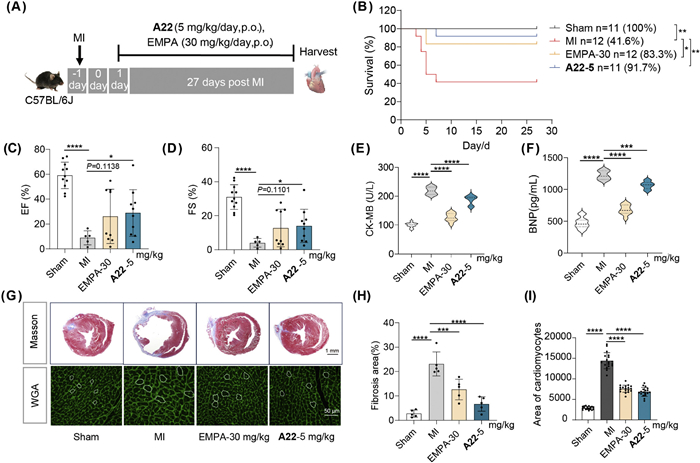
To further assess the therapeutic potential of A22, a post-myocardial infarction (MI) HF model was employed (Fig. 5A). Compared with Sham mice, untreated MI mice exhibited a low survival rate (41.6%), accompanied by severe cardiac dysfunction and ventricular remodeling. This was evidenced by reduced EF and FS, increased LVIDs and LVIDd, elevated serum creatine kinase-MB (CK-MB) and B-type natriuretic peptide (BNP) levels, increased myocardial fibrosis, and cardiomyocyte size in the MI border zone determined by wheat germ agglutinin (WGA) staining. Treatment of A22 (5 mg/kg) dramatically increased survival to 91.7% (Fig. 5B), with improved cardiac systolic function, manifested by significantly increased EF, FS (Figs. 5C and D) and slightly decreased LVIDs, LVIDd (Fig. S9 in Supporting information). Consistent with these findings, serum CK-MB and BNP levels were markedly decreased following A22 treatment (Figs. 5E and F). In addition, A22 significantly reduced myocardial fibrosis (Figs. 5G and H) and attenuated severe cardiomyocyte hypertrophy in MI mice (Figs. 5G and I). Functionally, A22 enhanced exercise tolerance, increased grip strength and prolonged latency to fall relative to untreated MI mice (Fig. S10 in Supporting information). Collectively, these results established A22 as a potent HF therapeutic candidate, achieving efficacy comparable to EMPA at a lower dose.
Figure 5
Figure 5.
Cardioprotective effect of A22 in MI-induced HF mice. (A) Schematic of the experimental design. (B) Survival curve of mice for 4 weeks. The cardiac function index of each group: (C) EF, (D) FS. (E) The level of CK-MB in serum. (F) The level of BNP in serum. (G) Representative images of Masson staining. Scale bar: 1 mm. Representative images of WGA staining. Scale bar: 50 µm. (H) Quantitative analysis of Masson staining. (I) Quantification of cardiomyocyte size from WGA staining. The data are shown as the mean ± SD (n = 5–12). *P < 0.05, ***P < 0.001, ****P < 0.0001 vs. MI group.
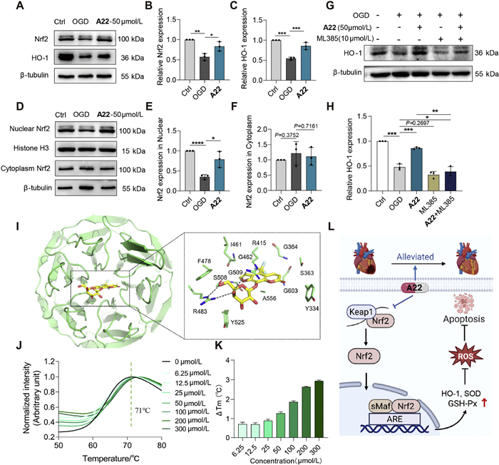
Our study demonstrated that A22 exerted cardioprotection in both ISO-induced HF and MI-induced HF models. However, the molecular mechanism of this cardioprotective benefit remains unknown. Psoralen derivatives have been reported to modulate the Kelch-like ECH-associated protein 1 (Keap1)–nuclear factor erythroid 2-related factor 2 (Nrf2)–antioxidant response element (ARE) pathway, which governs cellular antioxidant defense and is crucial in myocardial ischemic injury [29,30]. Given the structural similarity between A22 and psoralen, we hypothesize that A22 exerts its anti-HF effects via this pathway. Dihydroethidium (DHE) staining showed that A22 significantly reduced OGD-induced ROS accumulation (Fig. S11 in Supporting information), while azino-bis(3-ethylbenzothiazoline-6-sulfonic acid) (ABTS) assay results ruled out the possibility that A22 mitigated oxidative stress via direct ROS scavenging capacity (Fig. S12 in Supporting information). Mechanistically, the Keap1–Nrf2–ARE pathway functions as a master regulator of cellular antioxidant defense [31]. Under physiological conditions, Nrf2 is bound by Keap1 and degraded via the proteasome. In response to oxidative stress, Nrf2 is released and translocates into the nucleus, thus induces the expression of cytoprotective genes (including heme oxygenase 1 (HMOX1), superoxide dismutase (SOD), and glutathione peroxidase (GSH-Px)) [32]. In OGD-treated cells, Nrf2 and heme oxygenase-1 (HO-1) protein levels were significantly downregulated, whereas A22 treatment restored Nrf2 expression, and upregulated HO-1 expression (Figs. 6A–C). Furthermore, A22 attenuated the nuclear Nrf2 reduction induced by OGD injury, whereas the Nrf2 level in cytoplasm remained relatively unchanged. These findings suggested that A22 activated the nuclear Nrf2 accumulation (Figs. 6D–F). Besides, A22 reversed MI-triggered suppression of Nrf2 and HO-1 in MI mice (Figs. S13A–C in Supporting information). Similarly, serum levels of GSH-Px and SOD were significantly restored by A22 treatment in MI mice (Figs. S13D and E in Supporting information). To confirm the involvement of the Keap1–Nrf2–ARE pathway in A22-mediated antioxidant effects, we performed pharmacological experiments with ML385, a selective Nrf2 inhibitor that blocks Nrf2–ARE binding and downstream antioxidant gene transcription [33]. As illustrated in Figs. 6G and H and Fig. S14 (Supporting information), A22-induced upregulation of HO-1 protein levels and mRNA levels were markedly attenuated by co-treatment with ML385, confirming that A22 displayed antioxidant effects mainly via Keap1–Nrf2–ARE pathway. Given that excessive oxidative stress is a well-known trigger of apoptosis [34], we examined apoptotic signaling to clarify whether A22 exerts anti-apoptotic effects. Bax and Bcl-2 were quantified as key markers of mitochondrial apoptosis [35]. As shown in Fig. S15 (Supporting information), OGD treatment markedly downregulated Bcl-2 and upregulated Bax protein expression, indicating the activation of apoptotic pathway. A22 treatment markedly reversed these changes, whereas co-administration with ML385 partially abrogated its anti-apoptotic effects. Consistent with the above results, the ML385 cotreatment with A22 markedly attenuated the upregulated cell viability of A22 in OGD-induced injury model (Fig. S16 in Supporting information). These results indicated that A22 mitigated OGD-induced apoptosis partially through activation of the Nrf2 pathway. To investigate the mechanism of Nrf2 pathway activation by A22, we examined its effect on the Keap1–Nrf2 interaction. Given the critical role of the Keap1 Kelch domain in binding Nrf2 [36], molecular docking studies were employed to evaluate the interaction between A22 and the Keap1 Kelch domain (PDB ID: 8IVG). The results revealed that A22 occupied the canonical ETGE-binding pocket, with the C6–OH and C5-OH of the β-d-galactosyl moiety of A22 forming key hydrogen bonds with R415 and R483, the key residues that are responsible for recognizing the E79 residue of the Nrf2 ETGE motif [37]. In addition, hydrophobic interactions between A22 and Y334, F478, I461, and Y525 further stabilized the binding. These residues overlapped with the Nrf2-binding interface identified in the co-crystal structure of the Keap1–Nrf2 complex, which suggested a competitive inhibition mechanism against Nrf2 binding (Fig. 6I). Subsequently, the binding affinity between Keap1 and A22 was validated by differential scanning fluorimetry (DSF) assay. The results revealed that A22 directly bound to Keap1 and increased its melting temperature (ΔTm) in a concentration-dependent manner, indicating enhanced thermal stability of the protein upon ligand binding (Figs. 6J and K). The distinct interaction mode between A22 and Keap1 Kelch domain provides a structural explanation for the inferior cardioprotective effects of A21 and A23. Specifically, the reversed configuration of the C5-OH group in A21 and the absence of the C6-OH group in A23 limit their ability to form key interactions with specific residues within the Keap1 binding pocket. Collectively, these findings suggested that A22 inhibited the Keap1–Nrf2 interaction, thereby stabilizing and activating Nrf2, providing a mechanistic explanation for its cardioprotective effects by alleviating oxidative stress and suppressing apoptosis (Fig. 6L).
Figure 6
Figure 6.A22 exerted anti-oxidant effects via activating Keap1–Nrf2–ARE pathway. (A) Representative Western blot (WB) bands of Nrf2 and HO-1 in primary cardiomyocytes. (B, C) The protein quantification of Nrf2 and HO-1 in group 6A, respectively. (D) Representative WB bands for nuclear Nrf2 and cytoplasm Nrf2. (E, F) The protein quantification of nuclear Nrf2 and cytoplasm Nrf2 respectively. (G) WB of HO-1 in H9c2 cell lines. (H) The protein quantification of HO-1. (I) Molecular docking analysis of A22 with the Keap1 Kelch domain (PDB ID: 8IVG). The protein backbone is shown in green; the ligand is shown in yellow; oxygen atoms are represented in red; hydrogen bonds are indicated by dashed lines. (J) DSF melting curves of Keap1 at different concentrations of A22. (K) Concentration-dependent thermal stabilization of Keap1 by A22. (L) The proposed mechanistic pathway of A22 in cardioprotection. Independent experiments and data are presented as means ± SD (n = 3). *P < 0.05, **P < 0.01, ***P < 0.001 vs. OGD group.
To assess the potential of A22 as an anti-HF candidate drug, we evaluated its safety and pharmacokinetics in mice. Acute and subacute tests at high doses showed no mortality, morbidity, or organ toxicity, with hematoxylin and eosin (HE) staining and serum biochemistry confirming no hepatorenal damage (Figs. S17 and S18 in Supporting information). A22 also exhibited no human Ether-à-go-go gene (hERG) inhibition (Table S3 in Supporting information), indicating minimal cardiotoxicity. Pharmacokinetically, A22 demonstrated good liver microsomal stability (> 70% parent remaining after 60 min), no significant cytochrome P450 (CYP) inhibition at 10 µmol/L (Table S3 in Supporting information), and an oral bioavailability of 40.51% in mice (Table S4 in Supporting information). With an acceptable half-life (T1/2 = 1.32 h) and moderate clearance, A22 presents a promising profile for further development as an anti-HF candidate.
In conclusion, Synotis nagensium was identified as a novel herb with cardioprotective effect, with xanthotoxin characterized as its principal active constituent. Rational structural optimization was carried out to overcome the shortcomings of XAT, leading to the development of derivative A22, which exhibited no phototoxicity, improved solubility, and potent cardioprotective efficacy in both ISO and MI-induced HF models at 5 mg/kg. Mechanistic studies revealed that A22 activated the Keap1–Nrf2–ARE pathway by inhibiting the interaction between Keap1 and Nrf2, leading to the reduction in oxidative stress and cardiomyocyte apoptosis. Furthermore, comprehensive acute and subacute toxicity studies as well as pharmacokinetic studies confirmed the favorable safety and pharmacokinetic profiles of A22. Although the acute and subacute toxicity studies demonstrated a favorable safety profile, the long-term toxicity and metabolic fate of A22 remain to be elucidated in future investigations to better support its clinical translation.
Together, this study not only offers a novel strategy for mitigating phototoxicity of compounds, but also establishes A22 as a promising HF therapeutic candidate with favorable efficacy, safety, and pharmacokinetic profiles, meriting further preclinical evaluation and potential development for clinical use.
CRediT authorship contribution statement
Xiao Li: Writing – original draft, Methodology, Formal analysis, Data curation. Manjiong Wang: Writing – review & editing, Funding acquisition, Formal analysis. Yunpeng Guo: Methodology, Formal analysis, Data curation. Luoyifan Zhou: Methodology, Formal analysis, Data curation. Yue Yao: Formal analysis, Data curation. Xiaobo Guo: Formal analysis, Data curation. Conglong Xia: Resources, Methodology, Data curation. Hanfang Liu: Formal analysis, Data curation. Zihan Wang: Formal analysis, Data curation. Tiankuo Zhang: Formal analysis, Data curation. Chao Zhang: Formal analysis, Data curation. Yi Ge: Formal analysis, Data curation. Xiaokang Li: Project administration, Conceptualization. Jian Li: Resources, Funding acquisition, Conceptualization. Yixiang Xu: Writing – review & editing, Project administration, Funding acquisition, Conceptualization.
Declaration of competing interest
The authors declare that they have no known competing financial interests or personal relationships that could have appeared to influence the work reported in this paper.
Acknowledgments
This work was supported by the National Natural Science Foundation of China (Nos. 22477025, 22307038, 32121005), National key R&D Program of China (No. 2021YFA0804904), Shanghai Sailing Program (No. 24YF2708800, China), Shanghai Morning Light Program (No. 20CG36, China), Shanghai Frontier Science Center of Optogenetic Techniques for Cell Metabolism (No. 2021 Sci & Tech 03–28), Innovative Research Team of High-level Local Universities in Shanghai (No. SHSMU-ZDCX20212702), Chinese Special Fund for State Key Laboratory of Bioreactor Engineering (No. 2060204).
Supplementary materials
Supplementary material associated with this article can be found, in the online version, at doi:10.1016/j.cclet.2025.112287.
[1]
P.M. Becher, L.H. Lund, A.J.S. Coats, et al., Eur. Heart J. 43 (2022) 3005–3007. doi: 10.1093/eurheartj/ehac248
[2]
S.J. Greene, J. Bauersachs, J.J. Brugts, et al., J. Am. Coll. Cardiol. 81 (2023) 413–424.
[3]
G. Savarese, F. Lindberg, R.M. Christodorescu, et al., Eur. J. Heart Fail. 26 (2024) 1408–1418. doi: 10.1002/ejhf.3214
F. Begnini, S. Geschwindner, P. Johansson, et al., J. Med. Chem. 65 (2022) 3473–3517. doi: 10.1021/acs.jmedchem.1c01975
Figure 1
Xanthotoxin was identified as an active anti-HF constituent of Synotis nagensium. (A) The process of Synotis nagensium discovery and XAT isolation. (B) The cardiomyocyte protection of XAT on H9c2 cell lines (n = 3). Nicorandil, 100 µmol/L. (C) The schematic diagram of ISO-induced HF in C57BL/6J mice (n = 10). The cardiac function parameters of mice, EF for (D) and FS for (E). The data are shown as the mean ± SD. *P < 0.05, **P < 0.01, ****P < 0.0001 vs. the model group.
Figure 3
The cardiomyocyte protective effects evaluation of XAT-based derivatives in OGD-induced cell injury model. (A) The scatter plots of maximal cardiomyocyte protective effects of derivatives A1–A17. (B) The scatter plots of maximal cardiomyocyte protective effects of derivatives A18–A26. (C) Representative images of crystal violet-stained H9c2 cells subjected to OGD and treated with DMSO or derivatives. Scale bar: 100 µm. (D) The protective effects of derivatives A1, A22 and A26 on primary cardiomyocytes. The data are shown as the mean ± SD (n = 3). *P < 0.05, **P < 0.01, ***P < 0.001, ****P < 0.0001 vs. the model group.
Figure 4
Cardioprotective effect of derivatives A22 and A26 in ISO-induced HF mice. (A) Schematic of the experimental design. (B) The survival curve of mice. The cardiac function parameters of mice in each group: (C) EF, (D) FS. (E) The representative images of Masson staining. Scale bar: 1 mm. (F) Quantitative analysis of Masson staining. (G) Exercise tolerance of mice by rotarod test. Data are shown as the mean ± SD (n = 10). *P < 0.05, **P < 0.01, ***P < 0.001, ****P < 0.0001 vs. the model group.
Figure 5
Cardioprotective effect of A22 in MI-induced HF mice. (A) Schematic of the experimental design. (B) Survival curve of mice for 4 weeks. The cardiac function index of each group: (C) EF, (D) FS. (E) The level of CK-MB in serum. (F) The level of BNP in serum. (G) Representative images of Masson staining. Scale bar: 1 mm. Representative images of WGA staining. Scale bar: 50 µm. (H) Quantitative analysis of Masson staining. (I) Quantification of cardiomyocyte size from WGA staining. The data are shown as the mean ± SD (n = 5–12). *P < 0.05, ***P < 0.001, ****P < 0.0001 vs. MI group.
Figure 6A22 exerted anti-oxidant effects via activating Keap1–Nrf2–ARE pathway. (A) Representative Western blot (WB) bands of Nrf2 and HO-1 in primary cardiomyocytes. (B, C) The protein quantification of Nrf2 and HO-1 in group 6A, respectively. (D) Representative WB bands for nuclear Nrf2 and cytoplasm Nrf2. (E, F) The protein quantification of nuclear Nrf2 and cytoplasm Nrf2 respectively. (G) WB of HO-1 in H9c2 cell lines. (H) The protein quantification of HO-1. (I) Molecular docking analysis of A22 with the Keap1 Kelch domain (PDB ID: 8IVG). The protein backbone is shown in green; the ligand is shown in yellow; oxygen atoms are represented in red; hydrogen bonds are indicated by dashed lines. (J) DSF melting curves of Keap1 at different concentrations of A22. (K) Concentration-dependent thermal stabilization of Keap1 by A22. (L) The proposed mechanistic pathway of A22 in cardioprotection. Independent experiments and data are presented as means ± SD (n = 3). *P < 0.05, **P < 0.01, ***P < 0.001 vs. OGD group.
Table 1.
The PIF value and aqueous solubility of selected derivatives.
Comp.
IC50 (-UVA)
IC50 (+UVA)
PIFa
Aqueous solubility
XAT
-b
69.6
> 11.5
64.4 µg/mL
A1
66.3
50.4
1.3
/c
A3
-
104.8
> 7.6
/
A14
586.5
560.6
1.0
/
A15
123.0
24.7
5.0
/
A17
235.9
158.9
1.5
/
A22
-
-
1*
183.3 µg/mL
A26
-
564.4
0.7
104.3 µg/mL
a For cells with cytotoxicity both non-irradiation and irradiation: PIF < 2, no phototoxicity; 2 < PIF < 5, potential phototoxicity; PIF ≥ 5, phototoxicity. For cells without non-irradiation cytotoxicity: PIF > 1, phototoxicity. For cells with no cytotoxicity both non-irradiation and irradiation: PIF = 1*, no phototoxicity. b No cytotoxicity. c No test.


 DownLoad:

 下载:


 下载:

