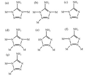
Scheme 1.
Coordination modes of HATZ

In the last year, Okamura et al. reported a pentanuclear iron cluster which can be catalyst water oxidation[1]. Polynuclear iron(III) clusters continue to attract the intense interest of many inorganic groups[2-4]. In the molecular magnet system, high-spin FeIII ions have a large number of unpaired electrons (3d5, S = 5/2) and usually display strong antiferromagnetic interactions among FeIII ions[5, 6], but part of high nuclearity iron(III) clusters which possess appropriate topologies and large spin (S) values in their ground states can even occasionally function as single-molecule magnets (SMMs)[7, 8].
Careful selection of appropriate organic ligand with certain characteristics, such as variable bonding modes and the ability to perform supramolecular interactions, is helpful for tailoring and constructing clusters with desirable properties[9-13]. In recent years, many complexes constructed with 5-amino-1, 2, 3, 4-tetrazole (HATZ) have been reported[14-19]. From the structural point of view, HATZ possesses two interesting characteristics: (1) the ATZ possesses four N atoms of tetrazole group and one amino group with two hydrogen atoms and might be utilized as versatile linker in constructing interesting coordination polymers with abundant hydrogen bonds[5, 14-19]. (2) HATZ shows abundant coordination modes (Scheme 1), such as μ2: 1η1: 4η1[14], μ3: 1η1: 2η1: 4η1[15], μ1: 1η1[16], μ3: 1η1: 2η1: 3η1: 4η1[17], μ2: 2η1: 3η1[18] and μ3: 1η1: 2η1: 3η1[19]. But the complexes of HATZ Schiff base are rarely reported[5]. Herein, using a new Schiff base H2timb synthesized by 5-bromo-2-hydroxybenzaldehyde and HATZ, a pentanuclear iron(III) cluster [HN(C2H5)3]· [Fe5(timb)4(ATZ)4(μ3-O)2]·(H2O)5 (1) was synthesized through micro-vial reactions.
A mixture of 5-bromo-2-hydroxybenzaldehyde (20 mmol, 4.021 g), HATZ (20 mmol, 1.701 g) and EtOH (30 mL) in a 100 mL flask was refluxed at 80 ℃ for 2 h, and then dried at 50 ℃ for 24 h after filtrated, yellow-greenish powder had been gained (yield: 3.706 g, ca. 98%, based on HATZ). Anal. Calcd. for H2timb: C8H6N5OBr (Mr = 268.09), Calcd.: C, 35.81; H, 2.34; N, 26.11%. Found: C, 35.78; H, 2.38; N, 26.14%. IR data (KBr, cm–1, Fig. S1): 3379w, 1613s, 1555s, 1470s, 1274s, 1174s, 1063m, 728m.
A mixture of FeCl3·6H2O (0.5 mmol, 0.135 g), H2timb (0.5 mmol, 0.134 g) and anhydrous acetonitrile (10 mL) with a pH adjusted to 7 by addition of triethylamine was stirred for 15 min at room temperature. The mixture was poured into a micro-vial (20 mL) and then heated at 80 ℃ for 72 h. Bright black cuboid crystals of 1 were collected by filtration, washed with anhydrous acetonitrile (5 mL × 3) and dried in air (yield: 0.152 g, ca. 88.74% based on FeIII). Anal. Calcd. for 1: C42H50Br4Fe5N41O11 (Mr = 1904.04): C, 26.49; H, 2.65; N, 30.15%. Found: C, 26.35; H, 2.74; N, 30.22. IR data for 1 (KBr, cm–1, Fig. S1): 3357m, 1601s, 1574w, 1444s, 1299s, 1178s, 829w, 586m.
The diffraction data were collected on an Agilent G8910A CCD diffractometer with graphite monochromatic Mo-Kα radiation (λ= 0.71073 Å) using an ω-θ scan mode in the range 3.74≤θ≤25.10°. Raw frame data were integrated with the SAINT program[20]. The structure of 1 was solved by direct methods using SHELXS-97 and refined by full-matrix least-squares on F2 using SHELXS-97[20]. An empirical absorption correction was applied with the program SADABS[20]. All non-hydrogen atoms were refined anisotropically. All hydrogen atoms were positioned geometrically and refined as riding. Calculations and graphics were performed with SHELXTL[20]. Disordered solvent molecules are removed by squeeze program. The solvent molecules and countervailing cations were determined by elemental and thermogravimetric analyses. Selected bond lengths and bond angles for 1 are listed in Table 1, and hydrogen bond lengths and bond angles in Table 2.
 DownLoad:
CSV
DownLoad:
CSV
| Bond | Dist. | Bond | Dist. | Bond | Dist. | ||
| Fe(1)–O(1) | 1.919(3) | Fe(2)–O(1)i | 1.880(6) | Fe(1)–N(6) | 2.078(6) | ||
| Fe(1)–O(2) | 1.907(5) | Fe(2)–O(1) | 1.880(6) | Fe(1)–N(7)iii | 2.145(6) | ||
| Fe(1)–N(1) | 2.191(6) | Fe(2)–N(2) | 2.113(6) | Fe(2)–N(2)ii | 2.113(6) | ||
| Fe(2)–N(2)iii | 2.113(6) | ||||||
| Angle | (°) | Angle | (°) | Angle | (°) | ||
| O(1)–Fe(1)–N(1) | 99.70(18) | O(1)–Fe(2)–N(2)ii | 86.48(14) | N(7)iii–Fe(1)–N(1) | 87.0(2) | ||
| O(1)–Fe(1)–N(3)i | 82.99(19) | O(1)–Fe(2)–N(2) | 93.52(14) | N(7)iii–Fe(1)–N(3)i | 165.6(2) | ||
| O(1)–Fe(1)–N(6) | 85.0(2) | O(1)–Fe(2)–N(2)i | 93.52(14) | O(2)–Fe(1)–N(1) | 172.9(2) | ||
| O(1)–Fe(1)–N(7)iii | 83.6(2) | O(1)–Fe(2)–N(2)iii | 93.52(14) | O(2)–Fe(1)–N(3)i | 96.4(2) | ||
| O(2)–Fe(1)–O(1) | 174.68(18) | N(2)i–Fe(2)–N(2)iii | 90.216(18) | O(2)–Fe(1)–N(6) | 89.8(2) | ||
| N(6)–Fe(1)–N(1) | 172.9(2) | O(1)i–Fe(2)–O(1) | 180.0 | O(2)–Fe(1)–N(7)iii | 97.4(2) | ||
| N(6)–Fe(1)–N(3)i | 95.8(2) | O(1)–Fe(2)–N(2)i | 86.48(14) | O(1)i–Fe(2)–N(2) | 86.48(14) | ||
| N(2)iii–Fe(2)–N(2) | 173.0(3) | N(2)ii–Fe(2)–N(2)i | 173.0(3) | O(1)i–Fe(2)–N(2)ii | 93.52(14) | ||
| N(2)i–Fe(2)–N(2) | 90.217(18) | O(1)i–Fe(2)–N(2)iii | 86.48(14) | ||||
| Symmetry transformation: (i) y, –x+1, –z; (ii) –y+1, x, –z; (iii) –x+1, –y+1, z | |||||||
DownLoad:
CSV
| D–H···A | d(D–H) | d(H···A) | d(D···A) | ∠DHA |
| N(10)–H(10A)···N(5)iv | 0.8600 | 2.4300 | 2.9126 | 116.00 |
| N(10)–H(10B)···O(2) | 0.8600 | 2.3800 | 3.0351 | 133.00 |
| N(10)–H(10B···N(4)iv | 0.8600 | 2.4200 | 2.9320 | 119.00 |
| C(2)–H(2)···N(5) | 0.9300 | 2.3300 | 2.7485 | 107.00 |
| N(9)–H(9)···Br(1)v | 0.86 | 2.80 | 3.516 | 141.6 |
| N(10)–H(10A)···Br(1)v | 0.86 | 2.95 | 3.672 | 142.9 |
| Symmetry codes: (iv) –y+0.5, −x, z−1/4; (v) 0.5−y, x−0.5, 0.5–z | ||||
Molecular Hirshfeld surface calculations were performed by using the CrystalExplorer program[21]. When the CIFs file of 1 are read into the CrystalExplorer program, all hydrogen bond lengths were automatically modified to typical standard neutron values (C–H = 1.083 Å, N–H = 1.009 Å and O–H = 0.983 Å)[22]. In this study, all the Hirshfeld surfaces were generated using a high (standard) surface resolution. The 3D dnorm surfaces were mapped by using a fixed color scale of 0.76 (red) to 2.4 (blue). The 2D fingerprint plots were displayed by using the standard 0.4~3.0 Å view with the de and di distance scales displayed on the graph axes.
Single-crystal X-ray structure analysis reveals that 1 crystallizes in tetragonal system space group I

Complex 1 further constructed a 1D chain through double receptor N···H–N hydrogen bonds (N(4)a···H(10a)i–N(10)i, 2.931 Å, N(5a)···H(10b)i–N(10)i, 2.913 Å symmetry code: (i) x, 0.5–y, 0.25–z) and double donor N– H···N hydrogen bonds (N(10)– H(10a)ii···N(4)ii, 2.931 Å, N(10)–H(10b)ii···N(5)ii, 2.913 Å symmetry code: (ii) y, x–0.5, 0.25+z. Fig. S2) which further formed a 3D network through abundant hydrogen bonds: N–H···N, N–H···Br (N(9)–H(9)···Br(1)iii, 3.516Å, N(10)– H(10a)···Br(1)iii, 3.672 Å, symmetry code: (iii) 0.5–y, x–0.5, 0.5–z), π···π interactions (the distance between the plane of C(3)~C(8) and the plane of (C(3)~C(8))iv is 3.457 Å, symmetry code: (iv) x, 0.5–y, 0.25–z), N–H···O and C–H···N (Table 2, Fig. S3)[26, 27].
The magnetic susceptibilities of 1 were measured by using crushed single crystal samples. The dc susceptibilities of 1 were measured under an applied field of 1 kOe at temperature ranging from 2 to 300 K.
For complex 1, the five Fe(III) ions give rise to the χmT product of 6.53 cm3·K·mol–1 at room temperature (Fig. 2). The χmT values at room temperature are all much less than the spin-only value of 22 cm3·K·mol–1 for five non-interacting high-spin FeIII ions assuming g = 2.2[25]. The observed values of 1 are also lesser than those obtained for [Fe5(μ3-F)2(XDK)2(py)4(O2CPh)4] (~10.98 cm3·K·mol–1) and [Fe5(μ3-F)2(XDK)2(N-MeIm)4(O2CPh)4] (~10.38 cm3·K·mol–1)[23]. Upon decreasing T, the χmT values of 1 gradually decrease to the minimum of 2.57 at 2 K. Similar magnetic behaviors were observed for [Fe5(μ3-F)2(XDK)2(py)4(O2CPh)4] and [Fe5(μ3-F)2(XDK)2 (N-MeIm)4(O2CPh)4][23]. The results indicated the presence of strong antiferromagnetic interactions.

Examination of the bond lengths and angles between Fe(III) ions in 1 for magneto-structural correlations reveal two obvious antiferromagnetic interactions. The quantitative approximation of the exchange interactions for the 3D oxo-bridged Fe(III) systems was obtained by fitting the appropriate theoretical equation to the experimental data separately in 22~300 K, through using the program Origin8.6. The Heisenberg spin-Hamiltonian (Eq. 1):
|
|
(Eq.1) |
Where J and J' characterize the exchange of Fe(1)···Fe(1)c and Fe(1)···Fe(2) through µ3-O bridge for 1 (inset Fig. 2). The best fitting gave: g = 2.026, J = –2.066 cm–1, J' = –1.80 × 10⁻⁴ cm–1, and R = ∑ [(χobs – χcalc) 2/∑(χobs) 2 = 1.20 × 10⁻⁵. These results further support intracluster antiferromagnetic coupling between the Fe(III) ions in 1 through μ3-O bridges.
The χm–1-T curve follows the Curie-Weiss law [χm = C/(T–θ)] (Fig. S4) severally in 70~300 K with a Weiss constant of θ to be –70.59 K and Curie constant of C being 7.90 cm3⋅K⋅mol–1. The negative Weiss constant suggests dominant intracluster antiferromagnetic coupling between adjacent Fe(III) ions through μ3-O bridges.
Hirshfeld surface analysis is a useful tool for describing the surface characteristics of the molecules[28-31], which was performed to visualize the different intermolecular interactions in crystal structures by employing 3D molecular surface contours. The Hirshfeld surfaces in the crystal structure of a particular complex are constructed on the basis of the electron distribution calculated as the sum of spherical atom electron densities. Inside the Hirshfeld surface the electron distribution due to a sum of spherical atoms for the molecule (the pro-molecule) dominates the corresponding sum over the crystal (the pro-crystal), and the Hirshfeld surface is defined as the ratio of the pro-molecule to pro-crystal electron densities equal to 0.5.
One of the useful supplements for Hirshfeld surface analysis is the 2D fingerprint plot. It quantitatively analyzes the nature and type of intermolecular interaction between the molecules inside the crystals. The fingerprint plots can be decomposed to highlight particularly close contacts between the elements (Fig. 3). The Br···H interaction is one of the most significant contacts for 1.
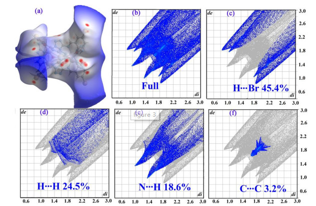
For 1, the Br···H interactions are represented by four blades in the fingerprint plot. So we can infer that there are significant N–H···Br (N(9)–H(9)···Br(1)iii, 3.516 Å, N(10)– H(10a)···Br(1)iii, 3.672 Å, symmetry code: (iii) 0.5–y, x–0.5, 0.5–z) interactions observed in complex 1 (The percentage of Br…H contact of 1 is 45.4%). H···H contacts are the second major intermolecular interaction. Another main intermolecular interaction of 1 is N···H contacts which reflected in the middle of the scattered point of the 2-D fingerprint plots (The percentage of N···H contacts of 1 is 18.6%). In the solid state structure, abundant N–H···N hydrogen bonds (N(10)–H(10a)ii···N(4)ii, 2.931 Å, N(10)–H(10b)ii···N(5)ii, 2.913 Å symmetry code: (ii) y, x–0.5, 0.25+z) were observed. Also, the C···C contacts play important roles for 1. The percentage of C···C contacts of 1 is 3.2%. π···π interactions were also observed (π(C(3)–C(8)···π(C(3)–C(8)iv distance is 3.457 Å, symmetry code: (iv) x, 0.5–y, 0.25–z, Fig. S2).
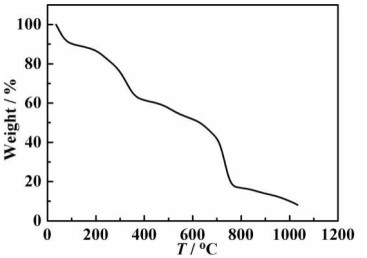
Considering the fact that multinuclear clusters based on tetrazolium are well-known for their high thermal stability, we also investigated the thermal analysis of 1 in this work. The sample was heated in a platinum crucible at a rate of 10 ℃⋅min–1 under a nitrogen atmosphere within the temperature range of 25 to 1030 ℃ (Fig. 4). At first, between 25 and 115 ℃, 1 loses 10.5% of the weight continuously, corresponding to the loss of five water molecules and a countercation [HN(C2H5)3] in 1. The weightlessness rate (10.5%) is consistent with the theoretical weightlessness rate (10.09%). The anionic [Fe5(timb)4(ATZ)4(μ3-O)2]– of 1 reveals high thermal stability, which begins to break down at 217 ℃.
In summary, a penta-nuclear Fe(III) cluster has been successfully synthesized with FeCl3·6H2O, together with H2timb Schiff base ligand. The Hirshfeld surface analysis results indicate that Br···H interactions play a most considerable role in stabilizing the self-assembly process. Magnetic studies reveal that complex 1 displays dominant antiferromagnetic intracluster interactions between FeIII ions through μ3-O bridges. In addition, the pentanuclear FeIII clusters manifest high stability.
Okamura, M.; Kondo, M.; Kuga, R.; Kurashige, Y.; Yanai, T.; Hayami, S.; Praneeth, V. K. K.; Yoshida, M.; Yoneda, K.; Kawata, S.; Masaoka, S. A pentanuclear iron catalyst designed for water oxidation. Nature 2016, 530, 465-468. doi: 10.1038/nature16529
Kitos, A. A.; Papatriantafyllopoulou, C.; Tasiopoulos, A. J.; Perlepes, S. P.; Escuer, A.; Nastopoulos, V. Binding of ligands containing carbonyl and phenol groups to iron(III): new Fe6, Fe10 and Fe12 coordination clusters. Dalton Trans. 2017, 46, 3240-3251. doi: 10.1039/C6DT04830G
Mokhtari, R.; Rezaeifard, A.; Jafarpour, M.; Farrokhi, A. Visible-light driven catalase-like activity of blackberry-shaped {Mo72Fe30} nanovesicles: combined kinetic and mechanistic studies. Sci. Technol. 2018, 8, 4645-4656.
Mayans, J.; Font-Bardia, M.; Escuer, A. Chiroptical and magnetic properties of star-shaped Fe4III complexes from chiral Schiff bases. Structural and magnetic correlations based on continuous shape measures. Dalton Trans. 2018, 47, 8392-8401. doi: 10.1039/C8DT01684D
Chen, Z.; Fan, Y.; Wang, J.; Yang, L.; Zhang, S. Pentanuclear Fe(III) cluster: synthesis, structure, magnetic properties and hirshfeld surface analysis. Chemi. Select. 2018, 3, 9841-9844.
Herold, S.; Lippard, S. J. Synthesis, characterization, and magnetic studies of two novel isostructural pentanuclear iron(II) complexes. Inorg. Chem. 1997, 36, 50-58. doi: 10.1021/ic960783i
Milios C. J.; Winpenny R. E. P. Cluster-based single-molecule magnets. Struct. Bond. 2015, 164, 1-110.
Barra, A. L.; Caneschi A.; Cornia, A.; Fabrizi de Biani, F.; Gatteschi, D.; Sangregorio, C.; Sessoli, R.; Sorace, L. Single-molecule magnet behavior of a tetranuclear iron(III) complex. The origin of slow magnetic relaxation in iron(III) clusters. J. Am. Chem. Soc. 1999, 121, 5302-5310. doi: 10.1021/ja9818755
Xiao, Y.; Liu, Y. Q.; Li, G.; Huang, P. Microwave-assisted synthesis, structure and properties of a co-crystal compound with 2-ethoxy-6-methyliminomethyl-phenol. Supramol. Chem. 2015, 27, 161-166. doi: 10.1080/10610278.2014.918268
Xiao, Y.; Qin, Y.; Yi, M.; Zhu, Y. A disc-like heptanuclear nickel cluster based on schiff base: Synthesis, structure, magnetic properties and hirshfeld surface analysis. J. Clust. Sci. 2016, 27, 2013-2023. doi: 10.1007/s10876-016-1059-y
Zhang, S. H.; Zhang, Y. D.; Zou, H. H.; Guo, J. J.; Li, H. P.; Song, Y.; Liang, H. A family of cubane cobalt and nickel clusters: syntheses, structures and magnetic properties. Inorg. Chim. Acta 2013, 396, 119-125. doi: 10.1016/j.ica.2012.10.032
Feng, C.; Zhu, Y. Q.; Huang, H. H.; Zhao, H. A triazolate-supported Fe3(μ3-O) core: crystal structure, fluorescence, and hirshfeld surface analysis. J. Clust. Sci. 2016, 27, 1181-1190. doi: 10.1007/s10876-016-0989-8
Schmitz, S.; Secker, T.; Batool, M.; Leusen, J.; Nadeem, M. A.; Kögerler, P. A planar decanuclear cobalt(II) coordination cluster. Inorg. Chim. Acta 2018, 482, 522-525. doi: 10.1016/j.ica.2018.06.005
Wang, X. W.; Chen, J. Z.; Liu, J. H. Photoluminescent Zn(II) metal-organic frameworks built from tetrazole ligand: 2D four-connected regular honeycomb (4363)-net. Cryst. Growth Des. 2007, 7, 1227-1229. doi: 10.1021/cg070330w
Yao, Y. L.; Xue, L.; Che, Y. X.; Zheng, J. M. Syntheses, structures, and characterizations of two pairs of Cd(II)-5-aminotetrazolate coordination polymers. Cryst. Growth Des. 2009, 9, 606-610. doi: 10.1021/cg8009157
Zhang, J. G.; Zhang, T. L.; Yu, K. B. Studies on synthesis, crystal structure and thermal decomposition mechanism of cadmium complex [Cd(ATZ)4(H2O)2](PA)2·2H2O. Acta Chim. Sinica 2001, 59, 84-90.
Lin, J. D.; Wang, S. H.; Cai, L. Z.; Zheng, F. K.; Guo, G. C.; Huang, J. S. Tetraalkylammonium cations as templates in the construction of two cadmium(II) metal-organic frameworks. CrystEngComm. 2013, 15, 903-910. doi: 10.1039/C2CE26213D
Kobrsi, I.; Bassioni, G. Bis(μ-5-diisopropylamino-1, 2, 3, 4-tetrazolido-κ2N2: N3)bis[(triisopropylphosphane)copper(I)]. Acta Crystal. 2011, E67, m975-m975.
He, X.; Lu, C. Z.; Yuan, D. Q. Two 3D porous cadmium tetrazolate frameworks with hexagonal tunnels. Inorg. Chem. 2006, 45, 5760-5766. doi: 10.1021/ic0520162
Sheldrick, G. M. Crystal structure refinement with SHELXL. Acta Cryst. 2015, C71, 3-8.
McKinnon, J. J.; Spackman, M. A.; Mitchell, A. S. Novel tools for visualizing and exploring intermolecular interactions in molecular crystals. Acta Cryst. 2004, B60, 627-668.
Allen, F. H.; Kennard, O.; Watson, D. G.; Brammer, L.; Orpen, A. G.; Taylor, R. Tables of bond lengths determined by X-ray and neutron diffraction. Part I. Bond lengths in organic compounds. J. Chem. Soc. Perkin Trans. 1987, 2, S1-S19.
Herold, S.; Lippard, S. J. Synthesis, characterization, and magnetic studies of two novel isostructural pentanuclear iron(II) complexes. Inorg. Chem. 1997, 36, 50-58. doi: 10.1021/ic960783i
Sertphon, D.; Harding, D. J.; Harding, P.; Murray, K. S.; Moubaraki, B.; Cashion, J. D.; Adams H. Anionic tuning of spin crossover in FeIII-quinolylsalicylaldiminate complexes. Eur. J. Inorg. Chem. 2013, 788-795.
Tabernor, J.; Jones, L. F.; Heath, S. L.; Muryn, C.; Aromi, G.; Ribas, J.; Brechin, E. K.; Collison, D. A centred, elongated 'ferric tetrahedron' with an S = 15/2 spin ground state. Dalton Trans. 2004, 33, 975-976.
Zhang, C.; Yang, L.; Chen, H.; Zhang, S. H. A novel dinuclear copper(II) complex: synthesis, crystal structure, properties and hirshfeld surface analysis. Chin. J. Struct. Chem. 2017, 36, 1904-1911.
Chen, G. H.; He, Y. P.; Zhang, S. H.; Zhang, L. A series of zirconium-oxo cluster complexes based on arsenate or phosphonate ligands. Inorg. Chem. Comm. 2018, 97, 125-128. doi: 10.1016/j.inoche.2018.09.013
Spackman, M. A.; Jayatilaka, D. Hirshfeld surface analysis. CrystEngComm . 2009, 11, 19-32. doi: 10.1039/B818330A
Feng, C.; Ma, Y. H.; Zhang, D.; Li, X. J.; Zhao, H. Highly efficient electrochemiluminescence based on pyrazolecarboxylic metal organic framework. Dalton Trans. 2016, 45, 5081-5091. doi: 10.1039/C5DT04740D
Zhao, H.; Zhu, Y. Q.; Feng, C. One novel Mn(II) complex with 1-substituted-1H-1, 2, 3-triazole-4-carboxylic acid: crystal structure, fluorescence and hirshfeld surface analysis. Chin. J. Struct. Chem. 2017, 36, 66-72.
Xiao, Y.; Qin, Y.; Yi, M.; Zhu, Y. A disc-like heptanuclear nickel cluster based on Schiff base: synthesis, structure, magnetic properties and hirshfeld surface analysis. J. Cluster Sci. 2016, 27, 2013-2023. doi: 10.1007/s10876-016-1059-y

Figure 1 Anionic structure of 1. The hydrogen atoms, solvent molecules and countervailing cations are omitted for clearly

Figure 2 Plots of χm-T and χmT-T for 1. The red solid line represents a fit of data in the temperature range of 22~300 K. Inset: The exchange pathway is between the Fe(III) ions

Figure 3 (a) dnorm of 1, (b) Full 2D Hirshfeld surface are, (c) Br···H (d) H···H, (e) N···H, (f) C···C fingerprint plots showing the percentages of contacts contributed to the total Hirshfeld surface area of 1
Table 1. Selected Bond Lengths (Å) and Bond Angles (°) for 1
| Bond | Dist. | Bond | Dist. | Bond | Dist. | ||
| Fe(1)–O(1) | 1.919(3) | Fe(2)–O(1)i | 1.880(6) | Fe(1)–N(6) | 2.078(6) | ||
| Fe(1)–O(2) | 1.907(5) | Fe(2)–O(1) | 1.880(6) | Fe(1)–N(7)iii | 2.145(6) | ||
| Fe(1)–N(1) | 2.191(6) | Fe(2)–N(2) | 2.113(6) | Fe(2)–N(2)ii | 2.113(6) | ||
| Fe(2)–N(2)iii | 2.113(6) | ||||||
| Angle | (°) | Angle | (°) | Angle | (°) | ||
| O(1)–Fe(1)–N(1) | 99.70(18) | O(1)–Fe(2)–N(2)ii | 86.48(14) | N(7)iii–Fe(1)–N(1) | 87.0(2) | ||
| O(1)–Fe(1)–N(3)i | 82.99(19) | O(1)–Fe(2)–N(2) | 93.52(14) | N(7)iii–Fe(1)–N(3)i | 165.6(2) | ||
| O(1)–Fe(1)–N(6) | 85.0(2) | O(1)–Fe(2)–N(2)i | 93.52(14) | O(2)–Fe(1)–N(1) | 172.9(2) | ||
| O(1)–Fe(1)–N(7)iii | 83.6(2) | O(1)–Fe(2)–N(2)iii | 93.52(14) | O(2)–Fe(1)–N(3)i | 96.4(2) | ||
| O(2)–Fe(1)–O(1) | 174.68(18) | N(2)i–Fe(2)–N(2)iii | 90.216(18) | O(2)–Fe(1)–N(6) | 89.8(2) | ||
| N(6)–Fe(1)–N(1) | 172.9(2) | O(1)i–Fe(2)–O(1) | 180.0 | O(2)–Fe(1)–N(7)iii | 97.4(2) | ||
| N(6)–Fe(1)–N(3)i | 95.8(2) | O(1)–Fe(2)–N(2)i | 86.48(14) | O(1)i–Fe(2)–N(2) | 86.48(14) | ||
| N(2)iii–Fe(2)–N(2) | 173.0(3) | N(2)ii–Fe(2)–N(2)i | 173.0(3) | O(1)i–Fe(2)–N(2)ii | 93.52(14) | ||
| N(2)i–Fe(2)–N(2) | 90.217(18) | O(1)i–Fe(2)–N(2)iii | 86.48(14) | ||||
| Symmetry transformation: (i) y, –x+1, –z; (ii) –y+1, x, –z; (iii) –x+1, –y+1, z | |||||||
 下载: 导出CSV
Table 2. Hydrogen Bond Lengths (Å) and Bond Angles (°) for 1
| D–H···A | d(D–H) | d(H···A) | d(D···A) | ∠DHA |
| N(10)–H(10A)···N(5)iv | 0.8600 | 2.4300 | 2.9126 | 116.00 |
| N(10)–H(10B)···O(2) | 0.8600 | 2.3800 | 3.0351 | 133.00 |
| N(10)–H(10B···N(4)iv | 0.8600 | 2.4200 | 2.9320 | 119.00 |
| C(2)–H(2)···N(5) | 0.9300 | 2.3300 | 2.7485 | 107.00 |
| N(9)–H(9)···Br(1)v | 0.86 | 2.80 | 3.516 | 141.6 |
| N(10)–H(10A)···Br(1)v | 0.86 | 2.95 | 3.672 | 142.9 |
| Symmetry codes: (iv) –y+0.5, −x, z−1/4; (v) 0.5−y, x−0.5, 0.5–z | ||||
下载: 导出CSV

 扫一扫看文章

扫一扫关注我们


 下载:
下载: