Figure 1.
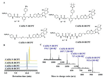
Schematic illustration of the molecular design of the CAIXi-HCPT SMDCs, the specific degradation mechanisms in tumor hypoxia microenvironment and specific structures of four HCPT SDMCs.
Carbonic anhydrase IX-targeted SMDCs for cancer precision treatment
Haolin Zhang , Kai Feng , Guisen Li , Weijiao Chen , Yuan Shao , Jiayu Ding , Mingming Zheng , Kai Yuan , Xiaolian Sun , Peng Yang

Although chemotherapy drugs are still widely used in the clinical treatment of various cancers, lack of specificity causes restrictions on the use of many chemotherapy drugs in the treatment of solid tumors [1-3]. The unordered amplification of solid tumors often leads to immaturity and deformity of its internal blood vessels, which can limit nutrient delivery and gas exchange for tumor cells to inhibit tumor cell amplification, on the other hand, it also results in tumor cells producing resistance to various tumor therapies (chemotherapy, radiotherapy targeted therapy, etc.) in many aspects: (1) Immaturity and deformity vessels significantly minimized the delivery of antitumor drugs; (2) low levels of metabolism to resistance dual pressures of nutrient deficiency and hypoxia; (3) gene mutations lead to high levels expression of drug-resistant genes [4]. In addition, the hypoxia environment of solid tumors also leads to the overexpression of hypoxia-inducible factor-1α (HIF-1α) and further activates downstream target genes to promote tumor growth and increase drug resistance [5].
As we all know, tumor microenvironment protects tumors and greatly promotes tumor growth, which seriously threatens human health, but fortunately, it also provides some excellent targets for the development of anti-tumor drugs [6,7]. Carbonic anhydrase IX (CAIX) is a downstream transmembrane protein of HIF-1α, overexpressed in triple-negative breast cancer, colorectal cancer, renal cell carcinoma and various cancer stem cells, especially under hypoxia conditions, to maintain extracellular pH homeostasis by hydration of CO2 to H+ and anion [8-10]. Besides, CAIX was also upregulated to promote tumor growth, migration, invasion and tolerance to various tumor therapies [11]. Some inhibitors of CAIX (CAIXi), such as acetazolamide (Ace) and arylsulfonamide derivatives, have been found to reduce extracellular acidification, which was beneficial for tumor treatment. Based above, some drug therapy strategies that are combined with CAIXi are already in clinical research [12-15]. Hence, the development of CAIXi targeting small molecule-drug conjugates (SMDCs) to improve tumor delivery and therapeutic effect has great prospects.
Tumor cells are remarkably heterogeneous, such as excessive reactive oxygen species (ROS) [16], reduced glutathione (GSH) [17], nitroreductase (NTR) [18], cathepsin B (CTSB) [19], azo reductase (AZOR) [20] and beyond [21,22]. Based on the abnormal expression of these metabolites and proteins, various types of prodrugs including SMDCs [23-26], antibody-drug conjugates (ADC) [27,28] and nanoconjugates [29-31], have been developed to improve the capability of tumor delivery and anti-tumor effective. Recently, Kim et al. have reported a series of CAIX-targeted SMDCs that couples cytotoxic molecules and CAIX-targeted warhead by NTR or GSH sensitive linkers, respectively, which exhibited more efficiency in tumor delivery and anti-tumor compared with unmodified drugs [32-34]. In addition, to improve traditional chemotherapy drugs, such as platinum and rhenium drugs, scientists are using CAIX inhibitors as axial ligands to target cancers, this strategy not only achieves effective and selective killing of solid tumors, it also has an regulatory effect on tumor microenvironment and metabolism [35-37]. However, these efforts were still insufficient to reply to the highly aggressive and rapidly developing tumors, more studies were urgently needed to explore the development direction of the CAIX-target strategy [38].
Herein, we developed a series of CAIXi-HCPT SMDCs including CAIXi-R-HCPT, CAIXi-N-HCPT, CAIXi-S-HCPT and CAIXi-B-HCPT to explore the delivery and anti-tumor efficiency of different SMDCs. As shown in Fig. 1, these molecules consist of a CAIX-targeting unit, cleavable linker and chemotherapeutics 10-hydroxycamptothecin (HCPT, a commonly used chemotherapy drug). The long-term exposure of tumor cells to a hypoxic microenvironment results in overexpression of CAIX on tumor cell membranes, while except for certain structures in the gastrointestinal (GI) tract and brain, normal cells are almost unexpressed [10]. Therefore, multifunctional molecules with CAIX warhead in the blood circulation system can be more captured and uptaken by tumor cells compared to normal cells [39-41]. Subsequently, these response factors, such as ROS, GSH, NTR, or CTSB activating corresponding cleavable linkers lead to the degradation of CAIXi-HCPT SMDCs and release HCPT, causing DNA damage in tumor cells [42]. In our work, all four SMDCs exhibited considerable stability in phosphate buffer solution (PBS) and fresh plasm, while rapidly degrading and releasing HCPT under the stimulation of corresponding response factors. In addition, the introduction of a CAIXi-targeting unit led to more CAIXi-HCPT SMDCs enriched on the tumor cell membranes and exhibiting considerable cytotoxicity to tumor cells. In subsequent in vivo evaluation, CAIXi-R-HCPT had a higher distribution ratio of tumor/normal tissues (T/N) and tumor inhibition rate relative to other SMDCs and irinotecan, a CAIX nontargeted molecule [43]. This indicates that CAIXi-targeting SMDCs are more suitable in combination with ROS-sensitive linkers. Overall, we investigated the tumor delivery and anti-tumor effects of the CAIXi-targeting strategy in combination with different response models and provided a suitable direction for the development of CAIX-targeting SMDCs.
SMDCs require appropriate stability and specificity to release cytotoxic compounds in the corresponding environments to kill tumors, while not influencing normal cells. Firstly, we evaluated the solubility of these SMDCs and HCPT, compared with HCPT, the solubility of CAIXi-R-HCPT, CAIXi-N-HCPT, and CAIXi-S-HCPT has been increased to a certain extent, while the structure of CAIXi-B-HCPT has a lower solubility due to the presence of more lipophilic amino acids (Fig. S1 in Supporting information). Subsequently, we also tested the stability of these SMDCs, which were dissolved in PBS (pH 7.4) at 37 ℃ and then tested by high performance liquid chromatography (HPLC). As shown in Figs. S2A–D (Supporting information), all four SMDCs exhibited considerable stability in PBS. After 12 h, all SMDCs showed no obvious degradation. Subsequently, we tested the releasing performance of all SMDCs in suitable conditions. It is expected that all SMDCs were degraded quickly under corresponding conditions and releasing HCPT. In the presence of 0.25 mmol/L H2O2, CAIXi-R-HCPT was completely degraded in PBS and released free HCPT within 2 h. It was seen in Fig. S3 (Supporting information) that the corresponding peak at 9.6 min for CAIXi-R-HCPT decreased and the new peak was formed at 9.2 min, consisting of the free HCPT (Fig. S3A). Subsequently, we tested the releasing efficiency of CAIXi-S-HCPT in PBS containing 1 mmol/L GSH, which was the same content of GSH in tumor cells according to previous literature [44]. The GSH could activate CAIXi-S-HCPT through a time-dependent form and release completely the free HCPT after 3 h (Fig. S3D). In addition, CAIXi-N-HCPT and CAIXi-B-HCPT also exhibited high release efficiency under 30 µg/mL NTR and 10 µg/mL CTSB, respectively (Figs. S3B and C). Degradation of SMDCs and release of free HCPT were also shown by fluorescence spectra (Figs. S3E–L). The fluorescence intensity of HCPT was reduced through the covalent conjugation, while restored upon the cleavage of the covalent bond. As shown in Figs. S3E–H, the fluorescence emission bands of all the HCPT SMDCs were around 450 nm. After 12 h, the fluorescence intensity showed no significant decrease in PBS (pH 7.4) at 37 ℃. After H2O2, NTR, CTSB and GSH were added to the above solutions, respectively (Figs. S3I–L), the fluorescence emission band around 450 nm of all the HCPT SMDCs were reduced with time-dependent, while an obvious appearance fluorescence emission band at 560 nm indicating the release of free HCPT.
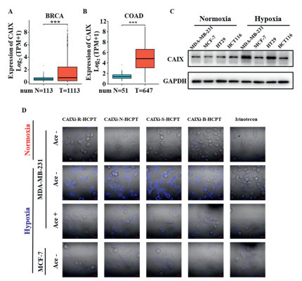
As a hypoxia-induced transmembrane protein, CAIX overexpressed on the surface of tumor cell membranes. We observed that CAIX was highly expressed in many malignant tumors such as breast cancer (Fig. 2A) and colorectal cancer (Fig. 2B) compared to normal tissues by analyzing the cancer genome atlas (TCGA) and Genotype-Tissue Expression (GTEx) databases, which was consistent with previous reports [45]. Subsequently, the protein-expressing level of CAIX in breast cancer and colon cancer cells was analyzed by Western blot. CAIX protein was upregulated significantly after hypoxia treatment for 24 h in MDA-MB-231 and HT29 cells, while not in MCF-7 and HCT116 cells (Fig. 2C and Fig. S4 in Supporting information). Based on these results, the cellular capture of CAIXi-HCPT SMDCs and Irinotecan (untargeted HCPT prodrug) was evaluated on MDA-MB-231 and MCF-7 breast cancer cells, respectively. As shown in Fig. 2D and Fig. S5 (Supporting information), the cellular capture of all four CAIXi-HCPT SMDCs was hypoxia- and CAIX-dependent in MDA-MB-231, while the uptaken of Irinotecan is not related to CAIX. After CAIXi-HCPT SMDCs treatment 2 h, strong fluorescence could be observed under hypoxia conditions (2%) O2), the Irinotecan treatment group had almost no fluorescence. When 50 µmol/L of Ace (an accepted CAIX inhibitor) was added, even under hypoxic conditions, the capture ability of MDA-MB-231 cells for CAIXi-HCPT SMDCs was lower, which indicated that Ace competitively inhibited the binding of SMDCs to CAIX. Similarly, low expression of CAIX in MCF-7 cells under hypoxia conditions also resulted in lower cellular capture. This result is similar to previous CAIX-targeted SMDCs [38]. Although there is no direct evidence to prove how compounds enter cells, CAIX can certainly increase the relative concentration of CAIX-targeted SMDCs on the cell membrane surface. Due to its low molecular weight, compounds can directly penetrate the phospholipid bilayer and enter the cell through locally high concentration gradients. Above all, these results suggested that CAIXi-HCPT SMDCs were more enriched and captured by MDA-MB-231 cells due to their binding to CAIX.
The cell cytotoxicity studies were performed in both CAIX low-expressing (MCF-7, HCT116) and high-expressing (MDA-MB-231, HT29) cells. As shown in Figs. S6A and B and Table S1 (Supporting information), CAIXi-N-HCPT, CAIXi-R-HCPT and CAIXi-S-HCPT had comparable cytotoxicity to HCPT in four types of cancer cells under normoxia and hypoxia condition, which suggested that these SMDCs can release sufficient amounts of HCPT in tumor cells. Although the expression of CAIX was lower under nonmovie conditions, long-term co-incubation of drugs and cells can also lead to enough uptake of SMDCs by cells. The CAIXi-B-HCPT has almost no cytotoxicity in vitro, this may attribute to the alkaline of the in vitro cell culture condition (pH 7.4), while the activation of CTSB usually requires acidic conditions (pH 5) [46]. In addition, four intermediates that did not couple with HCPT exhibited no cytotoxicity to all cell lines (Fig. S7 in Supporting information). Subsequently, we further investigated the cytotoxicity of all CAIXi-HCPT SMDCs for MCF-7, MDA-MB-231, HT29 and HCT116 in the condition of pretreated with 50 µmol/L of Ace under hypoxia (50 µmol/L of Ace in hypoxia condition exhibited negligible cytotoxicity (Fig. S8 in Supporting information)). Notably, the toxicity of all SMDCs was significantly decreased in MDA-MB-231 and HT29 cell compared with Ace negative group after treatment 24 h (Figs. S6C and S9 in Supporting information), the half maximal inhibitory concentration (IC50) values of Ace pretreated CAIXi-N-HCPT, CAIXi-R-HCPT, CAIXi-B-HCPT and CAIXi-S-HCPT decreased from 0.05, 0.07, 3.47 and 0.07 µmol/L against MDA-MB-231, respectively, to 0.13, 0.18, > 20 and 0.31 µmol/L. This trend is also similar with HT29, while not with MCF-7 and HCT116 (Table S2 in Supporting information). Overall, these cytotoxicity results suggested that the function of CAIXi-HCPT SMDCs was positively correlation with the level of CAIX expression.
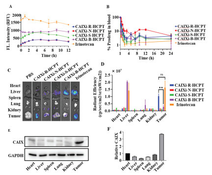
Although we have demonstrated that these CAIXi target SMDCs were enough stable in PBS (Figs. S3E–H in Supporting information), the components of plasma were much more complicated than in PBS. Therefore, we further performed in vitro and in vivo plasma stability experiments. The experimental process and animal use in this study were approved by the IACUC of China Pharmaceutical University. As shown in Fig. 3A, all compounds exhibited high-plasm stability and no significant degradation in vitro after being exposed to fresh plasma for 12 h. Subsequently, the blood clearances of CAIXi-HCPT SMDCs were investigated on Sprague-Dawley rats. Compared with Irinotecan, all the CAIXi-HCPT SMDCs exhibited extended blood circulation (Fig. 3B). In addition, we established an MDA-MB-231 tumor mice model to explore the biodistribution of the CAIXi-HCPT SMDCs after post-injection for 24 h. Among all of the SMDCs, CAIXi-R-HCPT exhibited higher selectivity to tumor tissues (Figs. 3C and D). CAIXi-B-HCPT was mainly enriched in liver and lung tissues, which attributed to the liver containing a large number of proteases, for example, CTSB, to cleave various peptide chains [47]. Although the Irinotecan treatment group also showed a high fluorescence signal in the tumor, there was also a strong fluorescence in the liver as it must be activated by carboxylesterase 2 (CES2) in the liver to release HCPT. The free HCPT enriched in the liver can be converted to inactive HCPT-glucuronide by glucuronosyltransferase 1A1 (UGT1A1) to reduce its toxic side effects. The UGT1A1 gene polymorphism (such as UGT1A1 * 28) can affect the glucuronidation of SN-38, leading to an increase in plasma concentration of SN-38 and an increased risk of toxic side effects. Therefore, the clinical application of Irinotecan has significant limitations. Compared with Irinotecan, CAIXi-R-HCPT can be directly activated by high concentrations of ROS in tumor tissue to exert its effects. These results indicated that CAIXi-HCPT SMDCs with ROS-sensitive linkers were more effective in targeting tumors. To further confirm the mechanism of the target, we analyzed the content of CAIX protein in major organs and tumor tissues. As shown in Figs. 3E and F, the CAIX protein in tumor tissues was 3–12 times more than in other tissues.
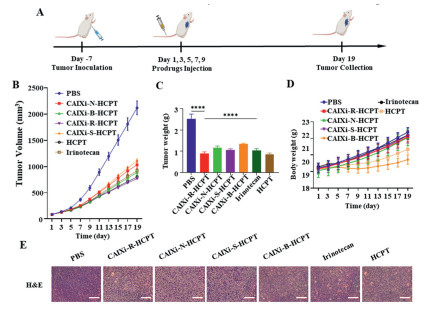
To further investigate the in vivo efficacy of CAIXi-HCPT SDMCs, we established a mouse model bearing MDA-MB-231 cell xenografts when tumor volume reached 80 mm3, mice were randomly assigned to five groups and injected with PBS, CAIXi-B-HCPT, CAIXi-N-HCPT, CAIXi-S-HCPT, CAIXi-R-HCPT, Irinotecan and HCPT every second day for five times (equivalent with HCPT 5 mg/kg) (Fig. 4A). The tumor volume and weight of mice treated with CAIXi-R-HCPT were significantly inhibited with a tumor inhibition rate of 63.8%) and equivalent to HCPT (62.3%)), which was much higher than CAIXi-N-HCPT (51.4%)), CAIXi-S-HCPT (47.6%)), CAIXi-B-HCPT (58.2%)) and Irinotecan (56.8%)) (Figs. 4B and C). We also monitored the body weight during treatment, except for CAIXi-B-HCPT and HCPT, the body weight of other treatment groups showed no noticeable weight loss, demonstrating the negligible systemic toxicity of SMDCs (Fig. 4D and Fig. S10 in Supporting information). These results suggested that CAIX targeted SMDCs not only can improve the efficacy of chemotherapy drugs but can also reduce toxic side effects. Subsequently, we performed histochemical analysis on the major organs (heart, liver, spleen, lung, kidney and tumors) of mice. The hematoxylin and eosin (H & E) staining showed that tumor cells were almost killed by CAIXi-HCPT SDMCs (Fig. 4E). Meanwhile, no apoptosis was found in normal tissue, indicating low systemic toxicity of CAIXi-HCPT SMDCs (Fig. S11 in Supporting information). These results prove that CAIXi-R-HCPT can effectively Safe and effective killing of tumors.
In conclusion, we have developed a series of CAIXi-HCPT SMDCs to explore which tumor microenvironment-triggered linker in combination with the CAIXi targeting strategy is more efficient for drug delivery. We demonstrated that our CAIXi-HCPT SMDCs could enriched in hypoxic cancer cells and fast-release HCPT triggered by high levels of corresponding enzymes and metabolites of cancer cells to generate CAIX-dependent cell cytotoxicity. Among all the SMDCs we designed, CAIXi-R-HCPT exhibited a higher level of T/N ratio and more efficient anti-cancer ability in the MDA-MB-231 bearing mice model to significantly inhibit tumor proliferation and promote apoptosis. In addition, no tissue damage or weight loss was observed in this group of mice, indicating that CAIXi-R-HCPT was safe and had no obvious side effects. Overall, our work provides direction for the development of CAIXi targeting tumor-specific delivery systems and we will further optimize this work in the future.
The authors declare that they have no known competing financial interests or personal relationships that could have appeared to influence the work reported in this paper.
Haolin Zhang: Writing – review & editing, Writing – original draft, Validation, Supervision, Software, Resources, Project administration, Methodology, Investigation, Formal analysis, Data curation, Conceptualization. Kai Feng: Writing – review & editing, Writing – original draft, Methodology, Investigation, Data curation. Guisen Li: Methodology. Weijiao Chen: Writing – review & editing, Methodology. Yuan Shao: Methodology. Jiayu Ding: Methodology. Mingming Zheng: Methodology. Kai Yuan: Writing – review & editing, Methodology. Xiaolian Sun: Writing – review & editing, Investigation, Funding acquisition, Conceptualization. Peng Yang: Writing – review & editing, Writing – original draft, Project administration, Investigation, Funding acquisition, Conceptualization.
This study was supported by the National Key R&D Program of China (No. 2022YFA1303803), the National Natural Science Foundation of China (Nos. 82425053, 82373738, 82321005), and the Natural Science Foundation of Jiangsu Province (No. BK20240013).
Supplementary material associated with this article can be found, in the online version, at doi:
G. Liu, W. Rui, X. Zhao, et al., Cell. Mol. Immunol. 18 (2021) 1085–1095. doi: 10.1038/s41423-021-00655-2
S. Wang, J. Sun, K. Chen, et al., BMC Med. 19 (2021) 140. doi: 10.1186/s12916-021-02006-4
X. Yu, L. Zhu, T. Wang, et al., Biochim. Biophys. Acta Rev. Cancer 1878 (2023) 188910. doi: 10.1016/j.bbcan.2023.188910
X. Jing, F. Yang, C. Shao, et al., Mol. Cancer 18 (2019) 157. doi: 10.1186/s12943-019-1089-9
V. Infantino, A. Santarsiero, P. Convertini, et al., Int. J. Mol. Sci. 22 (2021) 5037. doi: 10.3390/ijms22095037
Q. Liu, C. Guan, C. Liu, et al., Biomed. Pharmacother. 156 (2022) 113861. doi: 10.1016/j.biopha.2022.113861
S.H. Lee, M. Golinska, J.R. Griffiths, Cells 10 (2021) 2371. doi: 10.3390/cells10092371
N.S.P. Campos, B.S. Souza, G.C.P. Silva, et al., Cancers 14 (2022) 1392. doi: 10.3390/cancers14061392
C.T. Supuran, Nat. Rev. Drug Discov. 7 (2008) 168–181. doi: 10.1038/nrd2467
H.M. Becker, Br. J. Cancer 122 (2019) 157–167. doi: 10.2307/j.ctv9zcjt8.17
B. Muz, P. de la Puente, F. Azab, et al., Hypoxia 11 (2015) 83–92. doi: 10.2147/HP.S93413
M.Y. Mboge, J. Combs, S. Singh, et al., J. Med. Chem. 64 (2021) 1713–1724. doi: 10.1021/acs.jmedchem.0c02077
K. Eloranta, M. Pihlajoki, E. Liljeström, et al., Front. Oncol. 13 (2023) 1118268. doi: 10.3389/fonc.2023.1118268
S.J.A. van Kuijk, N.K. Parvathaneni, R. Niemans, et al., Eur. J. Med. Chem. 127 (2017) 691–702. doi: 10.1016/j.ejmech.2016.10.037
E. Andreucci, A. Biagioni, S. Peri, G. Versienti, et al., Cancer Lett. 571 (2023) 216338. doi: 10.1016/j.canlet.2023.216338
E.C. Cheung, K.H. Vousden, Nat. Rev. Cancer 22 (2022) 280–297. doi: 10.1038/s41568-021-00435-0
C. Deng, M. Zheng, S. Han, et al., Adv. Funct. Mater. 33 (2023) 2300348. doi: 10.1002/adfm.202300348
J. Qiao, M. Wang, M. Cui, et al., J. Pharm. Biomed. 203 (2021) 114199. doi: 10.1016/j.jpba.2021.114199
L. Dunsmore, C.D. Navo, J. Becher, et al., Nat. Chem. 14 (2022) 754–765. doi: 10.1038/s41557-022-00964-7
J. Zhu, T. Guo, Z. Wang, et al., J. Control. Release 345 (2022) 475–493.
L. Wu, J. Huang, K. Pu, et al., Nat. Rev. Chem. 5 (2021) 406–421. doi: 10.1038/s41570-021-00277-2
L. Gai, Y. Liu, Z. Zhou, et al., Coordin. Chem. Rev. 481 (2023) 215041. doi: 10.1016/j.ccr.2023.215041
A.M. Beekman, M.M.D. Cominetti, O.C. Cartwright, et al., MedChemComm 10 (2021) 2170–2174.
J. Zhu, Y. Xiong, X. Bai, et al., Chin. Chem. Lett. 36 (2025) 110799. doi: 10.1016/j.cclet.2024.110799
Y. Tong, H. Huang, H. Li, et al., Chin. Chem. Lett. 35 (2024) 109663.
S. Wang, K. Yu, Z. Yu, et al., Chin. Chem. Lett. 34 (2023) 108184. doi: 10.1016/j.cclet.2023.108184
L. Lu, Z. Niu, Z. Chao, et al., Cell. Mol. Life Sci. 80 (2023) 350. doi: 10.1007/s00018-023-04946-x
J.V. Leyton, Expert. Opin. Biol. Ther. 23 (2023) 1067–1076. doi: 10.1080/14712598.2023.2285996
B. Yu, Y. Wang, T. Bing, et al., Adv. Mat. 36 (2023) e2310456.
Z. Gao, S. Jia, H. Ou, et al., Angew. Chem. Int. Ed. 61 (2022) e202209793.
H. Shi, Y. Luo, S. Zhang, et al., Chin. Chem. Lett. 36 (2025) 110775. doi: 10.1016/j.cclet.2024.110775
J.H. Kim, S. Park, E. Jung, et al., Proc. Natl. Acad. Sci. U. S. A. 120 (2023) e2304081120. doi: 10.1073/pnas.2304081120
H.S. Jung, J. Han, H. Shi, et al., J. Am. Chem. Soc. 139 (2017) 7595–7602. doi: 10.1021/jacs.7b02396
J.H. Kim, P. Verwilst, M. Won, et al., J. Am. Chem. Soc. 143 (2021) 14115–14124. doi: 10.1021/jacs.1c03875
X. Su, W.J. Wang, Q. Cao, et al., Angew. Chem. Int. Ed. 61 (2022) e202115800. doi: 10.1002/anie.202115800
J. Yang, D.L. Chen, P.C. Wang, et al., Eur. J. Med. Chem. 243 (2022) 114702. doi: 10.1016/j.ejmech.2022.114702
Q. Cao, D.J. Zhou, Z.Y. Pan, et al., Angew. Chem. Int. Ed. 59 (2020) 18556–18562. doi: 10.1002/anie.202005362
P.C. McDonald, S.C. Chafe, C.T. Supuran, et al., Cancers 14 (2022) 3297. doi: 10.3390/cancers14143297
W. Huang, K. Wang, W. Huang, et al., Eur. J. Nucl. Med. Mol. Imaging 49 (2022) 4427–4439. doi: 10.1007/s00259-022-05922-6
K.T. Chen, Y. Seimbille, Int. J. Mol. Sci. 23 (2022) 6125. doi: 10.3390/ijms23116125
Y. Koba, M. Nakamoto, M. Matsusaki, ACS Appl. Mater. Interfaces 14 (2022) 51790–51797. doi: 10.1021/acsami.2c16454
L. Yang, J. Hong, J. Di, et al., Int. J. Nanomed. 12 (2017) 3681–3695. doi: 10.2147/IJN.S134005
P. Jaaks, E.A. Coker, D.J. Vis, et al., Nature 603 (2022) 166–173. doi: 10.1038/s41586-022-04437-2
S.K. Georgiou-Siafis, A.S. Tsiftsoglou, Antioxidants 12 (2023) 1953. doi: 10.3390/antiox12111953
Q. Wang, H. Cao, X. Hou, et al., Adv. Mat. 35 (2023) e2302916. doi: 10.1002/adma.202302916
M. Wei, L. Wang, Y. Wang, et al., Small 19 (2023) e2300015. doi: 10.1002/smll.202300015
J. Li, L. Chen, L. Du, et al., Chem. Soc. Rev. 42 (2013) 662–676. doi: 10.1039/C2CS35249D

Figure 1 Schematic illustration of the molecular design of the CAIXi-HCPT SMDCs, the specific degradation mechanisms in tumor hypoxia microenvironment and specific structures of four HCPT SDMCs.

Figure 2 In vitro cellular capture effects of CAIXi-HCPT SMDCs. (A) Comparison of CAIX expression between breast cancer susceptibility gene (BRCA) tumor samples (data are presented as 1.67 ± 2.03. n = 1113, from TCGA) and normal samples (data are presented as 0.57 ± 0.39, n = 113, from TCGA and GTEx). (B) Comparison of CAIX expression between colon adenocarcinoma (COAD) and rectal adenocarcinoma (READ) tumor samples (data are presented as 4.90 ± 2.30, n = 647, from TCGA) and normal samples (data are presented as 1.42 ± 0.58, n = 51, from TCGA and GTEx). Statistical analysis was performed using Wilcoxon rank sum test. (C) CAIX expression in MCF-7, MDA-MB-231, HT29 and HCT116 before or after 24 h hypoxia treatment (2%) O2). GAPDH, glyceraldehyde-3-phosphate dehydrogenase. (D) In vitro fluorescence of difference cells incubated with CAIXi-HCPT SMDCs under normoxia (21%) O2) and hypoxia (2%) O2). Scale bar: 20 µm.

Figure 3 Tumor targeting properties of CAIXi-HCPT SMDCs. (A) The fluorescence intensity changes of CAIXi-HCPT SMDCs and irinotecan in fresh plasma for 12 h. (B) The blood clearance profiles of CAIXi-HCPT SMDCs and irinotecan after intraperitoneal injection (HCPT-equivalent (eq), doses = 10 mg/kg, n = 3). The ex vivo fluorescence photographs (C) and semiquantitative analysis (D) of major organs after administration for 24 h (HCPT-eq, doses = 5 mg/kg, n = 3). Western blot assay (E) and semiquantitative analysis of CAIX (F) in normal and tumor tissues. Data represents mean ± SD. Statistical significance was calculated via one-way ANOVA. ns, no statistical significance. **P < 0.01.

Figure 4 In vivo antitumor effect of CAIXi-HCPT SMDCs. (A) Schematics of the establishment of the MDA-MB-231 xenograft nude mouse model and drug therapy. (B) Tumor growth curves of different groups (PBS, CAIXi-R-HCPT, CAIXi-N-HCPT, CAIXi-S-HCPT, CAIXi-B-HCPT, irinotecan and HCPT (5 mg HCPT-eq dose per kg)) (n = 5). (C) Tumor weight of each group on day 19 (n = 5). (D) Bodyweight curves of each group during treatment (n = 5). (E) Histological analysis of tumor sections with H & E staining (n = 3). Scale bar: 100 µm. Data represents mean ± SD. Statistical significance was calculated via one-way ANOVA in (C). ****P < 0.0001.

 扫一扫看文章

扫一扫关注我们
 DownLoad:
 下载:
下载:
 下载:
下载: