Table 1.
Crystallographic Parameters and Summary of Data Collection for 1 and 2
Citation:
Meng-Ying LV, Cong LV, Guo-Song YAN, Shi ZHOU, Wei JIANG, Bo LIU, Xiu-Mei LI. Syntheses, Crystal Structures and Fluorescent Properties of Two Zinc Complexes Assembled by Bis(imidazol) Ligands[J]. Chinese Journal of Structural Chemistry,
2020, 39(10): 1885-1891.
doi:
10.14102/j.cnki.0254–5861.2011–2709

Syntheses, Crystal Structures and Fluorescent Properties of Two Zinc Complexes Assembled by Bis(imidazol) Ligands
English
Syntheses, Crystal Structures and Fluorescent Properties of Two Zinc Complexes Assembled by Bis(imidazol) Ligands
Abstract:
Two new complexes [Zn(nph)(bix)]n·nH2O (1) and [Zn(nba)2(bix)]n (2) (H2nph = 4-nitrophthalic acid, Hnba = 4-nitrobenzoic acid, bix = 1, 4-bis(imidazol-1-yl)-benzene) have been hydrothermally synthesized and structurally characterized by elemental analysis, IR spectrum, fluorescence spectrum, single-crystal and power X-ray diffraction. Complex 1 exhibits a three-dimensional (3D) framework, which is stabilized through C–H···O hydrogen bonds and π-π stacking interactions. Complex 2 shows a zero-dimensional structure, which is further extended into a three-dimensional supramolecular structure through C–H···O and C–H···N hydrogen bonding interactions.
-
1. INTRODUCTION
The rational design and synthesis of novel complexes have been the subject of extensive investigation mainly due to their potential applications for new inorganic materials, chemical and biochemical catalytic systems, as well as their fascinating geometric aspects[1-5]. In general, the structure types of coordination polymers can be engineered via the ligand design to impart useful properties.
It has been our interest to construct coordination polymers with interesting properties using dicarboxylic acid and N-donor linker[6-8]. In our previous work, we have reported several coordination polymers which exhibit versatile coordination modes[9-11]. As a part of the research program on synthesizing various metal complexes, we herein report two new zinc complexes [Zn(nph)(bix)]n·nH2O (1) and [Zn(nba)2(bix)]n (2). In 1, the neighboring [Zn2(nph)] subunits are linked by bix ligands in trans-conformation bridging modes to form a 3D framework, which is stabilized by C–H···O hydrogen bonds and π-π interactions and extends into a 3D supramolecular architecture. In 2, two Zn(II) ions are bridged by two bix ligands to give crystallographical dimer with 28-numbered ring, and extend into a 3D supramolecular architecture by C–H···O and C–H···N hydrogen bonding interactions.
2. EXPERIMENTAL
2.1 General procedures
All reagents and solvents for synthesis were purchased from commercial sources and used without further purification. Elemental analyses (C, H and N) were performed on an Elementar Vario EL III elemental analyzer. IR spectra were recorded in the range of 4000~400 cm–1 on a Nicolet 6700 FT-IR spectrometer using KBr pellets. Power X-ray diffraction (PXRD) patterns were recorded on a Bruker D2 Phaser diffractometer with CuKα (λ = 1.5418 Å) radiation in the range of 5~50° at a scan speed of 0.1 º·s–1. Solid-state fluorescent spectra were taken on a Hitachi F-7000 fluorescence spectrophotometer.
2.2 Synthesis
[Zn(nph)(bix)]n·nH2O (1) A mixture of Zn(OAc)2·2H2O (0.2 mmol, 0.0439 g), H2nph (0.2 mmol, 0.0418 g) and bix (0.2 mmol, 0.048 g) was dissolved in 15 mL H2O/DMF. Suitable amount of NaOH was added to this solution to adjust the pH value to 7, followed by stirring at room temperature for 0.5 h until a homogeneous solution was obtained. Then it was sealed in a Parr Teflon-lined stainless-steel vessel (25 mL) under autogenous pressure at 140 ℃ for 5 days. After the reaction system was slowly cooled to room temperature, yellow block crystals were obtained by filtration and dried in air with the yield of 39% (based on Zn salt). Anal. Calcd. (%) for C22H19N5O7Zn: C, 49.78; H, 3.61; N, 13.19. Found (%): C, 49.03; H, 3.01; N, 12.95. IR (cm–1): 3425(s), 3139(w), 1617(s), 1589(m), 1574(s), 1524(s), 1479(w), 1432(w), 1410(w), 1357(s), 1245(s), 1121(w), 1104(w), 1088(w), 1068(w), 1030(w), 943(w), 852(w), 840(m), 793(w), 657(w), 596(w), 502(w), 484(w).
[Zn(nba)2(bix)]n (2) A mixture of Zn(OAc)2·2H2O (0.2 mmol, 0.0439 g), Hnba (0.2 mmol, 0.0334 g) and bix (0.2 mmol, 0.048 g) was dissolved in 15 mL H2O. Suitable amount of NaOH was added to this solution to adjust the pH value to 6.09 and it was stirred at room temperature for 0.5 h until a homogeneous solution was obtained. Then it was sealed in a Parr Teflon-lined stainless-steel vessel (30 mL) under autogenous pressure at 160 ℃ for 5 days. After the reaction system was slowly cooled to room temperature, pale yellow block crystals were obtained by filtration and dried in air. The yield of the target complex is ca. 41% (based on Zn salt). Anal. Calcd. (%) for C28H22N6O8Zn: C, 52.88; H, 3.49; N, 13.22. Found (%): C, 52.11; H, 3.01; N, 12.72. IR (cm–1): 3423(w), 3120(w), 2934(w), 1630(s), 1593(s), 1524(s), 1440(w), 1406(w), 1367(w), 1339(s), 1318(w), 1242(w), 1163(w), 1109(w), 1097(w), 1012(w), 954(w), 878(w), 830(m), 796(w), 769(w), 725(s), 658(w), 620(w), 581(w), 507(w), 484(w).
2.3 X-ray crystallography
Crystallographic measurements were made on a Bruker D8 QUEST CMOS diffractometer by using a graphite-monochromated MoKα (λ = 0.71073 Å) radiation. Absorption corrections were applied using multi-scan technique. The crystal structures of complexes 1 and 2 were solved by direct methods with SHELXS-97 program[12] and refined by fullmatrix least-squares techniques on F2 with SHELXL-97[13]. Metal atoms in each complex were located from the E-maps and non-hydrogen atoms were located in successive difference Fourier syntheses and refined with anisotropic thermal parameters. All non-hydrogen atoms were refined anisotropically and the hydrogen atoms of organic ligands were generated geometrically. A summary of the pertinent crystal data and structure refinement parameters for 1 and 2 are summarized in Table 1, and the selected bond distances and bond angles are listed in Table 2.
Table 1

 DownLoad:
CSV
DownLoad:
CSV
Parameter Complex 1 Complex 2 Empirical formula C22H19N5O7Zn C56H44N12O16Zn2 Fw (g/mol) 530.79 1271.77 Crystal system Monoclinic Triclinic Space group P21/c P $ \overline 1 $ a (Å) 13.4417(8) 10.019(3) b (Å) 10.9192(6) 12.752(4) c (Å) 14.9841(9) 13.324(4) α (°) 90.00 62.242(4) β (°) 90.348(2) 83.692(5) γ (°) 90.00 70.660(5) Volume (Å3) 1735.2(2) 1419.4(7) Z 4 1 Dc (g/cm3) 1.603 1.643 GOF 1.029 1.051 Reflns collected/unique 4647/2965 6170/4804 Rint 0.0235 0.0186 R (I > 2σ(I)) 0.0635 0.0386 wR 0.1340 0.0953 Table 2
Table 2. Selected Bond Lengths (Å) and Bond Angles (º) for 1 and 2DownLoad:
CSV
1 Bond Dist. Bond Dist. Bond Dist. Zn(1)–O(1) 2.453(8) Zn(1)–O(2) 2.275(8) Zn(1)–O(3A) 2.443(8) Zn(1)–O(4A) 2.278(9) Zn(1)–N(1) 2.273(8) Zn(1)–N(3) 2.251(8) Angle (°) Angle (°) Angle (°) N(3)–Zn(1)–N(1) 95.4(3) N(3)–Zn(1)–O(2) 96.5(3) N(1)–Zn(1)–O(2) 99.6(4) N(3)–Zn(1)–O(4A) 114.5(3) N(1)–Zn(1)–O(4A) 87.2(4) O(2)–Zn(1)–O(4A) 147.6(3) N(3)–Zn(1)–O(3A) 84.8(3) N(1)–Zn(1)–O(3A) 137.1(3) O(2)–Zn(1)–O(3A) 123.0(3) N(3)–Zn(1)–O(1) 150.3(3) N(1)–Zn(1)–O(1) 98.3(3) O(2)–Zn(1)–O(1) 55.3(3) O(4A)–Zn(1)–O(1) 92.5(3) O(3A)–Zn(1)–O(1) 102.3(3) 2 Bond Dist. Bond Dist. Bond Dist. Zn(1)–O(1) 1.9774(17) Zn(1)–O(5) 1.9499(18) Zn(1)–N(1) 2.0036(19) Zn(1)–N(4A) 2.0044(18) Angle (°) Angle (°) Angle (°) O(5)–Zn(1)–O(1) 112.27(8) O(5)–Zn(1)–N(1) 107.42(8) O(1)–Zn(1)–N(1) 114.72(7) O(5)–Zn(1)–N(4A) 97.38(8) O(1)–Zn(1)–N(4A) 110.55(7) N(1)–Zn(1)–N(4A) 113.18(8) Symmetry codes: 1: A: 1 – x, y + 1/2, –z + 1/2; 2: A: 1 – x, –y, 2 – z 3. RESULTS AND DISCUSSION
3.1 Description of the structure
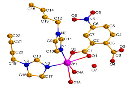
Single-crystal X-ray diffraction analysis reveals that complex 1 possesses a 3D framework and crystallizes in monoclinic space group P21/c. Its asymmetric unit contains one unique ZnII ion, one completely deprotonated nph2– ligand, one bix ligand and one crystal water molecule. As shown in Fig. 1, Zn(1) exhibits a distorted [ZnO4N2] octahedral geometry coordinated with four O atoms (O(1), O(2), O(3A), O(4A)) from two nph2– anions and two N atoms (N(1), N(3)) from two flexible bix ligands. The Zn–O bond distances lie in the range of 2.275(8)~2.453(8) Å and the Zn–N bond distances vary from 2.273(8) to 2.251(8) Å, all being normal for such coordination bonds[14]. The N(O)–Zn–O(N) angles fall in the 55.0(3)~150.3(3)º range.
Figure 1
 Figure 1. Coordination environment of the Zn(II) center in 1.
Symmetry code: (A) 1 – x, y + 1/2, –z+1/2
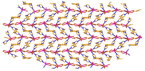
Each of all deprotonated nph2– ligand acts as a bidentatechelating connector bridging two ZnII centers in a μ2-kO, O: kO, O' mode. Notably, the flexible bix ligand adopts an anti-conformation bridging mode, in which the planes of two imidazole rings make a dihedral angle of 0°. The adjacent metal centers are linked by nph2– anions and bix ligands, leading to a 3D framework (Fig. 2).
Figure 2
 Figure 2. Three-dimensional network structure of 1
The related hydrogen-bonding geometries with symmetry codes are given in Table 3. All values involved with hydrogen bonding are within the normal ranges. The intermolecular C–H···O hydrogen bonding interactions between the carbon atoms and carboxylic oxygen atoms, nitro-oxygen atom and crystal water molecule stabilize the structure of complex 1. In addition, π-π interactions are found among imidazole rings of bix ligands. The imidazole ring of bix ligand centroid distance is 3.925(6) Å for N(1)C(10)C(9)N(2)C(11) and N(3)C(17)C(16)N(4)C(18) rings (x, –1/2–y, 1/2+z), with the vertical distance to be 3.321(4) Å, indicating the existence of π-π effect, so that the structure is more stable. Therefore, an interesting three-dimensional supramolecular network structure is formed by hydrogen-bonding and π-π stacking.
Table 3
Table 3. Hydrogen Bonds for Complexes 1 and 2DownLoad:
CSV
D–H···A d(D–H) d(H···A) d(D···A) ∠(DHA) Symmetry codes 1 C(5)–H(5A)···O(6) 0.93 2.55 3.251(1) 132 –x, –1/2 + y, 1/2 – z C(9)–H(9A)···O(7) 0.93 2.52 3.297(1) 141 1 – x, 1 – y, –z C(12)–H(12A)···O(2) 0.96 2.30 3.200(1) 155 x, 1/2 – y, –1/2 + z C(19)–H(19A)···O(4) 0.96 2.55 3.221(1) 127 1 – x, 1/2 + y, 1/2 – z 2 C(3)–H(3)···O(2) 0.96 2.46 2.801(4) 100 C(16)–H(16)···O(2) 0.87 2.60 3.195(4) 127 C(18)–H(18A)···O(3) 0.97 2.59 3.402(4) 141 –1 + x, 1 + y, z C(18)–H(18B)···O(6) 0.97 2.54 3.476(3) 162 1 – x, 1 – y, 1 – z C(24)–H(24)···N(2) 0.97 2.60 2.930(3) 100 C(26)–H(26)···O(1) 0.95 2.55 3.378(4) 146 1 – x, 1 – y, –z C(28)–H(28)···O(2) 0.92 2.55 3.409(4) 155 –1 + x, 1 + y, z Complex 2 crystallizes in triclinic space group P
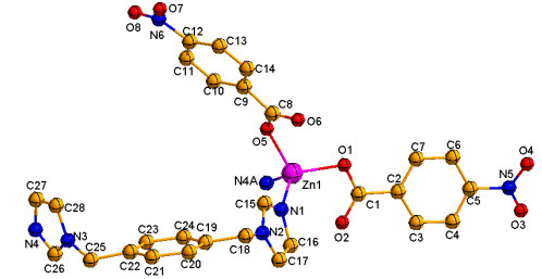
$ \overline 1 $ and exhibits a zero-dimensional structure. The coordination environment of the Zn(II) center is shown in Fig. 3. In the asymmetric unit of 2, there exist one crystallographically independent Zn(II) center, two nba- anions and a flexible bix molecule. The Zn(1) center adopts a four-coordinated tetrahedral geometry (ZnO2N2) by coordinating to two oxygen atoms of two different nba- anions and two nitrogen atoms of two different bix ligands. The Zn–O/N bond lengths fall in the range of 1.9499(18)~2.0044(18) Å (Table 2), and the N(O)–Zn–O(N) angles change from 97.38(8) to 114.72(7)º, which are all within the normal ranges generally found in the literature[15]. The completely deprotonated nba- ligand adopts a monodentate coordination mode, with the bix ligand exhibiting a bridging fashion. Since the carboxyl groups of each nba- ligand are only connected to one metal Zn(II) ion, complex 2 does not form an infinite one-dimensional long chain structure but a zero-dimensional coordination polymer with 28-numbered ring.Figure 3
 Figure 3. Coordination environment of the Zn(II) center in 2.
Symmetry code: (A) 1 – x, –y, 2 – z
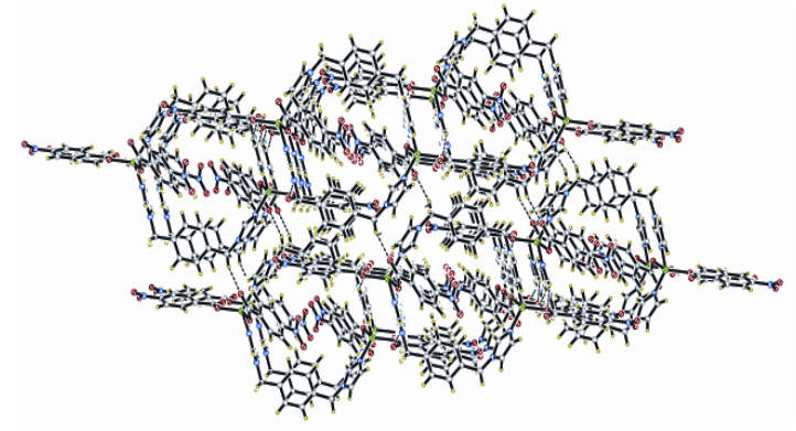
Hydrogen bonding interactions are frequently chief in the synthesis of supramolecular structure. There are existent C–H···O and C–H···N hydrogen bonding interactions in oxygen, nitrogen and carbon atoms in complex 2 (Table 3). Thus, through hydrogen bonds, a three-dimensional supramolecular architecture (Fig. 4) is formed, and they play an important role in stabilizing complex 2.
Figure 4
 Figure 4. Three-dimensional supramolecular structure of 2 through hydrogen bonding interactions
3.2 Spectral characterization of complexes 1 and 2
The main characteristic peaks of complex 1 are 3425, 3139, 1617, 1574, 1524, 1357, 1121, 943, 840, 657 and 502 cm–1, in which the strong peaks at 3440 cm–1 should be attributed to the stretching vibration absorption peak of O–H. The absorption peaks of asymmetric and symmetric stretching vibration are 1617 and 1357 cm–1[16], respectively, which indicates the presence of monodentate linkage of carboxylates in the dianions[17].
The main characteristic peaks of complex 2 are 3423, 3120, 1630, 1593, 1524, 1440, 1339, 1242, 830, 796, and 725 cm–1, and the strong peaks at 3423 and 3120 cm–1 should be attributed to the stretching vibration absorption peak of O–H. The absorption peaks of asymmetric and symmetric stretching vibration are 1593 and 1339 cm–1[18], respectively, showing the presence of monodentate linkage of carboxylates in the dianions[19].
In addition, X-ray diffraction analysis further indicates the bidentate (1) and monodentate (2) coordination manners of the carboxylate groups.
3.3 Powder X-ray diffraction (PXRD)
To confirm the phase purity of complexes 1 and 2, powder X-ray diffraction (PXRD) patterns were recorded, and they were comparable to the corresponding simulated patterns calculated from the single-crystal diffraction data (Fig. S1), indicating a pure phase of the bulky sample.
3.4 Fluorescence spectrum
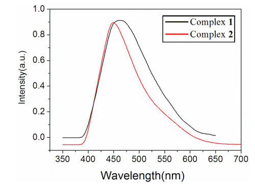
Metal-organic coordination polymers and conjugated organic linker have been researched because of their fluorescent properties and potential applications as fluorescentemitting materials, chemical sensors and electroluminescent displays[20]. Therefore, in the present work, the photoluminescent properties of H2nph, Hnba, bix, complexes 1 and 2 (Fig. 5) have been investigated in the solid state at room temperature. The free ligands H2nph, Hnba and bix show photoluminescence with the emission maximum at 440 (λex = 280 nm), 460 (λex = 207 nm) and 450 (λex = 350 nm) nm respectively, which can be assigned to intraligand (π → π*) transition[21]. Compared with the free H2nph, bix ligands, a wide range of emission with the maximum peaks at ca. 469 nm upon 275 nm excitation for complex 1 is observed, which is red-shifted. This emission band can be tentatively attributed to a ligand-to-metal charge transfer (LMCT)[22], while the emission peak of complex 2 (λem = 449 nm, λex = 350 nm) is blue-shifted as compared to that of the free ligand Hnba. Therefore, this emission band may be assigned as ligand-to-ligand charge transfer (LLCT) transitions[23, 24]. The fluorescence emissions of the complexes make them potentially useful photoactive materials.
Figure 5
 Figure 5. Solid-state emission spectra of 1 and 2 at room temperature
-
-
[1]
Zhu, Q. L.; Tian, C. B.; Shen, C. J.; Sheng, T. L.; Hu, S. M.; Xu, X. T. A three-dimensional coordination polymer based on linear trinuclear copper(II) clusters featuring a ferromagnetic exchange interaction. CrystEngComm. 2013, 15, 2120–2126. doi: 10.1039/c2ce26461g
-
[2]
Chow, T. W. S.; Liu, Y.; Che, C. M. Practical manganese-catalysed highly enantioselective cis-dihydroxylation of electron-deficient alkenes and detection of a cis-dioxomanganese(V) intermediate by high resolution ESI-MS analysis. Chem. Commun. 2011, 47, 11204–11206. doi: 10.1039/c1cc11999k
-
[3]
Wang, J.; Higgins, S. L. H.; Winkel, B. S. J.; Brewer, K. J. A new Os, Rh bimetallic with O2 independent DNA cleavage and DNA photobinding with red therapeutic light excitation. Chem. Commun. 2011, 47, 9786–9788. doi: 10.1039/c1cc11562f
-
[4]
Xie, L. X.; Hou, X. W.; Fan, Y. T.; Hou, H. W. Four three-dimensional coordination polymers constructed by 2-((1H-1, 2, 4-triazol-1-yl)methyl)-1H-imidazole-4, 5-dicarboxylate: syntheses, topological structures, and magnetic properties. Cryst. Growth Des. 2012, 12, 1282–1291. doi: 10.1021/cg201287t
-
[5]
Wen, Z. Z.; Wen, X. L.; Cai, S. L.; Zheng, S. R.; Fan, J.; Zhang, W. G. The construction of Cu(I)/Cu(II) coordination polymers based on pyrazine-carboxylate: structural diversity tuned by in situ hydrolysis reaction. CrystEngComm. 2013, 15, 5359–5367. doi: 10.1039/c3ce26817a
-
[6]
Li, X. M.; Sun, M.; Pan, Y. R. Synthesis, crystal structure and theoretical calculations of a nickel(II) coordination polymer assembled by 4, 4΄-oxydibenzoic acid and 1, 3-bis(imidazol-1-ylmethyl)-benzene ligands. Chin. J. Struct. Chem. 2015, 34, 710–718. http://manu30.magtech.com.cn/jghx/EN/abstract/abstract1237.shtml
-
[7]
Li, X. M.; Wang, Q. W.; Liu, B. Synthesis and crystal structure of a new nickel(II) complex with 2, 4-pyridinedicarboxylic acid and 1, 4-bis(imidazol-1-ylmethyl)-benzene) ligands. Chin. J. Struct. Chem. 2012, 31, 889–893. doi: 10.1007/s10870-008-9368-0
-
[8]
Li, G. F.; Wang, Y. N.; Wang, Q. W. Synthesis and crystal structure of two complexes of manganese, cadmium assembled by bis(imidazol) ligands. Chin. J. Inorg. Chem. 2014, 30, 2577–2583.
-
[9]
Li, X. M.; Pan, Y. R.; Liu, B.; Zhou, S. Syntheses, crystal structures and theoretical calculations of two complexes of cadmium assembled by bis(imidazol) ligands. Chin. J. Inorg. Chem. 2019, 35, 1275–1282.
-
[10]
Li, X. M.; Wang, Z. T.; Pan, Y. R.; Wang, Q. W.; Liu, B. Synthesis, crystal structure and theoretical calculations of two zinc, cobalt coordination polymers with 5-nitroisophthalic acid and 1, 4-bis(1H-benzimidazolyl)butane ligands. J. Inorg. Organomet. Poly. 2018, 28, 258–267. doi: 10.1007/s10904-017-0738-y
-
[11]
Pan, Y. R.; Li, X. M.; Ji, J. Y.; Wang, Q. W. Synthesis, crystal structure, and theoretical calculations of two cobalt, nickel coordination polymers with 5-nitroisophthalic acid and bis(imidazol) ligands. Aust. J. Chem. 2016, 69, 1296–1304. doi: 10.1071/CH16110
-
[12]
Sheldrick, G. M. SHELXS 97, Program for the Solution of Crystal Structure. University of Göttingen, Germany 1997.
-
[13]
Sheldrick, G. M. SHELXL 97, Program for the Refinement of Crystal Structure. University of Göttingen, Germany 1997.
-
[14]
Yang, E. C.; Liu, Z. Y.; Wang, X. G.; Batten, S. R.; Zhao, X. J. Three Zn(II)-triazole-H3btc complexes regulated by mixed ligands protonation upon stepwise crystallization. Cryst. Eng. Commun. 2008, 10, 1140–1143. doi: 10.1039/b804944k
-
[15]
Wang, Q. W.; Wang, Y. N.; Li, X. M.; Liu, B.; Kong, Z. G. Synthesis, crystal structure and fluorescent properties of zinc, cadmium coordination polymers. Chin. J. Inorg. Chem. 2014, 30, 2219–2224.
-
[16]
Li, X. M.; Wang, Z. T.; Pan, Y. R.; Wang, Q. W.; Liu, B. Synthesis, crystal structure and theoretical calculations of two zinc, cobalt coordination polymers with 5-nitroisophthalic acid and 1, 4-bis(1H-benzimidazolyl)butane ligands. J. Inorg. Organomet. Poly. 2018, 28, 258–267. doi: 10.1007/s10904-017-0738-y
-
[17]
Li, X. M.; Sun, M.; Pan, Y. R.; Ji, J. Y. Synthesis, crystal structure and theoretical calculations of a new Co(II) coordination polymer based on 5-nitroisophthalic acid and bis(imidazol) ligands. Chin. J. Struct. Chem. 2016, 35, 298–306.
-
[18]
Devereux, M.; Shea, D. O.; Kellett, A.; McCann, M.; Walsh, M.; Egan, D.; Deegan, C.; Kędziora, K.; Rosair, G.; Müller-Bunz, H. Synthesis, X-ray crystal structures and biomimetic and anticancer activities of novel copper(II)benzoate complexes incorporating 2-(4΄-thiazolyl)benzimidazole (thiabendazole), 2-(2-pyridyl)benzimidazole and 1, 10-phenanthroline as chelating nitrogen donor ligands. Inorg. Biochem. 2007, 101, 881–892. doi: 10.1016/j.jinorgbio.2007.02.002
-
[19]
Farrugia, L. J.; Wing, X. A. Windows Program for Crystal Structure Analysis. University of Glasgow, UK 1988.
-
[20]
Kreno, L. E.; Leong, K.; Farha, O. K.; Allendorf, M.; Duyne, R. P. V.; Hupp, J. T. Metal-organic framework materials as chemical sensors. Chem. Rev. 2012, 112, 1105–1125. doi: 10.1021/cr200324t
-
[21]
Lin, J. D.; Long, X. F.; Lin, P.; Du, S. W. A series of cation-templated, polycarboxylate-based Cd(II) or Cd(II)/Li(I) frameworks with second-order nonlinear optical and ferroelectric properties. Cryst. Growth Des. 2010, 10, 146–157. doi: 10.1021/cg9007476
-
[22]
Cui, Y. J.; Yue, Y. F., Qian, G. D.; Chen, B. L. Luminescent functional metal-organic frameworks. Chem. Rev. 2012, 112, 1126–1162. doi: 10.1021/cr200101d
-
[23]
Li, G. L.; Liu, G. Z.; Huang, L. L.; Li, L.; Zhang, X. Ancillary ligand-mediated syntheses, structures and fluorescence of three Zn/Cd(II) coordination polymers based on nitrobenzene dicarboxylate. J. Inorg. Organomet. Polym. 2014, 24, 617–623. doi: 10.1007/s10904-014-0024-1
-
[24]
Mohamed, G. G.; El-Gamel, N. E. A. Synthesis, investigation and spectroscopic characterization of piroxicam ternary complexes of Fe(II), Fe(III), Co(II), Ni(II), Cu(II) and Zn(II) with glycine and dl-phenylalanine. Spectrochim. Acta, Part A 2004, 60, 3141–3154. doi: 10.1016/j.saa.2004.01.035
-
[1]
-
Figure 1 Coordination environment of the Zn(II) center in 1.
Symmetry code: (A) 1 – x, y + 1/2, –z+1/2
Figure 3 Coordination environment of the Zn(II) center in 2.
Symmetry code: (A) 1 – x, –y, 2 – z
Figure 4 Three-dimensional supramolecular structure of 2 through hydrogen bonding interactions
Table 1. Crystallographic Parameters and Summary of Data Collection for 1 and 2
Parameter Complex 1 Complex 2 Empirical formula C22H19N5O7Zn C56H44N12O16Zn2 Fw (g/mol) 530.79 1271.77 Crystal system Monoclinic Triclinic Space group P21/c P $ \overline 1 $ a (Å) 13.4417(8) 10.019(3) b (Å) 10.9192(6) 12.752(4) c (Å) 14.9841(9) 13.324(4) α (°) 90.00 62.242(4) β (°) 90.348(2) 83.692(5) γ (°) 90.00 70.660(5) Volume (Å3) 1735.2(2) 1419.4(7) Z 4 1 Dc (g/cm3) 1.603 1.643 GOF 1.029 1.051 Reflns collected/unique 4647/2965 6170/4804 Rint 0.0235 0.0186 R (I > 2σ(I)) 0.0635 0.0386 wR 0.1340 0.0953  下载: 导出CSV
Table 2. Selected Bond Lengths (Å) and Bond Angles (º) for 1 and 2
1 Bond Dist. Bond Dist. Bond Dist. Zn(1)–O(1) 2.453(8) Zn(1)–O(2) 2.275(8) Zn(1)–O(3A) 2.443(8) Zn(1)–O(4A) 2.278(9) Zn(1)–N(1) 2.273(8) Zn(1)–N(3) 2.251(8) Angle (°) Angle (°) Angle (°) N(3)–Zn(1)–N(1) 95.4(3) N(3)–Zn(1)–O(2) 96.5(3) N(1)–Zn(1)–O(2) 99.6(4) N(3)–Zn(1)–O(4A) 114.5(3) N(1)–Zn(1)–O(4A) 87.2(4) O(2)–Zn(1)–O(4A) 147.6(3) N(3)–Zn(1)–O(3A) 84.8(3) N(1)–Zn(1)–O(3A) 137.1(3) O(2)–Zn(1)–O(3A) 123.0(3) N(3)–Zn(1)–O(1) 150.3(3) N(1)–Zn(1)–O(1) 98.3(3) O(2)–Zn(1)–O(1) 55.3(3) O(4A)–Zn(1)–O(1) 92.5(3) O(3A)–Zn(1)–O(1) 102.3(3) 2 Bond Dist. Bond Dist. Bond Dist. Zn(1)–O(1) 1.9774(17) Zn(1)–O(5) 1.9499(18) Zn(1)–N(1) 2.0036(19) Zn(1)–N(4A) 2.0044(18) Angle (°) Angle (°) Angle (°) O(5)–Zn(1)–O(1) 112.27(8) O(5)–Zn(1)–N(1) 107.42(8) O(1)–Zn(1)–N(1) 114.72(7) O(5)–Zn(1)–N(4A) 97.38(8) O(1)–Zn(1)–N(4A) 110.55(7) N(1)–Zn(1)–N(4A) 113.18(8) Symmetry codes: 1: A: 1 – x, y + 1/2, –z + 1/2; 2: A: 1 – x, –y, 2 – z
下载: 导出CSV
Table 3. Hydrogen Bonds for Complexes 1 and 2
D–H···A d(D–H) d(H···A) d(D···A) ∠(DHA) Symmetry codes 1 C(5)–H(5A)···O(6) 0.93 2.55 3.251(1) 132 –x, –1/2 + y, 1/2 – z C(9)–H(9A)···O(7) 0.93 2.52 3.297(1) 141 1 – x, 1 – y, –z C(12)–H(12A)···O(2) 0.96 2.30 3.200(1) 155 x, 1/2 – y, –1/2 + z C(19)–H(19A)···O(4) 0.96 2.55 3.221(1) 127 1 – x, 1/2 + y, 1/2 – z 2 C(3)–H(3)···O(2) 0.96 2.46 2.801(4) 100 C(16)–H(16)···O(2) 0.87 2.60 3.195(4) 127 C(18)–H(18A)···O(3) 0.97 2.59 3.402(4) 141 –1 + x, 1 + y, z C(18)–H(18B)···O(6) 0.97 2.54 3.476(3) 162 1 – x, 1 – y, 1 – z C(24)–H(24)···N(2) 0.97 2.60 2.930(3) 100 C(26)–H(26)···O(1) 0.95 2.55 3.378(4) 146 1 – x, 1 – y, –z C(28)–H(28)···O(2) 0.92 2.55 3.409(4) 155 –1 + x, 1 + y, z
下载: 导出CSV
-


 扫一扫看文章
计量
- PDF下载量: 1
- 文章访问数: 1091
- HTML全文浏览量: 46
 下载:
下载:
