Figure 1.
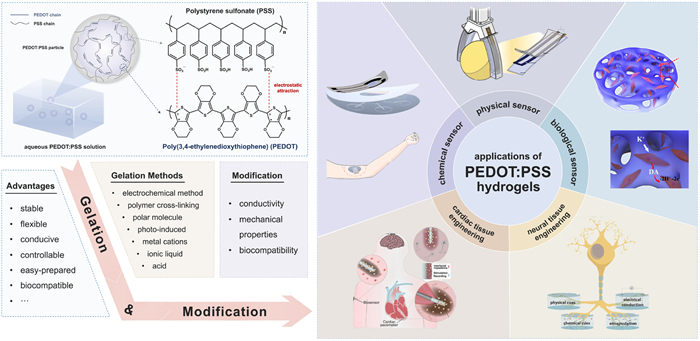
An overview of the properties and applications of PEDOT:PSS hydrogels. Through gelation and modification, PEDOT:PSS hydrogels can be applied for various sensors and tissue engineering.
Recent progress in the biomedical application of PEDOT:PSS hydrogels
Binhan Zhao , Zheng Li , Lan Zheng , Zhichao Ye , Yuyang Yuan , Shanshan Zhang , Bo Liang , Tianyu Li

PEDOT:PSS, a widely acclaimed conductive polymer material, has garnered immense attention owing to its remarkable conductivity, exceptional electrochemical stability and straightforward processing. Leveraging these attributes, PEDOT:PSS is frequently fabricated into films, showcasing promising applications in polymer solar cells [1-3], battery surface modification [4], organic photovoltaics [5,6] and supercapacitors [7]. To extend its utility into the biomedical realm, PEDOT:PSS necessitates structural adaptations to accommodate water-rich and soft biological tissues. Through relentless research endeavors aimed at structural regulation, PEDOT:PSS hydrogels have emerged as an ideal bioelectronic interface. These hydrogels offer an impeccable mechanical and electrical match, attributed to their biotissue-like properties, including superior flexibility, water-rich composition, high volumetric swelling ratios and outstanding biocompatibility [8,9].
The growing interest in gelation has led to the increasing maturity of PEDOT:PSS hydrogels formation. Ionically induced gelation methods represent the simplest and earliest approaches for creating PEDOT:PSS hydrogels. Originally, researchers employed metal cations (Mg2+, Ca2+, Fe2+/3+, Ru(NH3)62+/3+) to dope PEDOT:PSS solutions, inducing gelation by facilitating the phase separation of PEDOT and PSS [10]. Subsequent investigations have demonstrated that acid-induced gelation methods can also achieve the desired outcomes. The electrogelation method offers applicability for patterning PEDOT:PSS hydrogels on conductive templates [8,11]. To address the issue of hydrogel detachment from the electrode, a porous gold electrode has been introduced [12]. Ionic liquids play a role in increasing ion strength, shielding electrostatic repulsion between particles, and weakening the electrostatic interaction between PEDOT chains and PSS chains. Similarly, polar molecules induce the gelation process based on the same principle. In addition to the formation of single-network hydrogels, PEDOT:PSS can also combine with other polymers to form double-network hydrogels [13-15]. The process involves preparing an initial polymer network, which then serves as a template for the in-situ polymerization of PEDOT:PSS, resulting in the formation of a second network. These steps yield double-network/multiple-network PEDOT:PSS composite hydrogels. In the latest research, a photo-crosslinking gelation method has been developed, inducing selective phase separation of PEDOT:PSS [16-18]. This innovative approach represents a significant advancement in the field of PEDOT:PSS hydrogels formation.
Unmodified hydrogels often fall short of meeting the practical application requirements, prompting researchers to employ various methods for enhancing the electroconductivity and mechanical strength of hydrogels. In previous studies, the incorporation of PEDOT:PSS as an additive into other hydrogels at an appropriate proportion has proven effective in reducing impedance. Notably, the conductivity of PEDOT:PSS hydrogels can be further improved by introducing organic/inorganic conductive materials and implementing structural modifications [9,19,20]. This advancement opens avenues for the utilization of PEDOT:PSS hydrogels in biosensing and supercapacitors. However, PEDOT:PSS hydrogels with high water content and swelling characteristics often exhibit unsatisfactory mechanical strength. In response, scholars have undertaken molecular-level modifications of PEDOT:PSS hydrogels. Considerable attention has been directed towards double-network/multiple-network hydrogels composed of PEDOT:PSS, where interactions among polymers or monomers noticeably enhance the toughness and strength of the composite hydrogels [21-23].
Current research outcomes indicate promising advances in the application of PEDOT:PSS hydrogels. Thanks to the development of modification methods, PEDOT:PSS composite hydrogels with both satisfactory conductivity and toughness are now adaptable to the requirements of practical applications. Researchers are actively exploring the potential of PEDOT:PSS hydrogels in various multidisciplinary fields, including sensors and tissue engineering [24-26]. The proposed utilization of PEDOT:PSS hydrogels showcases their versatility and holds significant promise for future applications.
In this review, we have been dedicated to the exploration of PEDOT:PSS hydrogels. We commence with an overview of different gelation methods employed in hydrogel formation and compare these methods. Next, we systematically consolidates insights into the modification of PEDOT:PSS composite hydrogels, emphasizing aspects related to conductivity and mechanical strength, aiming to shed light on the diverse strategies employed in enhancing the performance of PEDOT:PSS composite hydrogels. Concluding this comprehensive exploration, the review meticulously introduces and appraises the applications of PEDOT:PSS composite hydrogels in the realms of sensors and tissue engineering, aiming to provide a panoramic view of the advancements and potentials witnessed in these application domains (Fig. 1).
PEDOT:PSS complex is composed of two parts, the conductive polymer PEDOT and the water-soluble polymer PSS. The conjugated backbone of PEDOT results in its high conductivity, which allows for the effective transmission of delocalized electrons through the π orbital system. Nevertheless, its inherent hydrophobicity imposes significant limitations on its applications in the realm of biomedicine. To enable the solubility of PEDOT in water, the hydrophilic anionic polyelectrolyte PSS is commonly used as a dopant, leading to the formation of PEDOT:PSS complex. In an aqueous solution, certain PEDOT chains are tightly connected to the PSS chains. This results in a scenario where the PEDOT chains are enveloped by the PSS chains, giving rise to core-shell assembly structures reminiscent of microgel particles. This innovative assembly ensures the solubility of PEDOT in water while preserving its conductive properties, opening up new possibilities for applications in biomedicine.
The gelation mechanism of PEDOT:PSS can be comprehended through a structural lens, considering two key perspectives. Firstly, in the aqueous dispersions of PEDOT:PSS, electrostatic repulsion exists between PSS− groups on the surface of microgel particles, fostering their mobility and flowability between particles [19]. The mitigation of electrostatic repulsion, achieved by shielding interactions between PSS− groups, results in reduced mobility between particles, facilitating their aggregation. Secondly and more prominently, the weakening the electrostatic attractions between the PEDOT and PSS chains induces phase separation. This separation allows the exposure of PEDOT+ chains to water. The exposed PEDOT+ chains undergo a conformational change, transitioning from a confined-coiled to an expanded-linear structure [9]. Subsequently, driven by π-π stacking and hydrophobic attractions, the physical crosslinking occurs between the aggregated PEDOT+ chains happens. This intricate interplay of electrostatic forces and conformational changes orchestrates the gelation process in PEDOT:PSS, unveiling a nuanced understanding of its structural dynamics.
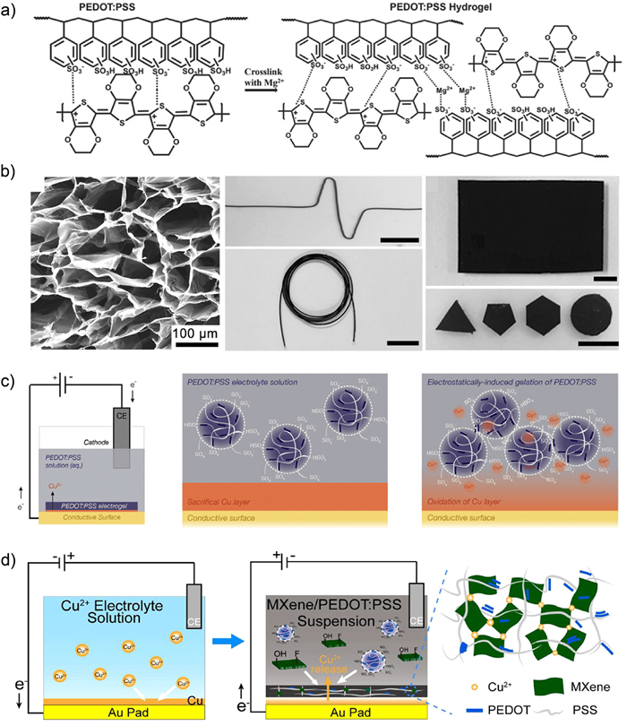
Directly incorporating metal cations is the simplest and earliest method of inducing the gelation of PEDOT:PSS. In 1998, Ghosh et al. [11] proposed that in PEDOT:PSS, an excess of PSS relative to PEDOT stabilizes microgel particles in an aqueous solution, and the excess negative charge can ionically cross-link with metal cations to form hydrogels. They cross-linked Mg2+, Ca2+, and negatively charged PSS, resulting in the gelation of conductive PEDOT:PSS networks in a PVP matrix. To investigate the effect of multivalent cation cross-linking, Vázquez et al. [22] printed PEDOT:PSS on Au electrodes and tested the ion cross-linking with Mg2+, Ca2+, Fe2+/3+, Ru(NH3)62+/3+. They found that the cross-linking with Ru(NH3)62+/3+ and Fe2+/3+ ions was highly efficient, while ion cross-linking in Mg2+ or Ca2+ solutions was not successful. Furthermore, electrodes treated with Ru(NH3)62+/3+ exhibited an acceptable potentiometric response and good potential stability, and no risk of polymer disintegration, making it a promising candidate for ion cross-linking. Yu et al. [23] investigated the method of stabilizing PEDOT hydrogels using Mg2+ ion cross-linking with PSS. They solve the problem that high-concentration Mg2+ solutions causing disintegration by initially slightly cross-linking with low concentrations of Mg2+ and then increasing the concentration to enhance cross-linking. It provides a strategy for ionic cross-linking of high concentrations of metal cations (Fig. 2a).
In addition to doping metal cations into the PEDOT:PSS aqueous dispersion, using EDOT monomers is a new method to prepare PEDOT:PSS hydrogels. Dai et al. [21] introduced a supramolecular self-assembly approach, physically cross-linking EDOT monomers, NaPSS, and Fe3+ cations to fabricate PEDOT:PSS hydrogels. In this method, an excess of Fe3+ serves both as an oxidizing agent to promote the polymerization of EDOT monomers and as an ion cross-linker. Fe3+ engages in electrostatic interactions with negatively charged PSS for gelation. Through this approach, Teng et al. [24] ultrasonically mixed EDOT monomers with NaPSS, added a solution of Fe(NO3)3·9H2O, and placed a Ti mesh substrate in the mixed solution to prepare PEDOT:PSS hydrogels network with hierarchical porous structures. This network demonstrated effective separation of various oil-water mixtures. This work introduces a novel approach to efficiently process and separate diverse oil-water mixtures.
Acid treatment, such as sulfuric acid, can also be employed for the preparation of PEDOT:PSS hydrogels, leading to a significant enhancement in conductivity. Acid treatment results in the partial protonation of PSS chains by H+, weakening the electrostatic attraction between PEDOT and PSS chains. This promotes π-π stacking between PEDOT chains, enabling PEDOT chains to assemble more tightly in the hydrogels. Better conductive pathways are formed, allowing charge carriers to traverse more effectively. Yao et al. [25] subjected a commercial PEDOT:PSS (PH1000) suspension to heat treatment in 0.1 mol/L sulfuric acid, followed by partial removal of its PSS component using concentrated sulfuric acid to prepare the PEDOT:PSS hydrogels (Fig. 2b). Similarly, Zhou et al. [26] injected a PEDOT:PSS suspension containing 0.05 mol/L H2SO4 into a glass capillary with a 0.5 mm inner diameter. After a 3-h treatment at 90 ℃, PEDOT:PSS hydrogel fibers with a highly conductive 3D scaffold was formed.
In recent years, electrochemical methods have been applied to ion-induced gelation of PEDOT:PSS. In comparison to traditional ion cross-linking methods, electrochemical methods allow for the precise positioning of metals, providing better control over the patterning of hydrogels. Feig et al. [19] proposed a novel electrochemical gelation method (involving the thermal vapor deposition of Cu film patterns on a substrate) (Fig. 2c). They used thermal evaporation to pattern Cu films on a substrate. Then they constructed an electrochemical cell comprising an aqueous PEDOT:PSS electrolyte, graphite rod counter electrode, and Ag/AgCl reference electrode. Finally, by applying a constant voltage, Cu was oxidized to Cu2+, inducing the gelation of PEDOT:PSS at controlled locations. This method not only obtains high-resolution patterned hydrogels but is also applicable to patterning PEDOT:PSS hydrogels on any conductive template, including curved surfaces. To address the issue of hydrogel detachment from electrodes, Li et al. [14] porousized the Au electrode substrate and electrochemically deposited Cu onto the porous Au electrode surface. After this processing, the PEDOT:PSS hydrogel network could penetrate the porous gold electrode and a close connection between the hydrogel and the electrode was formed. Additionally, this method can be employed for the patterned preparation of composite hydrogels. Zhang et al. [12] used electrochemical deposition to obtain a Cu-modified substrate. They placed it in an electrochemical cell filled with a Ti3C2T2 mixed MXene/PEDOT:PSS suspension, and applied a certain voltage or current to prepare a Ti3C2T2 mixed MXene/PEDOT:PSS composite hydrogels (Fig. 2d).
Leaf and Muthukumar suggest that, as the primary interaction between microgel particles was found to be electrostatic in nature, increasing ion strength facilitates the screen the electrostatic repulsions between particles. This allows them to physically cross-link through the π-π stacking of PEDOT, leading to the gelation of PEDOT:PSS [27]. Li et al. [28] mixed LiTFSI, PVA, and PEDOT:PSS solution and resulted in a highly stretchable PEDOT:PSS-LiTFSI-PVA hydrogel. Here, LiTFSI served as a doping agent inducing conformational changes in PEDOT chains. This enhanced interchain interactions, leading to the formation of a cross-linked 3D network. Additionally, Wang et al. [29] employed mature dry annealing and dehydration methods to introduce six ionic liquids (Li-TFSI, EMIM-TFSI, EMIM-TCM, EMIM-ES, EMIM-BF4, EMIM-Cl) into IL/PEDOT:PSS hydrogels, thereby improving their conductivity (Fig. 3a). Teo et al. [30] utilized MRIJP technology to collide and aggregate droplets of the ionic liquid EMIM:ES and PEDOT:PSS in air. This work achieves 3D printing and patterning of IL/PEDOT:PSS hydrogels on glass substrates (Fig. 3b).
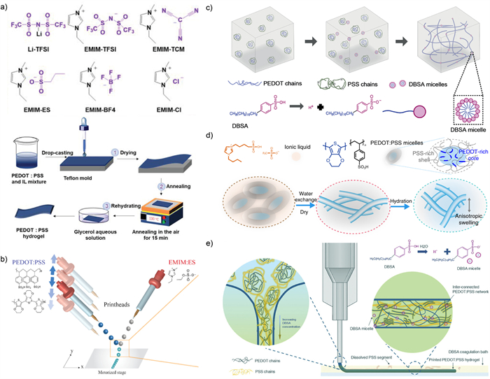
Dodecylbenzenesulfonic acid (DBSA) is an anionic surfactant with sulfonic acid groups, and its good water solubility makes it widely used in the doping of PEDOT:PSS by many research groups. High concentrations of DBSA can also form PEDOT:PSS hydrogels at room temperature. When the concentration of DBSA exceeds the critical micelle concentration, DBSA forms stable dispersed micelles in the solution, with hydrophobic groups (carbon chains) oriented inward and hydrophilic groups (-SO3−) outward in water. This weakens the electrostatic attraction between PEDOT+ and PSS−. Simultaneously, the partial protonation of PSS− weakens this electrostatic attraction, exposing PEDOT chains that reorganize with DBSA micelles. Thus, physical cross-linking is achieved through π-π stacking and hydrophobic interactions between PEDOT chains. Zhang et al. [9] used a syringe to inject a PEDOT:PSS suspension containing DBSA into a cavity by puncturing soft tissue at room temperature, leading to spontaneous hydrogel formation in about 10 min (Fig. 3c). Feig et al. employed the ionic liquid 4-(3-butyl-1-imidazolio)-1-butanesulfonic acid triflate to establish strong electrostatic interactions with PEDOT:PSS. Its gel phase boundaries was surpassed and PEDOT:PSS hydrogels were successfully formed [31]. Similarly, Liu et al. [32] utilized the same method to fabricate conductive hydrogels for the production of hydrogel-based microelectronic devices (Fig. 3d). Zheng et al. [33] developed a coagulation-bath assisted direct ink writing (DIW) method for 3D printable PEDOT:PSS hydrogels using a liquid-gel transition mechanism (Fig. 3e). They extruded PEDOT:PSS suspension into a coagulation bath with an appropriate DBSA concentration. This enables the fabrication of patterned PEDOT:PSS hydrogels. Li et al. [34] added PEDOT:PSS liquid onto filter paper, followed by the addition of DBSA, resulting in the formation of porous hydrogels on the paper’s surface. It is noteworthy that the —SO3H groups on PSS can form hydrogen bonds with paper fibers. In results, interaction is enhanced between PEDOT:PSS hydrogels and the paper substrate.
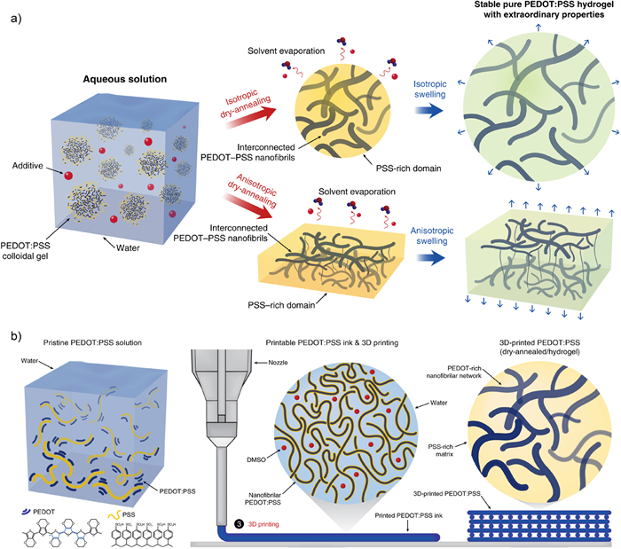
A polar molecule can be described as a molecule with a slight positive charge at one end and a slight negative charge at the other end. Typically, they exhibit an asymmetrical shape, leading to an uneven distribution of electrons. Theoretically, substances with high polarity could mitigate the electrostatic interactions between PEDOT and PSS, leading to the phase separation and gelation. Dimethyl sulfoxide (DMSO) is a high-boiling-point and highly polar organic solvent. DMSO is commonly used as a dopant to gel PEDOT:PSS. Lu et al. [8] prepared high-performance pure PEDOT:PSS hydrogels by mixing the volatile additive DMSO into the aqueous PEDOT:PSS solution, followed by controlled dry-annealing and rehydration (Fig. 4a). Yuk et al. [15] developed a paste-like conductive polymer ink for 3D printing. In this process, PEDOT:PSS solution cryogenically frozen, then lyophilized and controlled redispersed in a mixture of water and DMSO. This product enables the manufacturing of highly resolution (over 30 µm), high-aspect-ratio (over 20 layers) and highly repeatable conductive polymers (Fig. 4b).
Incorporating polymer monomers and oxidizing initiators into the aqueous PEDOT:PSS solution can form the network-structured PEDOT:PSS composite hydrogels. For example, pyrrole (Py) is a commonly used conductive polymer monomer. Yang et al. [35] mixed conductive polymer monomers (pyrrole, aniline, indole-4-NH2, indole-5-NH2, indole-6-NH2, indole-7-NH2 etc.) with aqueous PEDOT:PSS solution, and then added oxidation initiators (such as (NH4)2S2O8, NaIO4 and FeCl3) to polymerize the monomera, obtaining conductive polymer/PEDOT:PSS hydrogels within minutes (Fig. 5a). By in situ doping of PEDOT:PSS in PNAGA-PAAM hydrogels, Wu et al. [36] prepared a robust and highly stretchable supramolecular polymer conductive hydrogels, which featured double amide hydrogen bonding (Fig. 5b). Ren et al. [37] utilized the electrostatic interaction between the —SO3− group of PSS and the positively charged PPy to quickly mix PEDOT:PSS, Py monomers and APS. This work successfully produced PPy-PEDOT:PSS hybrid hydrogels with high conductivity (>867 S/m), excellent mechanical properties and a multi-level porous nanostructure.
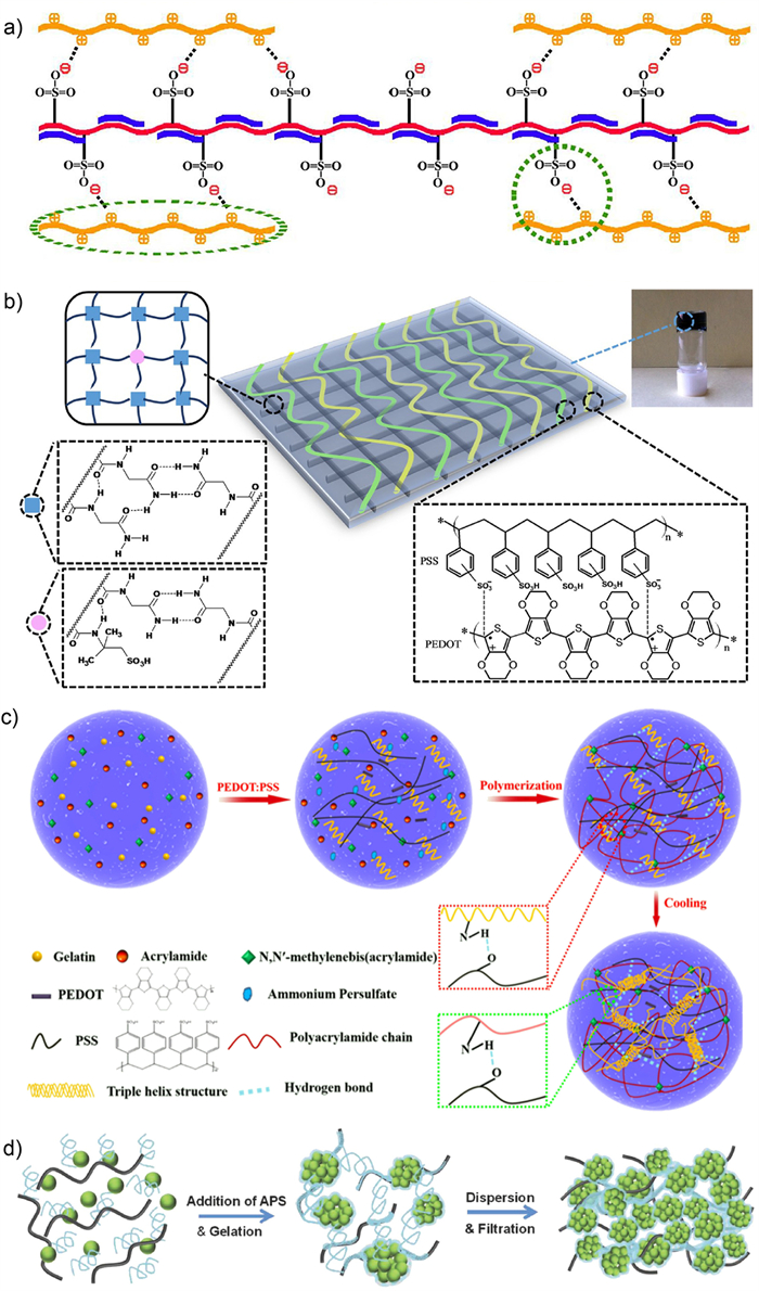
Aside from pyrrole, acrylamide (AAm) is also a commonly used monomer material in the preparation of conductive hydrogels. Aouada et al. [38] used sodium persulfate as the initiator to polymerized and crosslinked AAm and MBAAm (N,N’-methylenebisacrylamide) in aqueous PEDOT:PSS solution. The resulting crosslinked PAAm with intertwined PEDOT:PSS showed excellent mechanical properties and electrochemical stability. Similarly, Sun et al. [39] prepared double network composite hydrogels by adding the initiator ammonium persulfate (APS) to a mixture solution of AAm monomers, chemical crosslinker MBAAm and PEDOT:PSS (Fig. 5c). Yang et al. [40] prepared PEDOT:PSS composite hydrogels using aniline as the monomer and investigated their formation mechanism in detail. The researchers first prepared PANi dispersion by mixing aniline and phytic acid with ammonium persulfate, and then added the PANi dispersion into the PEDOT:PSS dispersion. This resulted in PEDOT/PANi hydrogels with significantly improved capacitance (112.6 F/g at 5 mV/s) and high compressive strength (41.6 kPa). Therefore, they proposed the formation mechanism of the PEDOT/PANi hydrogels. On one hand, phytic acid can provide a molecular bridge for PANi and PEDOT; on the other hand, acid induces partial removal of PSS and the conformational change of conjugated PEDOT chains from benzoid structure (coil) to quinoid structure (extended coil). As a result, the electrostatic repulsions between PSS chains are reduced and the π-π stacking interactions are enhanced.
In this approach, an initial polymer network is prepared first. Then it serves as a template for the in-situ polymerization of the second PEDOT:PSS polymer network. Through these steps, PEDOT:PSS composite hydrogels featuring dual or multiple networks are obtained. These composite hydrogels are often accompanied by good mechanical strength. Dai et al. [13] developed a special double network structure consisting of PAAm hydrogels as the first network and PEDOT:PSS semi-interpenetrating polymer network (semi-IPN) as the second network. The obtained hydrogels possess both mechanical toughness and electrical activity. In addition, they achieved a three-network structure by sequentially immersing the prepared PAAm double network hydrogels in EDOT, NaPSS, and Fe(NO3)3·9H2O. This PAAm/PEDOT:PSS three-network hydrogels not only maintained the excellent electrochemical properties of PEDOT:PSS hydrogels, but also exhibited superior mechanical strength(compressive fracture stress as high as 1.8 MPa) [41]. Naficy et al. [42] synthesized a tough and highly conductive PPEGMA1100-PAAM-PEDOT (PSS) hydrogels by polymerizing EDOT-PSS in a PPEGMA-PAAM double network hydrogels. Here, PPEGMA served as the first network and PAAm as the second network. Yan et al. [43] first reacted PVA with PSSH to produce PSS/PVA hydrogel films, and subsequently electropolymerized EDOT monomer in the PSS/PVA scaffold. The obtained PEDOT/PSS/PVA hydrogels film exhibit improved electrochemical stability and superior mechanical properties.
It is noteworthy that this method can also be applied in reverse order. We can firstly prepare the PEDOT:PSS hydrogels followed by doping with another polymer. Feig et al. [19] synthesized PEDOT:PSS hydrogels by initially using ionic liquid as an inducer. Subsequently, the gels were soaked in an aqueous solution of acrylic acid, bisacrylamide, and a thermal radical polymerization initiator. This resulted in the formation of two interpenetrating hydrogel networks, with polyacrylic acid serving as the secondary polymer network. The hydrogels exhibited dual electronic and ionic conductivity, as well as highly tunable mechanical properties that mimic biological tissue.
In addition to the use of organic polymers, inorganic materials can also serve as precursor templates for in situ polymerization. Chen et al. [44] added a gelator (such as APS) to a aqueous PEDOT:PSS solution doped with carbon nanotubes (CNTs). Whin approximately 5 min, they obtained a three-dimensionally interconnected hydrogel network composed of CNTs and PEDOT:PSS. This hydrogel network can be utilized for the fabrication of high-performance flexible lithium-ion battery electrodes (Fig. 5d).
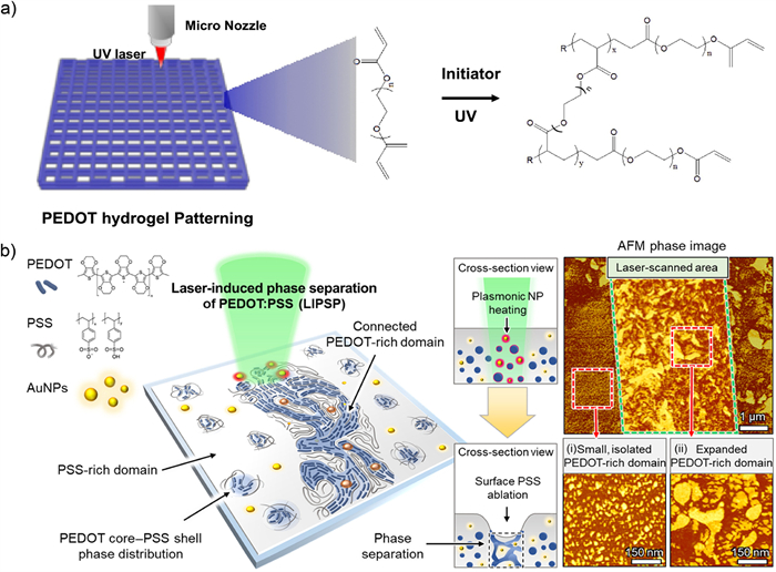
In recent years, several new methods have been developed for the formation of PEDOT:PSS hydrogels. Photo-induced gelation is one of the most popular gelation methods nowadays [45]. Heo et al. [46] dissolved PEDOT:PSS solid in a mixed solution of distilled water and ethylene glycol (8:1) and added polyethylene glycol diacrylate (PEGDA) containing 0.5 wt% photoinitiator (BAPO). Then they prepared photocrosslinked hydrogels through UV irradiation (Fig. 6a). Won et al. [47] employed the photothermal energy provided by ultrafast lasers and a strong electric field to induce selective phase separation of PEDOT:PSS (Fig. 6b). Photothermal energy caused local instability in the PEDOT and PSS biphasic system, and PEDOT and PSS separated under the influence of a strong electric field. After rapid cooling to room temperature, PEDOT:PSS was fixed in a separated state. Following laser-induced phase separation, the PEDOT-rich domains expanded and interconnected to form a robust network. This enhances both electrical conductivity and water stability and transforming PEDOT:PSS into water-stable hydrogels.
Owing to its impressive electrical conductivity, PEDOT:PSS has garnered substantial research attention in the realm of bioelectronics over the past three decades. To expand its application horizons, researchers have explored various conductive materials to enhance the conductivity of PEDOT:PSS. Noteworthy among these materials are inorganic options, including carbon materials such as graphene, carbon nanotubes (CNT), MXene, disulfides, nitrogen-based materials, metal-organic frameworks (MOFs), and nanomaterials like meta nanoparticles and silver nanowires. Furthermore, the Nobel Prize in Chemistry awarded to Hideki Shirakawa, Alan MacDiarmid and Alan Heeger in 2000 heightened global awareness of polyacetylene, sparking widespread interest in organic conjugated conductive polymer materials. In recent years, polymers like polypyrrole, polyaniline, polythiophene and their derivatives have gained prominence due to their metallic conductivity and versatility. Some research groups have ventured into doping PEDOT:PSS with these polymers, aiming to develop composite materials with enhanced performance. This strategic integration of diverse conductive materials underscores the ongoing efforts to optimize the conductivity of PEDOT:PSS and explore novel avenues for its application in bioelectronics.
An alternative approach to enhance conductivity involves modifying the inherent structure of PEDOT:PSS itself. The primary conductive mechanism in PEDOT:PSS is attributed to charge hopping among the PEDOT chains [48]. By strategically weakening the electrostatic interaction between PEDOT and PSS, a fraction of the PSS component is removed, inducing a transition in the structure of PEDOT from a confined-coiled to an expanded-linear conformation [9]. This transformation results in the creation of additional PEDOT-rich domains and reinforces the π-π stacking interactions between PEDOT chains. Consequently, the enhancement of charge hopping dynamics among PEDOT chains leads to improved conductivity. This structural refinement represents a nuanced strategy for optimizing the electrical properties of PEDOT:PSS. Conductivity range of hydrogels regulated by different substances is shown in Fig. 7 and Table S1 (Supporting information).
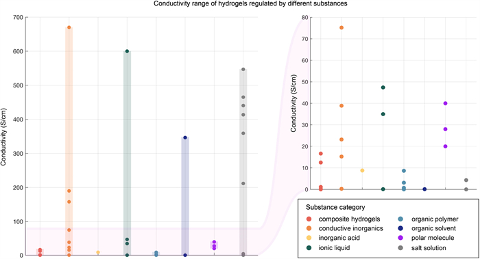
Doping inorganic conductive fillers is an important strategy to improve conductivity. Carbon-based fillers, such as CNTs and graphene, are wildly used inorganic conductive materials. Reduced graphene oxide (rGO) is a 2D material featured a large specific surface area, high chemical stability and carrier mobility. Taking advantage of the high conductivity of rGO, Kıranşan et al. [17] prepared a PEDOT:PSS/rGO composite sponge material with excellent sensing performance in hybrid DNA detection. The research showed that conductivity improves as the PEDOT:PSS content in the composite material increases. The one with the highest conductivity is PEDOT:PSS-10/rGO (190 S/cm) prepared with 10 mL PEDOT:PSS. To solve the problem of limited conductivity of PEDOT:PSS hydrogels at high water content, Liu et al. [18] used Ti3C2-MXene to develop an aqueous PEDOT:PSS ink with excellent 3D printing performance. Ti3C2-MXene is an emerging 2D transition metal carbide with both metallic conductivity and hydrophilicity due to its surface terminations (mainly —OH and —F). The study found that without the addition of MXene, the pure PEDOT:PSS hydrogels has a conductivity of 6.609 S/cm. And with the addition of MXene, the conductivity increases to 15.258 S/cm at a water content as high as 96.6 wt%. This material can be manufactured precisely and reproducibly via 3D printing, opening up new opportunities in the manufacturing of wearable and implantable devices. Besides, metal nanoparticles have been used more and more widely for its good performance in recent years. Won et al. [47] mixed AuNP ink with PEDOT:PSS and obtained PEDOT:PSS hydrogels through laser treatment. Its conductivity reaches a maximum of 670 S/cm, which greatly improves the electrical conductivity of PEDOT:PSS hydrogels.
Forming composite materials with organic conductive polymers can also improve conductivity. The derivatives of polypyrrole (PPy) and polyaniline (PANi) are commonly used organic conductive polymers nowadays. Ren et al. [37] prepared PPy-PEDOT:PSS hybrid hydrogels with a conductivity up to 8.67 S/cm. The interconnected conductive network formed between PPy and PEDOT:PSS can largely facilitate charge transport between hybrid hydrogels. Shih et al. [49] prepared PEDOT:PSS/PVA/PMAA composite hydrogels, which can be used as electrode material for stretchable supercapacitors. Its conductivity can be increased from 0.6 S/cm to 3.2 S/cm after water absorption (22.3 wt%).
Ionic liquids possess the capability to establish robust electrostatic interactions with PEDOT:PSS, inducing a phase separation between PEDOT and PSS chains. Part of the PSS is replaced by smaller anions, resulting in larger PEDOT-rich domains and thereby improves conductivity. Wang et al. [29] used six ionic liquids (Li-TFSI, EMIM-TFSI, EMIM-TCM, EMIM-ES, EMIM-BF4, EMIM-Cl) and glycerin to prepare PEDOT:PSS hydrogels, and obtained the maximum conductivity PEDOT:PSS/EMIM-TFSI hydrogels at 305.78 ± 36.18 S/cm. Liu et al. [32] used ionic liquid 4-(3-butyl-1-imidazolio)-1-butanesulfonic acid triflate to prepare ion gel film, followed by removing the added ionic liquid with water to obtain PEDOT:PSS hydrogels. Its electrical conductivity can reach 47.4 ± 1.2 S/cm. The research showed that part of the PSS is removed after washing, reducing the proportion of insulating polymers and improving the connectivity of the PEDOT polymer network, which together contribute to the high conductivity of the hydrogels. Zheng et al. [33] used a 3D-printing method to extrude the PEDOT:PSS suspension onto a coagulation bath containing DBSA. After dry-annealing, a high conductivity PEDOT:PSS hydrogels (~600 S/cm) is prepared. This method provides a new idea for patterned manufacturing of bioelectronic devices
Polar molecules, such as glycerol and DMSO, are frequently employed to enhance conductivity. It helps PEDOT form a highly ordered nanofiber structure and reduce the distance of π-π stacking. Research by Wang et al. [29] showed that due to the solvent effect, the conductivity of PEDOT:PSS/IL hydrogels increases as the concentration of the polar solvent glycerol increases. When the glycerol concentration increases from 0 to 90%, the conductivity of PEDOT:PSS/EMIM-TFSI increases from 94.28 ± 12.15 S/cm to 305.78 ± 36.18 S/cm. Lu et al. [8] doped DMSO into aqueous PEDOT:PSS solution, followed by controlled dry-annealing and rehydration. The resulting pure PEDOT:PSS hydrogels exhibits high conductivity up to 40 S/cm in DI water.
In salt solutions, ion exchange between metal cations and PEDOT:PSS plays an important role in promoting phase separation in hydrogels. However, the reason for the improvement in conductivity is still controversial, because it may also come from the conductivity of high concentrated salt solution itself. Besides, the chemical bond between metal cations and PSS is relatively weak, and the system is not tightly connected. Wang et al. [10] added metal halides (InCl3, CuCl2, NiCl2, MgCl2, NaCl, LiCl) to aqueous PEDOT:PSS solution containing DMSO, and obtained a series of PEDOT:PSS/MH hydrogels with ultra-high conductivity. The study found that the electrical conductivity increases with the increase of metal halides concentration. Besides, the hydrogels treated with metal halides of higher cationic charge has a greater maximum electrical conductivity, among which the PEDOT:PSS-InCl3 hydrogels exhibit the largest electrical conductivity (547 S/cm).
Hydrogels of superior mechanical strength promote fire-new applications in various fields. But high water content during the gelation often limits the toughness and stability of hydrogels. In response to the flawless, researchers adopted reformative gelation methods to improve hydrogel strength and toughness. The formation of composite hydrogels is also an advantageous approach. In previous studies, researchers make contributions to the double network/multi-network hydrogels, achieving highlights of mechanical strength enhancement [50]. Double network hydrogels are composed of two intertwined networks and multi-network hydrogels consist of more than two networks [51,52]. Compared with hydrogels with single network, double network hydrogels and multi-network hydrogels exhibit high toughness and stability [53,54]. In some circumstances, there are interactions between polymer chains of the two networks. Non-covalent interactions, such as hydrogen bond, are often reversible and more easily broken than covalent interactions, making the hydrogels self-healing [55]. Covalent interactions are more stable and irreversible. Hydrogels with covalent interactions are chemically cross-linked, which contributes to superior mechanical strength [56]. Double network/multi-network hydrogels with interactions between different polymers are called interpenetrating network hydrogels (IPN) [57,58]. It is worth noting that the polymers in interpenetrating network hydrogels are cross-linking [56]. If the linear monomer or polymer is diffused in other cross-linked polymers, the hydrogels are called semi-interpenetrating hydrogels [38,59]. In the previous studies, the mechanical strength modification of PEDOT:PSS hydrogels improves evidently with various experimental strategy. This section emphatically introduces the mechanism of improving the PEDOT:PSS hydrogels mechanical properties at the molecular level and the latest research progress [53,56].
Hydrogen bond is a category of non-covalent cross-linking. A hydrogen atom that is covalently bound to a high electronegative atom will form a hydrogen bond when it is close to a high electronegative atom with a small radius. Compared with covalent bond, hydrogen bond is dynamically reversible [60]. The bond energy of hydrogen bond mostly ranges from 25 kJ/mol to 40 kJ/mol, much less than covalent bonds. The reversibility associates with the self-healing capacity of hydrogels [61-63]. PVA/PEDOT:PSS composite hydrogels exhibit excellent tensile and compression properties. The networks consist of crosslinking PVA and linear PEDOT:PSS. Addition of PEDOT:PSS evidently improves the electroconductivity for the potential application for supercapacitor. The hydrogen bond between PEDOT:PSS and PVA contributes to the mechanical strength of composite hydrogels [64-66]. PNAGA-PAMPS/PEDOT/PSS composite hydrogels demonstrate high toughness and stretchability as a result of dual amide hydrogen bond [36]. Composite hydrogels composed of poly(2-vinyl-4,6-diamino-1,3,5-triazine-co-polyethylene glycol diacrylates) (PVDT-PEGDA) hydrogels with PEDOT/PSS demonstrate satisfying compressive strength and tensile strength by hydrogen bond between molecules [67]. In virtue of multiple hydrogen bonding, γ-PGA/PEDOT:PSS conductive hydrogels demonstrate high adhesion capacity and self-healing, as well as high mechanical strength [68]. Alginate/PEDOT:PSS hydrogels enhance mechanical strength through hydrogen bonds and hydrophobic forces. Hydrogen bond enhances the interactions between different polymer chains and bestow hydrogels an efficient energy-consuming mechanism [69]. So the composite hydrogels with hydrogen bonds exhibits excellent mechanical properties.
Hydrogels based on covalent bonds exhibit stable molecular structure and high toughness. In the system of PEDOT:PSS composite hydrogels, covalent bonds tend to form between materials other than PEDOT:PSS according to the molecular structure. Existence of covalent bonds obviously enhances the mechanical toughness of the materials, but the hydrogels are often not self-healing. Zhu et al. [52] demonstrated P(AMPS-Na)/PAM/PEDOT:PSS hydrogels. Covalent bonds evidently promote the mechanical strength and addition of PEDOT:PSS builds up the conductivity. Chen et al. [70] combined covalent crosslinking networks and PEDOT:PSS hydrogels to form composite materials. Hydrogels with high toughness makes up for the disadvantage of brittle hydrogels, pave the way to future application in more fields.
Biocompatibility is crucial for the application of hydrogels in the biomedical field. Combined with substances of well-known biocompatibility, composite PEODT:PSS hydrogels exhibit satisfying biocompatibility. Zeng et al. [71] utilized hyaluronic acid, dopamine and PEDOT:PSS to synthesize particle-free hydrogels, which can be used in biosensing safely. PVA/PEDOT:PSS composite hydrogels also exhibit good biocompatibility as well as sensitive to strain paving the way for further utilization potentiality in biosensing [72,73].
In recent decades, PEDOT:PSS hydrogels have garnered substantial attention. Composite PEDOT:PSS hydrogels exhibit remarkable electrical conductivity and mechanical toughness. At the cellular level, PEDOT:PSS composite hydrogels demonstrate the capability to facilitate cell adhesion, diffusion and proliferation across different tissues types. On a macro level, as a biointerface, PEDOT:PSS composite hydrogels ensure the high fidelity of signal transmission and harmless to organisms. The advancement presents a novel avenue for the potential repair of internal organs in humans. This section succinctly outlines several applications of PEDOT:PSS hydrogels, particularly in the realms of sensors and tissue engineering.
Sensor are designed to monitor specific indicators, converting detected parameters into electrical signals or other requisite forms for real-time monitoring. In this review, we classify the sensors into physical sensors, chemical sensors and biosensors according to the properties of target analyst. Physical sensors detect physical quantities, such as strain, polarized light, electricity, utilizing specific physical effects to convert measured parameters into easily quantifiable electrical signals or other forms [74]. Typically, there exists a distinct functional relationship between the output signal and the input signal. Chemical sensors, on the other hand, exhibit sensitivity to concentration, monitoring chemical substance concentrations by transforming them into precise electrical signals. Biosensor, a subset of biological sensors, comprise a biological sensing element and a signal transducer. The exceptional charge transfer properties and strain sensitivity of PEDOT:PSS composite hydrogels render them well-suited for sensor applications [75-78]. The unique characteristics of PEDOT:PSS hydrogels make them capable of contributing significantly to the field of sensors.
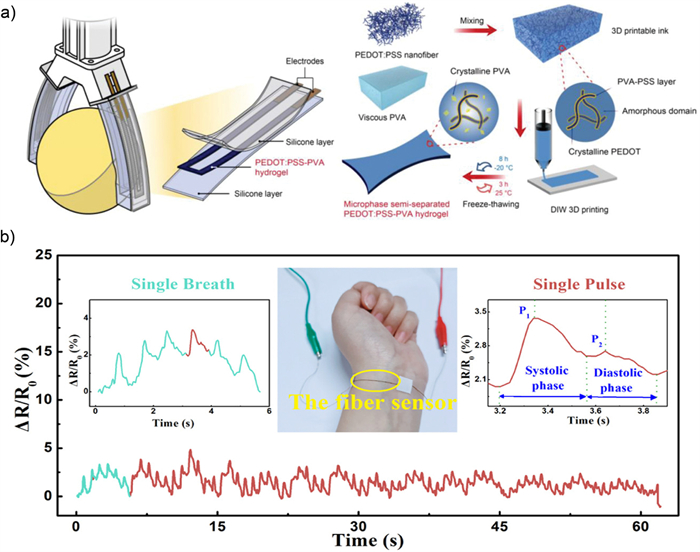
Numerous studies have concentrated on the monitoring human body movements using composite hydrogels containing PEDOT:PSS, encompassing activities such as swallowing, joint movement, respiratory movement. encompassing activities such as the performance of motion monitoring sensors. Zhang et al. [79] constructed a composite hydrogel containing polyacrylamide (PAM), lithium chloride and PEDOT:PSS coated cellulose nanofibril (CNF). The addition of CNF makes it possible to increase the formation of hydrogen bond. The strength of the hydrogen bond enhances the conductivity and mechanical strength of the composite hydrogel. The use of lithium oxide as an additive material can enhance the interaction between the colloidal phase and water molecules, which improves the freezing resistance of the sensor without influencing its accuracy and sensitivity for motion monitoring. Cao et al. [64] synthesized PEDOT:PSS-PVA composite hydrogel as a strain sensor, which proved to have good self-healing property. The properties of PEDOT: PSS-PVA composite hydrogel can be further improved by chemical treatment. Prameswati et al. [80] treated PEDOT:PSS-PVA composite hydrogel with NaCl, and the end-product has good mechanical properties and sensitivity. Shen et al. [72] demonstrated PVA/PEDOT:PSS composite hydrogels which can measure multiple physiological signals (Fig. 8a). Shi et al. [73] increased the resistance of PEDOT:PSS-PVA composite hydrogel by adding glycerin, and the sensor proves to have high sensitivity and strong resistance even under low temperature conditions (Fig. 8b). Xu et al. [81] used hydroxypropyl methylcellulose (HPMC), poly(3,4-vinyldioxythiophene), PEDOT:PSS and PAM to enhance the tensile property of the hydrogel, the composite hydrogel has strong sensitivity and low hysteresial, and can be used to monitor the intense strenuous exercise components of human activities and fine activities. For humidity, Bian et al. [82] used carboxycellulose nanofibers (CCNF) to distribute the opaque conductive PEDOT:PSS into the cross-linked polyacrylamide network, which enables the sensor to have sensitive humidity response.
Li et al. [34] used porous PEDOT-PSS hydrogels to monitor the concentration of glucose in sweat during exercise in real time (Fig. 9a). Xu et al. [59] interwoven PEDOT:PSS conductive polymer with amphoteric ionic PSBMA network to prepare a new PSBMA/PEDOT:PSS semi-interpenetrating hydrogel, which can accurately determine the tryptophan content (Trp) in sweat. Serafini et al. [83] synthesized a PEDOT:PSS/IrOx Ps/hydrogel sensor on a flexible plastic foil, inventing a wearable sensor capable of monitoring ammonia at room temperature. This type of sensor can be used for real-time oxygen detection. Xu et al. [84] developed a microfluidic-based wearable electrochemical sensor for durable and precise detection of UA (uric acid) in sweat by integrating a conductive PEDOT:PSS hydrogel with a flexible CPE (Fig. 9b). Odinotski et al. [85] utilized hydrogel forming microneedles to realize the pH measurement (Figs. 9c and d). Decataldo et al. [86] made hydrogel-based organic electrochemical transistors (OECTs) with PEDOT:PSS, which in turn synthesize wearable sensors.
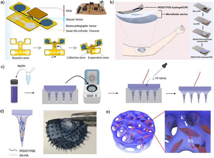
The biosensors synthesized by using PEDOT-PSS hydrogel as a component can be used to monitor the secretion of dopamine in PC12 cells. Ren et al. [37] prepared composite hydrogel by mixing the pyrrole monomer with PEDOT:PSS hydrogels. By adjusting the ratio of raw materials, the obtained material has high conductivity and good biocompatibility. The composite hydrogel is verified to be highly sensitive to monitoring the dopamine secretion of PC12 cells, and achieve the expected results in both enhancement and inhibition of the cell secretion. On the basis of the same cellular secretory system, Yang et al. [87] compared several conductive materials that can form hybrid hydrogel with PEDOT:PSS (including pyrrole, aniline, indole, 4-aminoindole, 5-aminoindole, 6-aminoindole, 7-aminoindole) (Fig. 9e). After measuring the conductivity and three-dimensional structure of different materials, the study finally selects the best-performing 5-aminoindole as the conductive polymer monomer. They synthesize an AuNPs/PEDOT:PSS/PIn-5-NH2/ITO electrode which proves to have good biocompatibility, high sensitivity for monitoring the dopamine secretion of PC12 cells and strong anti-interference ability. In addition to cellular level research, PEDOT:PSS is also feasible for sensing and monitoring of human macro biological indicators. Li et al. [34] used porous PEDOT-PSS hydrogel to assemble hydrogel paper patches on paper fibers, which can simultaneously monitor electrocardiogram (ECG) signals in real time (Fig. 9a). Lee et al. [88] used alginate/PEDOT:PSS conductive hydrogel to realize conformal brain interface and monitoring of cortical electrical layer in rodent models. Lee et al. [89] added natural crosslinking agent and PEDOT:PSS to gelatin hydrogel, enhancing the conductivity and mechanical properties of the composite hydrogel by adjusting the proportion of each component, constructing a porous hydrogel electrode to realize an ECG sensor with little skin irritation and good compatibility.
In the previous studies, scholars utilize hydrogel three-dimensional structure to observe the growth of inorganic substance expertly. It lays a foundation for the further use of hydrogel networks. Building on the researchers’ previous work, tissue engineering in hydrogel systems is becoming possible. Tissue engineering integrates cytobiology and materials science. Researchers culture cells derived from different tissues or organs in vitro and then mix cells and materials with good biocompatibility and 3D structure adapted to the tissue [16,90]. The cells adhere to the scaffold to form cell-material compound. The cell-material compound is planted in the injured parts to achieve tissue repair. So the materials need to be free of cytotoxicity and biodegradable in vivo. In humans, the regenerative capacity of individual organs varies greatly. The liver can revive as before in the 70% liver cut model. But cardiac and neural tissue has poor regenerative capacity in adults. So when myocardial damage or nervous disorder occurs, such as Alzheimer’s and arrhythmia, the diseases are usually devastating. Tissue engineering provides refractory disease with a feasible therapy. PEDOT:PSS composite hydrogels have been confirmed with good biocompatibility. Through electron microscope, the hydrogels have three-dimensional porous network structure similar to extracellular matrix. These characteristics make the application of PEDOT:PSS composite hydrogels in tissue engineering probable.
Cardiac tissue engineering promotes artificial repairment of the heart. This provides possibility for the treatment of myocardial infarction. Myocardial infarction is one of the fatal diseases in the cardiovascular field. Because the necrotic cardiomyocytes are replaced by fibrous scars instead of newborn myocardial cells during healing. The fibrous scars relate coordination of the heart beating, increasing the risk of ventricular remodeling [91-93]. The properties of PEDOT:PSS composite hydrogel provide a new idea for the treatment of heart disease [94,95]. Xue et al. [96] demonstrated conducting polymer hydrogel coated conventional metallic bioelectrodes to reduce the pacing threshold voltage (Figs. 10a–c). Casella et al. [97,98] demonstrated that conductive hydrogels without cell-binding sites promote cell adhesion compared with non-conductive hydrogels. In the PEDOT:PSS composite hydrogels system, the proper proportion of PEDOT:PSS mainly improves the conductivity, enhancing the adhesion and multiplication of cells. Testore et al. [99] prepared photo-crosslinked electroconductive hydrogels containing PEDOT:PSS with good biocompatibility and biodegradability (Fig. 10d). In vitro tests demonstrate the composite hydrogels promote human cardiac fibroblasts adhesion and potential use in cardiac tissue engineering. Han et al. [20] prepared PEDOT:PSS hydrogels based biointerfaces. The results demonstrate that the beating of the heart can be photo-controlled via this optoelectronic biointerfaces suggesting new method of cardiac implants. Roshanbinfar et al. [100] developed a biohybird hydrogel containing PEDOT:PSS. The hydrogel systems mimic the 3D structure of extracellular matrix and exhibit satisfying electrical coupling. Notably the addition of PEDOT:PSS improves the conductivity of rat cardiomyocytes and the biohybird hydrogels promote cell maturation. It is a progressive step to cure arrhythmia by using bioconductive materials.
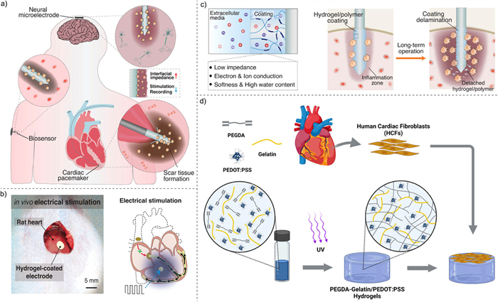
In the field of neurobiology, overcoming the difficulties of nerve tissue regeneration will solve the problem of incurable neurodegenerative diseases to a great extent. Neurodegenerative diseases and demyelinating diseases leave a very negative impact on the quality of life of older people. At present, doctors mainly use symptomatic treatment and neurotrophic factors to control patients’ symptoms. However, it is still difficult to fundamentally repair the function of neurons. Tissue engineering makes it possible to repair injury in the nervous system. Chik et al. [101] developed a flat neural probe based on a highly flexible microelectrode array. The electrodeposited hydrogels containing PEDOT:PSS onto the array lower impedance. In vivo tests demonstrate enhancement activity of hippocampus neuron in a long term. Furlani et al. [102] developed a new injectable electroconductive hydrogels containing PEDOT:PSS which improve the conductivity and mechanical strength. The developed hydrogels promote adhesion and growth of primary rat cortical astrocytes. Javadi et al. [103] used polyether based polyurethane (PU), PEDOT:PSS and liquid crystal graphene oxide (LCGO) to form biocompatible conductive hard hydrogels, which can support the differentiation of human neural stem cells into neurons and support glial cells. Ferlauto et al. [104] invented a conductive hydrogel microelectrode composed of PEDOT:PSS and alginate to achieve the recording of neuronal electrical signals. Han et al. introduced an integrated photoelectric biological interface synthesized by PEDOT:PSS and other materials, which can stimulate a single hippocampal neuron through safe and stable current. This mechanism suggests potential application in neurodegenerative diseases [20].
In recent decades, notable strides have been made in the synthesis and modification of PEDOT:PSS hydrogels. With their inherent advantages of portability, high conductivity, and biocompatibility, PEDOT:PSS hydrogels have emerged as compelling biointerface materials for applications in sensors and tissue engineering.
This paper comprehensively reviews the gelation methods, modification techniques, and diverse applications of PEDOT:PSS hydrogels. Initially, we elucidate the principle underlying the gelation of PEDOT:PSS hydrogels, detailing the transformation of PEDOT:PSS aqueous solution into hydrogels through phase separation. Subsequently, we provide an overview of various gelation methods employed in PEDOT:PSS hydrogels synthesis, highlighting the distinctive characteristics of the resulting products. This review subsequently enumerates diverse modification approaches for PEDOT:PSS hydrogels, considering their impact on electrical conductivity and mechanical strength. Comprehensive charts are constructed to facilitate a comparative analysis of hydrogel performance across the aforementioned studies. Finally, the paper delves into the practical applications of PEDOT:PSS hydrogels in sensors and tissue engineering. Concluding the discussion, we offer summaries and outlooks for further research in this dynamic field.
In terms of the preparation of PEDOT:PSS hydrogels in biomedical applications, the following recommendations are summarized.
(1) Operational convenience: Using mild and convenient conditions is an ideal strategy for gelation and modification. Conditions such as high temperature, high pressure and extremely low temperature will pose great challenges to the processing of PEDOT:PSS. Methods conducted at room temperature and normal pressure, such as electrochemical gelation, are preferred for their practicality and ease of implementation. Additionally, the utilization of volatile substances like DMSO can streamline the gelation process, facilitating the acquisition of purer products.
(2) High-resolution and patterning: Patterning PEDOT:PSS conductive inks to fabricate functional components has demonstrated escalating potential across diverse fields, including neural implantable devices, high-performance sensors and soft robotics. As 3D printing technology advances, an array of methods such as stereolithography and direct ink writing have emerged, enabling the fabrication of PEDOT:PSS hydrogels into high-resolution and precisely patterned devices.
In the realm of medicine, the future trajectory of PEDOT:PSS hydrogels revolves around achieving controllable performance to adapt to diverse biological tissues. This adaptability is facilitated through the utilization of various substances, rendering PEDOT:PSS hydrogels as on-demand and adjustable biomaterials. Consequently, the modification and regulation strategies of PEDOT:PSS hydrogels are poised to focus on the following critical aspects.
(1) Biocompatibility: Biocompatibility is paramount, ensuring stable interaction with biointerfaces and non-toxicity to organisms. Enhancing biocompatibility can be achieved through cross-linking with bio-based materials such as organics, biomacromolecules. This approach enhances utility across applications ranging from the cellular interactions to organ implants. However, caution is warranted with substances like ionic liquids and certain nanoparticles due to their potential toxicity, while strong acids may induce skin corrosion, thereby limiting their application in bioelectronic devices.
(2) Stretchability: The intrinsic chemical structure of PEDOT:PSS hydrogels offers avenues for mechanical modification. Enhancing stretchability at the molecular level can be achieved through the introduction of hydrogen bonds or covalent bonds. Additionally, cross-linking PEDOT:PSS hydrogels with other polymers to form multi-network structures enhance their mechanical properties. Given the wide variation in Young’s modulus across different tissues, aligning materials to match known modulus values and optimizing ratios allows for better control of mechanical strength, tailored to specific tissue requirements.
(3) Conductivity: The primary conductive mechanism in PEDOT:PSS involves charge hopping among PEDOT chains. Strengthening conductivity entails weakening the electrostatic interaction between PEDOT and PSS through doping with other substances, reinforcing π-π stacking interactions between PEDOT chains and improve the conductivity. Incorporating other conductive materials into PEDOT:PSS hydrogels also offers a viable strategy for enhancing conductivity. However, the selection of doped materials must adhere to biocompatibility and mechanical strength criteria specific to intended application scenarios.
Through meticulous design and thoughtful material selection, PEDOT:PSS hydrogels hold the promise of evolving into an ideal material with desirable electrical, mechanical and biological properties.
However, despite notable advancements, several key challenges persist, hindering their practical application.
(1) Enhanced biotissue-like characteristics: While PEDOT:PSS composite hydrogels demonstrate favorable conductivity and mechanical strength, there is a pressing need for additional modification methods to enhance biocompatibility. This is crucial to meet the stringent biosecurity requirements in applications such as biosensors and tissue engineering.
(2) Long-term stability: Hydrogels face challenges related to decreased adhesion and structural integrity over prolonged usage, impeding their practical utility. Addressing issues such as water evaporation prevention and developing composite conductive hydrogels with enhanced fatigue resistance and self-healing capabilities is imperative to overcome these limitations.
(3) Improved electrical conductivity: Further research is warranted to enhance the conductivity of hydrogels, as high conductivity is essential for ensuring high-fidelity signal transmission. Refinement of modification methods is necessary to enhance electronic transmission fidelity and electron transport efficiency.
(4) Comprehensive understanding of gelation mechanism: The structure and molecular interactions within double-network or multiple-network hydrogels remains poorly understood. In-depth exploration of the gelation mechanism is vital for discerning how to select additives tailored to specific application scenarios.
Undeniably, PEDOT:PSS hydrogels demonstrate immense potential in applications such as sensors and tissue engineering. Given the continuous evolution of materials science and chemistry, more researches on higher electrical conductivity, good biocompatibility and more stable structure of PEDOT:PSS hydrogels are expected in the future. The journey ahead holds promising prospects for the continued development and biomedical application of PEDOT:PSS hydrogels.
The authors declare that they have no conflict of interest.
Binhan Zhao: Writing – original draft. Zheng Li: Writing – original draft. Lan Zheng: Writing – original draft. Zhichao Ye: Project administration. Yuyang Yuan: Project administration. Shanshan Zhang: Project administration. Bo Liang: Writing – review & editing. Tianyu Li: Writing – review & editing.
We gratefully thank National Natural Science Foundation of China (No. 82272120), Natural Science Foundation of Zhejiang Province, China (Nos. LQ20F010011, LY18H180006), Key Research and Development Program of Zhejiang Province, China (No. 2022C03002). This work was financially supported by MOE Key Laboratory of Macromolecular Synthesis and Functionalization, Zhejiang University (No. 2022MSF**) and the open research fund of Guangdong Provincial Key Laboratory of Advanced Biomaterials.
Supplementary material associated with this article can be found, in the online version, at doi:
D. Gupta, M.M. Wienk, R. Janssen, Adv. Energy Mater. 3 (2013) 782–787. doi: 10.1002/aenm.201201061
D. Alemu, H.Y. Wei, K.C. Ho, et al., Energy Environ. Sci. 5 (2012) 9662–9671. doi: 10.1039/c2ee22595f
Z.Y. Hu, J.J. Zhang, Z.H. Hao, et al., Sol. Energy Mater. Sol. Cells 95 (2011) 2763–2767. doi: 10.1016/j.solmat.2011.04.040
F. Wu, J.R. Liu, L. Li, et al., ACS Appl. Mater. Interfaces 8 (2016) 23095–23104. doi: 10.1021/acsami.6b07431
Z.Y. Hu, J.J. Zhang, Y.J. Zhu, Renew. Energy 62 (2014) 100–105. doi: 10.1016/j.renene.2013.06.042
D.A. Rider, B.J. Worfolk, K.D. Harris, et al., Adv. Funct. Mater. 20 (2010) 2404–2415. doi: 10.1002/adfm.201000304
G.S. Gund, J.H. Park, R. Harpalsinh, et al., Joule 3 (2019) 164–176. doi: 10.1016/j.joule.2018.10.017
B.Y. Lu, H. Yuk, S.T. Lin, et al., Nat. Commun. 10 (2019) 1043. doi: 10.1038/s41467-019-09003-5
S.M. Zhang, Y.H. Chen, H. Liu, et al., Adv. Mater. 32 (2020) 1904752. doi: 10.1002/adma.201904752
H. Wang, T.T. Zhuang, J. Wang, et al., Adv. Mater. 35 (2023) 2302919. doi: 10.1002/adma.202302919
S. Ghosh, J. Rasmusson, O. Inganas, Adv. Mater. 10 (1998) 1097. doi: 10.1002/(SICI)1521-4095(199810)10:14<1097::AID-ADMA1097>3.0.CO;2-M
S.S. Zhang, T.T. Tu, T.Y. Li, et al., ACS Appl. Mater. Interfaces 14 (2022) 23877–23887. doi: 10.1021/acsami.2c03350
T.Y. Dai, X.T. Qing, H. Zhou, et al., Synth. Met. 160 (2010) 791–796. doi: 10.1016/j.synthmet.2010.01.024
T.Y. Li, Z.C. Ye, Y. Cai, et al., J. Electroanal. Chem. 911 (2022) 116183. doi: 10.1016/j.jelechem.2022.116183
H. Yuk, B.Y. Lu, S. Lin, et al., Nat. Commun. 11 (2020) 1604. doi: 10.1038/s41467-020-15316-7
J. George, C.C. Hsu, L. Nguyen, et al., Biotechnol. Adv. 42 (2020) 107370. doi: 10.1016/j.biotechadv.2019.03.009
K.D. Kiransan, E. Topçu, ACS Appl. Nano Mater. 3 (2020) 5449–5462. doi: 10.1021/acsanm.0c00782
J. Liu, L. Mckeon, J. Garcia, et al., Adv. Mater. 34 (2022) 2106253. doi: 10.1002/adma.202106253
V.R. Feig, H. Tran, M. Lee, et al., Adv. Mater. 31 (2019) 1902869. doi: 10.1002/adma.201902869
M. Han, E. Yildiz, H.N. Kaleli, et al., Adv. Healthc. Mater. 11 (2022) 2102160. doi: 10.1002/adhm.202102160
T. Dai, X. Jiang, S. Hua, et al., Chem. Commun. (2008) 4279–4281. doi: 10.1039/b807116k
M. Vázquez, P. Danielsson, J. Bobacka, et al., Sens. Actuator B 97 (2004) 182–189. doi: 10.1016/j.snb.2003.08.010
C.C. Yu, C.Y. Wang, X. Liu, et al., Adv. Mater. 28 (2016) 9349. doi: 10.1002/adma.201601755
C. Teng, X.Y. Lu, G.Y. Ren, et al., Adv. Mater. Interfaces 1 (2014) 1400099. doi: 10.1002/admi.201400099
B. Yao, H. Wang, Q. Zhou, et al., Adv. Mater. 29 (2017) 1700974. doi: 10.1002/adma.201700974
Q.Q. Zhou, W.L. Teng, Y.H. Jin, et al., Electrochim. Acta 334 (2020) 135530. doi: 10.1016/j.electacta.2019.135530
M.A. Leaf, M. Muthukumar, Macromolecules 49 (2016) 4286–4294. doi: 10.1021/acs.macromol.6b00740
J.H. Li, W.R. Yan, G.P. Zhang, et al., J. Mater. Chem. C 9 (2021) 1685–1692. doi: 10.1039/d0tc05270a
J. Wang, Q. Li, K.C. Li, et al., Adv. Mater. 34 (2022) 2109904. doi: 10.1002/adma.202109904
M.Y. Teo, N. RaviChandran, N. Kim, et al., ACS Appl. Mater. Interfaces 11 (2019) 37069–37076. doi: 10.1021/acsami.9b12069
V.R. Feig, H. Tran, M. Lee, et al., Nat. Commun. 9 (2018) 2740. doi: 10.1038/s41467-018-05222-4
Y.X. Liu, J. Liu, S.C. Chen, et al., Nat. Biomed. Eng. 3 (2019) 58–68. doi: 10.1038/s41551-018-0335-6
Y. Zheng, Y.D. Wang, F. Zhang, et al., Adv. Mater. Technol. 7 (2022) 2101514. doi: 10.1002/admt.202101514
T.Y. Li, B. Liang, Z.C. Ye, et al., Biosens. Bioelectron. 198 (2022) 113855. doi: 10.1016/j.bios.2021.113855
T.T. Yang, C. Xu, C.L. Liu, et al., Chem. Eng. J. 429 (2022) 132430. doi: 10.1016/j.cej.2021.132430
Q. Wu, J.J. Wei, B. Xu, et al., Sci. Rep. 7 (2017) 41566. doi: 10.1038/srep41566
X.N. Ren, M. Yang, T.T. Yang, et al., ACS Appl. Mater. Interfaces 13 (2021) 25374–25382. doi: 10.1021/acsami.1c04432
F.A. Aouada, M.R. Guilherme, G.M. Campese, et al., Polym. Test. 25 (2006) 158–165. doi: 10.1016/j.polymertesting.2005.11.005
H.L. Sun, Y. Zhao, C.F. Wang, et al., Nano Energy 76 (2020) 105035. doi: 10.1016/j.nanoen.2020.105035
Z.K. Yang, J. Ma, B.R. Bai, et al., Electrochim. Acta 322 (2019) 134769. doi: 10.1016/j.electacta.2019.134769
T. Dai, X. Qing, Y. Lu, et al., Polymer 50 (2009) 5236–5241. doi: 10.1016/j.polymer.2009.09.025
S. Naficy, J.M. Razal, G.M. Spinks, et al., Chem. Mater. 24 (2012) 3425–3433. doi: 10.1021/cm301666w
M.Y. Yan, L.L. Wang, Y.Y. Wu, et al., ACS Appl. Mater. Interfaces 15 (2023) 41310–41323. doi: 10.1021/acsami.3c07189
Z. Chen, J. To, C. Wang, et al., Adv. Energy Mater. 4 (2014) 1400207. doi: 10.1002/aenm.201400207
Y. Wang, S. Zhang, J. Wang, Chin. Chem. Lett. 32 (2021) 1603–1614. doi: 10.1016/j.cclet.2020.11.073
D.N. Heo, S.J. Lee, R. Timsina, et al., Mater. Sci. Eng. C 99 (2019) 582–590. doi: 10.1016/j.msec.2019.02.008
D. Won, J. Kim, J. Choi, et al., Sci. Adv. 8 (2022) eabo3209. doi: 10.1126/sciadv.abo3209
N.A. Shahrim, Z. Ahmad, A.W. Azman, et al., Mater. Adv. 2 (2021) 7118–7138. doi: 10.1039/d1ma00290b
C.C. Shih, Y.C. Lin, M.Y. Gao, et al., J. Power Sources 426 (2019) 205–215. doi: 10.1016/j.jpowsour.2019.04.030
Z. Zhao, X. Fan, S. Wang, et al., Chin. Chem. Lett. 34 (2023) 107892. doi: 10.1016/j.cclet.2022.107892
X. Cheng, S. Peng, L. Wu, et al., Chem. Eng. J. 470 (2023) 144058. doi: 10.1016/j.cej.2023.144058
D. Zhu, M. Miao, X. Du, et al., Eur. Polym. J. 174 (2022) 111327. doi: 10.1016/j.eurpolymj.2022.111327
M.G. Azar, J.M. Dodda, P. Belsky, et al., Polym. Int. 70 (2021) 1523–1533. doi: 10.1002/pi.6232
P. Li, H. Wang, Z. Ju, et al., ACS Nano 18 (2024) 2906–2916. doi: 10.1021/acsnano.3c07233
Q. Li, C. Liu, J. Wen, et al., Chin. Chem. Lett. 28 (2017) 1857–1874. doi: 10.1016/j.cclet.2017.05.007
H. Huang, Z. Dong, X. Ren, et al., Nano Res. (2023) 3475–3515. doi: 10.1007/s12274-022-5129-1
X. Zhao, L. Zhao, Q. Xiao, et al., Chin. Chem. Lett. 32 (2021) 1363–1366. doi: 10.1016/j.cclet.2020.10.008
Z.H. Zhao, X. Yuan, Y.C. Huang, et al., New J. Chem. 45 (2021) 208–216. doi: 10.1039/d0nj04867d
Z.Y. Xu, Y.Y. Liu, M.R. Lv, et al., Anal. Chim. Acta 1283 (2023) 341948. doi: 10.1016/j.aca.2023.341948
J.W. Wu, L.M. Tang, K. Chen, et al., Chin. Chem. Lett. 17 (2006) 1388–1390. https://www.chinchemlett.com.cn/EN/Y2006/V17/I10/1388
D. Herschlag, M.M. Pinney, Biochemistry 57 (2018) 3338–3352. doi: 10.1021/acs.biochem.8b00217
S. Nordholm, G.B. Bacskay, Molecules 25 (2020) 2667. doi: 10.3390/molecules25112667
S.K. Singh, A. Das, Phys. Chem. Chem. Phys. 17 (2015) 9596–9612. doi: 10.1039/C4CP05536E
J. Cao, Z. Zhang, K. Li, et al., Nanomaterials 13 (2023) 2465. doi: 10.3390/nano13172465
F.E. Jurin, C.C. Buron, E. Frau, et al., Sensors 24 (2024) 570. doi: 10.3390/s24020570
Y. Zhang, M. Guo, Y. Zhang, et al., Polym. Test. 81 (2020) 106213. doi: 10.1016/j.polymertesting.2019.106213
Q. Wu, B. Xu, J.J. Wei, et al., Chin. J. Polym. Sci. 35 (2017) 1222–1230. doi: 10.1007/s10118-017-1960-3
C. Zhang, M. Wang, C. Jiang, et al., Nano Energy 95 (2022) 106991. doi: 10.1016/j.nanoen.2022.106991
N.M. Badawi, M. Bhatia, S. Ramesh, et al., Polymers 15 (2023) 571. doi: 10.3390/polym15030571
Y. Chen, L. Chen, B. Geng, et al., SmartMat (2023), https://doi.org/10.1002/smm2.1229. doi: 10.1002/smm2.1229
M. Zeng, D. Wei, J. Ding, et al., Carbohydr. Polym. 302 (2023) 120403. doi: 10.1016/j.carbpol.2022.120403
Z. Shen, Z. Zhang, N. Zhang, et al., Adv. Mater. 34 (2022) 2203650. doi: 10.1002/adma.202203650
W. Shi, Z. Wang, H. Song, et al., ACS Appl. Mater. Interfaces 14 (2022) 35114–35125. doi: 10.1021/acsami.2c09556
M. Esashi, Jpn. J. Appl. Phys. 51 (2012) 080001. doi: 10.1143/JJAP.51.080001
J.M. Dodda, M.G. Azar, P. Belsky, et al., Cellulose 29 (2022) 6697–6717. doi: 10.1007/s10570-022-04686-4
J. Fu, Q. Gao, S. Li, Biosensors 13 (2023) 812. doi: 10.3390/bios13080812
S. Li, Y. Zhang, Y. Wang, et al., InfoMat 2 (2020) 184–211. doi: 10.1002/inf2.12060
H.H. Qazi, A.B. Bin Mohammad, M. Akram, Sensors 12 (2012) 16522–16556. doi: 10.3390/s121216522
M. Zhang, Y.X. Wang, K. Liu, et al., Carbohydr. Polym. 305 (2023) 120567. doi: 10.1016/j.carbpol.2023.120567
A. Prameswati, S.A.N. Entifar, J.W. Han, et al., ACS Appl. Mater. Interfaces 15 (2023) 24648–24657. doi: 10.1021/acsami.3c02407
L.L. Xu, S.D. Liu, L.F. Zhu, et al., Int. J. Biol. Macromol. 236 (2023) 123956. doi: 10.1016/j.ijbiomac.2023.123956
Z.Y. Bian, Y.H. Li, H.L. Sun, et al., Carbohydr. Polym. 301 (2023) 120300. doi: 10.1016/j.carbpol.2022.120300
M. Serafini, F. Mariani, I. Gualandi, et al., Sensors 21 (2021) 7905. doi: 10.3390/s21237905
Z. Xu, J. Song, B. Liu, et al., Sens. Actuator B 348 (2021) 130674. doi: 10.1016/j.snb.2021.130674
S. Odinotski, K. Dhingra, A. GhavamiNejad, et al., Small 18 (2022) 2200201. doi: 10.1002/smll.202200201
F. Decataldo, F. Bonafè, F. Mariani, et al., Polymers 14 (2022) 1022. doi: 10.3390/polym14051022
T.T. Yang, M. Yang, C. Xu, et al., J. Mat. Chem. B 11 (2023) 3226–3235. doi: 10.1039/d3tb00014a
S.J. Lee, K. Park, J. Kum, et al., Polymers 15 (2023) 84.
Y. Lee, S.G. Yim, G.W. Lee, et al., Sensors 20 (2020) 5737. doi: 10.3390/s20205737
A. Sun, X. He, X. Ji, et al., Chin. Chem. Lett. 32 (2021) 2117–2126. doi: 10.1016/j.cclet.2021.01.048
S.G. Alamdari, A. Alibakhshi, M. de la Guardia, et al., Adv. Healthc. Mater. 11 (2022) 2200526. doi: 10.1002/adhm.202200526
B. Pena, M. Laughter, S. Jett, et al., Macromol. Biosci. 18 (2018) e1800079. doi: 10.1002/mabi.201800079
T. Wu, W. Liu, Npg Asia Mater. 14 (2022) 9. doi: 10.1038/s41427-021-00330-y
G. Camci-Unal, N. Annabi, M.R. Dokmeci, et al., NPG Asia Mater. 6 (2014) e99. doi: 10.1038/am.2014.19
Z. Li, J. Guan, Polymers 3 (2011) 740–761. doi: 10.3390/polym3020740
Y. Xue, X. Chen, F. Wang, et al., Adv. Mater. 35 (2023) 2304095. doi: 10.1002/adma.202304095
A. Casella, J. Lowen, K.H. Griffin, et al., Adv. Healthc. Mater. (2023), doi: 10.1002/adhm.202302500.
A. Casella, A. Panitch, J.K. Leach, J. Biomed. Mater. Res. A 111 (2023) 596–608. doi: 10.1002/jbm.a.37503
D. Testore, A. Zoso, G. Kortaberria, et al., Front. Bioeng. Biotechnol. 10 (2022) 897575. doi: 10.3389/fbioe.2022.897575
K. Roshanbinfar, L. Vogt, B. Greber, et al., Adv. Funct. Mater. 28 (2018) 1803951. doi: 10.1002/adfm.201803951
G. Chik, N. Xiao, X.D. Ji, et al., Adv. Mater. Technol. 7 (2022) 2200143. doi: 10.1002/admt.202200143
F. Furlani, M. Montanari, N. Sangiorgi, et al., Biomater. Sci. 10 (2022) 2040–2053. doi: 10.1039/d2bm00116k
M. Javadi, Q. Gu, S. Naficy, et al., Macromol. Biosci. 18 (2018) 1700270. doi: 10.1002/mabi.201700270
L. Ferlauto, A.N. D’Angelo, P. Vagni, et al., Front. Neurosci. 12 (2018) 648. doi: 10.3389/fnins.2018.00648

Figure 1 An overview of the properties and applications of PEDOT:PSS hydrogels. Through gelation and modification, PEDOT:PSS hydrogels can be applied for various sensors and tissue engineering.

Figure 2 Ionically induced gelation methods. (a) Cross-linking process of PEDOT:PSS and Mg2+. Copied with permission [23]. Copyright 2016, John Wiley and Sons. (b) Cross-section SEM images of PEDOT:PSS hydrogels and PEDOT:PSS hydrogels with different geometric shapes. Copied with permission [25]. Copyright 2017, John Wiley and Sons. Preparation of (c) PEDOT:PSS hydrogels and (d) porous MXene/PEDOT:PSS composite hydrogels using copper as sacrificial metal layer. Copied with permission [19]. Copyright 2019, John Wiley and Sons. Copied with permission [12]. Copyright 2022, American Chemical Society.

Figure 3 Ionic liquid induced gelation methods. (a) The chemical structures of ionic liquids (Li-TFSI, EMIM-TFSI, EMIM-TCM, EMIM-ES, EMIM-BF4, EMIM-Cl) and the preparation process of PEDOT:PSS hydrogels. Copied with permission [29]. Copyright 2022, John Wiley and Sons. (b) Direct patterning of PEDOT:PSS/IL Hydrogels by microreactive inkjet printing. Copied with permission [30]. Copyright 2019, American Chemical Society. (c) Process of DBSA induced PEDOT:PSS gelation in room temperature. Copied with permission [9]. Copyright 2019, John Wiley and Sons. (d) The stepwise PEDOT:PSS hydrogels synthesis process. Copied with permission [32]. Copyright 2019, Nature Publishing Group. (e) Coagulation-bath assisted direct ink writing for PEDOT:PSS hydrogels patterning. Copied with permission [33]. Copyright 2022, John Wiley and Sons.

Figure 4 Polar molecule induced gelation methods. (a) Dry-annealing and swelling processes of PEDOT:PSS with DMSO as the additive. Copied with permission [8]. Copyright 2019, Nature Publishing Group. (b) 3D printable conducting PEDOT:PSS ink. Copied with permission [15]. Copyright 2020, Nature Publishing Group.

Figure 5 Polymer cross-linking gelation methods. (a) Gelation process of PEDOT:PSS with conductive polymer. Copied with permission [35]. Copyright 2022, Elsevier. (b) Network structures crosslinked by dual amide hydrogen bonds and doped with PEDOT:PSS. Copied with permission [36]. Copyright 2017, Nature Publishing Group. (c) Synthetic procedures of PAM/gelatin/PEDOT:PSS hydrogels. Copied with permission [39]. Copyright 2020, Elsevier. (d) Interpenetrating network of active nanoparticles, CNT and PEDOT:PSS. Copied with permission [44]. Copyright 2014, John Wiley and Sons.

Figure 6 Photo induced gelation methods. (a) Micro-patterning by SLA 3D printer and photocrosslinking. Copied with permission [46]. Copyright 2019, Elsevier. (b) Laser-induced phase separation of PEDOT:PSS. Copied with permission [47]. Copyright 2022, American Association for the Advancement of Science.


Figure 9 Chemical sensors and biosensors. (a) Hydrogel paper patches as biosensors. Copied with permission [34]. Copyright 2022, Elsevier. (b) PEDOT:PSS hydrogel based sensor for uric acid. Copied with permission [84]. Copyright 2021, Elsevier. (c) Fabrication of PEDOT:PSS based microneedles. Copied with permission [85]. Copyright 2022, John Wiley and Sons. (d) Structure of microneedles based sensors. Copied with permission [85]. Copyright 2022, John Wiley and Sons. (e) PEDOT:PSS hydrogels based biosensors for the secretion of cells. Copied with permission [87]. Copyright 2023, Royal Society of Chemistry.

Figure 10 PEDOT:PSS-based cardiac and neural tissue engineering. (a) Hydrogels based implantable bioelectronics. Copied with permission [96]. Copyright 2023, John Wiley and Sons. (b) Conducting polymer hydrogel-coated for pace-making. Copied with permission [96]. Copyright 2023, John Wiley and Sons. (c) Conductive hydrogels coated metallic electrodes. Copied with permission [96]. Copyright 2023, John Wiley and Sons. (d) PEGDA-Gelatin/PEDOT:PSS Hydrogels with cardiomyocyte compatibility. Copied with permission [99]. Copyright 2022, Frontiers Media S.A.

 扫一扫看文章

扫一扫关注我们
 DownLoad:
 下载:
下载:

 下载:
下载: