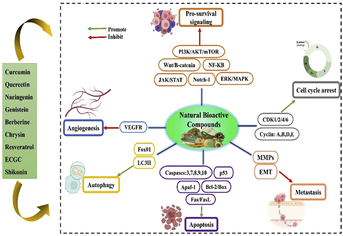
Figure 1.
Molecular-level mechanism of action of natural bioactive compounds to prevent breast cancer.

Natural bioactive compounds loaded chitosan nanocarriers: A promising strategy for breast cancer therapy
Guru Prasanna Sahoo , Tushar Kanti Rajwar , Jitu Halder , Ajit Mishra , Ritu Mahanty , Ivy Saha , Bibhanwita Satpathy , Rakesh Kumar Sahoo , Deepak Pradhan , Vineet Kumar Rai , Priyanka Dash , Chandan Das , Manoj Kumar Sarangi , Saroj Kumar Rout , Biswakanth Kar , Goutam Ghosh , Goutam Rath


Breast cancer remains one of the foremost prevalent malignant tumors in women globally, and it has drawn more attention from the medical and public health communities. According to the American Cancer Society, it will impact the lives of around 316,950 people with invasive breast cancer, and approximately 59,080 new cases of ductal carcinoma in situ will be diagnosed. It has been projected that in 2025, 42,170 women will lose their lives to breast cancer. Currently, hormone therapy, radiation, chemotherapy, surgery, and a combination of these are the available treatment options for breast cancer [1,2]. However, these conventional therapy approaches have several adverse consequences; surgery and radiotherapy are extremely intrusive procedures with poor specificity. Chemotherapy has serious side effects since the drugs are harmful to healthy cells that are growing [3]. In addition, the lack of a targeting mechanism, rapid systemic metabolism, and poor bioavailability of conventional dosage forms have all been associated with poor therapeutic efficacy [4]. Moreover, drug inactivation and efflux of drugs from cancer cells are caused by multi-drug resistance (MDR), in which the target develops structural and functional resistance, which is another drawback of traditional chemotherapy drugs that might lead to treatment failure [5]. As a result, research into safer, less harmful, and more efficient treatments for people with breast cancer is always pressing. During the last few decades, natural products have grown in popularity as active ingredients because synthetic drugs have undesirable qualities and adverse effects.
Natural bioactive compounds have gained more attention for treating breast cancer because of their safe toxicological profile in comparison to synthetic chemotherapeutic drugs in recent years [6]. Natural bioactive compounds are secondary metabolites derived from marine species, fungi, microbes, and plants. Several studies have indicated that they have important anticancer effects through apoptosis induction, stopping the proliferative cell cycle of cancer cells, preventing cancer cell migration and invasion, and inhibiting angiogenesis [7]. Notably, 85 (49%) of the 175 anticancer molecules released between 1981 and 2014 came from natural sources and their derivatives [8]. Additionally, specific bioactive compounds can improve the body's immune response, which advances cancer immunotherapy and has indirect anticancer benefits. Recently, natural products have been utilised as modulators of multidrug resistance and sensitisers for chemotherapy and radiation therapy. The most studied natural bioactive compounds for breast cancer treatment are polyphenols, alkaloids, terpenoids, saponins, glycosides, etc. [9,10]. For example, by targeting cyclin-dependent kinases (CDKS), flavopiridol, a semi-synthesised flavonoid derivative derived from rohitukine, suppresses cell cycle progression and cell proliferation [11]. As a result, natural bioactive compounds open the door for novel advancements in low-cost, highly effective natural therapies that improve breast cancer patients' quality of life and survival duration.
Despite their promise, the poor tumor targetability, high gastrointestinal instability, poor aqueous solubility, and low systemic bioavailability of many anticancer natural bioactive compounds significantly restrict their treatment efficiency [12]. For example, when given intravenously to rats, curcumin exhibits a half-life of 11.137 ± 1.620 h and a Cmax of 16.250 ± 0.596 ng/mL [13]. The physical barriers in the human body, including the gastrointestinal (GI) tract, blood-brain barrier, endothelial lining of blood vessels, and mucosa, are difficult to pass through due to their size and hydrophobic nature. Besides, they are broken down by enzymes in the digestive tract [14]. For instance, the efficiency of quercetin is decreased by its rapid breakdown by stomach acid or digestive enzymes [15]. In addition, natural compounds like shikonin and paclitaxel harm healthy tissues [16,17]. Therefore, introducing nanocarriers to encapsulate or conjugate these bioactive compounds may be an innovative approach to improve their bioefficacy by affecting their solubility, rate of absorption, and GI stability. Several nanoparticle systems developed using biomaterials loaded with anticancer drugs have been approved by the United States Food and Drug Administration (USFDA) or currently in clinical trials [18]. In the twenty-first century, these bio-nanocarriers have evolved into an international frontier field because they are biocompatible and highly effective at activating immune reactions in the human body. These are evidently due to the multiple benefits that nanocarriers provide over free drugs and conventional dosing forms. The benefits of the nanocarrier system include multiple drug loading, site-specific delivery, controlled and extended drug release, longer circulation half-life, minimal non-specific cell uptake, etc. [19,20]. With the development of the administration of drugs, numerous nanocarriers have been developed using natural polymeric materials. Chitosan-based nanocarriers are among the most promising nanocarriers for delivering chemotherapeutic or anti-tumor drugs [21,22].
Chitosan is a polymeric nanocarrier that has been employed and is considered safe for human use. The USFDA subsequently approved chitosan polymer due to its biocompatible and biodegradable properties and continuous release mechanism [23]. In addition to its many other biological functions, chitosan also acts as an immune-adjuvant, osteoblast accelerator, anti-angiogenic, anti-inflammatory, haemostatic, anti-microbial, anti-tumor, and connective gum tissue regenerator [24,25]. The amine and hydroxyl functional groups of chitosan's backbone allow easy modification to change the substance's stability, solubility, bioactivity, and mucoadhesion [26]. To increase the stability and loading efficiency of the drugs, the amine groups might be functionalised with other compounds [27]. Novel nanocarriers based on chitosan have been developed for the targeted delivery of drugs, demonstrating their potential to enhance the results of breast cancer treatments. For instance, nanoparticles containing chitosan provide versatility in surface modification, which facilitates tumor targeting. It exhibits surface flexibility and is configurable at the nanoscale, which enhances stability and makes it compatible with numerous anti-cancer drugs [28]. Additionally, it reduces unwanted side effects and shields the drugs from enzymatic destruction. These characteristics make chitosan-based nanocarriers ideal for encapsulating naturally occurring bioactive substances and boosting their anti-breast cancer therapeutic potential [29]. For example, Baksi et al. formulated the quercetin-loaded chitosan nanoparticles that demonstrated a sustained release profile (67.28%) over 12 h [30]. Similarly, the oral bioavailability of curcumin increased by 9.5-fold when delivered using N-carboxymethyl chitosan (NCC)-coated solid lipid nanoparticles (SLN) compared to a curcumin solution [31]. The MDA-MB-231 cells exhibited a 3–6-fold higher uptake efficiency of chitosan-coated Fe3O4 magnetic nanocomposites (NC) than free curcumin [32].
This review primarily highlights recent breakthroughs in chitosan nanocarrier-based delivery systems for natural bioactive compounds such as berberine, curcumin, quercetin, resveratrol, epigallocatechin-3-gallate (EGCG), with a focus on enhancing therapeutic efficacy and safety for breast cancer treatment, emphasising the use of chitosan-based nanoparticles, liposomes, SLN, micelles, and other nano-drug delivery approaches. Additionally, the challenges and future perspectives for chitosan-based nano carriers for treating breast cancer have been addressed. To the best of our knowledge, no comprehensive review particularly highlights the application of chitosan-based nanocarriers to deliver naturally occurring bioactive compounds in breast cancer treatment.
Several noteworthy developments in tumor biology research have been reported recently, yet conventional treatments are still incorporated. Fig. S1 (Supporting information) illustrates the challenges that conventional therapy confronts. Currently, breast cancer can be treated with chemotherapy, radiation treatment, hormone therapy, and surgery [33]. The primary form of therapy is surgery, removing the tumor and surrounding diseased tissues. The recurrence of cancer after a few years, because of cancer cells that were removed, is the sole risk associated with lumpectomy [34]. After surgery, radiation therapy is another local treatment that kills breast cancer cells by exposing them to high-energy radiation [35]. Multiple studies have demonstrated that while short-term radiation exposure during radiation therapy affects and kills breast cancer cells, long-term radiation exposure can raise the risk of dying from ischaemic heart failure. The primary challenge in traditional treatment is chemotherapeutic drug resistance brought on by the cancer cells' increasing potential [36]. Drug-resistant cancer cells' intracellular efflux and p-glycoprotein expression stopped the chemotherapeutics from penetrating the cells. Drug resistance during tumor formation may also prevent the chemotherapy drugs from killing the cancer cells by apoptosis. Several adverse effects, including myeloid leukaemia and neoplasia, are brought on by alkylating drugs. Additionally, it has been shown that several patients have infertility following gonadotox injections. Additional adverse effects, including dry mouth, water retention, digestive issues, dental issues, and emotional challenges, have been reported with standard therapy [37].
Molecular biology and tumor biology have advanced rapidly, altering anti-cancer treatment approaches. Since time immemorial, natural bioactive compounds have been an essential source of therapeutic options to cure ailments, and recently, they have gained popularity as a possible breast cancer treatment alternative. More than 5000 distinct natural bioactive compounds have reportedly been identified in grains, fruits, and vegetables [38]. Polyphenols, alkaloids, terpenoids, anthraquinones, etc., are the various natural bioactives extensively studied for their anticancer potential. They demonstrate anti-migration, anti-proliferative, anti-metastatic, anti-invasive, and anti-angiogenic properties. An increasing body of research has shown that natural bioactives can effectively target breast cancer and stop its growth by altering cellular signalling pathways [39]. Additionally, it has been demonstrated that phytoconstituents cause breast cancer cells to undergo apoptosis via a variety of pathways. They can restrict tumor growth and cancer spread by inducing cell death in cancer cells by activating proteins that facilitate apoptosis and blocking molecules that hinder it. These substances can also cause cell cycle disruption, which results in cell cycle arrest and further prevents cancer cells from proliferating [40]. The ability of bioactive compounds to combat drug resistance, a frequent problem with traditional treatments, is another important feature of their use in the treatment of breast cancer. Targeting the tumor microenvironment (TME) and breast cancer stem cells (CSCs), which are essential for the initiation, spread, and resistance to treatment of cancer, has proven to be a promising use for natural bioactives. By blocking self-renewal pathways like Wnt/β-catenin, Hedgehog, and Notch, which limit the cells' capacity to replicate, phytoconstituents attack CSCs in several ways [41]. They not only target CSCs but also alter the TME, which is made up of fibroblasts, immune cells, blood vessels, and extracellular matrix [42]. By stimulating cytotoxic T cells and natural killer cells and suppressing immunosuppressive cells such as regulatory T cells and myeloid-derived suppressor cells, phytochemicals improve the immune response [43]. Furthermore, they limit the function of cancer-associated fibroblasts in the invasion and metastasis of cancer cells by inhibiting them.
A variety of natural compounds were assessed by Islam and his team, who confirmed their effectiveness compared to conventional breast cancer treatments [44]. They observed that these compounds could enhance outcomes and lower mortality from breast cancer, and that developing these natural products could eventually lead to the disease's cure. For example, curcumin has become one of the phytochemicals that has been studied the most in the treatment of breast cancer, as curcumin causes radiation protection, chemosensitisation, and cell death. According to reports, curcumin's regulatory effect on breast cancer apoptosis and proliferation involves blocking fatty acid synthase. The MDA-MB-231 and MCF-7 breast cancer cell lines had a half maximal inhibitory concentration (IC50) value of 75 µmol/L for curcumin. Also, quercetin's anticancer properties are mostly attributed to its suppression of oncogenic kinases and control of pertinent signalling pathways. For instance, in MDA-MB-231 cells, quercetin downregulated p53 and stopped the cell cycle in the G2/M phase at a dosage of 7 µg/mL [45]. According to recent studies, changing the structure is a practical way to boost quercetin's bioavailability. The three quercetin derivatives, which are QD1, QD2, and QD3 that Lewińska et al. synthesised, significantly enhanced the removal of senescence brought on by the apoptotic drug etoposide, along with the downregulation of p27, interleukin-1 beta (IL-1β), IL-8, and heat shock protein 70 (HSP70) [46]. EGCG inhibits oxidative stress, inflammation, angiogenesis, apoptosis, and metastasis through a variety of biological and pharmacological actions. Wei et al. demonstrated that EGCG influenced glucose metabolism in a time-dependent and dose-dependent manner, reducing the proliferation of 4T1 cells and causing cell death within 24 h [47]. Additionally, EGCG reduced the tumor weight of breast cancer, lactate and glucose levels, and vascular endothelial growth factor (VEGF) expression in a dose-dependent manner in the in vivo breast cancer model. In addition to these phytoconstituents, several promising anticancer drugs have been discovered, including derivatives of camptothecin (CPT). The treatment of tumors has seen the application of a rising variety of CPT derivatives. Semi-synthetic CPT derivatives, such as topotecan and irinotecan, for instance, have been authorised to treat advanced breast cancer. The anti-cancer properties of CPT derivatives are demonstrated by their excellent specificity for targeting deoxyribonucleic acid (DNA) topoisomerase I (TOP1) [48]. Fig. S2 (Supporting information) shows various phases of development for natural bioactives' anticancer potential against breast cancer [49]. The figure provides a pathway from natural compounds to cancer treatment alternatives and describes the development process of bioactive compounds as possible anticancer agents.
Natural bioactive compounds have shown an incredible ability to target breast cancer cells in several in vitro and in vivo studies. Significant bioactive compounds with anti-cancer properties, including berberine, curcumin, quercetin, resveratrol, EGCG, and others, have been found because of extensive research into the effectiveness of natural bioactives for the prevention and treatment of breast cancer. Table S1 (Supporting information) provides an overview of some important natural bioactive compounds. They exhibit anti-cancer potential by regulating several signalling pathways and apoptotic mechanisms (Fig. 1). Their primary mechanisms include suppression of DNA synthesis, angiogenesis, metastasis, induction of cell death, heightened susceptibility to chemotherapy, immune function control, and drug resistance reversal. The genetically controlled process of planned cell death is called apoptosis. The extrinsic (death receptor) and internal (mitochondrial) pathways are the two well-known processes that trigger apoptosis [50]. The B-cell lymphoma 2 (Bcl-2) signalling pathway and the Fas/FasL signalling pathway are significant intrinsic and extrinsic mechanisms, respectively. Fas is a crucial death receptor located on the cell surface that, upon binding to its ligand FasL, triggers the activation of the death receptor pathway, leading to apoptosis. After passing the apoptotic signalling through the downstream caspase, it ultimately triggers apoptosis. Pro-apoptotic (Bax, Bak) and anti-apoptotic (Bcl-2, Bcl-XL) proteins make up the Bcl-2 family of proteins. For example, Kaempferol, curcumin, and resveratrol efficiently block Bcl-2 family proteins and cause apoptosis. Quercetin is a well-studied natural substance with anti-cancer effects linked to apoptosis in breast cancer. Fas, FasL, and Bax are upregulated in MCF-7 and MDA-MB-435 cells, while Bcl-2 is dose-dependently decreased. A cell cycle, which is divided into phases G1, S, G2, and M, is the complete process from the end of one division to the subsequent one. The primary cell cycle machinery that drives the cell cycle forward is processed in the nucleus and consists of cyclins and their catalytic partners, CDKS. To regulate the essential cell cycle mechanisms, they produce the cyclin-CDK complexes. Many naturally occurring compounds made from plants could stop the advancement of the cell cycle, which ultimately leads to the death of tumor cells. For instance, cell growth is inhibited by EGCG administration, which causes cell cycle arrest in the G1/S or G2/M phase of MCF-7 cells. Quercetin can stop the cell cycle and induce apoptosis in breast cancer cells at the S phase.
Additionally, the potential of natural products to prevent breast cancer cells from invading and spreading has been the subject of much research. Matrix metalloproteinases (MMPs) are essential for physiological and pathological processes. MMPs such as MMP-2 and MMP-9 are intimately associated with the invasion and migration of tumor cells inside the TME [51]. For instance, resveratrol decreased breast cancer cell migration and invasion via inhibiting phosphatidylinositol 3-kinase (PI3K)/protein kinase B (AKT) signalling, which in turn downregulated MMP-2. Through inhibiting MMP-9-related signalling pathways, kaempferol inhibited the invasion of MCF-7 and MDA-MB-231 cells. Epithelial-mesenchymal transition (EMT) is an essential step in the metastasis of breast cancer, in addition to the critical role that MMPs play in the invasion and migration of breast cancer [52]. During the EMT process, downregulated E-cadherin and increased vimentin and N-cadherin cause the invasion of breast cancer cells. The development of new blood vessels from preexisting vascular tissue and their interaction with pro-angiogenic growth factors is known as pathological angiogenesis, and it is one of the characteristics that sets cancer apart. Tumor cells produce a class of cytokines called VEGFs, which are important modulators of pathological angiogenesis and are triggered by growth factors, oncogenes, and hypoxia stimulation. Vascular endothelial growth factor receptor 1 (VEGFR-1), VEGFR-2, and VEGFR-3 are significant angiogenesis targets. For example, by blocking VEGF synthesis and preventing VEGF from interacting with breast cancer receptors, luteolin demonstrated anti-cancer properties by preventing angiogenesis [40].
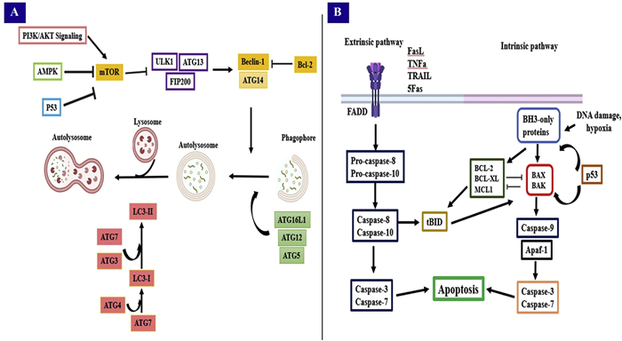
Moreover, autophagy is necessary for breast cancer tumor cells to survive. It could cause one of two cell survival or death pathways, which would either encourage or prevent the growth of tumors [53]. Mechanistic target of rapamycin complex 1 (mTORC1), along with Unc-51-like kinase 1(ULK1), mATG13, and FIP200, which is the commonly recognized sensor to trigger autophagy. mTORC1 and ULK1 are directly regulated by AMP-activated protein kinase (AMPK) [54]. Many cellular processes are modulated by the PI3K/AKT/mTORC1 signalling pathway, which is inhibited by natural products [55]. Breast cancer cell proliferation was impacted by ginsenosides RK1 and luteolin, which inhibited the PI3K signalling pathway and controlled autophagy. A crucial gene in the development of autophagosome membranes is Beclin-1 (BECN1). Autophagy is controlled through its interaction with autophagy-related 14 (ATG14) [56]. When Beclin-1 is downregulated, autophagy activity and level decrease, and breast cancer cells become more invasive [57]. Berberine is a naturally occurring substance that stimulates autophagy by upregulating Beclin-1. The schematic illustration of the autophagy and apoptosis mechanisms against breast cancer is shown in Fig. 2.
Natural bioactive compounds have become a popular alternative therapy choice worldwide in recent decades. Even in the United States, the United Kingdom, and the European Union, where the USFDA and the European Medicines Agency (EMA) extensively enforce laws for the safety assessment of pharmaceuticals and nutraceuticals, around 80% of the world's population depends on natural remedies for primary healthcare. They are regarded as innovative therapeutic agents for breast cancer since these selective bioactive compounds are affordable, widely accessible, and effective with minimal side effects. Given that oral administration is the most popular method, most traditional formulations of bioactive compounds have low bioavailability. The literature reported that natural products have a low melting point, a short biological half-life, poor absorption, and low water solubility [58]. Additionally, natural medicinal compounds may be quickly eliminated by a fast metabolism or destroyed by an acidic gastrointestinal environment. These substances have challenges passing across the mucosa, GI tract, blood vessel endothelium, and blood-brain barrier with efficiency [59]. However, there is no established validation procedure or evidence of the clinical use of natural bioactives in breast cancer treatment, which is one of the main drawbacks of this therapy.
The quality of natural components plays a major role in the effectiveness and safety of herbal medicines. Both extrinsic factors related to proper herbal collection procedures, such as plant selection, cultivation, and environmental conditions, and intrinsic ones associated with genetics, determine the quality of herbal medicines. As a result, evaluating them for thorough quality checks of natural products is quite challenging. However, because there is a lack of regulations governing natural remedies, there is an inadequate amount of information regarding their safety, and no long-term safety profile. Furthermore, it is challenging to accurately identify and gather drugs for treatment due to the lack of taxonomical understanding among herbal drug makers. Therefore, the lack of therapy at the site of action would restrict the herbal bioactive's potency. Alternatively, seeking the desired pharmacological response by boosting the dose may result in unfavourable toxicities. It can occasionally become questionable when drugs exhibit effective mechanistic effects in vitro cell culture models but fail to produce the expected impact in the target environment in vivo at the appropriate dosage [60]. For example, plant alkaloids called paclitaxel and docetaxel, which are taxane-based drugs, might result in hypersensitivity reactions that include redness, itching, and dyspnoea. Despite being thought to be safer than synthetic drugs, natural bioactives still need to be studied to determine the ideal dosage for treatment to be a more successful choice. Research has indicated that upon absorption, polyphenols undergo three different conjugation processes within the body: Methylation, sulfation, and glutaronidation. Other reports on natural drugs suggest the inclusion of harmful ingredients that could cause consumers to experience genotoxicity and cellular toxicity.
The fact that bioactive compounds exhibit a biphasic dose response is another important characteristic. As a result, several naturally active ingredients are researched and referred to as hormetic compounds since they exhibit markedly varied effects at various dosages. For instance, resveratrol has demonstrated differing effects in malignant and healthy cells at low and high dosages. Curcumin, quercetin, genistein, and numerous other bioactive compounds exhibit nonmonotonic dosage responses in various cancer cell lines. Considering these problems with natural components, multidisciplinary approaches are used more frequently to safely and efficiently provide natural bioactives. The challenges involved in administering antibiotic drugs through natural bioactive compounds have encouraged the development of novel drug carriers, especially those based on nanotechnology, known as nanocarriers. This suggests that using nanocarriers to combine bioactive substances with another anticancer drug is a more successful way to treat breast cancer than using bioactive compounds alone.
The naturally occurring mucopolysaccharide chitosan, which is made up of N-acetylglucosamine and d-glucosamine units, is obtained by deacetylating chitin. The degree of polymerisation and acetylation is a key factor in regulating the structural and functional characteristics used in nanocarrier engineering. The presence of the main amino group in the chemical structure of chitosan gives it a cationic character in contrast to other biodegradable polymers. This primary amine group also gives the nanocarriers important mechanistic characteristics, such as enhanced permeability, transfection, mucoadhesion, and controlled drug release [61]. In addition to all of this, chitosan may result in decreased toxicity and immunogenicity. The reactive amino and hydroxy groups give chitosan a variety of useful properties, including metal chelation, gel formation, antioxidant, anti-inflammatory, anti-angiogenic, anti-cancer, and antibacterial properties [62].
Regarding tumor targeting, chitosan-based nanocarriers are far more effective than many experimental polymeric nanoparticles. The main mechanism of tumor targeting is the special feature of pH-responsive drug delivery, since the pH of the tumor location is marginally lower than the physiological pH [63]. A natural benefit of chitosan nanoparticles as a novel drug delivery method is their mucoadhesive qualities. This results in a longer retention period for the chitosan-based nanocarriers at the intended location [64]. Chitosan nanoparticles are also more likely to be absorbed by tumor cells when there is a longer circulation time and greater cellular penetration because of the protonation of the amino group in an acidic environment [65]. Chitosan also greatly increases paracellular permeability because it can get across the tight junctions of epithelial cells. Since chitosan targets the defective and fragmented vascular system of tumor tissues, it also exhibits decreased reticuloendothelial system absorption because of its smaller particle size [66]. Chitosan particularly targets hyperactive receptors in breast cancer cells, such as human epidermal growth factor receptor 2 (HER2) and oestrogen receptors, to disrupt genes linked to tumor growth and drug resistance while facilitating the movement of siRNA and mRNA, two nucleic acids [67].
Chitosan's surface has undergone several changes, such as thiolation or PEGylation, to achieve certain drug delivery benefits. The thiolation of chitosan provides a triggered drug release profile, mechanical strength, and swelling behaviour. By chemically adding carboxymethyl and quaternary ammonium groups, chitosan can be made more soluble in water. The most prevalent modification of all the chitosan derivatives is acylated chitosan. Organic acids and their derivatives were modified by the acylation reaction of chitosan, improving biocompatibility. These characteristics are essential to chitosan's performance as a carrier material in drug delivery systems [68]. Since they provide absorption-mediated transcytosis and are a great vehicle for delivering anti-cancer treatments, chitosan has been used as a coating agent over several nanocarriers that operate as cationic ligands. Furthermore, chitosan's regenerative characteristics aid in tissue repair and wound healing for patients recovering from mastectomy or other breast cancer treatments; they may improve tissue regeneration, encourage collagen production, and aid in recovery after radiation or surgery. Chitosan compounds exhibit great promise in the treatment of breast cancer through several functions, such as the induction of apoptosis and the prevention of cell proliferation [69]. As hydrophobic natural compounds are poorly absorbed and metabolised before reaching tumor sites. These hydrophobic substances are encapsulated in chitosan nanoparticles, which increase their water dispersibility by making them nano-sized particles [22].
Several nanocarriers, including microspheres, sponges, rods, micelles, emulsions, membranes, and nanoparticles, can be synthesised with chitosan and employed to carry drugs. The literature provides several published methods for developing chitosan-based nanocarriers. These techniques are precipitation, coacervation, emulsion cross-linking, and ionic gelation. A readily adjustable site with a high potential for cancer theragnostic development is present in chitosan. Overall, chitosan-based nanocarriers enhance the solubility, bioavailability, and specificity of natural bioactive compounds in breast cancer therapy, offering a promising avenue for developing safer and more effective anticancer treatments. Table S2 (Supporting information) presents a comparative analysis between the chitosan-based nanocarrier and conventional delivery methods for breast cancer.
Bioactive compounds encapsulation using chitosan-based nanocarriers is a state-of-the-art pharmaceutical research approach offering significant advancements in drug delivery systems. Chitosan's properties make it an ideal material for drug encapsulation because they enable more controlled and targeted drug release, improved therapeutic efficacy, and higher drug solubility. Using the ionic gelation process, positively charged chitosan interacts with negatively charged counterions such as sodium sulphate or tripolyphosphate to form chitosan nanoparticles [70]. When a non-solvent is added during nanoprecipitation, chitosan is precipitated from a solvent, producing nanoparticles. As part of the emulsion cross-linking process, chitosan in an oil-in-water emulsion system is cross-linked using the proper cross-linking agent. These methods allow for the incorporation of both hydrophilic and hydrophobic drugs into chitosan nanoparticles, offering flexibility in the development of drug delivery systems for a range of drugs. The distinct release mechanisms exhibited by chitosan-based nanoparticles and nanocarriers dictate the controlled distribution of encapsulated drugs or active ingredients. These processes are influenced by the environment, the composition of the contained payload, and the physicochemical properties of the nanoparticles. Understanding these release pathways is crucial for improving drug delivery strategies and tailoring nanoparticles for specific therapeutic applications. One of the primary release mechanisms observed in chitosan-based nanoparticles is diffusion-controlled release. This method states that the encapsulated drugs or molecules are released from the nanoparticles by a diffusion process brought on by concentration gradients [71]. A few examples of factors that significantly affect diffusion rate are particle size, porosity, and polymer density. By adjusting the degree of deacetylation or adding pH-sensitive moieties, chitosan-based nanoparticles' pH sensitivity can be adjusted by researchers to alter drug release kinetics for precise administration to tissues or cell types. Enzyme-mediated release is another crucial method that chitosan-based nanoparticles use. Two examples of enzymes found in bodily fluids that may degrade chitosan enzymatically are chitinases and lysozymes. When chitosan nanoparticles contact these enzymes and progressively break down, the encapsulated payload is released. This method has been specifically researched in drug distribution to capitalise on the presence of certain enzymes in disease areas [72].
Surface modification of chitosan-based nanocarriers is crucial for optimising their properties for drug delivery applications. Nanoparticles can have their surface modified with a range of ligands, targeting moieties, or coatings to improve their stability, biocompatibility, and accurate interactions with target cells or tissues [73]. One of the main strategies for enhancing the targeting of chitosan-based nanoparticles is the conjugation of specific ligands on their surface [74]. The process of attaching ligands such as aptamers, peptides, antibodies, or small molecules to a nanoparticle's surface is called ligand conjugation [75]. These ligands can attach themselves specifically to biomarkers or overexpressed receptors on the surface of target cells or tissues. Chitosan-based nanoparticles can be coated with pH-sensitive materials that change their charge or solubility in response to pH variations. Given the somewhat acidic pH of TME, the coating may encourage the breakdown of nanoparticles, leading to medication release and enhanced cellular absorption [76]. This pH-responsive action reduces drug loss in circulation and enhances treatment outcomes by enabling controlled drug release at the intended site. The surface charge of nanoparticles can be altered by introducing charged polymers or altering the pH during nanoparticle formation. To boost cellular uptake, negatively charged cell membranes may interact with positively charged nanoparticles. On the other hand, negatively charged nanoparticles may interact with positively charged regions on target cells or tissues to enable site-specific delivery [77].
Pharmaceutical firms can employ these nanoscale carriers in a variety of ways to improve the efficacy of treatments and deliver drugs exactly where they are required. For the transportation of bioactive compounds, we look at some of the primary applications of chitosan-based nanocarriers. When drugs are encapsulated in chitosan nanoparticles, they are protected against enzyme degradation and harsh physiological conditions, which increases stability and bioavailability [78]. Furthermore, chitosan nanoparticles might aid in controlling drug release, allowing for accurate and prolonged drug administration. This is advantageous for drugs that require prolonged therapy or have short half-lives. This controlled release method keeps the drug concentration at the target area steady and efficient, with fewer doses and potential side effects [79]. By enhancing their selectivity and accumulation in tumor tissue, active targeting strategies, such as functionalising chitosan nanoparticles with tumor-specific ligands, enhance therapeutic outcomes with decreased systemic toxicity [80]. It is anticipated that continued research and development in this field will eventually result in innovative and efficient drug delivery methods. Multifaceted chitosan nanocarrier drug delivery is shown in Fig. 3.
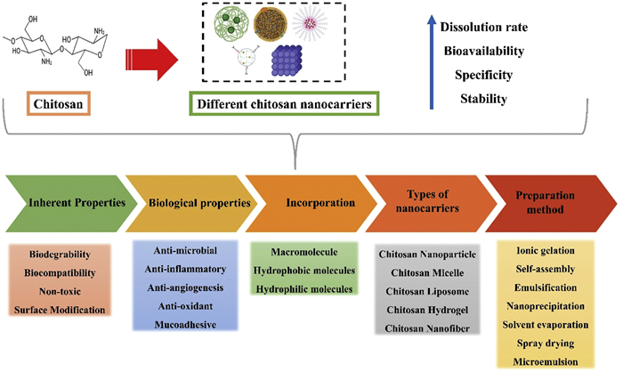
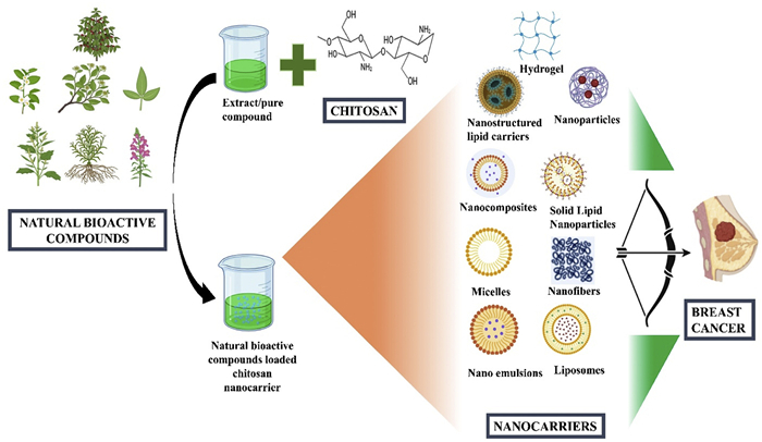
Since 1959, applying chemistry, materials science, engineering, and biology to nanotechnology has suggested a multidisciplinary strategy that has effectively expanded its biomedical science and healthcare research fields. The USFDA has authorised the commercialisation of over 100 nano-formulations over the past few years. Any anticancer formulation must aim for a prolonged, tumor-specific drug action with the least amount of systemic toxicity possible. Certain drugs have cytotoxic effects during delivery, making the use of nanocarriers in drug delivery systems necessary. Potential natural bioactive compounds administration via nano-drug delivery systems has emerged as a research hotspot in recent years. For efficient drug administration in the system, nanocarriers such as liposomes, dendrimers, micelles, hydrogel, metal nanoparticles, mesoporous polymers, and even protein-drug conjugates are employed. Chitosan-based nanocarriers exhibit improved stability, bioavailability, and targeted therapeutic effects when encapsulating these compounds. In the following sections, we will be discussing the various types of chitosan nanocarriers (Fig. 4) that have been developed, each with unique characteristics and applications. Therefore, it is believed that the developing nanotechnology science presents a remarkable, game-changing chance to improve cancer diagnosis and therapy significantly. Table 1 [81-131] summarizes the different applications of chitosan-based nanocarriers for the treatment of breast cancer.

 DownLoad:
CSV
DownLoad:
CSV
| Natural bioactive compound | Nanocarrier | Preparation method | Cell line | Anticancer mechanism | Outcome | Ref. |
| Curcumin | Folate-chitosan nanoparticle | Self-assembling process | MCF-7 | Induces apoptosis | pH-dependent sustained drug release, significant reduction in the viability, and enhanced tumor-site delivery | [81] |
| Chitosan nanoparticles | Ionic gelation method | MDA-MB-231 | Inhibited PI3K and AKT activation and VEGF and osteopontin (OPN) expression in the cells | Significant inhibition of cell viability, enhanced therapeutic efficacy, excellent tumor targeting abilities | [82] | |
| Chitosan-protamine nanoparticles | Ionic gelation method | MCF-7 | Reduction of cell viability, nuclear factor-kappa B (NF-kB), tumor necrosis factor-α (TNF-α), and IL-6 levels, downregulated the expression of the Bcl-2 | Enhancing cellular uptake and retention, suppressing inflammatory signalling, promoting apoptosis | [83] | |
| Selenium-chitosan NC | Ionic-gelation | 4T1 | Reduced cell proliferation rate | pH-dependent release, greater tumor and metastasis reduction in mice | [84] | |
| Chitosan-coated liposomes | Thin-film hydration | MCF-7 | ROS-mediated apoptosis | Controlled drug delivery, higher encapsulation efficiency, stronger growth-inhibition | [85] | |
| Chitosan-coated liposomal hydrogels | Thin-film hydration | MCF-7 | Significantly inhibits the growth of MCF-7 cells, inducing apoptosis | Sustained drug delivery, excellent tumoricidal effect, effective cellular uptake | [86] | |
| Polycaprolactone-chitosan core-shell nanofibers | Coaxial electrospinning | MCF-7, 4 T1 | Significant cytotoxicity, tumor growth suppression | pH-responsive and sustainable release, remarkable anti-tumor efficacy, localised delivery | [87] | |
| Chitosan/starch/MoS2 NC | Hydrothermal method & double emulsion | MCF-7 | Induces apoptosis | pH-responsive release, strong anticancer activity | [88] | |
| Chitosan/carbon quantum dots/Fe2O3 NC | Double emulsion method | MCF-7 | Cell viability was drastically decreased, inducing apoptosis | pH-sensitive release, significant cytotoxicity | [89] | |
| Chitosan-loaded SLN | Cold dilution of microemulsion | MCF-7, MDA-MB-231 | Inhibition of the AKT/IKKα-β/NF-kB axis | Increasing intracellular retention, reducing ROS, significantly reduced tumor growth and mass | [90] | |
| Chitosan-based nanoemulsions | Emulsification coating | 4T1 | Reducing cell viability | pH-dependent drug release, improved anti-tumor activity, reduced organ toxicity | [91] | |
| Fe3O4/chitosan/agarose nanoemulsion | Double emulsion | MCF-7 | High apoptosis | Sustained drug delivery, strong tumor cell apoptosis, and minimal side effects | [92] | |
| Quaternized chitosan derivative nanofibers | Electrospinning | 4T1 | Excellent anticancer activity | Controlled release profiles, strong cytotoxicity against cancer cells | [93] | |
| Chrysin | Alginate-chitosan hydrogel | Ionic gelation | T47D | Induce apoptosis, G2/M causes arrest of the cell cycle | Reduce the viability, enhance the anticancer drug delivery system | [94] |
| Quercetin | Chitosan nano micelles | Ultrasound method | MCF-7 | Inducing apoptosis | pH-responsive release, promotes cellular uptake, enhances inhibitory effect on tumor cells | [95] |
| Chitosan, halloysite, and graphitic-carbon nitride hydrogel | Water in oil in water emulsification process | MCF-7 | Induces apoptosis | pH-responsive release, significant cytotoxicity against cancer cells | [96] | |
| Chitosan functionalized copper oxide nanoparticles | Chemical precipitation | MCF-7 | Induced apoptosis via increased p53 gene, increased both cytochrome and caspase-3 levels | Sustain release, enhance anticancer, antioxidant activity | [97] | |
| Fe2O3/chitosan/montmorillonite NC | Ultrasonication | MCF-7 | Induces apoptosis | pH-dependency and controlled release, decrease side effects | [98] | |
| DEAE-chitosan nanoparticles | Ionic gelation | MCF-7 | Induces apoptosis | Controlled and sustained drug release, synergistic effect on the control of cell viability | [99] | |
| CS/PVP/γ-alumina NC hydrogel | Double oil-in-water emulsification method | MCF-7 | Significant cytotoxicity, enhanced apoptosis | Controlled release, excellent drug loading, and anticancer activity | [100] | |
| Chitosan nanoparticles | Ionic gelation method | MCF-7 | Induces apoptosis | Sustained release, stronger antioxidant effects, significant cytotoxic activity | [101] | |
| Amphiphilic chitosan nanoparticles | Self-assembling process | MCF-7 | Induce apoptosis | pH-sensitive release, biocompatible platform for targeted quercetin delivery | [102] | |
| Chitosan-functionalized Fe3O4 magnetic nanoparticles | Chemical co-precipitation | MCF-7 | More cell cytotoxicity on cell lines, more antiproliferative activity | Good loading efficiency and improved anticancer activity | [103] | |
| Chitosan-coated zein nanoparticle hydrogel composite | Nanoprecipitation and ionic crosslinking | MCF-7 | Apoptosis induction | Enhancing effect of the dual release system, increased cytotoxic action by 20.7-fold | [104] | |
| Oleyl chitosan NC | Self-assembly | MDA-MB-231 | Significant cytotoxicity | Enables dual imaging (MRI and CT) plus therapeutic (anticancer) functions, significant anti-cancer effect on tumor cell lines | [105] | |
| Curcumin and quercetin | Quaternary ammonium-chitosan nano-micelles | Dialysis method | MCF-7, MDA-MB-231 | Increase cell cycle arrest in the G2/M phase, inducing apoptosis | pH-responsive drug release, enhanced uptake, and anticancer activity | [106] |
| Sorafenib and quercetin | Aptamer and folic acid-targeted chitosan-poly lactic-co-glycolic acid | Emulsion solvent-evaporation | MCF-7 | Caspase-9 and P53 gene expression were increased, reduction was observed for Bcl-2 expression | pH-sensitive control release, high targeting specificity, and synergistic effectiveness | [107] |
| Naringenin | Chitosan nanoparticles | Ionic gelation method | MDA-MB-231 | Increase in intracellular ROS levels induces apoptosis and increases the activation of procaspase-3 | Reduced cell viability, cell proliferation | [108] |
| Chitosan and dextran sulfate nanoparticles | Complex coacervation technique | MCF-7 | Significant cytotoxic activity | Achieved high loading of naringenin, enhanced chemotherapy efficacy, and reduced dose-related side effects | [109] | |
| Naringin | Chitosan-coated poly(lactic-co-glycolide) nanoparticles | Double-emulsion solvent-evaporation technique | MCF-7 | Apoptosis induction | Sustained release profiles, synergistic anticancer effects, enhanced cytotoxicity against cancer cells | [110] |
| CGC | Chitosan nanoparticle with folic acid | Ionic gelation | MCF-7 | Excellent inhibitory effect on breast cancer cells | Excellent colloidal stability, showing good cytocompatibility | [111] |
| Chitosan-coated nanoliposomes | Sonication method | MCF-7 | Induced apoptosis, inhibited MCF-7 cell proliferation | Improved sustained release, higher intracellular EGCG levels, enhancing cellular uptake and anticancer activity | [112] | |
| Genistein | Chitosan-coated nanoliposomes | Ethanol injection method | MCF-7 | Enhanced cytotoxicity | Excellent drug loading, controlled release, enhanced absorption, potent antioxidant, and anticancer effects | [113] |
| Chitosan nanoparticles | Ionic-crosslinking method | MCF-7 | Enhanced cytotoxicity, greater apoptosis, and reduction in cell viability | Sustained release, enhancing synergistic anticancer activity | [114] | |
| Berberine | Chitosan-zinc oxide nanoparticles | Ionic gelation | MCF-7 | Elevated ROS levels, inducing apoptosis in the sub-G1 phase, caspase-8 and caspase-9 in MCF-7 cells significantly increased, Bcl-2 gene was reduced, increased Bax gene expression | Induce cell-cycle arrest and activate the apoptotic pathways | [115] |
| Curcumin and berberine | Zein-chitosan nanoparticles | Anti-solvent precipitation method | MDA-MB-231 | Induced elevated cytotoxic effect, and apoptosis with significant inhibition of IL-8 pro-inflammatory cytokines | pH-sensitive drug release, increased cellular uptake, stronger anticancer efficiency | [116] |
| Piperine | Chitosan-coated liposomes | Thin-film hydration | MCF-7 | Enhanced growth inhibitory effects in MCF-7 cells | Controlled release, better permeability, and enhanced anticancer effects | [117] |
| Chitosan-based microgels | Inverse miniemulsion polymerization | 4T1 | Exhibited high cytotoxicity | Enhances solubility, delivery efficiency, intracellular release, and anticancer activity | [118] | |
| Curcumin with piperine | Chitosan-zein and Fe3O4 nanoparticles | Anti-solvent precipitation and layer-by-layer deposition | MCF-7 | Significant synergistic cytotoxicity against MCF-7 cells | Higher drug release, targeted delivery, strong synergistic anticancer effects | [119] |
| Chitosan and zein nanoparticles | Electrospraying | MDA-MB-231 | AKT-1, IL-6, Bcl-2 revealed decreased expression | Robust anticancer activity and molecular suppression of tumor-promoting pathways | [120] | |
| Camptothecin | Chitosan-based nanoemulsion | Emulsification | MCF-7, BALB/c mice | Significant cytotoxicity, facilitated passive targeting | Extended drug release, strong antimicrobial activity, and promoted fibroblast proliferation | [121] |
| Glycol chitosan nanoparticles | Dialysis method | MDA-MB-231 | Significant antitumor effects and high tumor targeting ability | Sustained release, high efficiency, improved stability | [122] | |
| Kaempferol | Chitosan/silver NC | Green synthesis | MDA-MB-231 | Significant in vitro cytotoxic effects, induction of apoptosis | Strong anticancer activity and effective antibacterial properties | [123] |
| Chitosan-based nanostructured lipid carriers | Hot homogenization method | MDA-MB-468 | Stopped cells in Sub G1, significant in vitro cytotoxic effects | Improve cellular uptake, boost the efficacy of paclitaxel in breast cancer cells | [124] | |
| Resveratrol | Chitosan nanoparticles | Ionic gelation method | MDA-MB- 231 | Increased Bcl-2-associated x (BAX), BAX/Bcl-2 ratio, P53 expressions, reduced Bcl-2, and upregulated caspases 3, 8, and 9 | Sustained release, stronger anti-proliferative effects, stimulated intrinsic apoptotic pathway | [125] |
| Chitosan-based injectable in situ forming hydrogels | Nanoprecipitation method | MCF-7 | Decrease in cancer cell viability | Strong synergistic efficacy, localized, minimally invasive treatment | [126] | |
| Chitosan nanoparticles | Ionic gelation method | MDA-MB-231 | Increased Beclin-1, ATG-5, ATG-7 expression | Higher anticancer effect and stimulated autophagy | [127] | |
| Luteolin | Chitosan-based nanostructured lipid carriers | Melt emulsification ultrasonication technique | MDA-MB-231, MCF-7 | Enhanced dose and time-dependent cytotoxicity | Sustained release, higher antioxidant activity, and higher intestinal permeation | [128] |
| Chitosan/alginate-Fe3O4-nanoparticle | Layered encapsulation | MCF-7, MDA-MB-231 | Significantly enhanced cytotoxicity | Targeted, sustained delivery, and enhanced anti-breast cancer activity | [129] | |
| Chrysin | Chitosan-based liposomes | Thin film hydration and electrostatic deposition technique | MCF-7 | Strong cytotoxic potential | Exhibited improved stability and controlled release behavior, a 5-fold increase in chrysin bioavailability | [130] |
| Zataria multiflora | Chitosan-based nanoemulsion | Mild emulsification | MCF-7, T47D, MDA-MB-231 | Induces apoptosis, generates ROS, and triggers mitochondrial membrane permeabilization | Controlled delivery, stability, and triggering multiple cancer cell death pathways | [131] |
Nanoparticles have revolutionised the treatment of breast cancer by offering novel approaches to immune system activation, targeted delivery of drugs, and avoiding drug resistance [132]. Chitosan nanoparticles have numerous characteristics that make them appropriate for application in nanomedicine, such as stability, immunogenicity, biodegradability, biocompatibility with live tissue, and the absence of harmful byproducts. Several functional groups, including amino and hydroxyl groups, allow chitosan nanoparticles to crosslink with polyanionic polymers to develop nanoparticles [133]. Furthermore, the presence of reactive amino and hydroxyl groups makes it easier to modify the surface to bind with the imaging agent and targeting ligand and to form ionic bonds with the biological membrane [134]. These nanoparticles efficiently encapsulate bioactive compounds such as curcumin, quercetin, berberine, enhancing their solubility and stability. Chitosan nanoparticles are responsive to various stimuli, including pH, temperature, light, and magnetic fields, to release the encapsulated drugs in a regulated way [135]. Chitosan nanoparticles, therefore, hold significant promise for improving cancer patients' quality of life and therapeutic results [136]. The curcumin, a naturally occurring substance with well-established anticancer effects that is constrained by low bioavailability and poor water solubility, was loaded into chitosan nanoparticles to develop a smart drug delivery system for targeted breast cancer therapy. They showed considerable tumor suppression in vivo in mouse models and dramatically enhanced anticancer efficacy in vitro against breast cancer cells. Effective tumor targeting was validated by imaging, and toxicity evaluations showed a good safety profile with no appreciable negative effects on blood parameters or major organs [82]. In another study, quercetin was loaded into chitosan nanoparticles with enhanced drug release properties and encapsulation efficiency using the ionic gelation process. The IC50 value of the prepared nanoparticle was substantially lower than that of free quercetin in the in vitro cytotoxicity assay. When mice were treated with the prepared formulation, their antioxidant potential was noticeably higher than that of the free quercetin-treated group. Therefore, it could be a different kind of chemopreventive strategy to counteract cytotoxic conventional chemotherapeutics [30]. Ramya Devi et al. fabricated naringenin-incorporated chitosan nanoparticles through the ionic gelation method. When the drug carrier was administered to breast cancer cells, fluorescence pictures revealed a rise in reactive oxygen species (ROS) levels, which may trigger apoptosis. Western blot was used to assess the activity of caspase-3, and the results indicated that, in comparison to free naringenin, the higher concentration of the drug carrier had enhanced procaspase-3 activation [108]. A pH-responsive nanocarrier that delivers quercetin has been developed with amphiphilic chitosan polymers. The grafting agent used was 2‑chloro‑N,N-diethylethylamine hydrochloride (DEAE). When compared to free drugs, the study found that quercetin-loaded nanocarriers had a noticeably greater inhibitory effect on the growth of MCF-7 cells. Remarkably, the cancer cells were likewise inhibited by the blank nanoparticles. The additional positive charges of DEAE groups, which gave chitosan nanoparticles pro-apoptotic effects, can be used to explain this. The drug release at lower pH was substantially faster than at physiological pH, according to the study [99]. Folate receptor upregulation in numerous cancer cell types, particularly breast cancer, makes folate receptor targeting a well-known method for enhancing the specificity and effectiveness of anticancer medicines. The study found that encapsulating curcumin into chitosan nanoparticles might be a promising therapeutic formulation for the treatment of breast cancer. Using the ionic gelation method, the nanoparticles were prepared, and the optimised curcumin-chitosan nanoparticles were found to be suitable for drug delivery since their size was significantly smaller than 150 nm. After 74 days, no discernible agglomeration was seen, indicating the great stability of the prepared nanoparticle, according to an analysis of their long-term colloidal stability. By enhancing cellular absorption, folate conjugation enables the nanoparticles to target cancer cells that overexpress folate receptors [81].
Metal and magnetic chitosan nanoparticles have emerged as promising platforms in breast cancer therapy because of their special characteristics, which combine the biocompatibility of chitosan with the improved functionality offered by metal or magnetic cores. By enabling the regulated release of drugs in response to external stimuli, such as magnetic fields, these nanoparticles present the potential to achieve targeted drug delivery, reduce systemic toxicity, and enhance therapeutic outcomes. The magnetic alginate/chitosan delivery system was modified to include curcumin to increase its cytotoxicity and bioavailability against MDA-MB-231 cells. Electrostatic forces have been used to apply chitosan and alginate to magnetic nanoparticles. The absorption was substantially higher with an external magnetic field than with free curcumin. The nanocarriers' small size and strong water solubility may be the reason for the increased uptake of the nanoformulation when compared to the free drug. On the other hand, curcumin will form large aggregates that are more challenging to internalise because of its poor solubility in aqueous solutions. The MTT test revealed that, in comparison to free drugs, drug-loaded carriers exhibited noticeably more cytotoxicity towards MDA-MB-231 cells [32]. Additionally, Elsayed et al. investigated the synergistic anti-cancer activity of quercetin with chitosan functionalised copper oxide nanoparticles both in vitro and in vivo. In contrast to copper oxide nanoparticles and quercetin, the in vitro data demonstrated the strong anticancer activity of the drug delivery system. The treatment caused apoptosis by upregulating the p53 gene, stopping the cell cycle, and raising the amounts of caspase-3 and cytochrome c, which ultimately resulted in the death of breast cancer cells [97]. Zare et al. developed a new functionalised Fe3O4 nanoparticle using chitosan for delivering quercetin. When compared to quercetin-free magnetic nanoparticles, quercetin-loaded magnetic nanoparticles demonstrated higher toxicity against MCF-7 cells. Also, quercetin encapsulation into the prepared nanoparticle can help with magnetically guided drug delivery to tumor tissues and imaging applications [103]. Chitosan-coated iron oxide loaded with curcumin and piperine was developed by combining layer-by-layer deposition with anti-solvent precipitation. The nanocarrier showed an intriguing degree of pH sensitivity, releasing curcumin and piperine more quickly at the extracellular pH of surrounding tumors (6.5) than in normal tissues (7.4). They further demonstrated a notable synergistic cytotoxicity against MCF-7 cells in the MTT experiment when compared to other groups. These findings show that dual-drug loaded polymeric magnetic nanoparticles have potential for treating cancer cells specifically [119]. Bulatao et al. fabricated alginate iron oxide/chitosan nanoparticles loaded with luteolin. To address the challenges of delivering luteolin for a successful therapy of breast cancer, they have employed Fe3O4 as a magnetic-responsive method to localise the delivery of luteolin with the help of a permanent magnet. The optimised drug delivery system was biocompatible and showed a 4-fold increase in cytotoxicity against breast cancer MCF-7 cells when exposed to a permanent magnet, indicating the possibility of using this drug delivery system as a magnetically targeted delivery system for breast cancer. According to the study, covering Fe3O4 with chitosan and alginate is crucial for maintaining the nanoparticle's stability and mechanical integrity [129].
Chitosan can be used as a drug carrier with various surface chemistries to coat its surface. In a study, quercetin was encapsulated into novel pH-sensitive self-assembled amphiphilic chitosan nanoparticles to provide a drug delivery mechanism. The findings demonstrated that at an acidic pH of 5.0, the payload release is greater than at a pH level of 7.4. After encapsulation, the results also showed that quercetin inhibits MCF-7 cell metabolism. Nanoparticles aggregated on the cell surface, although few were internalised, according to cellular uptake assays. The hemocompatibility tests indicate that the nanoparticles have appropriate blood compatibility for biological uses [102]. In another study, CPT was readily encapsulated using a dialysis technique into hydrophobically modified glycol chitosan nanoparticles to improve drug stability and tumor targeting in cancer treatment. Subcutaneously implanting MDA-MB231 human breast cancer xenografts in nude mice demonstrated the prepared nanoparticles' strong anticancer effects as well as their ability to target tumors. In aqueous conditions, the prepared nanoparticles generated nanoscale self-aggregates and demonstrated a week-long sustained release of CPT [122].
The liposome, a closed spherical structure formed by a phospholipid bilayer in an aqueous medium, was initially identified by Dr. A.D. Bangham in 1965. The potential of liposomes to encapsulate both hydrophilic and hydrophobic drugs led to substantial research on their usage. Liposomes preserve the lead compound from deterioration and lessen the toxicity associated with drugs, making them a preferred option for drug delivery systems. Its stability in colloidal solution, biodegradability, and biocompatibility make it an excellent candidate for a drug carrier [137]. A liposome is typically a spherical vesicle made up of one or more phospholipid bilayers that can be derived from either plants or animals. Incorporating chitosan into the surface of liposomes enhances their ability to interact with the negatively charged membranes of cancer cells, prolongs their circulation time, and permits pH-responsive, controlled drug release [138]. Additionally, chitosan-modified liposomes show enhanced internalisation by cancer cells, which makes them appealing for the targeted administration of herbal medicinal substances in breast cancer treatment [139]. Several natural bioactives have been effectively added to liposomal systems modified with chitosan to treat breast cancer.
For example, the therapeutic application of curcumin, a naturally occurring substance with well-established anti-inflammatory and anticancer effects, is restricted by its low water solubility and bioavailability. The study uses lipid bilayer-based spherical vesicles called liposomes as curcumin transporters to get around these restrictions. For added durability, improved cellular absorption, and sustained drug release, these liposomes are additionally coated with chitosan, a biocompatible and biodegradable polymer. Additionally, chitosan coating of nanoliposomes allowed for a greater growth-inhibitory effect on MCF-7 breast cancer cells as well as a higher loading efficiency of curcumin (88% for coated liposomes against 65% for non-coated liposomes) [85]. Clinical use of EGCG is limited due to its poor stability and low absorption under physiological circumstances. The study uses nanoliposomes, which are tiny, lipid-based vesicles, as delivery vehicles to encapsulate and shield EGCG, enhancing its solubility, stability, and cellular uptake to overcome these difficulties. The MCF7 cells' cellular EGCG content and nanoliposome absorption were both markedly increased by this drug delivery system. At concentrations of 10 mmol/L or less, it dramatically reduced MCF7 cell viability and caused apoptosis in the cells. This research may provide a foundation for the application of delivery systems mediated by biodegradable and biocompatible nanoparticles to improve the stability, solubility, bioavailability, and payload of chemopreventive drugs [112].
Sharma et al. developed a liposomal carrier coated in chitosan and containing exemestane and genistein using the ethanol injection method that would be administered orally to treat breast cancer. The sustained release pattern using the Korsmeyer-Peppas model was determined to be the best-fitting model with Fickian diffusion in the in vitro release studies. In comparison to the uncoated liposomes, the produced chitosan-coated liposomes demonstrated increased% cytotoxicity and improved antioxidant activity. When compared to the plain drugs, the MTT assay demonstrated improved cytotoxicity of the chitosan-coated nanoliposomes, indicating improved drug penetration and bioavailability within cells [113]. Imam et al. intended to assess and prepare liposomes coated with piperine and chitosan for oral administration. Compared to pure piperine, the chitosan-coated liposomes had comparable antioxidant activity and strong mucoadhesive properties. Additionally, as compared to pure piperine, chitosan-coated liposomes had improved efficacy in lowering the IC50 in MCF7 cells [117]. Deshmukh et al. encapsulated chrysin in liposomal form, utilising the electrostatic deposition approach, for protection and additional bioavailability improvement. To protect chrysin from degradation and improve its biocompatibility, biocompatible and biodegradable biological macromolecules, called chitosan and soy lecithin, were employed. These molecules form a polymeric nanoshell. An in vivo pharmacokinetic investigation demonstrated that the prepared liposomes' relative bioavailability increased by more than five times. The outcomes of the in silico molecular docking investigation showed that two polymers interacted electrostatically. According to the current study, chitosan may shield and encapsulate chrysin, ultimately increasing both its cytotoxicity and bioavailability [130].
To enhance drug delivery methods and drug targeting in the human body, SLN, colloidal carriers, emerged in 1991. Surfactants, lipid matrix, and sometimes co-surfactants are used to develop SLN. The nanoparticles are referred to as SLN because the lipid matrix solidifies at physiological temperatures. One of the crucial factors in creating SLN is choosing the right lipids for the solid core. The structure of lipids may undergo polymorphic changes during the recrystallisation process following the synthesis of SLN. This alteration in lipid crystal structure can alter the size or form of the particle, result in drug expulsion, and reduce the effectiveness of drug entrapment. Three main mechanisms, passive, active, and codelivery, are used by SLN to administer drugs during cancer treatment. Blood vessels formed at tumor locations are utilised by SLN in the passive process, as was previously mentioned. The fast angiogenesis process causes the epithelium of these blood vessels to typically be discontinuous. The permeability and availability of SLN to the target tumor locations are increased by these 100–800 nm gaps between epithelial cells. Furthermore, the lymphatic system of tumor tissues typically develops poorly, which increases the collection and retention of SLN at the tumor site [140]. It exploits the overexpression of transporters and receptors on cancer cells in the active process. After that, the surface of the nanoparticles is altered to make them more specific to certain cancer cells, which can help lessen the negative effects of anticancer treatment on patients. For the treatment of breast cancer, chitosan-coated SLNs have synergistic benefits when paired with herbal bioactive substances. Chitosan's positive charge increases the stability of nanoparticles, promotes cellular uptake, extends systemic circulation, and allows for targeted distribution to tumor tissues through enhanced permeability and retention (EPR) effects [141,142]. Although herbal substances with strong anticancer properties, including curcumin, quercetin, and berberine, have low bioavailability, poor solubility, and volatility. These phytochemicals' solubility is greatly increased, their breakdown is prevented, and their targeted distribution to breast cancer cells is made easier by encapsulating them in chitosan-modified SLNs. Baek et al. fabricated NCC-coated curcumin-loaded SLN to improve bioavailability and prevent curcumin's rapid release in an acidic environment. In simulated gastric fluid, the NCC-SLN showed reduced burst release, whereas in simulated intestinal fluid, sustained release was noted. Additionally, NCC-SLN showed enhanced cellular uptake and cytotoxicity on MCF-7 cells. Compared to curcumin solution, NCC-SLN was found to have a 6.3-fold higher lymphatic absorption and a 9.5-fold better oral bioavailability. These findings imply that NCC-SLN may be a successful oral curcumin administration method [31]. In another study, curcumin was also loaded into SLN with chitosan coating, boosting cellular absorption, stability, and uniform water dispersibility. In Pgp-expressing triple-negative breast cancer (TNBC), curcumin-loaded SLNs increased the intracellular retention and toxicity of doxorubicin 5–10 times more effectively than free curcumin. Reductions in intracellular ROS, resulting in suppression of the AKT/IκB kinase alpha-beta (IKKα-β)/NF-kB axis, and decreased transcriptional activation of the Pgp promoter by p65/p50 NF-kB were the causes of the effect. These findings imply that doxorubicin and curcumin-loaded SLNs combined therapy is a safe and efficient way to treat Pgp-mediated chemoresistance in TNBC [90].
The second generation of lipid-based nanocarriers, known as nanostructured lipid carriers (NLC), emerged to get around the drawbacks of SLNs. NLCs are composed of a mixture of liquid and solid lipids, including ethyl oleate and glyceryl tricaprylate, whereas SLNs are composed of solid lipids. Advanced features of NLC make them an effective nano-carrier, including their ability to deliver drugs in a controlled, sustained manner, a high entrapment efficiency, and biocompatibility with biological systems. Surface modification using chitosan has been thoroughly investigated to further enhance the targeting, stability, and therapeutic efficacy of NLC. Chitosan is very helpful for delivering herbal bioactives in breast cancer therapy because of its positive surface charge and bioadhesive qualities, which can improve the mucoadhesion, cellular absorption, and circulation time of NLCs [143]. In a study, chitosan-coated NLC encapsulated with luteolin were developed by employing the melt emulsification ultrasonication method. When NLCs were coated with chitosan, the zeta potential, encapsulation efficiency, and particle size all increased noticeably. Additionally, the prepared nanocarrier system attained a slow-release rate of luteolin during the 24-h research period. In addition, compared to uncoated and pure luteolin, the prepared nanocarrier system demonstrated dose- and time-dependent cytotoxicity against breast cancer cells as well as statistically increased antioxidant capability. The developed nanocarrier system offers superior mucoadhesive nanoformulations to address the difficulties of luteolin for better distribution and therapeutic effectiveness against breast cancer [128]. Aghazadeh et al. developed kaempferol loaded into NLCs to promote paclitaxel-dependent apoptosis, cytotoxicity, and effectiveness in breast cancer cells. In conjunction with NLCs loaded with kaempferol, paclitaxel may increase the synergistic anticancer behaviour by suppressing cancer cell cycle arrest in Sub G1 arrest, blocking apoptotic signalling, and downregulating the levels of its anti-apoptotic Bcl-2 family genes. When all outcomes are considered, co-treating breast cancer with paclitaxel and kaempferol-loaded NLCs as an adjuvant can result in a better effective treatment [124].
NC is a multi-component material in which one or more constituents have at least one nanoscale dimension(s). A polymer of chitosan with particles that are typically smaller than 100 nm is referred to as chitosan NC. As a result, this composite retains exceptional qualities of both the polymer and nanoparticles. For many years, chitosan, a naturally occurring polymer, has been employed in biological applications. However, researchers have mostly viewed chitosan NC as an emergent species. Furthermore, they may be efficiently loaded with relatively high concentrations of drugs and enzymes to develop a stable and biocompatible nanoscaled medium because of their extremely wide surface areas [144]. By improving their solubility, stability, and bioavailability, the addition of herbal substances like curcumin, thymoquinone, and berberine to chitosan-based NC has demonstrated great promise in the treatment of breast cancer. The study developed curcumin-loaded selenium-chitosan NC and reported their inhibitory effect on tumor-bearing mice and 4T1 cell lines. At pH 5.5, the cumulative proportion of curcumin released from NC was almost 1.8 times greater than at pH 7.4. When compared to pure curcumin, the in vitro cytotoxicity study conducted on 4T1 cells demonstrated that the developed drug delivery system significantly decreased the rate of cell growth. Furthermore, the in vivo investigation verified that they outperformed net curcumin in terms of tumor volume reduction. More significantly, histological research showed that they had a stronger inhibitory effect on tumor growth and liver metastasis than free curcumin. Thus, this drug delivery system may hold promise for future tumor management given its pH-dependent release, enhanced inhibitory impact on tumor cells, and exceptional effectiveness in the animal model [84]. Omrani et al. formulated a nanocarrier made of chitosan, starch, and molybdenum disulphide that was used to cure breast cancer in MCF-7 cells. Drug loading and trapping were found to have respective efficiencies of 46.5% and 87.25%, which are respectable numbers in comparison to prior investigations. The prepared NC has a 94% cell survival rate, indicating its high biocompatibility and lack of toxicity. When compared to the prepared NC, the early apoptosis rate of free curcumin dropped from 20.1% to 4.9%, indicating that the curcumin was properly loaded in the nanocarriers. Due to its non-toxicity and pH sensitivity, this nanocarrier is crucial for delivering drugs and medical applications [88].
Ahmadi et al. synthesised the NC of Fe2O3/chitosan/montmorillonite incorporating curcumin for targeted delivery of drugs in the treatment of breast cancer. The drug's loading and encapsulation efficiencies in the prepared drug delivery system were determined to be roughly 57% and 94%, respectively, for quercetin. The Weibull kinetic model was followed by the release profile of quercetin in various media, which showed pH dependence and controlled release of the NC. By using the MTT assay and flow cytometry, the drug delivery system's biocompatibility with MCF-7 cells was demonstrated [98]. Hemalatha et al. developed the NC based on quercetin that has theranostic potential. Quercetin NC showed efficacy as a contrast agent for computed tomography (CT) phantom images and magnetic resonance imaging (MRI), respectively. The bioimaging capabilities of iron oxide and gold nanoparticles, as well as the intrinsic cytotoxic capability of quercetin, were retained in the quercetin-loaded NC. The findings show that quercetin NC has a strong anti-cancer effect on tumor cell lines and can be employed as a dual modality contrast agent for both MRI and CT [105].
Nanoemulsions are colloidal dispersions of two immiscible liquid phases; depending on their categorisation, they can be categorised as either water-in-oil or oil-in-water emulsions. Additionally, the emulsifying agent stabilizes by lowering the surface tension between two incompatible phases. An ideal emulsifying agent can quickly absorb at the interface and lessen steric hindrance, thus lowering interfacial tension. Researchers and users alike find this nanoemulsion platform intriguing and appealing due to its nanometric size and large surface area, notable thermodynamic stability, with a simple preparation method. Compared to traditional administration methods, these formulations are known to generate greater efficacy and can be administered by a variety of routes. Over the years, research on drug delivery systems based on nanotechnology has increased because of their demonstrated benefits as superior drug carriers over traditional cancer therapy methods [145]. Nanoemulsions coated with chitosan were investigated for passive medication delivery to breast tumors. To stabilise the formulation of the nanoemulsion, chitosan is utilised. Targeting efficacy, circulation half-life, and solubility are all improved by the produced chitosan-coated nanoemulsion. As chitosan encouraged oil phase fragmentation and homogeneous globule dispersion, it seemed to be more polydisperse [146]. Encapsulated hydrophobic bioactive chemicals are well transported by the hydrophobic core. This reduced size increases the drug's solubility and stability, which facilitates absorption at the site of contact. For example, because nanoemulsion has more surface area for binding sites available for the enzyme reaction, its digestion rate is higher than that of conventional emulsions. Furthermore, the nanoemulsion encapsulation method prolongs the drug's half-life in the bloodstream, lessening side effects and plasma fluctuations. Natesan et al. formulated a chitosan-stabilised CPT nanoemulsion to increase CPT's effectiveness in addressing breast cancer. When tested against MCF-7 cancer cells, the nanocarrier system had a consistent droplet size distribution, prolonged drug release, acceptable haemolytic potential, considerable cytotoxicity (178 ± 4.3 ng/mL), and minimal DNA damage to lymphocytes. An in vivo biodistribution research using 4T1-breast tumor xenograft BALB/c mice revealed that, in comparison to the non-stabilised nanoemulsion, this nanocarrier system passively targeted breast cancer with 2495.22 ± 174.66 ng/gm of CPT. Passive targeting of developing it may therefore offer a viable strategy for effective treatment of breast cancer [121]. Salehi et al. developed Zataria Multiflora essential oil loaded into chitosan nano-emulsification against breast cancer. The results showed that the prepared formulation enhances the rate at which breast cancer cells are inhibited from proliferating, as demonstrated by MTT, morphological alterations, DNA fragmentation, and flow cytometry studies. Exposure to the formulation causes DNA damage, apoptosis, ROS production, and mitochondrial membrane permeabilisation without endangering healthy cells [131]. A study was conducted to develop a chitosan-stabilized nanoemulsion for the administration of methotrexate and curcumin. The MTT test showed that the drug delivery system outperformed both the free drugs and bare formulation in lowering the cell viability of 4T1 cells. Furthermore, mice that received treatment showed a 2.6-fold decrease in relative tumor volume when compared to the group that received no drug treatment. The produced nanostructures had a more significant inhibitory effect on tumor growth and lung metastasis than free drugs, according to histopathological investigations. Free drugs caused severe damage to the liver, kidney, lung, and spleen, but no discernible toxicity was seen in the animals taking this drug delivery system. Overall, this may show promise in the treatment of breast cancer due to its pH-dependent drug release, enhanced anti-tumor efficacy, and decreased organ toxicity [91]. To provide an improved drug loading capacity, prolonged release, and apoptotic-inducing effects of the anti-cancer drug, the study sought to load curcumin into a double emulsion of Fe3O4/chitosan/alginate and create a pH-responsive drug delivery system. Curcumin's release profile from the nanocarrier showed a pH-responsive, prolonged release, preventing overdosage and reducing adverse effects. Ultimately, a high apoptosis percentage from flow cytometry analysis demonstrated that the effect of the developed drug delivery system on MCF-7 cell lines had been programmed, and the cytotoxicity of the generated NC led to the conclusion that the nanocarrier is biocompatible [92].
Chitosan-based nanofibers are a flexible and promising platform for the targeted and controlled administration of natural bioactive substances in the treatment of breast cancer. The most popular method of developing these nanofibers is electrospinning, which yields fragile fibres with superior porosity, high surface area-to-volume ratios, and adjustable mechanical characteristics [147-149]. Introducing natural compounds such as curcumin, quercetin, and paclitaxel into chitosan nanofibers has demonstrated better anticancer activity, stability, and prolonged drug release. Chitosan's natural bioadhesive and biocompatible qualities enable the nanofibers to adhere to tumor tissues efficiently, increasing the therapeutic agent's localised concentration while reducing systemic side effects. Using the coaxial electrospinning technique, Rakhshani et al. developed a pH-sensitive core-shell nanofiber made of polyethylene oxide-chitosan/polycaprolactone-chitosan. Curcumin (hydrophobic anti-cancer) and doxorubicin hydrochloride (hydrophilic anti-cancer) have been inserted into the shell and core sections of the produced pH-sensitive coaxial nanofibers, respectively. According to the findings of the in vitro release investigation, pH had a significant impact on the release of doxorubicin hydrochloride and curcumin from the produced nanofibers. According to the MTT experiment, the two drugs' combined effects on MCF-7 cell inhibition showed that the 1:5 ratio had the strongest synergistic impact, with a combination index of 0.00492. The developed pH-sensitive core-shell nanofibers showed exceptional anti-tumor activity, according to the findings of in vivo investigations [87]. Curcumin, a natural drug, and polyvinyl alcohol were successfully used to produce a new quaternized chitosan derivative that was electrospun. The produced nanofibers exhibit outstanding anticancer efficacy against 4T1 breast cancer cells, according to the MTT assay results, and have no harmful effects on healthy cells [93].
Micelles are spherical nanostructures aimed at first-line technology to deliver hydrophobic ingredients. This is because their specified molecular architectures have made manufacturing them easier than with other formulations. Micelles are made up of water-soluble surface-active agents, which are compounds with a hydrophilic head and a hydrophobic tail. The amphiphilic character of the colloid causes it to aggregate and self-assemble at higher concentrations, which allows micelles to form. The amphiphilic character of the colloid causes the micelles to aggregate and self-assemble as the concentration rises. Typically consisting of 50–200 monomers, micelles have a hydrophilic surface and a hydrophobic interior [150]. Herbal chemotherapeutic drugs generally are highly toxic, have a short half-life, and are water-insoluble. Utilizing the polymeric micelle platform for cancer-targeted drug delivery is expected to enhance the solubility and half-life of chemotherapeutic drugs. For therapeutic or imaging applications, the hydrophobic cores of the micelles aid in the delivery of non-polar drugs. The pH-responsiveness of chitosan micelles is one of their main benefits. To reduce systemic side effects, micelles disintegrate in the acidic TME and release their payload at the cancer site. Additionally, chitosan micelles can be designed to carry several herbal agents or herbal-drug combinations simultaneously, providing anticancer effects that work in concert [151]. As nanocarriers for the effective delivery of natural bioactives that are poorly soluble in water, chitosan-based micelles have drawn more attention in the treatment of breast cancer. A study focused on MDR mitigation and intracellular drug accumulation in quercetin based on chitosan and citrate anhydride pH-responsive nano-micelles to formulate an efficient delivery system, especially for MDR cancer cells. Doxorubicin and quercetin were released at higher rates at pH 4.5 than at pH 7.4. Most significantly, the prepared nanomicelles exhibited the ability to break free from lysosomes and quickly release quercetin and doxorubicin into the cytoplasm, which improved their ability to suppress tumor cells, particularly MCF-7. The findings demonstrated the nanomicelles' great potential for tumor therapy linked to multidrug resistance [95]. In another study, to develop safe and stable curcumin-loaded nano-micelles with excellent selectivity and targeting of the tumor cell lines, a study described the synthesis of a quercetin-quarternized chitosan combination. In comparison to pure curcumin, the curcumin-loaded nano-micelles demonstrated improved internalisation, decreased toxicity to normal cardiomyocytes, increased cell cycle arrest at the G2/M phase of the breast cancer cell lines, and induced apoptosis with high intensity [106].
"Hydrogen" describes a three-dimensional (3D) structure composed of hydrophilic polymers. The advantages of hydrogel include its regulated drug release, drug-loading capacity, biodegradability, and biocompatibility. After being implanted, the hydrogel has been found to have no immunostimulatory activity and no harmful systemic effects on the body [152,153]. Chitosan is a great matrix for hydrogel formation because of its intrinsic bioactivity, mucoadhesiveness, biodegradability, and natural biocompatibility [154]. Researchers have improved the solubility, stability, and targeted distribution of chitosan hydrogels by adding herbal substances like resveratrol, chrysin or curcumin. This has reduced systemic side effects and increased therapeutic efficacy. The pH-sensitive or thermo-responsive designs of these devices allow for site-specific and regulated release of drugs in response to the acidic environment of tumor tissues. Incorporating bioactive compounds from plants into chitosan hydrogel matrix also creates opportunities for combination therapy, such as co-loading chemotherapeutics with herbal antioxidants to lessen side effects and drug resistance. In a study, the liposomes that contained curcumin were subsequently coated with thiolated chitosan to develop the liposomal hydrogels. The thermosensitive hydrogels had in situ injectable capability; they gelled rapidly at 37 ℃ and were fluidic at room temperature. According to the cytotoxicity test, the liposomal hydrogels exhibited good cytocompatibility; however, after curcumin was encapsulated, MCF-7 cells were significantly inhibited and died after 72 h. The prepared gel prevented breast cancer recurrence following tumor resection and tissue healing in the gel group, according to the in vivo breast cancer recurrence experiment. The outcomes demonstrated that the drug-loaded liposomal hydrogels had a strong tumoricidal effect both in vitro and in vivo, and could continuously administer curcumin [86]. In another study, an investigation into the anticancer effects of chrysin and curcumin loaded into the alginate-chitosan hydrogel on T47D breast cancer cells. The hydrogels of the prepared formulation could considerably decrease vitality and trigger apoptosis. Furthermore, G2/M stops the cell cycle in the cell lines [94]. For the first time, injectable in situ forming hydrogels based on ionotropically crosslinked chitosan that include resveratrol and dopamine-reduced graphene oxide were developed. The developed hydrogels demonstrated good cytocompatibility, appropriate physicochemical characteristics, injectability, and in situ gelation. The prepared hydrogel-mediated chemo-photothermal therapy impact reduces the viability of cancer cells to 31% [126].
Overall, chitosan-based delivery systems present a diverse toolkit for enhancing the therapeutic performance of natural bioactive compounds in breast cancer treatment. Nanoparticles offer precise targeting and improved intracellular delivery; hydrogels provide localized, sustained release; liposomes enhance encapsulation efficiency and biocompatibility; and nanofibers support implantable delivery. Each formulation leverages the intrinsic properties of chitosan, such as its biodegradability, cationic nature, and tumor-targeting potential, to overcome key limitations of natural compounds, including poor solubility and low bioavailability. The tailored application of these systems holds great promise for maximizing anticancer activity while minimizing adverse effects in breast cancer therapy.
Chitosan-based nanocarriers are gaining attention as promising candidates for the clinical management of breast cancer, owing to their excellent biocompatibility, biodegradability, and potential for targeted drug delivery [155]. Despite extensive preclinical evidence demonstrating the efficacy of chitosan-based nanocarriers in enhancing natural anticancer agents' delivery and therapeutic performance, their clinical translation in breast cancer treatment remains limited. For example, a phase II, double-blind, randomised, controlled clinical study assessed how well a formulation of chamomile microparticles coated with chitosan prevented radiodermatitis in patients with breast cancer receiving radiation therapy [156]. With its innovative, natural approach to reducing radiation-induced skin damage in patients with breast cancer, this trial demonstrates the clinical promise of chitosan-based delivery systems in supportive cancer therapy. Additionally, another clinical trial (NCT02967146) assessed the safety and anti-adhesive properties of MedicloreⓇ, a thermosensitive hydrogel made of gelatin, chitosan, and poloxamer, in patients having their axillary lymph nodes removed for breast cancer. It demonstrated better shoulder mobility after surgery and a decrease in the size of adhesions, indicating that it could be a useful anti-adhesive agent in breast cancer surgery. However, challenges such as batch reproducibility, regulatory approval, and scale-up for clinical use persist. Continued efforts in optimizing formulation, enhancing biocompatibility, and conducting large-scale trials are essential for translating these promising carriers into effective clinical tools for breast cancer management.
There is growing interest in using drug carriers based on chitosan for the treatment of breast cancer. Nevertheless, there are currently few clinical trials conducted for the use of natural bioactives loaded in chitosan nanocarriers for breast cancer prevention and therapy, despite their effectiveness and the scientific community's strong interest in them. Even though nanotechnology has shown promise for boosting the bioavailability of natural compounds and avoiding many of the drawbacks of conventional breast cancer therapy, the use of optimised nanoformulations faces several obstacles necessary for preclinical and clinical trials, including industrial production, ensuring the long-term stability of nanotherapeutics, physiological barriers, safety concerns, and regulatory issues. The drug discovery pipeline is a lengthy and costly process that includes pre-clinical and clinical trials, chemical isolation, characterisation, and biological activity assessment. According to a recent US Securities and Exchange Commission report, the median cost of creating a single cancer drug was $648.0 million. Additionally, it is estimated that pharmaceutical companies must develop 8.5 drugs before receiving one authorised drug, which might take up to 11–12 years on average, due to all the difficulties encountered during the drug development process [157,158]. Translating laboratory-scale manufacturing to large-scale, cost-competitive, and safe production is one of the requirements for introducing nanotherapeutics into the pharmaceutical market. Complex and sophisticated formulations, nanomedicines rely heavily on their physicochemical characteristics (particle size, surface charge, shape, and hydrophobicity) for clinical use [159]. The pharmacological activity and toxicological profile of nanomaterials can be influenced by the notable variations in pharmacokinetic processes that occur when introduced into living organisms. Predicting the in vivo performance trends of many nanomaterial kinds is therefore an enormous problem, as each nanoparticle may have unique pharmacokinetic qualities that depend on its physicochemical attributes.
Chitosan is especially suitable for treating breast cancer because of its biological characteristics that prevent angiogenesis and metastasis. Chitosan is poorly soluble in neutral and alkaline pH; it is difficult to fabricate electrospun nanofibers from pure chitosan; increasing chitosan acetylation increases toxicity; and increased Mw of chitosan has detrimental effects on cells. As an example, the current situation lacks a standard operating procedure, improved in vivo models, and safety variables to assess the harmful and hazardous impacts of nanoparticles. Several issues, including the lack of data on interspecific chemical variety and non-standard herbal plants, have limited the use of nanoparticle technology in natural medicine. Among the many considerations that need to be made at this point, the worldwide availability of the plants that will be utilised for the phytofabrication of the nanoparticles should be carefully considered. Seasonal growth, geography, and extraction technology all influence the bioactive components of herbal extracts, which limit how often nano-herbal formulations may be made [160]. The effective way to administer nanoparticles is another problem, and there is a lack of information on how they are absorbed and metabolised in the gastrointestinal tract [161]. After administration, a major problem is the biological fate and clearance of chitosan nanoparticles. Scalability and reproducibility issues with chitosan nanoparticle manufacture present another difficulty. Additionally, large-scale synthesis of chitosan nanoparticles made by ionic gelation is hampered by batch-to-batch variability [162]. The greatest worry is the potential for long-term toxicity from extended exposure and buildup, which would become an issue in the long run and be challenging to examine. On the other hand, their large surface area and small size could interact with biological systems, leading to bioaccumulation and long-term safety issues. The absence of regulatory guidelines for nanotherapeutics is another potential obstacle to the development of nanomedicines for clinical use.
A noteworthy development in the administration of natural bioactives is the incorporation of chitosan nanoparticles into breast cancer treatment. These technologies provide a substantial improvement over conventional treatments by utilising their capacity to target specific cancer cells, regulate release rates, and improve drug solubility. Additionally, the facilities and strategies that will be used for the phytofabrication of nanoparticles should be adaptable enough to enable rapid change between sources as necessary. Future research should also investigate the synthesis and characterisation of new natural drugs derived from microorganisms and plants using improved screening techniques. To develop health-promoting pharmaceuticals, such as anti-cancer ones, regulatory bodies such as the FDA and the World Health Organisation must have a close connection and collaboration with the pharmaceutical industry globally. Before clinical usage, chitosan nanoparticle-based treatments must undergo extensive safety testing. Immune responses, systemic toxicity, and off-target effects should all be evaluated in preclinical research [163]. International standardisation of the assessment of natural extract composition, efficacy, safety, and quality, as well as regulatory procedures and approval, is therefore necessary. Additionally, large-scale clinical trials and commercialisation need the manufacture of chitosan nanoparticles in a scalable and repeatable manner. Cancer treatment diagnosis, monitoring, and evaluation could be completely transformed by chitosan nanoparticle delivery systems with state-of-the-art imaging and diagnostic techniques. In a nutshell, it can be noted that the development of nanotechnology has made significant advancements in improving the therapeutic effects of phytoconstituents by delivering them more precisely, which can then minimise the toxicity and complications associated with multidrug resistance [164,165].
Future applications of nanotechnology can be expanded by reducing the cost-benefit ratio. As studies continue, these nanoparticle systems may be further optimised, and novel combinations and formulations may be investigated, eventually leading to safer and more effective treatments for breast cancer that will enhance patient outcomes and quality of life. To develop targeted drugs, it is necessary to identify specific targets of active ingredients, which calls for extensive research and financial investments. Encouragement for collaboration among scientists, physicians, and pharmaceutical firms to expedite the development and advancement of natural bioactive-based treatments for breast cancer.
This review systematically addresses how natural bioactives are potentially encapsulated in chitosan nanocarriers for the prevention and treatment of breast cancer. Nowadays, a lot of chemotherapy drugs are being used in breast cancer treatments; however, their clinical use is limited by the possibility of adverse effects and multidrug resistance brought on by a lack of selectivity and sensitivity. Therefore, to halt the disease's progression, it is imperative to develop more effective and targeted methods of therapy. Natural bioactive compounds have shown great promise in the development of new pharmaceuticals because of their varied biological activity, multiple targets, minimal toxicity and adverse effects, and widespread availability. However, most of them have off-target effects, limited systemic bioavailability, poor stability, and poor solubility, which significantly limit their potential uses. A next-generation therapeutic approach is needed to overcome these constraints. This can be accomplished through pharmaceutical nano-drug delivery devices, which have caught researchers' attention. Notably, chitosan-based novel nanocarrier systems offer suitable approaches for the treatment of breast cancer and yield noticeably superior results in terms of tumor growth suppression. Chitosan-formulated nanoparticles and their derivatives, with their biocompatible, biodegradable, mucoadhesive, anti-microbial, anti-tumor, trapping, and permeation enhancement characteristics, have proven to be an excellent backbone material of nanoparticulate carriers for anticancer drug administration. Considering this, this study offers a comprehensive review of every aspect of delivering phytochemicals through chitosan-based nanocarriers for the treatment of breast cancer. Researchers have been able to enhance the therapeutic effect of base vehicles by integrating several activities because of their multipotentiality. Even though these drug carriers have not been successfully translated into clinical settings, chitosan-based nanocarriers are under investigation and hold promise for the development of innovative therapeutic strategies for the treatment of breast cancer in the near future.
The authors declare that they have no known competing financial interests or personal relationships that could have appeared to influence the work reported in this paper.
Guru Prasanna Sahoo: Writing – original draft. Tushar Kanti Rajwar: Data curation. Jitu Halder: Formal analysis. Ajit Mishra: Visualization. Ritu Mahanty: Formal analysis. Ivy Saha: Visualization. Bibhanwita Satpathy: Methodology. Rakesh Kumar Sahoo: Validation. Deepak Pradhan: Investigation. Vineet Kumar Rai: Investigation. Priyanka Dash: Visualization. Chandan Das: Investigation. Manoj Kumar Sarangi: Data curation. Saroj Kumar Rout: Formal analysis. Biswakanth Kar: Investigation. Goutam Ghosh: Validation. Goutam Rath: Conceptualization.
The authors acknowledge the financial support of the Department of Biotechnology (DBT), Govt. India. This work was supported by the DBT Builder Project (No. BT/INF/22/SP45078/2022), New Delhi, India.
Supplementary material associated with this article can be found, in the online version, at doi:
A. Bhushan, A. Gonsalves, J.U. Menon, Pharmaceutics 13 (2021) 723. doi: 10.3390/pharmaceutics13050723
L. Liu, Z. Zhao, F. Zou, W. Liu, Y. Lu, Chin. Chem. Lett. 36 (2025) 111451. doi: 10.1016/j.cclet.2025.111451
M.F. Prihantono, Ann. Med. Surg. 70 (2021) 102793. doi: 10.1016/j.amsu.2021.102793
S. Habibi, S. Bahramian, S. Zare Jalise, et al., Crit. Revi. Oncol. /Hematol. 211 (2025) 104715. doi: 10.1016/j.critrevonc.2025.104715
L. Gao, F. Meng, Z. Yang, et al., Biomed. Pharmacother. 179 (2024) 117327. doi: 10.1016/j.biopha.2024.117327
K. Ganesan, B. Du, J. Chen, Pharm. Res. 178 (2022) 105974. doi: 10.1016/j.phrs.2021.105974
R. Singh, Front. Biosci. 12 (2020) 137–160. doi: 10.2741/s544
D.J. Newman, G.M. Cragg, J. Nat. Prod. 79 (2016) 629–661. doi: 10.1021/acs.jnatprod.5b01055
J. Zhang, Y. Wu, Y. Li, et al., Phytomedicine 129 (2024) 155600. doi: 10.1016/j.phymed.2024.155600
A.A. Mokashi, N.M. Bhatia, Curr. Bioact. Compd. 19 (2023) 38–67.
M.W. Saif, E. Tytler, F. Lansigan, D.M. Brown, A.J. Husband, Expert Opin. Investig. Drugs 18 (2009) 469–479. doi: 10.1517/13543780902762835
C. Melim, M. Magalhães, A.C. Santos, E.J. Campos, C. Cabral, Expert Opin. Drug Deliv. 19 (2022) 179–197. doi: 10.1080/17425247.2022.2041599
P. Ji, L. Wang, Y. Chen, et al., Biomater. Sci. 8 (2020) 462–472. doi: 10.1039/C9BM01605H
R. Ahmad, S. Srivastava, S. Ghosh, S.K. Khare, Colloids Surf. B 197 (2021) 111389. doi: 10.1016/j.colsurfb.2020.111389
U. Rajesh R, D. Sangeetha, Phytomedicine 133 (2024) 155902. doi: 10.1016/j.phymed.2024.155902
D. Ling, X. Jia, K. Wang, et al., Acta Pharm. Sin. B 14 (2024) 365–377. doi: 10.1016/j.apsb.2023.08.012
S. Yadav, A. Sharma, G.A. Nayik, et al., Front. Pharmacol. 13 (2022) 905755. doi: 10.3389/fphar.2022.905755
S. Arora, P. Narayan, C.L. Osgood, et al., Clin. Cancer Res. 28 (2022) 1072– 1086. doi: 10.1158/1078-0432.CCR-21-2600
V.P. Chavda, A.B. Patel, K.J. Mistry, et al., Front. Oncol. 12 (2022) 867655. doi: 10.3389/fonc.2022.867655
K.M. Yap, M. Sekar, S. Fuloria, et al., Int. J. Nanomedicine 16 (2021) 7891–7941. doi: 10.2147/IJN.S328135
G. Wang, R. Li, B. Parseh, G. Du, Carbohydr. Polym. 268 (2021) 118192. doi: 10.1016/j.carbpol.2021.118192
Y. Herdiana, N. Wathoni, S. Shamsuddin, I.M. Joni, M. Muchtaridi, Polymers 13 (2021) 1717. doi: 10.3390/polym13111717
A. Muxika, A. Etxabide, J. Uranga, P. Guerrero, K. De La Caba, Int. J. Biol. Macromol. 105 (2017) 1358–1368. doi: 10.1016/j.ijbiomac.2017.07.087
S. Kim, Int. J. Polymer Sci. 2018 (2018) 1–13.
H.S. Adhikari, P.N. Yadav, Int. J. Biomater. 2018 (2018) 2952085.
I. Aranaz, A.R. Alcántara, M.C. Civera, et al., Polymers 13 (2021) 3256. doi: 10.3390/polym13193256
Y. Yang, M. Aghbashlo, V.K. Gupta, et al., Int. J. Biol. Macromol. 236 (2023) 123954. doi: 10.1016/j.ijbiomac.2023.123954
J. Jhaveri, Z. Raichura, T. Khan, M. Momin, A. Omri, Molecules 26 (2021) 272. doi: 10.3390/molecules26020272
M. Fathi, P. Sahandi Zangabad, S. Majidi, et al., Bioimpacts 7 (2017) 269– 277. doi: 10.15171/bi.2017.32
R. Baksi, D.P. Singh, S.P. Borse, et al., Biomed. Pharmacother. 106 (2018) 1513–1526. doi: 10.1016/j.biopha.2018.07.106
J.S. Baek, C.W. Cho, Eur. J. Pharm. Biopharm. 117 (2017) 132–140. doi: 10.1016/j.ejpb.2017.04.013
W. Song, X. Su, D.A. Gregory, et al., Nanomaterials 8 (2018) 907. doi: 10.3390/nano8110907
B. Costa, I. Amorim, F. Gärtner, N. Vale, Eur. J. Pharm. Sci. 151 (2020) 105401. doi: 10.1016/j.ejps.2020.105401
T.A. King, M. Morrow, Nat. Rev. Clin. Oncol. 12 (2015) 335–343. doi: 10.1038/nrclinonc.2015.63
L.M. Spring, G. Fell, A. Arfe, et al., Clin. Cancer Res. 26 (2020) 2838–2848. doi: 10.1158/1078-0432.CCR-19-3492
N.O. Halloran, A. Lowery, C. Curran, et al., Clin. Breast Cancer 19 (2019) 377–382. doi: 10.1016/j.clbc.2019.04.011
V. Martelli, M.M. Latocca, T. Ruelle, et al., Breast Cancer 13 (2021) 341–351.
S.T. Asma, U. Acaroz, K. Imre, et al., Cancers 14 (2022) 6203. doi: 10.3390/cancers14246203
F. Majolo, L.K. De Oliveira Becker Delwing, D.J. Marmitt, I.C. Bustamante-Filho, M.I. Goettert, Phytochem. Lett. 31 (2019) 196–207. doi: 10.1016/j.phytol.2019.04.003
M. Yuan, G. Zhang, W. Bai, et al., Oxid. Med. Cell Longev. 2022 (2022) 1429869. doi: 10.1155/2022/1429869
P.R. Dandawate, D. Subramaniam, R.A. Jensen, S. Anant, Semin. Cancer Biol. 40–41 (2016) 192–208.
P. Gupta, M. Saraff, R. Gahtori, et al., Pharmaceuticals 14 (2021) 676. doi: 10.3390/ph14070676
J. Lee, Y. Han, W. Wang, et al., Biomolecules 11 (2021) 1107. doi: 10.3390/biom11081107
Md. R. Islam, F. Islam, M.H. Nafady, et al., Molecules 27 (2022) 2165. doi: 10.3390/molecules27072165
M.A. Avila, J.A. Velasco, J. Cansado, V. Notario, Cancer Res. 54 (1994) 2424–2428.
A. Lewinska, P. Przybylski, J. Adamczyk-Grochala, et al., Cancers 14 (2022) ´ 605. doi: 10.3390/cancers14030605
R. Wei, L. Mao, P. Xu, et al., Food Funct. 9 (2018) 5682–5696. doi: 10.1039/C8FO01397G
C. Tesauro, A.K. Simonsen, M.B. Andersen, et al., BMC Cancer 19 (2019) 1158. doi: 10.1186/s12885-019-6371-0
C.F. Chiu, Y.Q. Lin, J.M. Park, et al., Biomed. Pharmacother. 128 (2020) 110309. doi: 10.1016/j.biopha.2020.110309
M. Oladnabi, M. Amir Mishan, M. Rezaeikanavi, et al., J. Ophthalmic Vis. Res. 16 (2021) 202–211.
M. Hashemi, H.Z. Arani, S. Orouei, et al., Biomed. Pharmacother. 155 (2022) 113774. doi: 10.1016/j.biopha.2022.113774
M.J. Kwon, Front. Oncol. 12 (2023) 1108695. doi: 10.3389/fonc.2022.1108695
X. Zhou, G. Yue, S. Tsui, et al., Curr. Cancer Drug Targets 18 (2018) 239–255. doi: 10.2174/1568009617666170330124819
A. Martelli, M. Omrani, M. Zarghooni, et al., Cancers 14 (2022) 5839. doi: 10.3390/cancers14235839
J.P. Shiau, Y.T. Chuang, J.Y. Tang, et al., Antioxidants 11 (2022) 1845. doi: 10.3390/antiox11091845
M. Liao, R. Qin, W. Huang, et al., J. Hematol. Oncol. 15 (2022) 44. doi: 10.1186/s13045-022-01260-0
H. Xiang, J. Zhang, C. Lin, et al., Acta Pharm. Sin. B 10 (2020) 569–581. doi: 10.1016/j.apsb.2019.10.003
L.K. Caesar, J.J. Kellogg, O.M. Kvalheim, N.B. Cech, J. Nat. Prod. 82 (2019) 469–484. doi: 10.1021/acs.jnatprod.9b00176
P. Garcia-Oliveira, P. Otero, A.G. Pereira, et al., Pharmaceuticals 14 (2021) 157. doi: 10.3390/ph14020157
H. Aryan, Galen Med. J. 7 (2018) e1179.
S.G. Kou, L.M. Peters, M.R. Mucalo, Int. J. Biol. Macromol. 169 (2021) 85–94. doi: 10.1016/j.ijbiomac.2020.12.005
S. Islam, M.A.R. Bhuiyan, M.N. Islam, J. Polym. Environ. 25 (2017) 854–866. doi: 10.1007/s10924-016-0865-5
S. Karimifard, N. Rezaei, E. Jamshidifar, et al., ACS Appl. Nano Mater. 5 (2022) 8811–8825. doi: 10.1021/acsanm.2c00861
M. Motiei, S. Kashanian, L.A. Lucia, M. Khazaei, J. Control. Release 260 (2017) 213–225. doi: 10.1016/j.jconrel.2017.06.010
X. Lang, T. Wang, M. Sun, X. Chen, Y. Liu, Int. J. Biol. Macromol. 154 (2020) 433–445. doi: 10.1016/j.ijbiomac.2020.03.148
V.S. Madamsetty, S. Tavakol, S. Moghassemi, et al., J. Control. Release 341 (2022) 733–752. doi: 10.1016/j.jconrel.2021.12.012
S. Niu, G.R. Williams, J. Wu, et al., Chem. Eng. J. 369 (2019) 134–149. doi: 10.1016/j.cej.2019.02.201
H. Peng, L. Qiao, G. Shan, et al., Carbohydr. Polym. 291 (2022) 119554. doi: 10.1016/j.carbpol.2022.119554
J. Lakkakula, G.K.P. Srilekha, P. Kalra, S.A. Varshini, S. Penna, Carbohydr. Res. 545 (2024) 109271. doi: 10.1016/j.carres.2024.109271
K. Jafernik, A. Ładniak, E. Blicharska, et al., Molecules 28 (2023) 1963. doi: 10.3390/molecules28041963
Y. Herdiana, N. Wathoni, S. Shamsuddin, M. Muchtaridi, Heliyon 8 (2022) e08674. doi: 10.1016/j.heliyon.2021.e08674
R. Safdar, A.A. Omar, A. Arunagiri, I. Regupathi, M. Thanabalan, J. Drug Deliv. Sci. Technol. 49 (2019) 642–659. doi: 10.1016/j.jddst.2018.10.020
J. Wang, S. Zhuang, J. Clean. Prod. 355 (2022) 131825. doi: 10.1016/j.jclepro.2022.131825
Y. Lv, X. Chen, Y. Shen, Carbohydr. Polym. 323 (2024) 121434. doi: 10.1016/j.carbpol.2023.121434
M.B. Kaczmarek, K. Struszczyk-Swita, X. Li, M. Szczęsna-Antczak, M. Daroch, Front. Bioeng. Biotechnol. 7 (2019) 243. doi: 10.3389/fbioe.2019.00243
X. Li, Y. Wang, C. Feng, H. Chen, Y. Gao, Biomacromolecules 23 (2022) 2197–2218. doi: 10.1021/acs.biomac.2c00184
M.S. Shakil, K.M. Mahmud, M. Sayem, et al., Polysaccharides 2 (2021) 795–816. doi: 10.3390/polysaccharides2040048
P. Baharlouei, A. Rahman, Mar. Drugs 20 (2022) 460. doi: 10.3390/md20070460
Q. Chen, C. Jia, Y. Xu, et al., Carbohydr. Polym. 290 (2022) 119518. doi: 10.1016/j.carbpol.2022.119518
A. Babu, R. Ramesh, Mar. Drugs 15 (2017) 96. doi: 10.3390/md15040096
S. Esfandiarpour-Boroujeni, S. Bagheri-Khoulenjani, H. Mirzadeh, S. Amanpour, Carbohydr. Polym. 168 (2017) 14–21. doi: 10.1016/j.carbpol.2017.03.031
B. Mishra, A.S. Yadav, D. Malhotra, et al., Nanomaterials 14 (2024) 1294. doi: 10.3390/nano14151294
M.A. Abdel-Hakeem, S. Mongy, B. Hassan, O.I. Tantawi, I. Badawy, J. Pharm. Sci. 110 (2021) 3298–3305. doi: 10.1016/j.xphs.2021.06.004
Z. Karami, E. Aghajani, M. Salami, et al., BioNanoSci 14 (2024) 4679–4691. doi: 10.1007/s12668-024-01431-y
M. Hasan, K. Elkhoury, N. Belhaj, et al., Mar. Drugs 18 (2020) 217. doi: 10.3390/md18040217
R. Li, Z. Lin, Q. Zhang, et al., ACS Appl. Mater. Interfaces 12 (2020) 17936–17948. doi: 10.1021/acsami.9b21528
A. Rakhshani, S. Maghsoudian, N.M. Ejarestaghi, et al., Int. J. Biol. Macromol. 305 (2025) 141191. doi: 10.1016/j.ijbiomac.2025.141191
Z. Omrani, M. Pourmadadi, F. Yazdian, H. Rashedi, Int. J. Biol. Macromol. 250 (2023) 125897. doi: 10.1016/j.ijbiomac.2023.125897
M. Zoghi, M. Pourmadadi, F. Yazdian, et al., Int. J. Biol. Macromol. 249 (2023) 125788. doi: 10.1016/j.ijbiomac.2023.125788
G.E. Fathy Abd-Ellatef, E. Gazzano, D. Chirio, A.R. Hamed, et al., Pharmaceutics 12 (2020) 96. doi: 10.3390/pharmaceutics12020096
M. Nikpour, Z. Karami, S. Rafieenia, et al., Pharm. Dev. Technol. 30 (2025) 57–68. doi: 10.1080/10837450.2024.2448335
M. Pourmadadi, M. Ahmadi, F. Yazdian, Int. J. Biol. Macromol. 235 (2023) 123786. doi: 10.1016/j.ijbiomac.2023.123786
R. Sedghi, M. Gholami, A. Shaabani, M. Saber, H. Niknejad, Eur. Polym. J. 123 (2020) 109421. doi: 10.1016/j.eurpolymj.2019.109421
F. Abbasalizadeh, E. Alizadeh, S.M. Bagher Fazljou, M. Torbati, A. Akbarzadeh, Curr. Drug Deliv. 19 (2022) 600–613. doi: 10.2174/1567201818666210813142007
Y. Mu, G. Wu, C. Su, et al., Carbohydr. Polym. 223 (2019) 115072. doi: 10.1016/j.carbpol.2019.115072
M. Sabzini, M. Pourmadadi, F. Yazdian, P. Khadiv-Parsi, H. Rashedi, Int. J. Biol. Macromol. 226 (2023) 159–171. doi: 10.1016/j.ijbiomac.2022.11.189
A.M. Elsayed, N.M. Sherif, N.S. Hassan, et al., Int. J. Biol. Macromol. 185 (2021) 134–152. doi: 10.1016/j.ijbiomac.2021.06.085
M. Ahmadi, M. Pourmadadi, S.A. Ghorbanian, F. Yazdian, H. Rashedi, Int. J. Biol. Macromol. 191 (2021) 738–745. doi: 10.1016/j.ijbiomac.2021.09.023
R. De Oliveira Pedro, F.M. Goycoolea, S. Pereira, C.C. Schmitt, M.G. Neumann, Int. J. Biol. Macromol. 106 (2018) 579–586. doi: 10.1016/j.ijbiomac.2017.08.056
E. Nematollahi, M. Pourmadadi, F. Yazdian, et al., Int. J. Biol. Macromol. 183 (2021) 600–613. doi: 10.1016/j.ijbiomac.2021.04.160
K.V. Jardim, J.L.N. Siqueira, S.N. Báo, A.L. Parize, J. Drug Deliv. Sci. Technol. 74 (2022) 103561. doi: 10.1016/j.jddst.2022.103561
R. De Oliveira Pedro, S. Hoffmann, S. Pereira, et al., Eur. J. Pharm. Biopharm. 131 (2018) 203–210. doi: 10.1016/j.ejpb.2018.08.009
M. Zare, M.N. Sarkati, H. Tashakkorian, S. Rahaiee, J. Clust. Sci. 33 (2022) 449–455. doi: 10.1007/s10876-021-01982-0
S.D.P.D. Nascimento, R.R.M. De Souza, M.V. Sobral, et al., ACS Omega 9 (2024) 45190–45202. doi: 10.1021/acsomega.4c06404
T. Hemalatha, T. UmaMaheswari, K. Anbukkarasi, N. Ayyadurai, Mater. Lett. 279 (2020) 128496. doi: 10.1016/j.matlet.2020.128496
S. Nalinbenjapun, S. Sripetthong, A. Basit, et al., Int. J. Biol. Macromol. 281 (2024) 135904. doi: 10.1016/j.ijbiomac.2024.135904
A. Narmani, S. Ganji, M. Amirishoar, et al., Carbohydr. Polym. Technol. Appl. 9 (2025) 100695.
K.T. Ramya Devi, M.K. Jaganathan, M.R. Ganesh, K. Dharshene, Med. Oncol. 41 (2023) 3. doi: 10.1007/s12032-023-02227-y
S. Muralidharan, K. Shanmugam, J. Pharm. Innov. 16 (2021) 269–278. doi: 10.1007/s12247-020-09444-2
A. Alshememry, M.A. Kalam, A. Almoghrabi, et al., J. Drug Deliv. Sci. Technol. 68 (2022) 103036. doi: 10.1016/j.jddst.2021.103036
Y. Liu, S. Hu, Y. Feng, et al., J. Innov. Opt. Health Sci. 11 (2018) 1850018. doi: 10.1142/S1793545818500189
R.C.C. De Pace, X. Liu, M. Sun, et al., J. Liposome Res. 23 (2013) 187–196. doi: 10.3109/08982104.2013.788023
S. Sharma, P. Gupta, S.M. Kawish, et al., ACS Omega 9 (2024) 9735–9752. doi: 10.1021/acsomega.3c09948
Y. Xing, W. Shen, C. Sun, et al., J. Pharm. Sci. 113 (2024) 2575–2583. doi: 10.1016/j.xphs.2024.05.023
F. Esnaashari, H. Zamani, H. Zahmatkesh, et al., Sci. Rep. 15 (2025) 3185. doi: 10.1038/s41598-025-87445-2
N. Ghobadi-Oghaz, A. Asoodeh, M. Mohammadi, Int. J. Biol. Macromol. 204 (2022) 576–586. doi: 10.1016/j.ijbiomac.2022.02.041
S.S. Imam, S. Alshehri, M.A. Altamimi, et al., Molecules 26 (2021) 3281. doi: 10.3390/molecules26113281
X. Wang, J. Wang, H. Li, Int. J. Biol. Macromol. 253 (2023) 127019. doi: 10.1016/j.ijbiomac.2023.127019
F. Ahmadi, J. Akbari, M. Saeedi, et al., J. Drug Deliv. Sci. Technol. 86 (2023) 104624. doi: 10.1016/j.jddst.2023.104624
D.A. Jafari, Y. Baspinar, M. Ustundas, et al., Russ. J. Appl. Chem. 95 (2022) 135–142. doi: 10.1134/S1070427222010177
S. Natesan, A. Sugumaran, C. Ponnusamy, et al., Int. J. Biol. Macromol. 104 (2017) 1846–1852. doi: 10.1016/j.ijbiomac.2017.05.127
K.H. Min, K. Park, Y.S. Kim, et al., J. Control. Release 127 (2008) 208–218. doi: 10.1016/j.jconrel.2008.01.013
D. Bharathi, R. Ranjithkumar, J.G.T. Nandagopal, et al., Environ. Res. 238 (2023) 117109. doi: 10.1016/j.envres.2023.117109
T. Aghazadeh, N. Bakhtiari, I.A. Rad, F. Ramezani, Biointerface Res. Appl. Chem. 11 (2021) 14591–14601. doi: 10.33263/BRIAC116.1459114601
A. Bozorgi, Z. Haghighi, M.R. Khazaei, M. Bozorgi, M. Khazaei, IET Nanobiotechnol 17 (2023) 91–102. doi: 10.1049/nbt2.12108
B.L. Melo, R. Lima-Sousa, C.G. Alves, et al., Biochem. Eng. J. 185 (2022) 108529. doi: 10.1016/j.bej.2022.108529
M.R. Khazaei, M. Bozorgi, M. Khazaei, M. Aftabi, A. Bozorgi, Cell J. 26 (2024) 112–120.
S.J. Gilani, M. Bin-Jumah, Md. Rizwanullah, et al., Coatings 11 (2021) 158. doi: 10.3390/coatings11020158
B.P. Bulatao, N. Nalinratana, P. Jantaratana, et al., Int. J. Biol. Macromol. 242 (2023) 124673. doi: 10.1016/j.ijbiomac.2023.124673
P.K. Deshmukh, R.E. Mutha, S.J. Surana, Drug Dev. Ind. Pharm. 47 (2021) 809–819. doi: 10.1080/03639045.2021.1934873
F. Salehi, H. Behboudi, G. Kavoosi, S.K. Ardestani, Int. J. Biol. Macromol. 143 (2020) 382–392. doi: 10.1016/j.ijbiomac.2019.12.058
H. Hu, W. Zhang, L. Lei, et al., Chin. Chem. Lett. 35 (2024) 108765. doi: 10.1016/j.cclet.2023.108765
G. Lohiya, D.S. Katti, Carbohydr. Polym. 277 (2022) 118822. doi: 10.1016/j.carbpol.2021.118822
B. Sachdeva, P. Sachdeva, A. Negi, et al., Mar. Drugs 21 (2023) 211. doi: 10.3390/md21040211
M.Z. Ahmad, Md. Rizwanullah, J. Ahmad, et al., Int. J. Polym. Mater. Polym. Biomater. 71 (2022) 602–623. doi: 10.1080/00914037.2020.1869737
T.K. Rajwar, R.K. Sahoo, J. Halder, et al., Int. J. Biol. Macromol. 320 (2025) 145874. doi: 10.1016/j.ijbiomac.2025.145874
Q. Xiao, X. Li, C. Liu, et al., Chin. Chem. Lett. 33 (2022) 4191–4196. doi: 10.1016/j.cclet.2022.01.083
C. Sebaaly, A. Trifan, E. Sieniawska, H. Greige-Gerges, Processes 9 (2021) 445. doi: 10.3390/pr9030445
M.M. Mady, M.M. Darwish, S. Khalil, W.M. Khalil, Eur. Biophys. J. 38 (2009) 1127–1133. doi: 10.1007/s00249-009-0524-z
L. Bayón-Cordero, I. Alkorta, L. Arana, Nanomaterials 9 (2019) 474. doi: 10.3390/nano9030474
Y. Luo, Z. Teng, Y. Li, Q. Wang, Carbohydr. Polym. 122 (2015) 221–229. doi: 10.1016/j.carbpol.2014.12.084
F.Z. Wang, M.W. Zhang, D.S. Zhang, et al., J. Biomed. Res. 32 (2018) 411–423. doi: 10.7555/JBR.32.20160170
T.H. Truong, K.P. Alcantara, B.P.I. Bulatao, et al., Carbohydr. Polym. 288 (2022) 119401. doi: 10.1016/j.carbpol.2022.119401
A. Kumar, A. Kumar, Regen. Eng. Transl. Med. 10 (2024) 1–8.
P. Sahu, D. Das, V.K. Mishra, V. Kashaw, S.K. Kashaw, Mini Rev. Med. Chem. 17 (2017) 1778–1792.
E. Sánchez-López, M. Guerra, J. Dias-Ferreira, et al., Nanomaterials 9 (2019) 821. doi: 10.3390/nano9060821
R. Sedghi, A. Shaabani, Z. Mohammadi, F.Y. Samadi, E. Isaei, Carbohydr. Polym. 159 (2017) 1–10.
T. Zhang, D. Chen, N. Zhang, et al., Chin. Chem. Lett. 37 (2026) 111117. doi: 10.1016/j.cclet.2025.111117
G.P. Sahoo, V.K. Rai, D. Pradhan, et al., Int. J. Biol. Macromol. 320 (2025) 145947. doi: 10.1016/j.ijbiomac.2025.145947
B. Ghosh, S. Biswas, J. Control. Release 332 (2021) 127–147. doi: 10.1016/j.jconrel.2021.02.016
H. Zhu, F. Liu, J. Guo, et al., Carbohydr. Polym. 86 (2011) 1118–1129. doi: 10.1016/j.carbpol.2011.05.061
R. Tian, X. Qiu, W. Mu, et al., Chin. Chem. Lett. 35 (2024) 108343. doi: 10.1016/j.cclet.2023.108343
J. Wu, Y. Qu, K. Shi, et al., Chin. Chem. Lett. 29 (2018) 1819–1823. doi: 10.1016/j.cclet.2018.10.004
G.P. Sahoo, V.K. Rai, D. Pradhan, et al., RSC Adv. 14 (2024) 33952–33967. doi: 10.1039/D4RA06253A
A.M. Itoo, M. Paul, B. Ghosh, S. Biswas, Carbohydr. Polym. 282 (2022) 119108. doi: 10.1016/j.carbpol.2022.119108
D.C. Garbuio, V.D.S. Ribeiro, A.C. Hamamura, et al., Am. J. Clin. Oncol. 45 (2022) 183–189. doi: 10.1097/COC.0000000000000905
V. Prasad, S. Mailankody, JAMA Intern. Med. 177 (2017) 1569–1575. doi: 10.1001/jamainternmed.2017.3601
W. Wein, ESMO Open 1 (2016) e000033. doi: 10.1136/esmoopen-2016-000033
N. Desai, AAPS J. 14 (2012) 282–295. doi: 10.1208/s12248-012-9339-4
D. Kashyap, H.S. Tuli, M.B. Yerer, et al., Semin. Cancer Biol. 69 (2021) 5–23. doi: 10.1016/j.semcancer.2019.08.014
G. Smets, P. Rüdelsheim, N. Biotechnol. 42 (2018) 42–47. doi: 10.1016/j.nbt.2018.02.005
M.H. Rahman, M.I.H. Mondal, Heliyon 10 (2024) e39879. doi: 10.1016/j.heliyon.2024.e39879
A.K. Lunawat, S. Thakur, B.D. Kurmi, et al., J. Drug Deliv. Sci. Technol. 96 (2024) 105661. doi: 10.1016/j.jddst.2024.105661
A. Naeem, P. Hu, M. Yang, et al., Molecules 27 (2022) 8367. doi: 10.3390/molecules27238367
T. Andreani, R. Cheng, K. Elbadri, et al., Drug Deliv. Transl. Res. 14 (2024) 2845–2916. doi: 10.1007/s13346-024-01649-z

Figure 1 Molecular-level mechanism of action of natural bioactive compounds to prevent breast cancer.

Figure 2 Schematic illustrations of (A) autophagy mechanism, (B) apoptosis mechanism in breast cancer.

Figure 4 Schematic illustration of several chitosan nanocarriers with natural bioactive compounds for safer and more effective breast cancer treatment.
Table 1. Chitosan-based nanocarrier for the delivery of different natural bioactive compounds for breast cancer treatment.
| Natural bioactive compound | Nanocarrier | Preparation method | Cell line | Anticancer mechanism | Outcome | Ref. |
| Curcumin | Folate-chitosan nanoparticle | Self-assembling process | MCF-7 | Induces apoptosis | pH-dependent sustained drug release, significant reduction in the viability, and enhanced tumor-site delivery | [81] |
| Chitosan nanoparticles | Ionic gelation method | MDA-MB-231 | Inhibited PI3K and AKT activation and VEGF and osteopontin (OPN) expression in the cells | Significant inhibition of cell viability, enhanced therapeutic efficacy, excellent tumor targeting abilities | [82] | |
| Chitosan-protamine nanoparticles | Ionic gelation method | MCF-7 | Reduction of cell viability, nuclear factor-kappa B (NF-kB), tumor necrosis factor-α (TNF-α), and IL-6 levels, downregulated the expression of the Bcl-2 | Enhancing cellular uptake and retention, suppressing inflammatory signalling, promoting apoptosis | [83] | |
| Selenium-chitosan NC | Ionic-gelation | 4T1 | Reduced cell proliferation rate | pH-dependent release, greater tumor and metastasis reduction in mice | [84] | |
| Chitosan-coated liposomes | Thin-film hydration | MCF-7 | ROS-mediated apoptosis | Controlled drug delivery, higher encapsulation efficiency, stronger growth-inhibition | [85] | |
| Chitosan-coated liposomal hydrogels | Thin-film hydration | MCF-7 | Significantly inhibits the growth of MCF-7 cells, inducing apoptosis | Sustained drug delivery, excellent tumoricidal effect, effective cellular uptake | [86] | |
| Polycaprolactone-chitosan core-shell nanofibers | Coaxial electrospinning | MCF-7, 4 T1 | Significant cytotoxicity, tumor growth suppression | pH-responsive and sustainable release, remarkable anti-tumor efficacy, localised delivery | [87] | |
| Chitosan/starch/MoS2 NC | Hydrothermal method & double emulsion | MCF-7 | Induces apoptosis | pH-responsive release, strong anticancer activity | [88] | |
| Chitosan/carbon quantum dots/Fe2O3 NC | Double emulsion method | MCF-7 | Cell viability was drastically decreased, inducing apoptosis | pH-sensitive release, significant cytotoxicity | [89] | |
| Chitosan-loaded SLN | Cold dilution of microemulsion | MCF-7, MDA-MB-231 | Inhibition of the AKT/IKKα-β/NF-kB axis | Increasing intracellular retention, reducing ROS, significantly reduced tumor growth and mass | [90] | |
| Chitosan-based nanoemulsions | Emulsification coating | 4T1 | Reducing cell viability | pH-dependent drug release, improved anti-tumor activity, reduced organ toxicity | [91] | |
| Fe3O4/chitosan/agarose nanoemulsion | Double emulsion | MCF-7 | High apoptosis | Sustained drug delivery, strong tumor cell apoptosis, and minimal side effects | [92] | |
| Quaternized chitosan derivative nanofibers | Electrospinning | 4T1 | Excellent anticancer activity | Controlled release profiles, strong cytotoxicity against cancer cells | [93] | |
| Chrysin | Alginate-chitosan hydrogel | Ionic gelation | T47D | Induce apoptosis, G2/M causes arrest of the cell cycle | Reduce the viability, enhance the anticancer drug delivery system | [94] |
| Quercetin | Chitosan nano micelles | Ultrasound method | MCF-7 | Inducing apoptosis | pH-responsive release, promotes cellular uptake, enhances inhibitory effect on tumor cells | [95] |
| Chitosan, halloysite, and graphitic-carbon nitride hydrogel | Water in oil in water emulsification process | MCF-7 | Induces apoptosis | pH-responsive release, significant cytotoxicity against cancer cells | [96] | |
| Chitosan functionalized copper oxide nanoparticles | Chemical precipitation | MCF-7 | Induced apoptosis via increased p53 gene, increased both cytochrome and caspase-3 levels | Sustain release, enhance anticancer, antioxidant activity | [97] | |
| Fe2O3/chitosan/montmorillonite NC | Ultrasonication | MCF-7 | Induces apoptosis | pH-dependency and controlled release, decrease side effects | [98] | |
| DEAE-chitosan nanoparticles | Ionic gelation | MCF-7 | Induces apoptosis | Controlled and sustained drug release, synergistic effect on the control of cell viability | [99] | |
| CS/PVP/γ-alumina NC hydrogel | Double oil-in-water emulsification method | MCF-7 | Significant cytotoxicity, enhanced apoptosis | Controlled release, excellent drug loading, and anticancer activity | [100] | |
| Chitosan nanoparticles | Ionic gelation method | MCF-7 | Induces apoptosis | Sustained release, stronger antioxidant effects, significant cytotoxic activity | [101] | |
| Amphiphilic chitosan nanoparticles | Self-assembling process | MCF-7 | Induce apoptosis | pH-sensitive release, biocompatible platform for targeted quercetin delivery | [102] | |
| Chitosan-functionalized Fe3O4 magnetic nanoparticles | Chemical co-precipitation | MCF-7 | More cell cytotoxicity on cell lines, more antiproliferative activity | Good loading efficiency and improved anticancer activity | [103] | |
| Chitosan-coated zein nanoparticle hydrogel composite | Nanoprecipitation and ionic crosslinking | MCF-7 | Apoptosis induction | Enhancing effect of the dual release system, increased cytotoxic action by 20.7-fold | [104] | |
| Oleyl chitosan NC | Self-assembly | MDA-MB-231 | Significant cytotoxicity | Enables dual imaging (MRI and CT) plus therapeutic (anticancer) functions, significant anti-cancer effect on tumor cell lines | [105] | |
| Curcumin and quercetin | Quaternary ammonium-chitosan nano-micelles | Dialysis method | MCF-7, MDA-MB-231 | Increase cell cycle arrest in the G2/M phase, inducing apoptosis | pH-responsive drug release, enhanced uptake, and anticancer activity | [106] |
| Sorafenib and quercetin | Aptamer and folic acid-targeted chitosan-poly lactic-co-glycolic acid | Emulsion solvent-evaporation | MCF-7 | Caspase-9 and P53 gene expression were increased, reduction was observed for Bcl-2 expression | pH-sensitive control release, high targeting specificity, and synergistic effectiveness | [107] |
| Naringenin | Chitosan nanoparticles | Ionic gelation method | MDA-MB-231 | Increase in intracellular ROS levels induces apoptosis and increases the activation of procaspase-3 | Reduced cell viability, cell proliferation | [108] |
| Chitosan and dextran sulfate nanoparticles | Complex coacervation technique | MCF-7 | Significant cytotoxic activity | Achieved high loading of naringenin, enhanced chemotherapy efficacy, and reduced dose-related side effects | [109] | |
| Naringin | Chitosan-coated poly(lactic-co-glycolide) nanoparticles | Double-emulsion solvent-evaporation technique | MCF-7 | Apoptosis induction | Sustained release profiles, synergistic anticancer effects, enhanced cytotoxicity against cancer cells | [110] |
| CGC | Chitosan nanoparticle with folic acid | Ionic gelation | MCF-7 | Excellent inhibitory effect on breast cancer cells | Excellent colloidal stability, showing good cytocompatibility | [111] |
| Chitosan-coated nanoliposomes | Sonication method | MCF-7 | Induced apoptosis, inhibited MCF-7 cell proliferation | Improved sustained release, higher intracellular EGCG levels, enhancing cellular uptake and anticancer activity | [112] | |
| Genistein | Chitosan-coated nanoliposomes | Ethanol injection method | MCF-7 | Enhanced cytotoxicity | Excellent drug loading, controlled release, enhanced absorption, potent antioxidant, and anticancer effects | [113] |
| Chitosan nanoparticles | Ionic-crosslinking method | MCF-7 | Enhanced cytotoxicity, greater apoptosis, and reduction in cell viability | Sustained release, enhancing synergistic anticancer activity | [114] | |
| Berberine | Chitosan-zinc oxide nanoparticles | Ionic gelation | MCF-7 | Elevated ROS levels, inducing apoptosis in the sub-G1 phase, caspase-8 and caspase-9 in MCF-7 cells significantly increased, Bcl-2 gene was reduced, increased Bax gene expression | Induce cell-cycle arrest and activate the apoptotic pathways | [115] |
| Curcumin and berberine | Zein-chitosan nanoparticles | Anti-solvent precipitation method | MDA-MB-231 | Induced elevated cytotoxic effect, and apoptosis with significant inhibition of IL-8 pro-inflammatory cytokines | pH-sensitive drug release, increased cellular uptake, stronger anticancer efficiency | [116] |
| Piperine | Chitosan-coated liposomes | Thin-film hydration | MCF-7 | Enhanced growth inhibitory effects in MCF-7 cells | Controlled release, better permeability, and enhanced anticancer effects | [117] |
| Chitosan-based microgels | Inverse miniemulsion polymerization | 4T1 | Exhibited high cytotoxicity | Enhances solubility, delivery efficiency, intracellular release, and anticancer activity | [118] | |
| Curcumin with piperine | Chitosan-zein and Fe3O4 nanoparticles | Anti-solvent precipitation and layer-by-layer deposition | MCF-7 | Significant synergistic cytotoxicity against MCF-7 cells | Higher drug release, targeted delivery, strong synergistic anticancer effects | [119] |
| Chitosan and zein nanoparticles | Electrospraying | MDA-MB-231 | AKT-1, IL-6, Bcl-2 revealed decreased expression | Robust anticancer activity and molecular suppression of tumor-promoting pathways | [120] | |
| Camptothecin | Chitosan-based nanoemulsion | Emulsification | MCF-7, BALB/c mice | Significant cytotoxicity, facilitated passive targeting | Extended drug release, strong antimicrobial activity, and promoted fibroblast proliferation | [121] |
| Glycol chitosan nanoparticles | Dialysis method | MDA-MB-231 | Significant antitumor effects and high tumor targeting ability | Sustained release, high efficiency, improved stability | [122] | |
| Kaempferol | Chitosan/silver NC | Green synthesis | MDA-MB-231 | Significant in vitro cytotoxic effects, induction of apoptosis | Strong anticancer activity and effective antibacterial properties | [123] |
| Chitosan-based nanostructured lipid carriers | Hot homogenization method | MDA-MB-468 | Stopped cells in Sub G1, significant in vitro cytotoxic effects | Improve cellular uptake, boost the efficacy of paclitaxel in breast cancer cells | [124] | |
| Resveratrol | Chitosan nanoparticles | Ionic gelation method | MDA-MB- 231 | Increased Bcl-2-associated x (BAX), BAX/Bcl-2 ratio, P53 expressions, reduced Bcl-2, and upregulated caspases 3, 8, and 9 | Sustained release, stronger anti-proliferative effects, stimulated intrinsic apoptotic pathway | [125] |
| Chitosan-based injectable in situ forming hydrogels | Nanoprecipitation method | MCF-7 | Decrease in cancer cell viability | Strong synergistic efficacy, localized, minimally invasive treatment | [126] | |
| Chitosan nanoparticles | Ionic gelation method | MDA-MB-231 | Increased Beclin-1, ATG-5, ATG-7 expression | Higher anticancer effect and stimulated autophagy | [127] | |
| Luteolin | Chitosan-based nanostructured lipid carriers | Melt emulsification ultrasonication technique | MDA-MB-231, MCF-7 | Enhanced dose and time-dependent cytotoxicity | Sustained release, higher antioxidant activity, and higher intestinal permeation | [128] |
| Chitosan/alginate-Fe3O4-nanoparticle | Layered encapsulation | MCF-7, MDA-MB-231 | Significantly enhanced cytotoxicity | Targeted, sustained delivery, and enhanced anti-breast cancer activity | [129] | |
| Chrysin | Chitosan-based liposomes | Thin film hydration and electrostatic deposition technique | MCF-7 | Strong cytotoxic potential | Exhibited improved stability and controlled release behavior, a 5-fold increase in chrysin bioavailability | [130] |
| Zataria multiflora | Chitosan-based nanoemulsion | Mild emulsification | MCF-7, T47D, MDA-MB-231 | Induces apoptosis, generates ROS, and triggers mitochondrial membrane permeabilization | Controlled delivery, stability, and triggering multiple cancer cell death pathways | [131] |
 下载: 导出CSV

 扫一扫看文章


扫一扫关注我们

 下载:
下载: