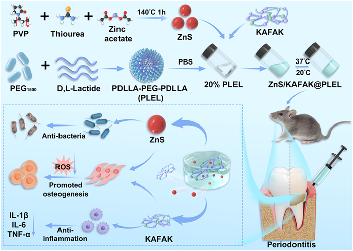
Scheme 1.
Schematic illustration of the synthesis of ZnS/KAFAK@PLEL hydrogel and its multiple function in promoting alveolar bone regeneration.

Thermosensitive composite hydrogel with multifunction for sustainable alveolar bone regeneration in periodontitis
Xinlong He , Shoushan Hu , Zejin Liu , Xicheng Li , Kun Shi , Jie Xiang , Xueman Zhou , Yating Yi , Zhenzhen Zhang , Ruichen Dan , Jiaqi Liu , Chengxinyue Ye , Jun Wang , Zhiyong Qian , Jin Liu


Periodontitis, a chronic inflammatory disease initiated by dental plaque [1], exhibited a pooled prevalence of approximately 61.6% in dentate adults (2011–2020), with severe cases accounting for 23.6% according to recent epidemiological studies [2]. The disease manifests through gingival symptoms (swelling, bleeding, recession) and alveolar bone resorption [3], while also demonstrating systemic implications for conditions including diabetes and Alzheimer's disease [4,5]. However, traditional treatment, such as guided tissue regeneration, has limited efficacy in sustained clearance of pathogens and periodontal regeneration [6]. Therefore, it is necessary to development novel therapeutic approaches to promote alveolar bone regeneration.
Human periodontal ligament stem cells (hPDLSCs) play a crucial role in alveolar bone regeneration owing to their osteogenic differentiation capacity [7,8]. But, within the local microenvironment of periodontitis, multiple adverse factors compromise the osteogenic differentiation potential of hPDLSCs, such as excessive reactive oxygen species (ROS) and elevated levels of inflammatory cytokines, such as interleukin-1β (IL-1β), IL-6, and tumor necrosis factor-α (TNF-α) [9–11]. Additionally, periodontal pathogens can aggravate inflammatory responses by secreting lipopolysaccharide (LPS), thereby disrupting the regeneration of periodontal tissues [12]. Therefore, eliminating local pathogens while reducing ROS and inflammatory cytokine levels can help restore the osteogenic differentiation capacity of hPDLSCs and promote periodontal regeneration [13–15]. However, conventional antibiotic therapy has several limitations, including inefficient drug delivery, the risk of antibiotic resistance, and the inability to modulate the periodontal microenvironment. These challenges have driven researchers to explore novel multifunctional bioactive materials that can bypass antibiotic resistance risks while promoting periodontal regeneration by restoring microenvironmental homeostasis [16,17].
Antimicrobial therapy for periodontitis currently faces significant challenges due to the growing issue of antibiotic resistance. While antibiotics remain a cornerstone of clinical treatment, the emergence of drug-resistant bacterial strains has substantially diminished their effectiveness, and the required higher dosages raise concerns about potential adverse effects. Consequently, there is an urgent need to develop novel treatment strategies that can overcome antibiotic resistance and improve clinical outcomes in periodontitis management [15,18,19]. As a zinc-based material, zinc sulfide (ZnS) recently have shown promise in biomaterials for tissue repair [20,21]. The relevant research reports their application in photothermal cancer therapy and regulation of redox homeostasis to promote skin wound healing [22–25]. ZnS nanoparticles can release zinc ions under inflammatory conditions, which exhibit antibacterial properties and contribute to stimulating osteoblastic differentiation [21,26]. Furthermore, compared to other Zn-based biomaterials like zeolitic imidazolate ZIF-8, ZnS will not generate harmful byproducts in degradation [27]. These advantages credit ZnS with potential for application in bacteria-rich oral environments. Apart from nanomaterials, it is noteworthy that there is an anti-inflammatory peptide (KAFAKLAARLYRKALARQLGVAA, KAFAK) for cartilage repair of knee arthritis. It is a cell-penetrating peptide that can reduce the secretion of inflammatory factors by inhibiting the effect of mitogen-activated protein kinase on activated protein kinase 2 [28]. Studies have confirmed that KAFAK peptides have reduced ex vivo inflammation and promoted the generation of cartilage extracellular matrix [29,30]. These evidences imply the potential of KAFAK peptides for bone defect repair in inflammatory environments.
However, drugs exhibit extremely limited retention time within the complex periodontal pocket, making it difficult to achieve sustained antibacterial, anti-inflammatory and osteoinducible effects. In recent years, drug-loaded hydrogels have emerged as a promising minimally invasive local drug delivery strategy for periodontitis, attracting significant attention due to their advantages in controlled release, excellent biocompatibility, and prolonged drug retention [31]. Among these, thermosensitive hydrogels offer unique therapeutic benefits as they can fully infiltrate deep periodontal pockets in a sol state and then undergo a sol-gel transition to enable sustained drug release [32,33]. Building on this concept, we developed a thermosensitive and highly biocompatible triblock copolymer hydrogel, poly(D,L-lactic acid)-block-poly(ethylene glycol)-block-poly(D,L-lactic acid) (PDLLA-PEG-PDLLA, PLEL). PLEL-based multifunctional composite hydrogels have already demonstrated broad therapeutic potential in osteoarthritis, skin defect repair, and tumor treatment [34–37]. Given these material properties and the pathological features of periodontitis, we fabricated a multifunctional hydrogel platform, ZnS/KAFAK@PLEL, by incorporating antibacterial/osteoinducible ZnS nanoparticles and the anti-inflammatory peptide KAFAK (Scheme 1). The key advancements of our ZnS/KAFAK@PLEL system are: (1) Antibacterial, anti-inflammatory, osteoinducible, and sustained-release properties are integrated into a single, injectable platform. This allows for simultaneous targeting of the key pathological facets of periodontitis, which encompass bacterial infection, inflammation and bone loss. (2) ZnS nanoparticles and KAFAK peptide are demonstrated to have a synergistic osteoinducible effect on hPDLSCs under oxidative stress. (3) The composite hydrogel is characterized by rapid gelation (< 1 min at 37 ℃) and suitable rheological properties, specifically designed to facilitate retention in the periodontal pocket. This study aims to evaluate the hydrogel's biocompatibility, antioxidant activity, antibacterial efficacy and ability to promote osteogenic differentiation of hPDLSCs, as well as its regenerative effects in a rat periodontitis model. Our findings may provide a novel comprehensive strategy for periodontitis treatment.
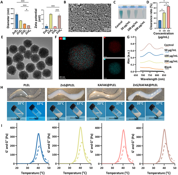
Specifically, to achieve this objective, we first focused on screening ZnS nanoparticles with optimal physicochemical properties. Four variants of ZnS nanoparticles (ZnSa-d) prepared by different synthesis methods were systematically characterized using dynamic light scattering and zeta potential analysis (Fig. 1A) to investigate the influence of synthesis processes on material characteristics and identify the optimal candidate. The data revealed hydrodynamic diameters of 570.0 ± 49.50 nm for ZnSa, 319.7 ± 6.31 nm for ZnSb, 191.8 ± 1.06 nm for ZnSc, and 124.6 ± 0.25 nm for ZnSd. The corresponding zeta potentials were 2.60 ± 1.767, 15.50 ± 0.85, 2.21 ± 0.13, and 13.87 ± 1.71 mV, respectively. Among the tested samples, ZnSd nanoparticles exhibited the smallest size, the narrowest size distribution, and demonstrated excellent monodispersity, homogeneity, stability, and batch-to-batch reproducibility. Given that smaller particle sizes typically confer a higher specific surface area and enhanced surface reactivity, ZnSd was selected for the subsequent construction of the composite material. The structural characteristics of the selected ZnSd nanoparticles were further confirmed through multiple analytical techniques. Size distribution analysis indicated good dispersity of the particles (Fig. S1A in Supporting information). Transmission and scanning electron microscopy (TEM and SEM) images revealed dense, spherical structures of uniform size (Figs. 1B and E). Elemental composition analysis (Fig. S1B in Supporting information) determined average atomic percentages of O: 27.79% ± 2.31%, S: 36.37% ± 4.65%, and Zn: 35.84% ± 3.91%. Elemental mapping (Fig. 1F) further confirmed the homogeneous distribution of S (red) and Zn (teal), as well as their co-localization in ZnS, collectively verifying the successful synthesis of the ZnS nanoparticles. Considering that the pathological progression of periodontitis is closely associated with excessive ROS production and subsequent oxidative stress damage in the periodontal microenvironment, we specifically evaluated the antioxidant capacity of the ZnS nanoparticles. The experimental results demonstrated a concentration-dependent ROS scavenging effect: With increasing concentrations, the ZnS nanoparticles significantly reduced ROS levels by 32%, 60%, and 84%, respectively (Figs. 1C, D, and G). These findings strongly indicate that ZnS nanoparticles function as efficient antioxidants, effectively scavenging ROS and providing a theoretical foundation for their application in periodontitis treatment. To verify the successful synthesis of the PLEL copolymer, we analyzed the copolymer using proton nuclear magnetic resonance (1H NMR) spectroscopy. In Fig. S2 (Supporting information), the spectrum exhibits characteristic peaks corresponding to hydrogen atoms at specific sites. This observation confirms the synthesis of the target PLEL copolymer according to the previous research [33]. Based on these findings, we proceeded to construct a ZnS/KAFAK@PLEL composite hydrogel system. The composite was prepared via simple physical mixing, which is a straightforward and highly reproducible method well-suited for developing injectable therapeutic systems. However, the precise nature of the molecular/ionic interactions involved (which may include electrostatic forces, coordination bonds, or hydrophobic interactions) requires further systematic investigation. To determine whether the incorporation of ZnS and KAFAK altered the physicochemical properties of the PLEL hydrogel, we systematically evaluated the thermosensitive behavior and antioxidant capacity of the composite hydrogels through rheological characterization and 2′-azinobis-(3-ethylbenzothiazoline-6-sulfonic acid) (ABTS) radical scavenging assays. As shown in Fig. 1H, all hydrogels maintained fluidity at 20 ℃ and could be easily extruded through a syringe. Upon heating to 37 ℃, the systems underwent a rapid sol-gel transition within 1 min. Furthermore, rheological data for the composite systems (Fig. 1I) indicated that the storage modulus (G') exceeded the loss modulus (G'') at 37 ℃ for all formulations, confirming their potential for gelation at body temperature, even with the incorporation of ZnS and KAFAK. The viscosity-temperature profile (Fig. S3A in Supporting information) further revealed negligible post-gelation viscosity changes for the ZnS/KAFAK@PLEL composite gel. Additionally, Fig. S3B (Supporting information) illustrates the sustained release profile of ZnS nanoparticles from the ZnS/KAFAK@PLEL system, a behavior that facilitates effective ROS scavenging and underscores the material's antioxidant functionality. These results collectively demonstrate that the incorporation of ZnS nanoparticles and the KAFAK peptide does not adversely affect the fundamental physicochemical properties of the thermo-sensitive PLEL hydrogel, thereby supporting its potential for subsequent investigation.
To evaluate the potential application of the composite hydrogel in treating periodontitis, we conducted a series of experiments to evaluate its antibacterial effect on Porphyromonas gingivalis and Staphylococcus aureus. The antibacterial properties of the materials were comprehensively analyzed using three complementary methods: Colony counting, live/dead bacterial staining, and SEM. Colony counting results (Fig. S4 in Supporting information) demonstrated that while the pure PLEL hydrogel and the KAFAK@PLEL group exhibited only limited inhibitory effects on both bacterial species, the experimental groups containing ZnS (ZnS@PLEL and ZnS/KAFAK@PLEL) displayed nearly complete bactericidal efficacy. This marked difference confirms that ZnS nanoparticles are the core component responsible for the antibacterial functionality of the composite system. To further validate this conclusion, we performed live/dead bacterial staining for direct visualization. Fluorescence images (Fig. S5 in Supporting information) revealed a predominance of green viable bacteria in the groups without ZnS, whereas the ZnS-treated groups exhibited extensive red fluorescence. This indicates that the membrane integrity of the vast majority of bacteria was compromised, resulting in bacterial death. Observations of bacterial ultrastructural changes via SEM (Fig. S6 in Supporting information) provided further insight into the antibacterial mechanism of ZnS. Bacteria in the control group displayed intact and smooth surfaces. In contrast, the ZnS-treated groups showed numerous nanoparticles adhered to the bacterial surfaces, causing irreversible damage such as membrane shrinkage and collapse. This morphological evidence directly confirms a direct interaction between the ZnS nanoparticles and the bacteria. In summary, multiple lines of experimental evidence collectively demonstrate that the ZnS/KAFAK@PLEL composite hydrogel, through the effective loading of ZnS nanoparticles, efficiently inhibits key periodontal pathogens. The antibacterial mechanism of the ZnS-containing hydrogel, involving zinc ion release and membrane interactions, warrants further investigation using methods like inductively coupled plasma mass spectrometry (ICP-MS) and high-resolution electron microscopy.
We further evaluate the effect of the composite hydrogel on the hPDLSCs, which were isolated from fresh human premolars extracted for orthodontic purposes from patients under 20 years old in the West China Hospital of Stomatology, Sichuan University (ethical approval No WCHSIRB-D-2024–354). Optical microscopy observation of hPDLSCs revealed that the cells exhibited a typical fibroblast-like spindle morphology in purified cultures and demonstrated robust proliferative capacity (Fig. S7A in Supporting information). Flow cytometric analysis confirmed their stem cell properties, demonstrating high expression (98.86%) of the mesenchymal stem cell marker CD90, whereas the expression of hematopoietic/endothelial markers (CD31 and CD45) was minimal (< 1%) (Fig. S7B in Supporting information). These results verified the high purity of the isolated hPDLSCs, meeting the requirements for subsequent experiments. Following validation of the cellular model, we assessed the biocompatibility of ZnS nanoparticles and KAFAK peptide with hPDLSCs. Cell counting kit-8 (CCK-8) assays revealed that ZnS nanoparticles at concentrations ≤400 µg/mL maintained cell viability comparable to controls, whereas higher concentrations (800–1600 µg/mL) surprisingly promoted cell proliferation (Fig. S8A in Supporting information). Live/dead staining further confirmed excellent biosafety, showing normal morphology and high survival rates even at the maximum concentration of 1600 µg/mL (Fig. S8B in Supporting information). For the KAFAK peptide, viability assays indicated no significant effects at concentrations ≤100 µg/mL. Although viability decreased to 81.30% ± 6.27% at 200 µg/mL, this reduction was not statistically significant (P = 0.068). However, significant cytotoxicity emerged at 400 µg/mL, reducing viability to 49.38% ± 2.70% (Fig. S9A in Supporting information). Live/dead staining visually confirmed this trend, showing diminished green fluorescence and morphological abnormalities at 200 µg/mL, and extensive cellular damage at 400 µg/mL (Fig. S9B in Supporting information). Furthermore, examination of hPDLSCs morphology after 24-h culture with hydrogel extracts showed no significant differences in cell density among groups (Fig. S10A in Supporting information). Cell viability assays demonstrated comparable survival rates between experimental and control groups without statistical significance (Fig. S10B in Supporting information). In brief, the abovementioned results collectively demonstrate that both ZnS nanoparticles (≤1600 µg/mL) and KAFAK peptide (≤100 µg/mL) exhibit excellent biocompatibility with hPDLSCs, and that the composite hydrogel itself exhibits outstanding biosafety. These findings provide a reliable concentration window for subsequent functional studies of the composite hydrogel system.
The excessive inflammatory response within the periodontal microenvironment is a central driver of tissue destruction. To effectively modulate this pathological process, we first evaluated the ability of the KAFAK peptide to regulate macrophage polarization. By quantifying the expression levels of key pro-inflammatory cytokines (IL-1β, IL-6, TNF-α) before and after LPS stimulation using quantitative real-time polymerase chain reaction (qPCR), we found that KAFAK treatment significantly modulated cellular inflammation levels. As shown in Fig. S11 (Supporting information), the mRNA expression of all three inflammatory cytokines (IL-1β, IL-6 and TNF-α) exhibited a clear dose-dependent decrease, with the most pronounced inhibitory effect observed at 100 µg/mL. This result not only confirms the capacity of the KAFAK peptide to modulate the inflammatory microenvironment but also provides a precise concentration basis for its application in periodontitis therapy.
Concurrently, the oxidative stress state within the periodontal microenvironment exacerbates tissue damage and further inhibits the osteogenic differentiation process. To address this limitation, we investigated the pro-osteogenic capacity of ZnS nanoparticles under both physiological and stress conditions. Under standard culture conditions, alkaline phosphatase (ALP) and Alizarin Red S (ARS) staining (Fig. S12A in Supporting information) revealed that ZnS exerted its most significant pro-osteogenic effect on hPDLSCs at 400 µg/mL. More importantly, in an oxidative stress environment induced by H2O2 (Fig. S12B in Supporting information), as the concentration of ZnS particles increased to 1200 µg/mL, the suppressed osteogenic differentiation capacity was restored to near-normal levels. The ARS semi-quantitative analysis results (Fig. S13 in Supporting information) further validated the efficacy of ZnS in mitigating oxidative stress damage.
Through systematic screening, this study has successfully identified the optimal concentration of the KAFAK peptide (100 µg/mL) for modulating the inflammatory microenvironment, as well as the optimal working concentrations for ZnS nanoparticles under both physiological (400 µg/mL) and stress (1200 µg/mL) conditions. These findings provide critical parameters and theoretical support for the subsequent development of a composite therapeutic system capable of simultaneously addressing the challenging inflammatory milieu and promoting bone regeneration.
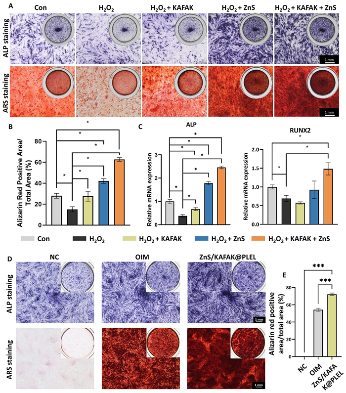
Based on these findings, we further employed ALP and ARS staining to evaluate the synergistic effects of the KAFAK peptide and ZnS nanoparticles on the osteogenic differentiation of hPDLSCs under ROS-induced stress conditions. As shown in Fig. 2A, under H2O2-induced oxidative stress, the staining intensity for both ALP and ARS was markedly reduced in the control group, indicating significant suppression of osteogenic differentiation. Treatment with KAFAK peptide alone partially reversed this inhibition, with ARS staining returning to near-baseline levels, primarily attributable to its anti-inflammatory properties. In contrast, ZnS nanoparticles demonstrated more potent pro-osteogenic effects, consistent with their antioxidant capacity and direct osteogenic activity. Notably, the combination of KAFAK and ZnS produced the most pronounced enhancement in staining, demonstrating a clear synergistic effect. Semi-quantitative analysis of ARS staining (Fig. 2B) provided statistical confirmation of this synergistic interaction. At the molecular level, analysis of osteogenic markers (Fig. 2C) revealed that the mRNA expression of both ALP and RUNX2 in the KAFAK+ZnS group was significantly higher than in any single-treatment group. This finding further validates their synergistic role in regulating osteogenic differentiation at the transcriptional level. Building upon these component-level findings, we established a sustained-releasing co-culture model to assess the pro-osteogenic capacity of the ZnS/KAFAK@PLEL composite hydrogel system. Following 21 days of osteogenic induction, the ZnS/KAFAK@PLEL group demonstrated significantly enhanced ALP activity and more intense ARS staining compared to the standard osteogenic induction medium group (Fig. 2D), indicating superior promotion of mineralized nodule formation. Semi-quantitative analysis of ARS staining intensity (Fig. 2E) confirmed that the differences between groups were statistically significant (P < 0.05).
The rat periodontitis model was used to evaluate the effect of the composite hydrogel (Fig. S14 in Supporting information). The animal study was approved by the Animal Ethics Committee of West China Hospital of Stomatology, Sichuan University (No. WCHSIRB-D-2024–541). After treatment, detailed three-dimensional reconstruction and quantitative analysis of the palatal alveolar bone surrounding the maxillary second molars in rats were conducted using micro computed tomography (micro-CT) scanning. Morphometric analysis (Fig. 3A) revealed a significant increase (P < 0.01) in the cementoenamel junction-to-alveolar bone crest (CEJ-ABC) distance in the untreated periodontitis group compared to the healthy control, confirming successful induction of alveolar bone loss. Among all treatment groups, the ZnS/KAFAK@PLEL group exhibited the smallest CEJ-ABC distance, demonstrating superior bone defect repair capacity. Further qualitative analysis (Figs. 3B and C) showed that the bone mineral density (BMD) and bone volume fraction (BV/TV) values in the ZnS/KAFAK@PLEL group were closest to those of the healthy control and significantly superior to single-component treatment groups. This indicates that the composite system not only restores macroscopic bone architecture but also effectively ensures the quality of newly formed bone. Three-dimensional reconstruction images (Fig. 3D) visually confirmed that the alveolar bone morphology in this treatment group most closely resembled that of the healthy control, validating its exceptional bone regeneration capability from a morphological perspective.
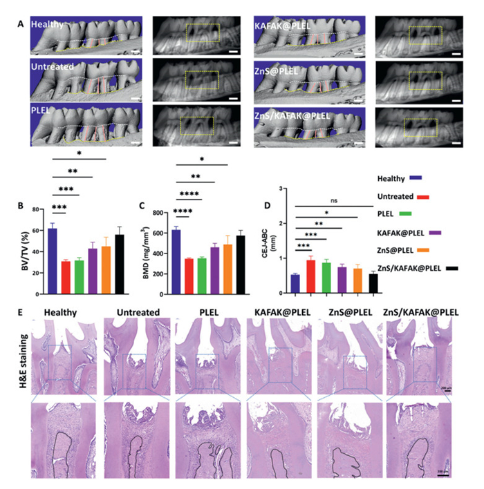
Histological analysis of rat alveolar bone samples using hematoxylin and eosin (H&E) staining further revealed the regenerative effects of the composite hydrogel on periodontal tissues at the microscopic level. As shown in Fig. 3E, both the untreated and pure PLEL groups exhibited significant alveolar bone resorption with irregular bone crest contours. In contrast, all active component treatment groups showed varying degrees of reduction in alveolar ridge height loss, consistent with the micro-CT quantitative results. These multi-level findings collectively demonstrate that the ZnS/KAFAK@PLEL nanocomposite hydrogel effectively promotes localized alveolar bone regeneration in the periodontitis microenvironment through synergistic anti-inflammatory, antioxidant and osteoinducible functions.
In summary, the ZnS/KAFAK@PLEL composite hydrogel system not only exhibits sustained ZnS nanoparticle release and excellent biocompatibility, but also effectively scavenges excess reactive oxygen species to alleviate oxidative stress damage. Furthermore, it demonstrates broad-spectrum antibacterial activity, thereby creating a favorable microenvironment for tissue regeneration. In vivo evaluation in a rat periodontitis model confirmed that the hydrogel treatment significantly inhibited alveolar bone resorption, restored bone mineral density, increased bone volume fraction, and promoted periodontal tissue regeneration. The successful validation of this synergistic biofunctional triad establishes an experimental foundation for developing next-generation periodontal regenerative biomaterials. Future work will employ transcriptomic approaches (e.g., RNA sequencing) to comprehensively elucidate the underlying molecular mechanisms, potentially clarifying the signaling networks and interdependent pathways responsible for these observed effects.
Xinlong He: Writing – review & editing, Writing – original draft, Visualization, Validation, Supervision, Software, Resources, Methodology, Investigation, Formal analysis, Data curation, Conceptualization. Shoushan Hu: Writing – review & editing, Writing – original draft, Visualization, Validation, Software, Resources, Project administration, Methodology, Investigation, Formal analysis, Data curation, Conceptualization. Zejin Liu: Methodology, Formal analysis, Data curation. Xicheng Li: Software, Methodology, Investigation. Kun Shi: Software, Resources, Methodology, Investigation. Jie Xiang: Software, Methodology, Data curation. Xueman Zhou: Software, Resources, Data curation. Yating Yi: Software, Methodology. Zhenzhen Zhang: Software, Methodology. Ruichen Dan: Software, Formal analysis. Jiaqi Liu: Visualization, Methodology. Chengxinyue Ye: Software, Resources. Jun Wang: Visualization, Validation, Supervision, Resources, Project administration, Methodology, Investigation, Funding acquisition, Conceptualization. Zhiyong Qian: Writing – review & editing, Visualization, Validation, Supervision, Software, Resources, Project administration, Methodology, Investigation, Funding acquisition, Formal analysis, Conceptualization. Jin Liu: Supervision, Project administration, Methodology, Investigation, Funding acquisition, Data curation, Conceptualization.
The authors declare that they have no known competing financial interests or personal relationships that could have appeared to influence the work reported in this paper.
This research was supported by National Natural Science Foundation of China (Nos. 82472149, U21A20417, 82471024, 82271019), Sichuan Science and Technology Program (No. 24ZDYF0099), the Nature Science Foundation of Sichuan Province (No. 2024NSFSC0046), the Noncommunicable Chronic Diseases-National Science and Technology Major Project (No. 2024ZD0522800 to P. Tian, No 2024ZD0522803 to Z. Qian) and the 1·3·5 Project for Disciplines of Excellence, West China Hospital, Sichuan University (No. ZYGD24003).
Supplementary material associated with this article can be found, in the online version, at doi:
B.L. Pihlstrom, B.S. Michalowicz, N.W. Johnson, Lancet 366 (2005) 1809–1820. doi: 10.1016/S0140-6736(05)67728-8
D. Trindade, R. Carvalho, V. Machado, et al., J. Clin. Periodontol. 50 (2023) 604–626. doi: 10.1111/jcpe.13769
M.S. Tonetti, S. Jepsen, L. Jin, J. Otomo-Corgel, J. Clin. Periodontol. 44 (2017) 456–462. doi: 10.1111/jcpe.12732
S.S. Dominy, C. Lynch, F. Ermini, et al., Sci. Adv. 5 (2019) eaau3333. doi: 10.1126/sciadv.aau3333
P.M. Preshaw, A.L. Alba, D. Herrera, et al., Diabetologia 55 (2012) 21–31. doi: 10.1007/s00125-011-2342-y
F.M. Chen, J. Zhang, M. Zhang, et al., Biomaterials 31 (2010) 7892–7927. doi: 10.1016/j.biomaterials.2010.07.019
B.M. Seo, M. Miura, S. Gronthos, et al., Lancet 364 (2004) 149–155. doi: 10.1016/S0140-6736(04)16627-0
A. Tomokiyo, N. Wada, H. Maeda, Stem Cells Dev. 28 (2019) 974–985. doi: 10.1089/scd.2019.0031
F.S.C. Sczepanik, M.L. Grossi, M. Casati, et al., Periodontol. 2000 84 (2020) 45–68. doi: 10.1111/prd.12342
C. Pouliou, C. Piperi, Curr. Med. Chem. 31 (2024) 6187–6203. doi: 10.2174/0109298673297545240507091410
J.M. Yang, Y. Zhu, D.Y. Duan, et al., Arch. Oral. Biol. 96 (2018) 234–242. doi: 10.1016/j.archoralbio.2017.03.006
J.Y. Liu, B. Chen, J. Bao, et al., Stem Cell Res. Ther. 10 (2019) 320. doi: 10.1080/00207721.2018.1552765
Z.Y. Sun, T.Y. Wang, E. Chen, et al., Mater. Horiz. 12 (2025) 608–622. doi: 10.1039/d4mh01137f
E.N. Chen, T.Y. Wang, Z.Y. Sun, et al., Regen. Biomater. 11 (2024) rbae058. doi: 10.1093/rb/rbae058
B.R. Zeng, Z.X. Mu, T.X. Shen, et al., Chin. Chem. Lett. 36 (2025) 110350. doi: 10.1016/j.cclet.2024.110350
C.H. Zheng, J. Xi, S.Y. Long, et al., Chin. Chem. Lett. 36 (2025) 110223. doi: 10.1016/j.cclet.2024.110223
J.Y. Luan, R.T. Li, W.G. Xu, et al., Acta Pharm. Sin. B 13 (2023) 2310–2333. doi: 10.1016/j.apsb.2022.10.026
D. Kanakaraju, A. Chandrasekaran, Sci. Total. Environ. 868 (2023) 161525. doi: 10.1016/j.scitotenv.2023.161525
J.J. Wang, X.X. Ge, Y.J. Xiang, et al., Chin. Chem. Lett. 36 (2025) 109819. doi: 10.1016/j.cclet.2024.109819
X.L. Qi, Y. Li, Y.J. Xiang, et al., Cell Biomater. 1 (2025) 100020. doi: 10.1016/j.celbio.2025.100020
Y.C. Su, I. Cockerill, Y.D. Wang, et al., Trends Biotechnol. 37 (2019) 428–441. doi: 10.1016/j.tibtech.2018.10.009
B. Han, W.H. Fang, S. Zhao, et al., Nanomed. Nanotechnol. Biol. Med. 29 (2020) 102263. doi: 10.1016/j.nano.2020.102263
J.Q. Yang, W. Guo, R. Huang, et al., Front. Immunol. 14 (2023) 1173487. doi: 10.3389/fimmu.2023.1173487
B. Zhou, B.X. Jia, M.J. Zhang, et al., Colloids. Surf. B: Biointerfaces 226 (2023) 113313. doi: 10.1016/j.colsurfb.2023.113313
J. Zhang, X. Liu, Y. Sun, et al., Int. J. Biol. Macromol. 304 (2025) 140787. doi: 10.1016/j.ijbiomac.2025.140787
K.S. Yuan, C.C. Deng, L.L. Tan, et al., Bioact. Mater. 35 (2024) 306–329.
X.Y. Wen, J. Wang, X.B. Pei, X. Zhang, J. Mater. Chem. B 11 (2023) 11405–11425. doi: 10.1039/d3tb01874a
J.L. Brugnano, B.K. Chan, B.L. Seal, A. Panitch, J. Control. Release 155 (2011) 128–133. doi: 10.1016/j.jconrel.2011.05.007
K. Zhou, C.L. Yang, K. Shi, et al., Biomaterials 295 (2023) 122036. doi: 10.1016/j.biomaterials.2023.122036
J.B. Lin, S. Poh, A. Panitch, Nanomed. Nanotechnol. Biol. Med. 12 (2016) 2095–2100. doi: 10.1016/j.nano.2016.05.010
R. Das, N. Suryawanshi, N. Burnase, et al., Dent. Mater. 41 (2025) 81–99. doi: 10.1016/j.dental.2024.10.017
Q.Q. Pan, Z.H. Zong, H.B. Li, et al., Int. J. Biol. Macromol. 284 (2025) 137893. doi: 10.1016/j.ijbiomac.2024.137893
K. Shi, Y.L. Wang, Y. Qu, et al., Sci. Rep. 6 (2016) 19077. doi: 10.1038/srep19077
J.N. Li, L. Li, T.K. Wu, et al., Small Methods 8 (2024) e2300843. doi: 10.1002/smtd.202300843
J.F. Liu, X. Li, J.W. Chen, et al., J. Nanobiotechnology 22 (2024) 338. doi: 10.54097/nqj90n37
Z.Q. Zheng, S.Q. Bian, Z.Q. Li, et al., Carbohydr. Polym. 249 (2020) 116826. doi: 10.1016/j.carbpol.2020.116826
F. Yang, K. Shi, Y. Hao, et al., Bioact. Mater. 6 (2021) 3036–3048.

Scheme 1 Schematic illustration of the synthesis of ZnS/KAFAK@PLEL hydrogel and its multiple function in promoting alveolar bone regeneration.

Figure 1 Thermosensitivity and antioxidant performance. (A) Size and zeta potential of ZnS nanoparticles. (B) SEM image of ZnS (scale bar: 500 nm). (C) Visual results of ABTS radical scavenging assay. (D) ROS clearance rates of ZnS nanoparticles at different concentrations. (E) TEM image (scale bar: 50 nm). (F) Elemental distribution of nanoparticles (scale bar: 20 nm). (G) UV–vis absorption spectra of ABTS. (H) Injectability at 20 ℃ and Sol-gel transition at 37 ℃. (I) G'/G" profiles. The images represent PLEL, ZnS@PLEL, KAFAK@PLEL, and ZnS/KAFAK@PLEL, respectively. *P < 0.05. ***P < 0.001. Data are presented as mean ± standard deviation (SD) (n = 3).

Figure 2 In vitro osteogenic promotion of hPDLSCs. (A) ALP and ARS staining images. (B) Semi-quantitative analysis of ARS staining. (C) Relative mRNA expression of ALP and RUNX2. (D) ALP and ARS staining. (E) Bar graph quantifying alizarin red positive area percentage. *P < 0.05, ***P < 0.001. Data are presented as mean ± SD (n = 3).

Figure 3 Therapeutic efficacy in rat periodontitis model. (A) Three-dimensional reconstructions and corresponding X-ray micrographs of rat maxillary second molar palatal alveolar bone across various groups (scale bar: 1 mm). (B) Bar graph of BV/TV across groups. (C) Bar graph of BMD across groups. (D) Bar graph of CEJ-ABC distance across groups. (E) H&E staining of periodontal tissues post-treatment across various groups (scale bar: 200 µm). ns, not significant. *P < 0.05, **P < 0.01, ***P < 0.001, ****P < 0.0001. Data are presented as mean ± SD (n = 5).

 扫一扫看文章


扫一扫关注我们
 DownLoad:
 下载:
 下载: