Citation:
Song Wei, Zhang Hai-Juan, Liu Ying-Hua, Ren Cui-Ling, Chen Hong-Li. A new fluorescence probing strategy for the detection of parathion-methyl based on N-doped carbon dots and methyl parathion hydrolase[J]. Chinese Chemical Letters,
2017, 28(8): 1675-1680.
doi:
10.1016/j.cclet.2017.05.001

A new fluorescence probing strategy for the detection of parathion-methyl based on N-doped carbon dots and methyl parathion hydrolase
English
A new fluorescence probing strategy for the detection of parathion-methyl based on N-doped carbon dots and methyl parathion hydrolase
Abstract:
A new facile fluorescence probing strategy, which was based on N-doped carbon dots (NCDs) and methyl parathion hydrolase (MPH), was developed for the determination of parathion-methyl (PM). The fluorescence intensity of NCDs-MPH system was proportional to PM concentration in the range of 2.38-73.78 μmol/L, with a detection limit of 0.338 μmol/L. Moreover, the present simple and facile method could be used to determine methyl parathion in environmental and agricultural samples successfully. Furthermore, the detection mechanism of this system is inner filter effect and molecular interactions between NCDs and p-nitrophenol, which is the hydrolysis product of PM catalyzed by methyl parathion hydrolase.
-
Key words:
- N-doped carbon dots
- / Parathion-methyl
- / Methyl parathion hydrolase
- / Inner filter effect
- / Fluorescence
-
1. Introduction
Parathion-methyl (O, O-dimethyl-O-4-nitrophenyl phosphorothioate, PM) is an organophosphorus insecticide that widely used for control insects of crops, fruits and vegetables. However, PM is classified to be ‘Category Ia' (extremely toxic) by the World Health Organization (WHO) and ‘Toxicity Category Ⅰ' (most toxic) by the United States Environmental Protection Agency (USEPA). The high toxicity of organophosphate pesticides (OPs) is owing to their disruption to mammal nervous system [1-3]. Furthermore, this pesticide can be concentrated in agricultural products and environment, thus threatening the environmental safety [4]. Therefore, it is significant to develop viable method to detect the PM residues in environmental and agricultural samples.
During the past decade, a large number of methods have been devised to detect PM, such as chromatography (including gas chromatography and liquid chromatography) methods [5-8] and electrochemical detection [9-14]. These methods have high sensitivity and adequate selectivity, nevertheless most of these assays are expensive, laborious and time-consuming. So more rapid, simple and selective method is still needed for the determination of PM. For these reasons, some new analytical techniques have been developed based on acetyl cholinesterase (AChE) [15, 16], but such sensors showed low selectivity to OPs because the activity of AChE can also be inhibited by carbamates pesticide [17]. Some other sensors were developed based on organophosphorus hydrolase (OPH) by detecting the hydrolysate of OPs, such as p-nitrophenol and dimethylthiophosphoric acid (DMPA) [18-20]. As one kind of OPH, methyl parathion hydrolase (MPH), which is isolated from the soil-dwelling bacterium Pseudomonas sp. WBC-3, can only degrade organophosphorus compounds, such as methyl parathion, chlorpyrifos, malathion. In addition, MPH have many advantages over other enzymes in pesticide detection, such as less background noise and high turnover number, which is favorable for sensitive detection [21-23].
Carbon dots (CDs) have attracted much attention in recent years because of their low cytotoxicity, biocompatibility, hydrophilicity, and non-blinking emission, but the relatively low quantum yield of CDs hindered their applications. In order to improve the quantum yield of CDs, doping and surface passivation methods have been reported. Among these materials, nitrogen-doped CDs (NCDs) have attracted much attention because N-doping can tune the chemical, physical, and electronic properties of CDs. The good solubility, robust chemical inertness, inexpensive nature and strong luminescence of NCDs have made them become an important fluorescent nanosensor [24].
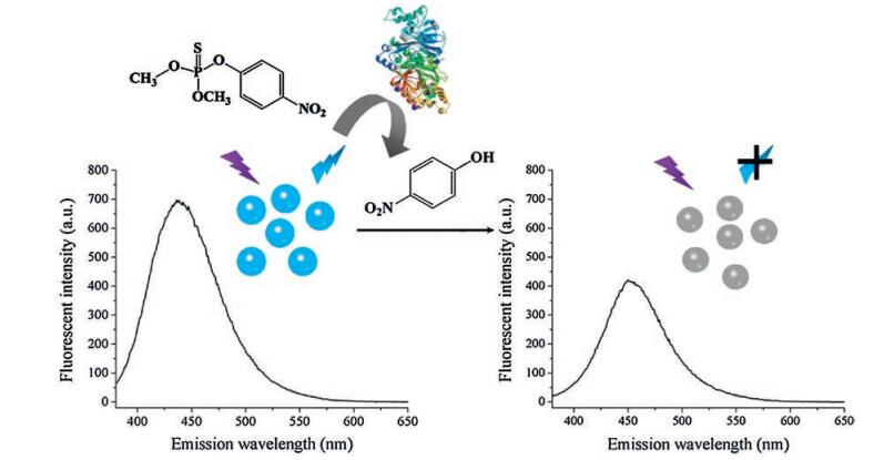
Hence, in this paper, NCDs and MPH were chosen as the sensing and detecting units for sensitive and selective detection of PM, respectively. The water-soluble NCDs were synthesized by solidphase synthesis. p-Nitrophenol (4-NP), the hydrolysis product of PM catalyzed by MPH, could efficient quench the fluorescence of the NCDs (Fig. 1). The detection linear range and detection limit were investigated. Furthermore, river water, soil and pear were chosen as the sample matrix to further investigate the practical application of the fluorescence sensor for PM detection.
图 1
2. Results and discussion
2.1 Characterization of NCDs
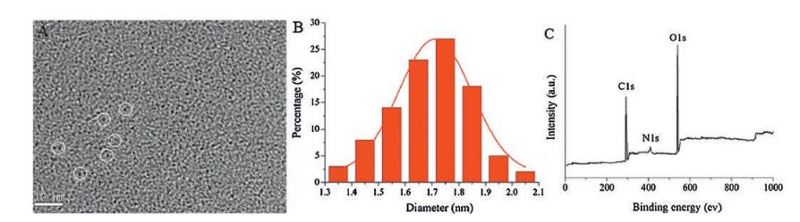
The morphology and size of the prepared NCDs were characterized by TEM (Fig. 2A), it clearly showed that the synthesized NCDs were monodisperse and uniform in size. Their average diameters were about 1.7 nm (Fig. 2B), which is in consistent with previous study [26]. In order to investigate the elemental composition for the resultant nanoparticles, XPS spectrum was recorded. As shown in Fig. 2C, it proved that the prepared carbon dots were composed of C, O and N, indicating the successful preparation of NCDs.
图 2
 图 2 TEM images (A) and the corresponding diameter distribution histograms (B) and wide XPS spectrum (C) of NCDs.Figure 2. TEM images (A) and the corresponding diameter distribution histograms (B) and wide XPS spectrum (C) of NCDs.
The optical properties of the prepared NCDs were characterized by UV-vis absorption and fluorescent spectra. As shown in Fig. S1 (Supporting information), the obtained NCDs have a strong absorption peak at 358 nm and a maximum emission at 435 nm excited at 360 nm. The fluorescence intensity of NCDs under different pH, ion concentration and temperature were shown in Fig. S2 (Supporting information). We found that the PL intensity of NCDs was relatively stable at different pH (1-10) (Fig. S2A) and ionic strength (Fig. S2B), but their fluorescent intensity decreased slightly when the temperature changed from 18 ℃ to 40 ℃ (Fig. S2C). These results suggest that the prepared NCDs have stable optical properties and can be used as good candidature for chemical sensing.
2.2 Optimization of the detection conditions
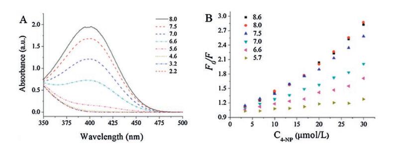
The adsorption of 4-NP varied obviously according the pH of buffer solution, as shown in Fig. 3A. The adsorption intensity of 4-NP at 400 nm gradually increased when the pH changed from 2.2 to 8.0. The influence of pH on the determination performance of NCDs towards 4-NP was also investigated, (Fig. 3B) though the optical properties of NCDs was stable in the pH range of 1-10. The fluorescence quenching efficiency (F0/F) by 4-NP was significantly affected by the pH of the solution. The fluorescence quenching efficiency was low when the pH was 5.7, and this response enhanced gradually when the pH increased from 5.7 to 8.6. Furthermore, previous work have reported that MPH can retain more than 90% of the maximum activity in the pH range from 7.0 to 9.0, and the optimal pH for MPH was 8.0 [27]. Therefore, pH 8.0 was chosen for further experiments.
图 3
 图 3 UV spectra of 4-NP at different pH (A) and fluorescence quenching efficiency (F0/F vs. 4-NP concentration) of NCDs by 4-NP at different pH values (B), where F0 and F are the fluorescence intensities of NCDs in the absence and presence of 4-NP, respectively.Figure 3. UV spectra of 4-NP at different pH (A) and fluorescence quenching efficiency (F0/F vs. 4-NP concentration) of NCDs by 4-NP at different pH values (B), where F0 and F are the fluorescence intensities of NCDs in the absence and presence of 4-NP, respectively.
The influences of PM and MPH to NCDs were investigated. As shown in Fig. S3 (Supporting information), the FL intensity of NCDs changed inconspicuously before and after the addition of various concentrations of MPH and PM, proving PM and MPH have little impacts on the optical property of NCDs. These results prove that the optical response of NCDs are stemming from 4-NP, the hydrolysate of PM. The effects of the addition sequence of PM and MPH were also investigated. As shown in Fig. S4 (Supporting information), the addition sequence of PM and MPH have little influences to the fluorescent intensity of NCDs.
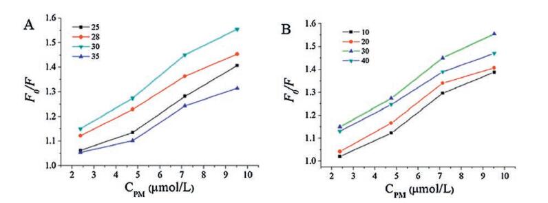
Temperature and reaction time is the other two important factors that affect the reaction between MPH and PM, so the effects of incubation time and temperature on the detection performance of NCDs were investigated. The results are shown in Fig. 4. As can be seen, F0/F was first increased when the temperature increased from 25 ℃ to 30 ℃ (Fig. 4A), then decreased when the temperature further increased to 35 ℃. This result was in consistence with the previous reports [22]. Therefore, the optimal incubation temperature for this system is 30 ℃. Then the incubation time was also optimized, the results are shown in Fig. 4B. We can see that the quenching efficiency reached maximum when the incubation time changed from 10 min to 30 min, and when the incubation time was prolonged to 40 min, the quenching efficiency was decreased. So the optimal incubation time and temperature was 30 min and 30 ℃, respectively.
图 4
 图 4 Fluorescence quenching efficiency (F0/F vs. PM concentration) of NCDs by PM when the NCDs-MPH system incubated at different temperature (A) and time (B).Figure 4. Fluorescence quenching efficiency (F0/F vs. PM concentration) of NCDs by PM when the NCDs-MPH system incubated at different temperature (A) and time (B).
2.3 Sensitive assay for PM
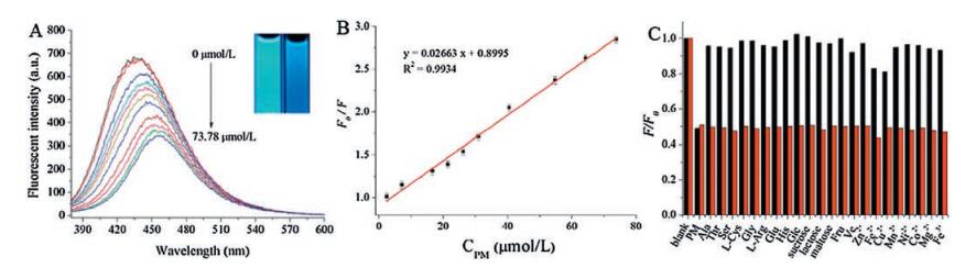
The feasibility of using NCDs as fluorescent probes for the detection of PM we explored. The fluorescence spectra of the mixture of NCDs and MPH with various concentration of PM were recorded (Fig. 5A) under the optimum conditions. Accordingly, the color of the mixture changed from bright blue to faint blue under UV irradiation (inset of Fig. 5A). The value of F0/F gradually increased upon the concentration of PM increased, and a good linearity was obtained when the PM concentration ranged from 2.38 μmol/L to 73.78 μmol/L with a coefficient of 0.9934 (Fig. 5B). In addition, the limit of detection was estimated to be 0.338 μmol/L, which was obtained based on 3 σ/κ (σ is the standard deviation of the blank measurements and κ is the slope of the calibration curve). This detection limit was comparable to or even lower than the previous reports. Further more, the presented method did not need complicated process, harsh conditions and expensive equipment (Table 1).
图 5
 图 5 Fluorescent spectra of mixture of NCDs and MPH upon addition of various concentrations of PM (from top to bottom 0, 2.38, 7.14, 16.6, 21.42, 26.18, 30.94, 40.46, 54.74, 64.26, 73.78 μmol/L), insets showed the photographs of the as prepared NCDs aqueous solutions in the absence and presence of 73.78 μmol/L PM under 365 nm UV light (A), dependence of F0/F on the concentration of PM within the range of 2.38-73.78 μmol/L (λem = 435nm) (B) and selectivity of NCDs for the detection of PM over other chemicals. The concentration of metal ions and other chemicals were 323.7 μmol/L, the concentration of PM was 40.46 μmol/L (C).Figure 5. Fluorescent spectra of mixture of NCDs and MPH upon addition of various concentrations of PM (from top to bottom 0, 2.38, 7.14, 16.6, 21.42, 26.18, 30.94, 40.46, 54.74, 64.26, 73.78 μmol/L), insets showed the photographs of the as prepared NCDs aqueous solutions in the absence and presence of 73.78 μmol/L PM under 365 nm UV light (A), dependence of F0/F on the concentration of PM within the range of 2.38-73.78 μmol/L (λem = 435nm) (B) and selectivity of NCDs for the detection of PM over other chemicals. The concentration of metal ions and other chemicals were 323.7 μmol/L, the concentration of PM was 40.46 μmol/L (C).
表 1
 表 1 Comparison of different methods for PM determination.Table 1. Comparison of different methods for PM determination.
 下载:
导出CSV
下载:
导出CSV

2.4 Anti-interference capability of the sensor
As this work is aimed at developing a new system for detecting PM residues, the anti-interference capability of the sensor was also estimated. The common coexisting substances in water, soil and fruit, including metal ions, amino acid and sugar, were chosen as interferences. As shown in Fig. 5C, only PM could quench the fluorescence of NCDs significantly (black column). Furthermore, the quenching efficiency of PM when these materials co-existed was used to evaluate the anti-interference abilities of NCDs (red column). These results revealed this NCDs based fluorescence probe possess high selectivity for detecting PM.
2.5 Quenching mechanism investigation
According to Fig. 5A, the fluorescence intensity of NCDs was gradually decreased with the concentration of PM increasing, the quenching mechanism may due to fluorescence resonance energy transfer (FRET) or internal filtering effect (IFE). FRET is a kind of nonradiation energy transfer, which can only occur over very short distances, accompanied by the fluorescence lifetime changed correspondingly [28, 29]. IFE refers to the decrease of fluorescence intensity caused by the absorption of nonfluorescent substance [30]. These two kinds of fluorescence quenching require that the donor's emission spectrum and the absorption spectrum of the acceptor have a very good overlap, which is the prerequisite for the higher FRET or IFE efficiency.
Fig. S3 proving the fluorescence quenching of NCDs was caused by 4-NP, the hydrolysate of PM. In order to study the fluorescence quenching mechanism of NCDs by 4-NP, the UV absorption spectra of 4-NP and the excitation and emission spectra of NCDs were investigated (Fig. 6A). It showed that 4-NP solution has a strong absorption in the range of 350-450 nm, which is greatly overlapped with the excitation and emission spectra of NCDs. Moreover, the fluorescence lifetime of NCDs before and after adding 4-NP was studied by time-resolved fluorescence spectroscopy. As shown in Fig. 6B, the fluorescence decay signals show a double exponential decay and the fluorescence lifetime of NCDs remained stable after the addition of 4-NP. This can exclude the occurrence of FRET between NCDs and 4-NP [31, 32], so IFE is considered as the main reason for fluorescence quenching [33]. On the other hand, the fluorescence emission peak of NCDs showed a gradual red shift with the concentrations of 4-NP increasing (Fig. 5A), which may stem from the molecular interactions (electrostatic interaction, hydrogen bonding interaction) [34-36]. So the possible mechanism for the fluorescence response of NCDs to 4-NP is the common effect of IFE and molecular interactions, the fluorescence quenching diagram is shown in Fig. 6C.
图 6
 图 6 UV-vis spectra (□) of 4-NP (pH = 8.0, blue line) and excitation (ο) and emission spectra (▲) of NCDs (A), time-resolved decays of the NCDs before (solid line) and after the addition of 4-NP (dash line) (B) and schematic illustration of the FL quenching mechanism of NCDs by 4-NP (C).Figure 6. UV-vis spectra (□) of 4-NP (pH = 8.0, blue line) and excitation (ο) and emission spectra (▲) of NCDs (A), time-resolved decays of the NCDs before (solid line) and after the addition of 4-NP (dash line) (B) and schematic illustration of the FL quenching mechanism of NCDs by 4-NP (C).
2.6 Application to real sample analysis
The excellent selectivitty and high sensitivity of the NCDsenzyme system towards PM demonstrate that it could be applied in real samples. Fruit, water and soil were used to evaluate the potential of this system for PM detection in various sample matrixes. A standard solution of PM (380 μmol/L) was added to the real samples at three different levels. As shown in Table 2, the recoveries were in the range of 95.1%-108%, which was satisfactory for application. These results further indicated this proposed method can be used as an alternative method for PM residues detection in real samples.
表 2
表 2 Detection results for PM in different samples by the proposed method.Table 2. Detection results for PM in different samples by the proposed method.下载:
导出CSV

3. Conclusion
A novel NCDs-MPH based sensing system was designed for the detection of PM. The fluorescence intensity of NCDs is proportional to PM concentration in the range of 2.38-73.78 μmol/L, with a detection limit of 0.338 μmol/L. Furthermore, our study demonstrates that FL quenching effect of p-nitrophenol on the NCDs is attributed to IFE and molecular interactions between them, while IFE is the main reasons. River water, soil and pear were chosen as the sample matrix to further investigate the practical application of the fluorescence sensing system for PM detection and the result is satisfactory, implying this sensing strategy is of prospective application for PM residue detection in foodstuffs and environmental samples.
4. Experimental
4.1 Materials and instrumentation
Polyoxyethylenebis (amine) (PEG-diamine, MW 2000) was purchased from Aladdin Chemistry Co., Ltd. (Shanghai, China). Citric acid was obtained from Tianjin Guangfu Chemical Reagents Co., Ltd. (Tianjin, China). The MPH was kindly supplied by State Key Laboratory of Virology, Wuhan Institute of Virology, Chinese Academy of Sciences. Methyl parathion (1.0 mg/mL, methanol) was purchased from J&K Scientific Ltd. (Beijing, China). All other reagents were of analytical grade and used without further purification. All solutions were prepared with deionized (DI) water. Soil samples were obtained from Qingyang, Gansu province, China. Water sample was obtained from Yellow River. Pear was bought from local market.
Morphology and size of the prepared NCDs was characterized by transmission electron microscope (TEM, Hitachi-600, Hitachi, Japan). Photoluminescent measurements were performed on an RF-5301PC fluorescence (FL) spectrophotometer using 5/5 nm slit width, and equipped with a 1 cm quartz cell (Shimadzu, Kyoto, Japan). The ultraviolet-visible (UV-vis) absorption spectra were acquired on a TU-1901 UV-vis Spectrophotometer with a 1 cm quartz cell (Beijing Purkinje General Instrument Co., Ltd., Beijing, China). Time-resolved fluorescence spectra were carried out in a time correlated single photon counting (TCSPC) system from FL920P spectrometer (Edinburgh Instruments, U.K.) with λex = 365 nm.
4.2 Synthesis of NCDs and fluorescence quenching experiments with PM
NCDs were prepared according to a previous reported method [26]. A certain amount of NCDs was added into 700 μL citric acid Na2HPO4 buffer solution (pH 8.0) and the fluorescent intensity was recorded as F0. For PM detection, 40 μL of MPH was added into the NCDs solution, and the mixture was incubated at 30 ℃ for 30 min. And then, 30 μL PM solutions with different concentrations, which were prepared by diluting different volume of PM standard solution using citric acid-Na2HPO4 buffer solution (pH 8.0), was added to the above reaction solution, and the fluorescent signal was recorded as F. The procedure for 4-NP detection was conducted by adding different concentration of 4-NP into the NCDs solution.
4.3 Analysis of practical samples
3 g Soil was extracted by 10 mL methanol with 10 min, after centrifugation, the supernatant was filtrated by 0.45 μm filtration membranes. Water sample was filtrated before measurement. 10 g pear were first chopped, and then mixed with 30 mL ether, the resulting mixture was filtered through a 0.45 μm membrane to remove the insoluble parts. The obtained filtrate was evaporated on a water bath, the residual was diluted by 3 mL methanol. The recovery test was carried out on all samples spiked with varying levels of PM. For each sample, three parallel experiments were conducted.
Acknowledgments
This work was supported by the National Natural Science Foundation of China (No. 21207057) and the Fundamental Research Funds for the Central Universities (No. lzujbky-2016-43). We specially thank Wuhan Institute of Virology, Chinese Academy of Sciences for providing MPH.
Appendix A. Supplementary data
Supplementary data associated with this article can be found, in the online version, at http://dx.doi.org/10.1016/j.cclet.2017.05.001.
-
-
[1]
Buratti F.M., Volpe M.T., Meneguz A., Vittozzi L., Testai E.. CYP-specific bioactivation of four organophosphorothioate pesticides by human liver microsomes[J]. Toxicol. Appl. Pharmacol., 2003, 186: 143-154. doi: 10.1016/S0041-008X(02)00027-3
-
[2]
Du D., Chen S., Cai J., Zhang A.. Immobilization of acetylcholinesterase on gold nanoparticles embedded in sol-gel film for amperometric detection of organophosphorous insecticide[J]. Biosens. Bioelectron., 2007, 23: 130-134. doi: 10.1016/j.bios.2007.03.008
-
[3]
Zhang H.X., Wei R.B., Chen C.Z., Tuo X.L., Wang X.G.. A novel fluorescent epoxy resin for organophosphate pesticide detection[J]. Chin. Chem. Lett., 2015, 26: 39-42. doi: 10.1016/j.cclet.2014.10.014
-
[4]
Remucal C.K.. The role of indirect photochemical degradation in the environmental fate of pesticides:a review[J]. Environ. Sci.:Processes Impacts., 2014, 16: 628-659. doi: 10.1039/c3em00549f
-
[5]
Beltran J., Pitarch E., Egea S., Lopez F.J., Hernandez F.. Gas chromatographic determination of selected pesticides in human serum by head-space solidphase microextraction[J]. Chromatographia, 2001, 54: 757-763. doi: 10.1007/BF02492495
-
[6]
Huang G.M., Ouyang J., Baeyens W.R.G., Yang Y.P., Tao C.J.. High-performance liquid chromatographic assay of dichlorvos, isocarbophos and methyl parathion from plant leaves using chemiluminescence detection[J]. Anal. Chim. Acta, 2002, 474: 21-29. doi: 10.1016/S0003-2670(02)01014-0
-
[7]
Cappiello A., Famiglini G., Palma P., Mangani F.. Trace level determination of organophosphorus pesticides inwater with the new direct-electron ionization LC/MS interface[J]. Anal. Chem., 2002, 74: 3547-3554. doi: 10.1021/ac015685f
-
[8]
Fernandez M., Pico Y., Girotti S., Manes J.. Analysis of organophosphorus pesticides in honeybee by liquid chromatography-atmospheric pressure chemical ionization-mass spectrometry[J]. J. Agric. Food Chem., 2001, 49: 3540-3547. doi: 10.1021/jf010238m
-
[9]
Huang B.A., Zhang W.D., Chen C.H., Yu Y.X.. Electrochemical determination of methyl parathion at a Pd/MWCNTs-modified electrode[J]. Microchim. Acta, 2010, 171: 57-62. doi: 10.1007/s00604-010-0408-z
-
[10]
Zhao L.J., Zhao F.Q., Zeng B.Z.. Electrochemical determination of methyl parathion using a molecularly imprinted polymer-ionic liquid-graphene composite film coated electrode[J]. Sens. Actuators B, 2013, 176: 818-824. doi: 10.1016/j.snb.2012.10.003
-
[11]
Li C.Y., Wang Z.G., Zhan G.Q.. Electrochemical investigation of methyl parathion at gold-sodium dodecylbenzene sulfonate nanoparticles modified glassy carbon electrode[J]. Colloids Surf. B:Biointerfaces, 2011, 82: 40-45. doi: 10.1016/j.colsurfb.2010.08.011
-
[12]
Gong J.M., Miao X.J., Zhou T., Zhang L.Z.. An enzymeless organophosphate pesticide sensor using Au nanoparticle-decorated graphene hybrid nanosheet as solid-phase extraction[J]. Talanta, 2011, 85: 1344-1349. doi: 10.1016/j.talanta.2011.06.016
-
[13]
Musameh M., Notivoli M.R., Hickey M.. Carbon nanotube-Web modified electrodes for ultrasensitive detection of organophosphate pesticides[J]. Electrochim. Acta, 2013, 101: 209-215. doi: 10.1016/j.electacta.2012.11.030
-
[14]
Fu J., Tan X.H., Li Y.H., Song X.J.. A nanosilica/exfoliated graphene composite film-modified electrode for sensitive detection of methyl parathion[J]. Chin. Chem. Lett., 2016, 27: 1541-1546. doi: 10.1016/j.cclet.2016.07.007
-
[15]
Gao X., Tang G.C., Su X.G.. Optical detection of organophosphorus compounds based on Mn-doped ZnSe d-dot enzymatic catalytic sensor[J]. Biosens. Bioelectron., 2012, 36: 75-80. doi: 10.1016/j.bios.2012.03.042
-
[16]
Meng X.W., Wei J.F., Ren X.L., Ren J., Tang F.Q.. A simple and sensitive fluorescence biosensor for detection of organophosphorus pesticides using H2O2-sensit quantum dots/bi-enzyme[J]. Biosens. Bioelectron., 2013, 47: 402-407. doi: 10.1016/j.bios.2013.03.053
-
[17]
Liu D.B., Chen W.W., Wei J.H.. A highly sensitive, dual-readout assay based on gold nanoparticles for organophosphorus and carbamate pesticides[J]. Anal. Chem., 2012, 84: 4185-4191. doi: 10.1021/ac300545p
-
[18]
Yan X., Li H.X., Yan Y., Su X.G.. Selectivedetection of parathion-methyl based on near-infrared CuInS2 quantum dots[J]. Food Chem., 2015, 173: 179-184. doi: 10.1016/j.foodchem.2014.09.152
-
[19]
Lan W.S., Chen G.P., Cui F.. Development of a novel optical biosensor for detectionof organophoshorus pesticides based on methyl parathion hydrolase immobilized by metal-chelate affinity[J]. Sensors, 2012, 12: 8477-8490. doi: 10.3390/s120708477
-
[20]
Kumar J., Jha S.K., Souza S.F. D'. Optical microbial biosensor for detection of methyl parathion pesticide using Flavobacterium sp whole cells adsorbed on glass fiber filters as disposable biocomponent[J]. Biosens. Bioelectron., 2006, 21: 2100-2105. doi: 10.1016/j.bios.2005.10.012
-
[21]
Yang W., Zhou Y.F., Dai H.P.. Application of methyl parathion hydrolase (MPH) as a labeling enzyme[J]. Anal. Bioanal. Chem., 2008, 390: 2133-2140. doi: 10.1007/s00216-008-1987-y
-
[22]
Leng Y., Wei H.P., Zhang Z.P.. Integration of a fluorescent molecular biosensor into self-assembled protein nanowires:a large sensitivity enhancement[J]. Angew. Chem.Int. Ed., 2010, 49: 7243-7246. doi: 10.1002/anie.v49:40
-
[23]
Yan X., Li H.X., Wang X.Y., Su X.G.. A novel fluorescence probing strategy for the determination of parathion-methyl[J]. Talanta, 2015, 131: 88-94. doi: 10.1016/j.talanta.2014.07.032
-
[24]
Chen C., Wu Z.L., Wang T.T.. Preparation of highly luminescent nitrogen and sulfur co-doped carbon nanoparticles for iron (Ⅲ) ions detection and cell imaging[J]. Chin. Chem. Lett., 2017, 28: 1385-1390. doi: 10.1016/j.cclet.2017.03.022
-
[25]
Dong Y.J., Bartlam M., Sun L.. Crystal structure of methyl parathion hydrolase from Pseudomonas sp. WBC-3[J]. J. Mol. Biol, 2005, 353: 655-663. doi: 10.1016/j.jmb.2005.08.057
-
[26]
Zhang H.J., Chen Y.L., Liang M.J.. Solid-phase synthesis of highly fluorescent nitrogen-doped carbon dots for sensitive and selective probing ferric ions in living cells[J]. Anal. Chem., 2014, 86: 9846-9852. doi: 10.1021/ac502446m
-
[27]
Yang J.J., Yang C., Jiang H., Qiao C.L.. Over expression of methyl parathion hydrolase and its application in detoxification of organophosphates[J]. Biodegradation, 2008, 19: 831-839. doi: 10.1007/s10532-008-9186-2
-
[28]
Yang Y.M., Zhao Q., Feng W., Li F.Y.. Luminescent chemodosimeters for bioimaging[J]. Chem. Rev., 2013, 113: 192-270. doi: 10.1021/cr2004103
-
[29]
Yu L.X., Liu Y., Chen S.C., Guan Y., Wang Y.Z.. Reversible photoswitching aggregation and dissolution of spiropyran-functionalized copolymer and light-responsive FRET process[J]. Chin. Chem. Lett., 2014, 25: 389-396. doi: 10.1016/j.cclet.2013.12.014
-
[30]
Chen C.X., Zhao D., Hu T., Sun J., Yang X.. Highly fluorescent nitrogen and sulfur co-doped graphene quantum dots for an inner filter effect-based cyanide sensor[J]. Sens. Actuators B., 2017, 241: 779-788. doi: 10.1016/j.snb.2016.11.010
-
[31]
Wang X., Sheng P.T., Zhou L.P.. Fluorescence immunoassay of octachlorostyrene based on Forster resonance energy transfer between CdTe quantum dots and rhodamine B[J]. Biosens. Bioelectron., 2014, 60: 52-56. doi: 10.1016/j.bios.2014.03.056
-
[32]
He Y.L., Tian J.N., Zhang J.N.. DNAzyme self-assembled gold nanorodsbased FRET or polarization assay for ultrasensitive and selective detection of copper (Ⅱ) ion[J]. Biosens. Bioelectron., 2014, 55: 285-288. doi: 10.1016/j.bios.2013.12.032
-
[33]
Shao N., Zhang Y., Cheung S.M.. Copper ion-selective fluorescent sensor based on the inner filter effect using a spiropyran derivative[J]. Anal. Chem., 2005, 77: 7294-7303. doi: 10.1021/ac051010r
-
[34]
Rong M.C., Lin L.P., Song X.H.. A label-free fluorescence sensing approach for selective and sensitive detection of 2, 4, 6-trinitrophenol (TNP) in aqueous solution using graphitic carbon nitride nanosheets[J]. Anal. Chem., 2015, 87: 1288-1296. doi: 10.1021/ac5039913
-
[35]
Nagarkar S.S., Desai A.V., Ghosh S.K.. A fluorescent metal-organic framework for highly selective detection of nitro explosives in the aqueous phase[J]. Chem. Commun., 2014, 50: 8915-8918. doi: 10.1039/C4CC03053B
-
[36]
Ding A.X., Yang L.M., Zhang Y.Y.. Complex-formation-enhanced fluorescence quenching effect for efficient detection of picric acid[J]. Chem. Eur. J., 2014, 20: 12215-12222. doi: 10.1002/chem.v20.38
-
[37]
Tang X.S., Zhang D., Zhou T.S.. Fe3O4@AuAu sphere molecular imprinting with self-assembled monolayer for the recognition of parathion-methyl[J]. Anal Methods, 2011, 3: 2313-2321. doi: 10.1039/c1ay05279a
-
[1]
-

Figure 1 Scheme illustration for the detection procedure of PM by the NCDs-MPH system, structure of MPH (1P9E) was according to reference [25].
Figure 2 TEM images (A) and the corresponding diameter distribution histograms (B) and wide XPS spectrum (C) of NCDs.
Figure 3 UV spectra of 4-NP at different pH (A) and fluorescence quenching efficiency (F0/F vs. 4-NP concentration) of NCDs by 4-NP at different pH values (B), where F0 and F are the fluorescence intensities of NCDs in the absence and presence of 4-NP, respectively.
Figure 4 Fluorescence quenching efficiency (F0/F vs. PM concentration) of NCDs by PM when the NCDs-MPH system incubated at different temperature (A) and time (B).
Figure 5 Fluorescent spectra of mixture of NCDs and MPH upon addition of various concentrations of PM (from top to bottom 0, 2.38, 7.14, 16.6, 21.42, 26.18, 30.94, 40.46, 54.74, 64.26, 73.78 μmol/L), insets showed the photographs of the as prepared NCDs aqueous solutions in the absence and presence of 73.78 μmol/L PM under 365 nm UV light (A), dependence of F0/F on the concentration of PM within the range of 2.38-73.78 μmol/L (λem = 435nm) (B) and selectivity of NCDs for the detection of PM over other chemicals. The concentration of metal ions and other chemicals were 323.7 μmol/L, the concentration of PM was 40.46 μmol/L (C).
Figure 6 UV-vis spectra (□) of 4-NP (pH = 8.0, blue line) and excitation (ο) and emission spectra (▲) of NCDs (A), time-resolved decays of the NCDs before (solid line) and after the addition of 4-NP (dash line) (B) and schematic illustration of the FL quenching mechanism of NCDs by 4-NP (C).
-


 下载:
下载:


 扫一扫看文章
计量
- PDF下载量: 0
- 文章访问数: 811
- HTML全文浏览量: 17
 下载:
下载:
