Key Laboratory of Eco-environments in Three Gorges Reservoir Region, Ministry of Education, College of Environment and Ecology, Chongqing University, Chongqing 400044, China
b.
State Key Laboratory of Urban Water Resource and Environment, School of Ecology and Environment, Harbin Institute of Technology Shenzhen, Shenzhen 518055, China
c.
Department of Applied Physics, The Hong Kong Polytechnic University, Hong Kong, China
d.
Department of Civil and Environmental Engineering, Water Technology Centre, Hong Kong Branch of Chinese National Engineering Research Centre for Control & Treatment of Heavy Metal Pollution, The Hong Kong University of Science and Technology, Hong Kong, China
Received Date:
22 September 2025 Accepted Date:
19 March 2026 Revised Date:
02 February 2026 Available Online:
15 November 2026
Abstract:
Patterns to generating complex and intensified oxidation pathways by activating heterojunctions remains unclear, and the deactivation mechanisms of antibiotic-resistant bacteria (ARB) under high oxidative stress also require further elucidation. Herein, a typical ternary heterojunction BiOCl/Bi5O7I/Bi2O3 completely eliminated ARB within 45 min with antimicrobial resistance inhibition above 80% and over 20% reduction in resistant gene dissemination. The synergistic action of multiple reactive oxygen species (ROS) generated from the ternary heterojunction demonstrated superiority efficacy over standalone oxidation pathway from binary heterojunctions. Particularly, the dual Z-scheme configuration illustrated the mechanism of complex ROS generation by energy band measurements and calculations. Extracellular ROS facilitated cell inactivation by cell membrane damage, intracellular ROS accumulation, and cellular repairing ceasing, ultimately establishing a lethal cycle of "ROS burst-repair failure" and metabolic malfunction. This work established a connection between the various oxidation pathways via photocatalysts configuration and the mechanisms responsible for ARB deactivation.
Current estimates indicate antimicrobial resistance caused about 1.14 million deaths in 2021 due to the abuse of antibiotics, with projections suggesting this figure could rise to 1.91 million by 2050 [1]. Antibiotic-resistant bacteria (ARB) rapidly accumulate and disseminate within the environments, posing significant threats to public health for harboring and propagating antibiotic resistance genes (ARGs), especially in developing countries [2]. Various advanced treatment are considered as crucial barriers to prevent the introduction of ARGs into natural ecosystems [3]. Among which photocatalyst optimization is capable of achieving high light-to-energy conversion while sustaining disinfection efficacy comparable to that of energy-intensive traditional UV treatments simultaneously at significantly lower energy costs [4]. Photocatalysis is capable of generating sustainable and active reactive oxygen species (ROS) that effectively deactivate ARB and ARGs, making it a promising approach to deal with antibiotic-resistance problems [5]. The construction of heterojunctions greatly influenced types and intensities of ROS, as it impacts visible light adsorption and conversion and influences photocatalytic mechanism [6]. As Allen J. Bard proposed in 1979, Z-scheme heterojunctions have been demonstrated to prevent photogenerated electron-hole recombination, which holds significant promise for facilitating complete photo-oxidation reactions [7]. Currently, dual Z-scheme heterojunctions was also designed to further elevate the separation of space charges through interfaces and modulate ROS generation patterns [8]. The deactivation of pathogenic bacteria in photocatalytic disinfection is largely due to the intensified and consistent ROS attacking, however, intracellular imbalance has often been overlooked [9]. It remains essential to investigate how ROS causes cellular imbalance from a microbial perspective, as well as clarifying the role of oxidative stress in metabolism and relevant in vivo characterization [10]. Cell inactivation through membrane destruction can lead to ARGs dissemination into aquatic environment, resulting in persistent accumulation of resistance risks [11]. Therefore, a major challenge currently confronting photocatalytic disinfection efforts against ARB lies in simultaneously achieving effective elimination of ARB while controlling or completely degrading leaked ARGs.
In this work, the ternary heterojunction Bi2O3/Bi5O7I/BiOCl demonstrated superior performance compared to the individual combinations in all aspects, including cell activation, inhibition of ARGs leakage, and degradation of antibiotics. The types and intensities of triggered ROS and the photoelectronic contributions from the heterojunction were accordingly revealed, simultaneously theoretical calculations demonstrated the advantages from dual Z-scheme configuration. Furthermore, the correlation between extracellular ROS presence and intracellular imbalance within cells through in vivo microbiological characterizations were analyzed, and the genetic expression analyses related to both metabolic processes and antibiotic-resistance were conducted to provide insights into the disinfection mechanisms at a microbiological level and assess potential risks associated with ARGs leakage.
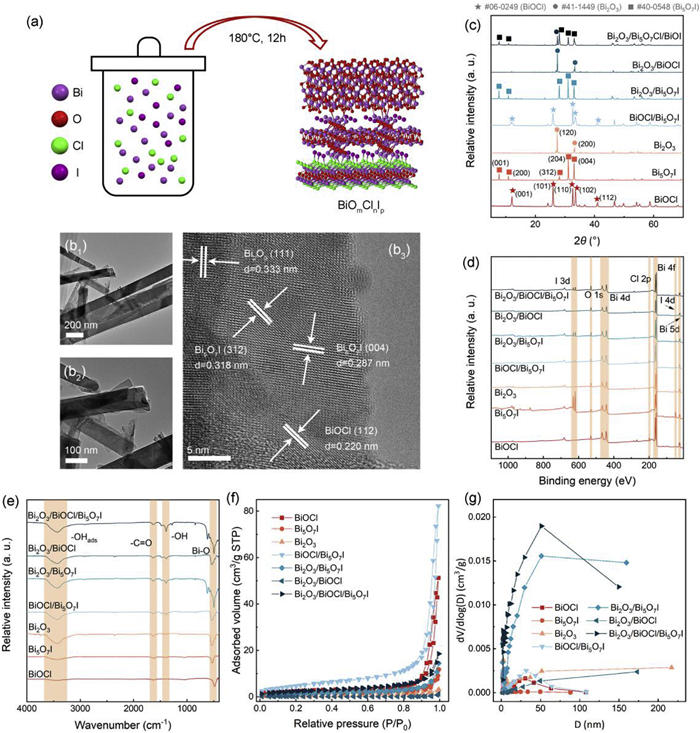
According to Fig. 1a, all heterojunctions were fabricated using hydrothermal methods. The photocatalysts were structured according to varying dosages of each element while maintain identical reaction condition (referred to materials section). As shown in Fig. 1b, TEM revealed the rod-like structure of the catalyst material presented ~100 nm thickness. High-resolution TEM (HR-TEM) analyses provided additional insights into the microstructure of Bi2O3/Bi5O7I/BiOCl, revealing three distinct crystal planes, 0.333, 0.287, and 0.220 nm, respectively corresponded to Bi2O3 (111), Bi5O7I (004) and (312), and BiOCl (112) [12]. The homogeneous distribution of lattices across these three components facilitated the formation of a dual heterojunction [13]. The detailed characterization results of heterojunctions were presented in Figs. 1c–g, and Text S1, Table S1, Figs. S1 and S2 (Supporting information).
Figure 1
Figure 1.
(a) X-ray diffraction patterns, (b) Fourier-transform infrared spectra, (c) X-ray photoelectron spectroscopies, (d) high-resolution TEM images (scale bars: (1) 200 nm, (2) 100 nm, and (3) 5 nm of crystal planes, (e) synthesizing and structuring scheme of heterojunctions, (f) nitrogen adsorption–desorption isotherms, and (g) pore-size distributions of heterojunctions.
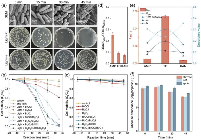
Fig. 2a presents the microscopic process of E. coli MG1655 inactivation under xenon lamp-simulated photocatalysis utilizing Bi2O3/Bi5O7I/BiOCl during the reaction. Most ARB was deactivated within the first 15 min, and completely eliminated by 45 min. In contrast, control experiments conducted in darkness showed no bacterial killing efficiency. The plates images demonstrated that photocatalytic disinfection effectively targeted ARB, while SEM images showed progressive disintegration of E. coli MG1655 as photocatalysis proceeded (Fig. 2b). The cells began to exhibit fragmentation and the catalyst material started intermixing with them after 30 min; by the end of the reaction, fragmentation intensified resulting in partial embedding of the material within the (inactivated) cells. According to Figs. 2b and c, standalone Bi-based materials presented weak disinfection performance (< 40%), while the efficiencies of photocatalysts were significantly enhanced when they reacted in heterojunction forms. Previous studies indicated that ARB appeared to be more robust than the regular E. coli [11], which further underscores the necessity for research and development of photocatalytic disinfection systems. Fig. S4 (Supporting information) further illustrated the sterilization efficacy of different heterojunctions, only the ternary heterojunction achieved complete deactivation of ARB in this study. Although both Bi2O3/BiOCl and Bi2O3/Bi5O7I reached degradation rates exceeding 95%, all three binary heterojunctions showed varying degrees of incomplete bacterial eradication according to the plate spreading results. Despite the accessibility to eliminate ARB during 45 min, the dual Z-scheme heterojunctions also achieved > 20% antibiotic resistance inhibition and 80% ARGs degradations (detailed in Figs. 2d–f, Text S2, Figs. S3 and S4 in Supporting information).
Figure 2
Figure 2.
(a) The antibacterial effects after visible light irradiation or dark storage for 0, 15, 30, and 45 min were presented as the result of SEM images and spread plates. Disinfection efficiency comparisons of all synthesized catalysts (b) under simulated solar light illumination and (c) in the dark. (d) The antibiotic resistance inhibition of E. coli in 30 min disinfection, assessed by 24-h bacterial growth observation. (e) Degradation rates (kobs) of antibiotics with Bi2O3/Bi5O7I/BiOCl photocatalytic degradation, and their values of and electrophilic index (N), nucleophilic index (ω), energy gap (Egap). (f) Change of ARGs abundance during the photocatalytic reactions. Experimental condition: [ARB] = 1 × 10⁷ – 1 × 10⁸ CFU/L, [antibiotics] = 20 mg/L [catalysts] = 100 mg/L.
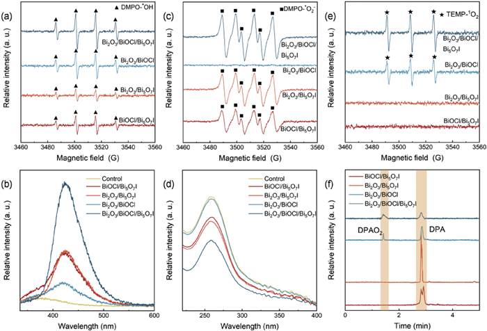
The composition of the material significantly influenced its photocatalytic oxidation performance, which in turn leads to differences in its disinfection and degradation effects. This phenomenon is owing to both the structure and composition of the heterojunction. To understand the photocatalytic oxidation mechanism of heterojunction structures, the generation and role of ROS are investigated. EPR was applied as a primary ROS identification method. Fig. 3a presented the existence of •OH under the photocatalysis of heterojunctions. In EPR tests, DMPO was employed to capture radicals, forming the more stable spin adduct DMPO-•OH. This adduct exhibits a longer half-life compared to the original free radicals, making it easier to detect via EPR spectroscopy [14]. All heterojunctions under solar-light irradiation were observed with characteristic peaks of DMPO-•OH adduct with a signal ratio of 1:2:2:1. Notably, Bi2O3/Bi5O7I/BiOCl generated the most intensive •OH signals, indicating superior activity among ternary heterojunctions compared to simple binary configurations. Chemical probes also served as identifiers for ROS, as shown in Fig. 3b, PTA presents the fluorescent activity when reacting with •OH [15]. Identically, Bi2O3/Bi5O7I/BiOCl presented the highest generation of ⋅OH among all tested heterojunctions. The observation of •OH is usually associated with presence of •O2–, which could also be detected by EPR with trapping agent of DMPO. Validation of •O2– was performed in a MeOH matrix for avoiding the interference from aqueous ⋅OH form the DMPO-•O2– adduct which exhibited typical peak in EPR spectra with intensities ratio of 2:2:1:2:1:2 [16]. All heterojunctions except Bi2O3/BiOCl showed significant characteristic DMPO-•O2– peaks, presumably owing to the enhanced conversion from •O2– to 1O2 (Fig. 3c) [17]. NBT also served as a chemical probe for detecting •O2–, decreases in peaks intensity indicated higher concentration of •O2– [18]. In Fig. 3d, the NBT probing test showed consistent results, indicating that Bi2O3/Bi5O7I/BiOCl presented the highest concentration of •O2–. In contrast, BiOCl/Bi5O7I and Bi2O3/Bi5O7I showed the relatively less concentration of •O2–. The existence of 1O2 is also potential because of its conversion from other ROS, and as illustrated in Fig. 3e, which shows that it can be trapped by TEMP to form identifiable TEMPO in EPR tests [19]. The disappearance of DMPO-•O2– characteristic peaks of Bi2O3/BiOCl could be explained as the complete conversion to 1O2. While Bi2O3/Bi5O7I/BiOCl maintained a high intensity of 1O2, indicating the oxidation performance of heterojunctions was strongly connected with the ROS generation. DPA was applied to capture 1O2 to oxide DPAO2 as a typical oxidation product (Fig. 3f and Fig. S7 in Supporting information). All experiments were observed with DPA but only Bi2O3/Bi5O7I/BiOCl and Bi2O3/BiOCl were discovered with DPAO2, with Bi2O3/Bi5O7I/BiOCl exhibiting the highest photocatalytic disinfection activity attributable to both enhanced intensity and broad-spectrum of ROS [20].
Figure 3
Figure 3.
Spin-trapping EPR spectra obtained using DMPO in H2O (a), DMPO in MeOH (c), and TEMP (e). (b, d) show the corresponding UV–vis detection results using NBT and PTA as probes, respectively. (f) Mass spectrometry analysis of diphenylanthracene (DPA) and its oxidation product (DPAO2).
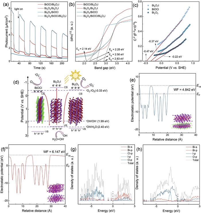
The influence of material composition on photocatalytic disinfection performance against ARB has been analyzed, highlighting critical points related to ROS regulation. However, factors influencing ROS formation should be explained from structural and photoelectronic properties inherent in the catalyst. As shown in Fig. 4a, the photo-responsiveness and recombination of photogenerated charges were assessed through transient photocurrent measurements [21]. All photocatalysts showed prompt and consistent photocurrent responses, while Bi2O3/Bi5O7I/BiOCl presented highest photocurrent density. Therefore, this ternary heterojunction facilitated high photo-electron conversion efficiency and presumably led to abundant ROS generation. As shown in Fig. S8 (Supporting information), EIS was also applied for characterizing the photoelectron transfer rates, the observed lowest resistance confirmed that Bi2O3/Bi5O7I/BiOCl exhibited superior photocatalytic efficiency [22]. UV–vis DRS displays the light-harvesting capabilities of all catalysts (Fig. S9 in Supporting information), indicating that Bi2O3/Bi5O7I/BiOCl has broaden its light absorption spectrum. Fig. 4b further exhibited the identification on the energies of band gap (Eg). The Eg values for Bi2O3/Bi5O7I/BiOCl, Bi2O3/BiOCl, Bi5O7I/Bi2O3, and BiOCl/Bi5O7I were calculated as 2.83 V, 2.56 V, 2.25 V, and 2.14 V (vs. SHE). A narrower Eg of Bi2O3/Bi5O7I/BiOCl effectively accelerated the migration process of photogenerated carriers, thereby enhancing photocatalytic activity. As shown in Fig. S10 (Supporting information), PL spectroscopies were conducted to evaluate the separation behavior by observing photogenerated electron-hole pairs within the photocatalysts. To illustrate how the heterojunction structure influenced its photogenerated electron transition capability and to assess the feasibility of specific reactions, Mott-Schottky analyses were deployed to measure the actual energies of conduction band (ECB). According to Fig. 4c, all Bi2O3, BiOCl, and Bi5O7I were identified as n-type semiconductors, with ECB calculated –0.22, –0.37, and –0.47 V (vs. SHE) [23]. The Eg values for these standalone semiconductors were calculated to 2.71, 3.07, and 2.88 V (vs. SHE) as shown in Fig. S11 (Supporting information), thus the energies of valence band (EVB) could be obtained according to EVB = ECB + Eg. Therefore, EVB of Bi2O3, BiOCl, and Bi5O7I were calculated to 2.49 V, 2.70 V, and 2.41 V (vs. SHE).
Figure 4
Figure 4.
(a) Visible-light-induced photocurrent. (b) Kubelka-Munk function derived from UV–vis diffuse reflectance results (E: photon energy; α: normalized adsorption coefficient). (c) Mott-Schottky measurement. (d) Reaction mechanism diagram of the BiOCl/Bi5O7I/Bi2O3 dual heterojunction. Electrostatic potential of (e) BiOCl/Bi5O7I heterojunction and (f) Bi5O7I/Bi2O3 heterojunction. Density of states (DOS) spectra for (g) BiOCl/Bi5O7I heterojunction and (h) Bi5O7I/Bi2O3 heterojunction.
The scheme of the ternary heterojunction is illustrated in Fig. 4d. The Bi2O3/Bi5O7I/BiOCl heterojunction is speculated to work as a dual Z-scheme, forming a sandwich-like structure where Bi5O7I is clamped by Bi2O3 and BiOCl. All standalone catalysts were able to generate •OH, but only Bi5O7I was accessible to generate •O2–, which explained why Bi2O3/BiOCl photocatalytic oxidation did not yield detectable levels of •O2–. The combination of heterojunctions was considered to represent a Z-scheme heterojunction instead of a conventional type II, which lack the potential to trigger required reactions [24]. All binary heterojunctions in this work were assumed to form Z-scheme structures, hence it follows that the ternary heterojunction could be structured as a dual Z-scheme [25]. This proposed structural model is further corroborated by the calculated work functions and interfacial electronic interactions, which collectively provide a coherent basis for the dual Z-scheme charge transfer pathway. This proposed dual Z-scheme structure illustrated the mechanism that ROS were generated in high intensities and in multiple types, ultimately clarifying why ternary heterojunction obtained superior oxidation performance. Although the proposed scheme for the heterojunction suggested a layer-structure, it did not imply that the ternary heterojunction was composed solely of layers or possessed that specific microstructure. Previous morphological characterization has confirmed only the close contact between these heterojunctions, hence, the diagram merely indicated an optimal structure or configuration during operation. The work function (WF) was quantified by calculating the value difference between the Fermi level (EF) and the vacuum level (Evac). This value corresponds to the minimum energy needed for electron addition or removal at the solid surface [26]. According to Figs. 4e and f, the electrostatic potential was individually calculated by separating Bi2O3/Bi5O7I/BiOCl into Bi2O3/Bi5O7I and Bi5O7I/BiOCl, resulting WFs were calculated at 4.842 and 6.147 eV, respectively. According to Fig. S12 (Supporting information), standalone measurements indicated WFs for Bi5O7I, BiOCl, and Bi2O3 at 1.283, 5.881, and −3.870 eV, which align with pathways of the Z-scheme charge transfer mechanisms [27]. The absence of an abrupt WF shift across the heterojunction interfaces suggests weak interfacial chemical bonding, dominated by van der Waals interactions, which is consistent with the hydrothermal growth process yet still facilitates effective charge separation via the proposed Z-scheme pathway. The total density of states (DOS) and projected DOS for Bi2O3/Bi5O7I and Bi5O7I/BiOCl were calculated to illustrate the electronic attributes of heterojunctions, as shown in Figs. 4g and h [28]. All orbitals were calculated but only valence orbitals showed distribution on shallow energy levels, namely, Bi-s, Bi-p, Cl-p. I-p, and O-p. Fig. S13 (Supporting information) further illustrated the change of electronic properties by comparing the DOS with Bi2O3, Bi5O7I, and BiOCl. The band gaps in the projected DOS were found to be > 2 eV, approximately equaling to the experimental Eg. In contrast, the band gaps of heterojunctions, especially Bi5O7I/Bi2O3, were significantly reduced, highlighting the effectiveness of semiconductor combinations, and further strengthen the potential for a Z-scheme configuration. The continuous DOS distribution near the Fermi level originates from the hybridized interface states between Bi5O7I and Bi2O3, which serve as efficient channels for charge transfer and are crucial for the enhanced Z–scheme charge separation. Therefore, it elucidated that the ternary heterojunction existed within a dual Z-scheme structure, enhancing photocatalytic oxidation through an increase in both the variety and intensity of ROS production.
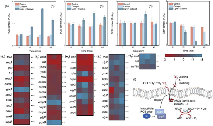
While the mechanism of catalysts catalyzing the production of ROS to eliminate bacteria has been revealed in previous studies, however, it is still unclear that how ARB experienced from metabolism disruption and membrane damage under ambient ROS attack [29]. To prevent interference from elevated extracellular ROS levels, cells were extracted at specific time points, lysed, and subsequently assayed for intracellular ROS levels. The results were shown as Fig. 5a, intracellular ROS levels in experimental groups increased by > 350%, while controlled groups remained relatively stable. The peak level of intracellular ROS reached at 30 min as the oxidative stress accumulated during light irradiation. However, as this level began to decline towards the end of the reaction period, it indicated that intensive oxidative stress accumulation caused membrane damage and eventually triggered an inevitable self-driven cell death process [30]. The characterization of intracellular ROS demonstrated effective oxidation effects from •OH, •O2–, and 1O2, simultaneously. Fig. 5b illustrated that the superoxide dismutase (SOD) contents were gradually increased from 0 min to 30 min and decayed at 45 min. The SOD production countered intracellular ROS, thereby maintaining ARB homeostasis. This further demonstrated that an effective oxidative stress response was triggered by Bi2O3/Bi5O7I/BiOCl [31]. Glutathione (GSH) also reflects the cellular resistance to the ambient oxidative capacity as a strong non-enzymatic antioxidant [32]. In the context of Bi2O3/Bi5O7I/BiOCl photocatalysis, GSH exhibited a similar trend of change as SOD, further confirming that ARB, under maximum oxidative stress (i.e., at 30 min of reaction), was unable to withstand this condition, resulting in its eventual deactivation (Fig. 5c). Antioxidant capacity and intracellular ROS exhibited closely correlated trends, notably, ARB was completely inactivated at 45 min, indicating that the oxidative stress exceeded its tolerance threshold. Adenosine triphosphate (ATP) levels reflected cellular bioactivity, demonstrating both metabolic activity and the maintenance of physiological integrity under external environmental stress [33]. Fig. 5d presented the continuous decrease in ATP levels as oxidation proceeded, culminating in the depletion of bioenergy. It was speculated that ARB sustained irreversible damage, which resulted from persistent external oxidation of the cells, or indirectly from the process of combating oxidation thus severely compromised other cellular functions. Therefore, regulation of ROS played a critical role in cellular inactivation, it also suggested that the intensity of ROS was the dominant factor influencing photocatalytic disinfection efficacy, and its regulation could lead to significantly enhanced disinfection performance.
Figure 5
Figure 5.
The intracellular (a) reactive oxygen species (ROS), (b) superoxide dismutase (SOD), (c) glutathione (GSH), and (d) adenosine triphosphate (ATP) changing in photocatalysis reaction. (e) For photocatalytic treatment comparison to control groups at 30 min, the heatmaps comparing the expression of (1) responses to oxidative stress, (2) membrane reparation, (3) DNA reparation, (4) ATP-related genes, and (5) antibiotic resistant genes in E. coli. (f) Mechanism of membrane reparation on E. coli and ROS intracellular mitigation mechanism. All samples were collected from experiments under optimal Bi2O3/Bi5O7I/BiOCl photocatalytic oxidation.
Fig. 5e and Tables S2–S6 (Supporting information) presented the gene expression regulation during the photocatalytic reaction. The significant upregulation of the thioredoxin gene trxA and the glutathione system gene grxA indicated that ARB tackled with oxidative damage by facilitating the reduction of other proteins through the cysteine thiol-disulfide exchange pathway [34]. Simultaneously, the synergistic activation of the superoxide sensing regulatory gene soxR and the hydrogen peroxide-decomposing gene katG confirmed that bacteria initiated an emergency defense mechanism involving a multilevel ROS scavenging strategy [35]. However, the inhibition of oxyR, gor, and sodA indicated a severe collapse in maintaining the homeostasis against ambient ROS. Additionally, the significant upregulation of biofilm formation gene pgaB indicated that biofilm formation was accelerated as a physical barrier against ROS penetration. However, synergistic suppression of membrane genes (lamB, bamA/B/C, ompF, tolC, yhiM, and yqjE) reflected membrane homeostasis disruption, which caused lipopolysaccharide assembly defects and transmembrane transport disorder [20]. Notably, the exceptionally high expression of porin ompG suggested a compensatory response enhancing nutrient uptake or toxic efflux through specific channel upregulation [36]. The significant upregulation of the yebG, cho, and recA/N confirmed DNA damage in ARB, indicated preferential repair of minor base damage, induced a transient SOS repair response. Additionally, significant upregulation of key SOS response genes umuC/D revealed a unique DNA damage tolerance strategy in bacteria, allowing DNA replication at lesion sites with this error-prone repair mechanism [37]. Furthermore, the downregulation of key ATP synthase genes atpA/C/D/E/F/G/H, the ATP-dependent protease gene clpX, and the protein-transporting ATPase gene secA reflected the bacterial stress strategy of actively shutting down non-essential energy-consuming pathways in response to reduced ATP synthesis [38]. Therefore, when photocatalysis-induced oxidative damage exceeded the bacterial repair threshold, energy metabolism collapse triggered by ATP synthesis inhibition impeded NADPH regeneration. This rendered reducing power-dependent antioxidant enzymes (e.g., gor and sodA) malfunction, ultimately establishing a lethal cycle of "ROS burst-repair failure". The molecular mechanism by which photocatalysis achieved highly effective disinfection through overwhelming bacterial compensatory capacity against oxidative stress was systematically clarified. Therefore, how related gene expression and cell membrane repair take effects when facing the generated extracellular oxidative stress (•OH, •O2–, and 1O2) is depicted in Fig. 5f.
In summary, Bi2O3/Bi5O7I/BiOCl was capable of producing high-intensity and diverse ROS, thereby achieving optimal efficacy in eliminating ARB. The cell membrane was progressively disrupted during photocatalytic process, while intracellular ATP levels gradually declined, and oxidative stress triggered by a sharp increase in ROS led to an intracellular imbalance. Transcriptomic analysis further revealed that ARB mobilized all available resources to repair their cell membranes and mitigate intracellular oxidative pressure, which concurrently suppressed other cellular functions. As the reaction proceeded, oxidative stress peaked until metabolic processes ultimately collapsed when ambient oxidative damage exceeded critical thresholds. Therefore, this work comprehensively examined how modulation of photocatalytic efficiency through heterojunction configuration influenced bacterial deactivation mechanisms, offering novel insights into photocatalytic disinfection on ARB.
The authors declare that they have no known competing financial interests or personal relationships that could have appeared to influence the work reported in this paper.
Acknowledgments
This investigation was supported by the National Natural Science Foundation of China (Nos. 52321005 and 52470032), the Guangdong Basic and Applied Basic Research Foundation (Nos. 2023A1515012383 and 2024A1515030138), the State Key Laboratory of Urban Water Resource and Environment (Harbin Institute of Technology) (No. 2024TS25), China Postdoctoral Science Foundation (No. 2024M764193), and Shenzhen Science and Technology Program (Nos. KQTD20190929172630447 and JCYJ20240813104840054).
Supplementary materials
Supplementary material associated with this article can be found, in the online version, at doi:10.1016/j.cclet.2026.112650.
Figure 2
(a) The antibacterial effects after visible light irradiation or dark storage for 0, 15, 30, and 45 min were presented as the result of SEM images and spread plates. Disinfection efficiency comparisons of all synthesized catalysts (b) under simulated solar light illumination and (c) in the dark. (d) The antibiotic resistance inhibition of E. coli in 30 min disinfection, assessed by 24-h bacterial growth observation. (e) Degradation rates (kobs) of antibiotics with Bi2O3/Bi5O7I/BiOCl photocatalytic degradation, and their values of and electrophilic index (N), nucleophilic index (ω), energy gap (Egap). (f) Change of ARGs abundance during the photocatalytic reactions. Experimental condition: [ARB] = 1 × 10⁷ – 1 × 10⁸ CFU/L, [antibiotics] = 20 mg/L [catalysts] = 100 mg/L.
Figure 3
Spin-trapping EPR spectra obtained using DMPO in H2O (a), DMPO in MeOH (c), and TEMP (e). (b, d) show the corresponding UV–vis detection results using NBT and PTA as probes, respectively. (f) Mass spectrometry analysis of diphenylanthracene (DPA) and its oxidation product (DPAO2).
Figure 5
The intracellular (a) reactive oxygen species (ROS), (b) superoxide dismutase (SOD), (c) glutathione (GSH), and (d) adenosine triphosphate (ATP) changing in photocatalysis reaction. (e) For photocatalytic treatment comparison to control groups at 30 min, the heatmaps comparing the expression of (1) responses to oxidative stress, (2) membrane reparation, (3) DNA reparation, (4) ATP-related genes, and (5) antibiotic resistant genes in E. coli. (f) Mechanism of membrane reparation on E. coli and ROS intracellular mitigation mechanism. All samples were collected from experiments under optimal Bi2O3/Bi5O7I/BiOCl photocatalytic oxidation.


 DownLoad:

 下载:


 下载:

