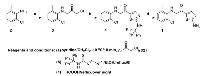
Scheme 1.
Synthesis route of compound 1

The human dental tissue-derived mesenchymal stem cells (MSCs) have been proved playing an important role in the clinic treatment[1], such as the dental pulp MSCs, exfoliated deciduous MSCs, periodontal ligament MSCs, dental follicle progenitor MSCs, alveolar bone-derived MSCs, apical papilla MSCs, tooth germ progenitor MSCs, and gingival MSCs[2, 3]. MSCs can produce a large number of biologically active substances which have the activities of hematopoietic and supporting, activate endogenous stem or progenitor cells, tissue damage repair, immune regulation, anti-apoptosis, anti-oxidation, anti-fibrosis and so on[4, 5]. The most important thing is that the DP-MSCs has the ability to differentiate into different tissue cells, such as neural cells, osteoblasts, adipocytes and chondrocytes[6]. So, it is reasonable for us to give this hypothesis that the DP-MSCs are suitable candidates for tissue regeneration.
Dasatinib is a protein tyrosine kinase inhibitor, a Src Kinase inhibitor, and is useful in the treatment of immunologic and oncological diseases[7], while 2-amino-N-(2-chloro-6-methylphenyl)thiazole-5-carboxamide (1) which is one of the most important intermediates in the synthesis of Dasatinib fascinated us[8]. Thus, much attention has been devoted to the synthesis, characterization and crystal structure of compound 1 which has never been reported before[9].
The present work deals with the synthesis and characterization of the title compound 1. 2-Chloro-6-methylaniline (2) was used as the starting material to obtain 2-chloro-N(2-chloro-6-methylphenyl) acetamide (3), which then reacted with (E)-N, N-dimethyl-N'-(tritylcarbamothioyl)formimidamide to get N-(2-chloro-6-methylphenyl)-2-(tritylamino)thiazole-5-carboxamide (4). Then 4 was cyclized with HCOOH (Scheme 1) to afford 1. In biological research, to evaluate the induction activity on the dental pulp mesenchymal stem cells (DP-MSCs) differentiation, the DP-MSCs colony formation ability assay and osteogenesis related genes expression assay were determined. The former results indicated that the compound could significantly reduce the colony numbers at the 7th and 14th days after compound incubation, and showed a dose dependent relationship. In addition, the data of RT-PCR also confirmed that 1 significantly influences the relative level of genes related with cell osteogenesis and proliferation. All the results indicated that the compound could affect the osteogenic differentiation of DP-MSCs.
IR spectra (400~4000 cm-1) were obtained using a Brucker Equinox-55 spectrophotometer. 1H NMR spectra were obtained using a Varian Inova-400 spectrometer (at 400 MHz). Mass spectra were obtained using a micrOTOF-Q II mass spectrometer. The melting points were taken on a XT-4 micro melting apparatus, and the thermometer was uncorrected. The human promyelocytic leukemia cancer HL-60 cell and the normal cell line HEK-293 were obtained from the Cell Bank of Shanghai Institte of Biochemistry and Cell Biology, Chinese Academy of Sciences (Shanghai, China). These two cell lines were cultured in Dulbecco's modified Eagle's medium (DMEM; Gibco, Life Technologies, Carlsbad, CA, USA) culture medium according to the protocol. The DMEM culture medium was supplemented with 2% L-glutamine, 10% heat-inactivated fetal bovine serum (FBS) and 1% penicillin-streptomycin solution (Gibco, Life Technologies). Cells were cultured in a standard cell culture environment (humidified incubator with 5% CO2 at 37 ℃). The culture media were changed according to the cells growth condition.
To a mixture of compound 2 (25.0 g, 176.6 mmol) in CH2Cl2 (200 mL) was added pyridine (28.6 mL, 353 mmol) at –10 ℃. After 10 min, chloroacetyl chloride (21.1 mL, 265 mmol) was added. The reaction mixture was allowed to warm to room temperature over a period of 2 h. A solution of 1 N HCl (500 mL) was added and the mixture was stirred for 10 min. The organic phase was separated and the aqueous layer was extracted with CH2Cl2 (2 × 300 mL). The combined organic extracts were washed with 1 N HCl solution (400 mL), H2O (400 mL) and brine (400 mL). It was dried over MgSO4, filtered and concentrated in vacuo to give the product 3 as a white solid (34.70 g, 90.13% yield). m.p: 162~163 ℃. 1H NMR (500MHz, CDCl3): δ 2.28 (s, 3H), 4.25 (s, 2H), 7.14~7.18 (m, 2H), 7.26~7.31 (m, 1H), 8.02 (bs, 1H). IR (KBr pellet, cm−1): 3442m, 3176w, 3054w, 2965m, 2875w, 1598w, 1540s, 1456m, 1397s, 792m, 740s.
A solution of compound 3 (32.7 g, 150 mmol) and (E)-N, N-dimethyl-N'-(tritylcarbamothioyl) formimidamide (56.1 g, 150 mmol) in EtOH (750 mL) was heated to reflux for 6 h. To the reaction solution obtained in the previous step was added 96% formic acid (150 mL). The reaction mixture was refluxed overnight. The reaction mixture was then partitioned between EtOAc and H2O (1:1, L). The organic phase was separated and concentrated in vacuo. The residue was subjected to column chromatography using EtOAc in hexane (20%~80%) to give the title compound 1 (24.38 g, 60.74% yield). m.p: 196~197 ℃. 1H NMR (300MHz, DMSO-d6): δ 2.21(s, 3H), 7.20~7.22 (m, 2H), 7.37(dd, J = 7.2 and 2.4Hz, 1H), 7.57 (s, 2H), 7.86 (s, 1H), 9.60 (s, 1H). HRMS (ESI +): m/z: calcd. for C11H10ClN3OS: 290.0131 [M + Na + ]; found: 290.0119. IR (KBr pellet, cm-1): 3447m, 3118w, 3088s, 2970m, 2882w, 1707m, 1605s, 1552s, 1418s, 1389m, 783m, 698m.
A suitable single crystal of compound 1 (obtained by slow volatilization of its CH2Cl2 solution) was carefully selected under optical microscope and glued on thin glass fibers. The intensity data of 1 were collected on an Oxford Xcalibur E diffractometer. The empirical absorption corrections were applied to the data using the SADABS system[10]. The structure was solved by direct methods and refined by fullmatrix least-squares techiniques on F2 using the SHELXS-97 program. All non-hydrogen atoms of 1 were refined anistropically, and all hydrogen atoms attached to carbon atoms were fixed at their ideal positions. Compound 1 (C11H10ClN3OS, Mr = 267.73 g/mol) belongs to monoclinic space group P21/n with a = 16.7208(7), b = 8.6098(5), c = 16.9367(8) Å, β = 93.496(4)°, V = 2433.7(2) Å3, Z = 8, T = 296.15 K, μ(MoKα) = 0.471 mm-1 and Dc = 1.461 g/cm3. For 1, a total of 11205 reflections were collected (4.80≤2θ≤58.67°), of which 5708 were unique (Rint = 0.0340, Rsigma = 0.0566) and used in all calculations. The final R = 0.0529 (I > 2σ(I)) and wR = 0.1403 (all data).
To induce the differentiation of dental pulp mesenchymal stem cells (DP-MSCs), the cells collected were seeded into cell culture plates at the concentration of 1 × 10⁴ cell/mL, then divided into the control and differentiation groups. The cells in the latter group were cultured in culture medium supplemented with transforming growth factor-β (TGF-β) and compound for 20 days, while those in the former group were incubated in basal medium[11].
To evaluate the inhibitory of compound on DP-MSCs differentiation, the cell colony formation assay was performed in this present research. Briefly, the DP-MSCs collected as described were seeded onto 6 well plates at the final destiny of 1 × 10⁵ cell/well, followed by the compound attrition at the concentration of 1, 5 and 10 μg/mL for indicated treatment. At the 7th and 14th days after compound treatment, the cells were washed with PBS solution, and the cells remaining on the plates were labeled with crystal violet staining and the numbers were counted. This preformation was conducted at least three times.
Real-time RT-PCR was conducted in this experiment to detect the relative expression of the genes with relationship with the cell osteogenesis and proliferation. In short, the DP-MSCs were isolated from the tissues collected in our hospital, and then the cells were seeded into 24-well plates at the final destiny of 1 × 10⁴ cells/mL. The differentiation of DP-MSCs was induced with the incubation of 5 ng/mL TGF-β, then the compound was added into cells at the concentration of 1, 5 and 10 μg/mL. After 20 days treatment, the cells were harvested, washed with PBS solution and the TRIzol reagent was used for the total RNA isolation. The total RNA was reverse-transcribed into cDNA with cDNA Synthesis Kit after concentration measurement. Subsequently, the Real-Time PCR System was used for the Real-time RT-PCR preformation, and the relative gene expression levels were measured and the data were expressed as mean±SD from three repeats[12].
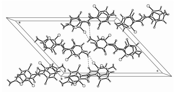
The structure of the title compound was measured by X-ray diffraction crystal structural analysis. The result indicates that the compound is of monoclinic system with space group P21/n. It displays a one-dimensional chain structure through two kinds of hydrogen bonds (N–H···N and N–H···O). The molecular structural unit is shown in Fig. 1, in which the angle between the six-membered ring (C(1)~C(6)) and the adjacent five-membered ring (S(1), C(8)~C(10) and N(2)) is 58.06(2)°. The bond lengths of C–C, C–N and C–S fall in the ranges of 1.353(3)~1.453(3), 1.304(3)~1.422(4) and 1.727(3)~1.745(3) Å, respectively. The bond distances of C– F, C–O and C–Cl are 1.651, 1.242 and 1.691 Å, respectively, all falling in their normal scopes[13-15].

In addition, two kinds of hydrogen bonds (N–H···N and N–H···O) could be found in the packing structure, and their detailed information is given in Table 1. Based on these them, a one-dimensional chain structure is generated, with its crystal packing structure depicted in Fig. 2.
 DownLoad:
CSV
DownLoad:
CSV
| D–H···A | d(D–H) | d(H···A) | d(D···A) | ∠DHA |
| N(1)–H(1)···O(1)a | 0.86 | 2.17 | 2.928(4) | 148 |
| N(3)–H(3A)···N(2)b | 0.87 | 2.41 | 3.092(6) | 136 |
| N(3)–H(3B)···O(2) | 0.86 | 2.43 | 3.265(3) | 163 |
| N(4)–H(4A)···O(2)c | 0.86 | 2.36 | 3.085(5) | 143 |
| N(6)–H(6A)···N(5)d | 0.87 | 2.35 | 3.065(5) | 140 |
| N(6)–H(6B)···O(1)e | 0.86 | 2.57 | 3.160(5) | 126 |
| Symmetry codes: (a) 1/2 – x, 1/2 + y, 1/2 – z; (b) 1 – x, 1 – y, 1 – z; (c) 1/2 – x, 1/2 + y, 3/2 – z; (d) 1 – x, 1 – y, 2 – z; (e) 1/2 + x, 1/2 – y, 1/2 + z | ||||
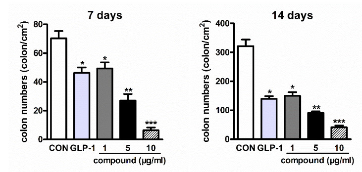
After the synthesis of the compound, its induction activity DP-MSCs differentiation was evaluated. Firstly, the colony formation assay was conducted to evaluate the influence of compound on DP-MSCs differentiation. After treated with the influence of compound on DP-MSCs differentiation, the cells remaining on the plates were labeled with crystal violet staining. The results in Fig. 3 indicated that the number of cell colonies was much lower than the control group after compound treatment, and the inhibitory effect of the compound on the DP-MSCs colony formation ability was dose dependent. In addition to this, we can also see from the results that the positive control drug GLP-1 showed a slightly weaker activity than the compound. All the results suggested strongly that the colony formation ability of DP-MSCs is significantly reduced by compound.
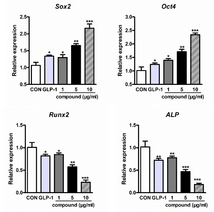
In the previous research, we have confirmed the reduced colony formation ability of DP-MSCs after compound treatment, while the biological activity needs further exploration. The ability of cell proliferation is the classical character of stem cells. Thus, in this experiment, the RT-PCR was performed to detect the relative expression of cell proliferation associated genes, such as Sox2 and Oct4, which are reported as the key genes involved in the regulation of stem cells proliferation, and the inhibition of them will cause cells apoptosis. Besides, the osteogenesis related transcriptional factors Runx2 and ALP were also measured by RTPCR to detect the induction of osteogenesis of DP-MSCs influenced by compound. As shown in Fig. 4, the relative expression of Runx2 and ALP was significantly up-regulated and the level of cell proliferation associated genes Sox2 and Oct4 was reduced obviously after compound treatment. All the above results indicated the compound could induce the osteogenic differentiation of DP-MSCs.
In conclusion, we synthesized a heterocyclic derivative and characterized it via IR, 1H NMR, HRMS, and single-crystal X-ray diffraction analysis. In biological research, we aimed to explore the effect of compound in regulating the osteogenesis of DP-MSCs. The results of the colon formation assay indicated that the compound could significantly reduce the colony formation ability of DP-MSCs. In addition, the RT-PCR results indicated the compound significantly up-regulates the expression of osteogenic related transcription factors. All these confirmed that the synthetic compound affects the osteogenic differentiation of DP-MSCs, which laid solid foundation for the regeneration of dentin.
Terunuma, A.; Ashiba, K.; Takane, T.; Sakaguchi, Y.; Terunuma, H. Comparative transcriptomic analysis of human mesenchymal stem cells derived from dentalpulp and adipose tissues. J. Stem. Cells Regen. Med. 2019, 15, 8–11.
Tang, X.; Li, W.; Wen, X.; Zhang, Z.; Chen, W.; Yao, G.; Chen, H.; Wang, D.; Shi, S.; Sun, L. Transplantation of dental tissue-derived mesenchymal stem cells ameliorates nephritis in lupus mice. Ann. Transl. Med. 2019, 7, 132. doi: 10.21037/atm.2019.02.41
Jin, Q.; Yuan, K.; Lin, W.; Niu, C.; Ma, R.; Huang, Z. Comparative characterization of mesenchymal stem cells from human dental pulp and adipose tissue for bone regeneration potential. Artif. Cells Nanomed. Biotechnol. 2019, 47, 1577–1584. doi: 10.1080/21691401.2019.1594861
Vahedi, P.; Jarolmasjed, S.; Shafaei, H.; Roshangar, L.; Soleimani Rad, J.; Ahmadian, E. In vivo articular cartilage regeneration through infrapatellar adipose tissue derived stem cellin nanofiber polycaprolactone scaffold. Tissue Cell 2019, 57, 49 – 56. doi: 10.1016/j.tice.2019.02.002
Dave, J. R.; Tomar, G. B. Dental tissue-derived mesenchymal stem cells: applications in tissue engineering. Crit. Rev. Biomed. Eng. 2018, 46, 429–468.
Xing, Y.; Zhang, Y.; Wu, X.; Zhao, B.; Ji, Y.; Xu, X. A comprehensive study on donor-matched comparisons of three types of mesenchymal stem cells-containing cells from human dental tissue. J. Periodontal Res. 2019, 54, 286–299. doi: 10.1111/jre.12630
Nakaya, A.; Fujita, S.; Satake, A.; Nakanishi, T.; Azuma, Y.; Tsubokura, Y.; Hotta, M.; Yoshimura, H.; Ishii, K.; Ito, T.; Nomura, S. Clinical significance of dasatinib-induced pleural effusion in patients with de novochronic myeloid leukemia. Hematol. Rep. 2018, 10, 7474.
Naqvi, K.; Kantarjian, H. M. Reply to starting with a lower daily dose of dasatinib in patients with chronic myeloid leukemia in chronic phase: less is more, or is it? Cancer 2018, 124, 4261. doi: 10.1002/cncr.31683
Wehrstedt, S.; Kubis, J.; Zimmermann, A.; Bruns, H.; Mayer, D.; Grieshober, M.; Stenger, S. The tyrosine kinase inhibitor dasatinib reduces the growth of intracellular Mycobacterium tuberculosis despite impairing T-cell function. Eur. J. Immunol. 2018, 48, 1892–1903. doi: 10.1002/eji.201847656
Sheldrick, G. M. SHELXL-97, Program for Solution Crystal Structure and Refinement. University of Göttingen: Göttingen, Germany 1997.
Igarashi, Y.; Chosa, N.; Sawada, S.; Kondo, H.; Yaegashi, T.; Ishisaki, A. VEGF-C and TGF-β reciprocally regulate mesenchymal stem cell commitment to differentiation into lymphatic endothelial or osteoblastic phenotypes. Int. J. Mol. Med. 2016, 37, 1005–1013. doi: 10.3892/ijmm.2016.2502
Ajlan, S. A.; Ashri, N. Y.; Aldahmash, A. M.; Alnbaheen, M. S. Osteogenic differentiation of dental pulp stem cells under the influence of three different materials. BMC Oral Health 2015, 15, 132. doi: 10.1186/s12903-015-0113-8
Liu, W.; Zhou, J.; Zheng, Y.; Qi, F.; Zhang, H.; Qian, H.; Wang, J.; Cheng, Y.; Gust, R. Design, synthesis and antiproliferative activity of 2-acetamidothiazole-5-carboxamide derivatives. Med. Chem. 2012, 8, 587–594. doi: 10.2174/157340612801216418
Singh, K. S.; Devi, P.; Sawant, S. G.; Kaminsky, W. Arene ruthenium(II) complexes with 2-acetamidothiazole derived ligands: synthesis, structural studies, antifouling and antibacterial properties. Polyhedron 2015, 100, 321–325. doi: 10.1016/j.poly.2015.08.016
Hatfield, J. M.; Eidell, C. K.; Stephens, C. E. Mono- and trifluorination of the thiazole ring of 2, 5-diarylthiazoles using N-fluorobenzenesulfonimide (NFSI). Tetra. Lett. 2013, 54, 1025–1028. doi: 10.1016/j.tetlet.2012.12.052

Figure 3 Reduced colony formation ability of DP-MSCs after compound treatment. The DP-MSCs were collected and seeded into plates, followed by compound treatment at different concentrations. The colony formation ability of DP-MSCs was determined by crystal violet staining assay. This experiment was repeated at least three times

Figure 4 Regulated expression of osteogenesis and cell proliferation associated genes after compound treatment. The DP-MSCs were cultured in basal medium supplemented with 5 ng/mL TGF-β, then different concentrations of compound(1, 5, 10 mg) was added for osteogenic induction for 20 days. The expression of osteogenesis and cell proliferation associated genes were measured by RT-PCR. All the data were expressed as mean±SD
Table 1. Hydrogen Bonds for the Title Compound
| D–H···A | d(D–H) | d(H···A) | d(D···A) | ∠DHA |
| N(1)–H(1)···O(1)a | 0.86 | 2.17 | 2.928(4) | 148 |
| N(3)–H(3A)···N(2)b | 0.87 | 2.41 | 3.092(6) | 136 |
| N(3)–H(3B)···O(2) | 0.86 | 2.43 | 3.265(3) | 163 |
| N(4)–H(4A)···O(2)c | 0.86 | 2.36 | 3.085(5) | 143 |
| N(6)–H(6A)···N(5)d | 0.87 | 2.35 | 3.065(5) | 140 |
| N(6)–H(6B)···O(1)e | 0.86 | 2.57 | 3.160(5) | 126 |
| Symmetry codes: (a) 1/2 – x, 1/2 + y, 1/2 – z; (b) 1 – x, 1 – y, 1 – z; (c) 1/2 – x, 1/2 + y, 3/2 – z; (d) 1 – x, 1 – y, 2 – z; (e) 1/2 + x, 1/2 – y, 1/2 + z | ||||
 下载: 导出CSV

 扫一扫看文章

扫一扫关注我们


 下载:
下载: