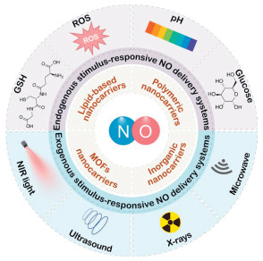
Figure 1.
Schematic illustration of NO delivery systems for precision therapeutics.

Recent advances in nitric oxide delivery systems for precision therapeutics
Hui Yu , Ziyu He , Bochuan Liu , Tianyou Du , Hai-Yan Wang , Yi Liu


Nitric oxide (NO) is an endogenous gaseous signaling molecule that plays a pivotal role in multiple physiological processes, including vasodilation, neurotransmission, immune responses, and inflammation regulation [1-4]. The biosynthesis of NO is primarily catalyzed by nitric oxide synthase (NOS) that converts L-arginine (L-Arg) into L-citrulline and releases NO [5-7]. Studies have shown that NO exhibits broad therapeutic potential in promoting wound healing, enhancing tumor treatment sensitivity, and exerting antibacterial and antibiofilm effects, especially in the fields of cancer, chronic infection, cardiovascular disease and tissue regeneration [8-12]. A notable feature of NO is its concentration-dependent biological activity [13]. At low concentrations (10−9–10−6 mol/L), NO could activate the soluble guanylate cyclase (sGC)-cGMP pathway [14]. Then the elevated cGMP levels trigger the activation of cGMP-dependent protein kinase G, leading to a reduction in intracellular calcium concentration [15]. This process mediates the regulation of vascular tone and increases vascular permeability. Furthermore, NO can induce angiogenesis, promote cell survival and proliferation [16]. At high concentrations (>10−6 mol/L), NO could induce tumor cell apoptosis and bacterial biofilm disintegration through oxidative and nitrosative stress [17,18]. The cytotoxicity is mainly due to the reaction of NO with superoxide to generate highly cytotoxic peroxynitrite (ONOO−) that mediates toxic effects [19-21]. Additionally, as the reservoir of NO, the metabolic reaction products of NO, such as nitrite, nitrate, S- or N-nitroso protein derivatives, and iron-nitrosyl complexes, can be reduced to NO in blood and tissues through various reactions, forming a recycling pathway for NO regeneration [22]. The NO is transported to distant tissues through circulation for utilization by receptor tissues [23]. Therefore, the bioavailability of NO depends not only on free NO radicals but also on NO-releasing compounds. However, the gaseous nature of NO results in rapid diffusion, a short half-life (<5 s) and a limited diffusion radius (40–200 µm), severely limiting its practical application [24-26]. Therefore, achieving precise and controlled NO release is crucial for advancing its clinical therapeutic applications.
With advances in nanotechnology, the construction of NO nanoplatforms has emerged as a highly promising strategy for achieving precise control and targeted delivery of NO [27,28]. Given the diverse biological activities of NO, various NO delivery systems have been developed for in vivo and in vitro applications. Prior to exploring therapeutic applications, it is essential to clarify key parameters of these carriers, including NO release rate, loading capacity, and release mechanism. To achieve effective regulation of NO release, researchers have developed responsive strategies based on endogenous and exogenous stimuli. By modulating the stimulation intensity, these methods can flexibly control the release of NO, thereby achieving the diversification of NO release patterns. Among them, the endogenous stimulus-responsive NO delivery nanomaterials can utilize specific features of the disease microenvironment (e.g., glutathione (GSH), acidic pH, reactive oxygen species (ROS), and glucose) for achieving targeted release of NO in specific sites, offering tissue specificity [29-31]. While the exogenous stimulus-responsive NO nanosystems rely on external physical stimuli (e.g., light, ultrasound (US), X-rays and microwaves) for high-precision spatiotemporal control of NO release, enhancing the operability and spatiotemporal accuracy of the release process [32-34]. Given the differences in control logic and application characteristics between these two release mechanisms, this provides a key basis for rationally designing NO therapeutic strategies according to different medical needs, ultimately allowing for precise spatial and temporal control of NO release.
In this review, we systematically summarize the recent advances in NO delivery systems for precision therapeutics, elucidating the therapeutic potential and applications of NO as a bioactive molecule in different disease interventions (Fig. 1). The review classifies NO delivery systems based on the stimulus-responsive mechanisms and therapeutic strategies, and comprehensively summarizing the current applications in precision medicine. We particularly focus on the construction methods and controllable release mechanisms of NO delivery systems, as well as their applications in specific diseases such as cancer, bacterial infections, inflammatory bowel disease, wound healing, renal fibrosis, and atherosclerosis. Finally, this review also analyzes and discusses the key challenges in NO delivery systems and outlines their future directions. We anticipate that this review will contribute to the further development of NO delivery systems and facilitate the clinical translation of NO-based gas therapy.
NO, as a small molecule gas messenger, has high reactivity and holds significant therapeutic potential in various medical conditions. However, the precise delivery of NO remains a major challenge due to its extremely short half-life, high diffusivity, and difficulty in maintaining effective concentrations at the target sites. Therefore, the development of effective NO delivery systems is of paramount importance. Nanocarriers, with their high loading capacity, protective effects on NO active molecules, enhanced penetration and retention effects, surface functionalization, and stimulus-responsive design, have emerged as an ideal platform for achieving precise NO delivery [35,36]. The selection of carrier material is a critical determinant of the release efficiency, targeting accuracy, and therapeutic efficacy in NO gas therapy.
Lipid-based nanocarriers, such as liposomes, nanostructured lipid carriers (NLCs), and solid lipid nanoparticles (SLNs), hold immense potential in drug delivery due to their excellent biocompatibility, degradability, enhanced drug permeation and ease of surface functionalisation [37-39]. The design strategies for NO delivery systems based on lipid-based nanocarriers typically involve encapsulating hydrophobic NO donors within the lipid bilayer or incorporating hydrophilic NO donors into the aqueous core [40,41]. Lipid-based NO delivery systems exhibit low toxicity, ease of preparation, and straightforward surface modification, which are widely utilized in biomedical delivery, particularly for cancer and inflammation therapies. For instance, Wu et al. constructed an US-responsive cerasomal nanoplatform (DOX@PC-SNO) by co-assembling cerasome-forming lipid, porphyrin lipid, unsaturated lipid dioleoylphosphatidylcholine (DOPC), doxorubicin (DOX) and DSPE-PEG2000-SNO as the NO donor. Under the US irradiation, porphyrin generates ROS, which simultaneously oxidizes DOPC to release doxorubicin (DOX) and cleaves S-nitrosothiol bonds for controlled NO release, enabling deep penetration and combination therapy [42].
Polymeric nanocarriers, such as polylactic-co-glycolic acid (PLGA), polycaprolactone (PCL), chitosan (CS), possess excellent biodegradability, biocompatibility, controllable release kinetics and modifiability, and are widely used to construct multifunctional NO delivery systems in the application of cancer therapy, skin wound healing and bone regeneration [43-45]. However, the hydrolysis byproducts of the biodegradable polyester polymer may result in inflammatory responses [43]. The design strategies of this type of system mainly include the following methods. The hydrophilic and hydrophobic NO donors can be encapsulated via nanoprecipitation or emulsion diffusion methods [46]. Alternatively, NO donors can be attached to the polymer backbone as a side chain or a terminal group through chemical bonding [47-49]. Furthermore, polymers can be further cross-linked into hydrogel networks for loading NO donors, enabling sustained localized NO release [50,51]. By adjusting the molecular weight of the polymer, the composition of the copolymer, and the degradation behavior, the kinetics of drug release can be further optimized [52,53]. Additionally, polymer nanocarriers can also simultaneously encapsulate chemotherapy drugs or imaging agents to achieve integrated diagnosis and treatment. For instance, Kim et al. constructed a thermosensitive copolymer hydrogel (F127-g-Gelatin) by conjugating Pluronic F127 with polymers gelatin for sustained release of the NO donor S-nitroso-L-glutathione (GSNO) and antibody blocking immune checkpoint cytotoxic T-lymphocyte-associated protein-4. Upon injection, the hydrogel forms micelles in situ that are optimized for lymphatic uptake, enhancing drug retention and delivery in tumors and draining lymph nodes, thereby improving local and systemic antitumor immune responses [50].
Inorganic nanocarriers, such as mesoporous silica nanoparticles (MSN), gold nanoparticles (AuNPs), manganese dioxide (MnO2) nanosheets, upconversion nanoparticles (UCNPs) and graphene etc., possess advantages including high loading capacity, high structural rigidity, tunable size and morphology, excellent biocompatibility, tunable surface chemistry and ease of functionalization [54-56]. Leveraging their high specific surface area, pore structure, energy conversion ability, catalytic or physical adsorption effects, chemical modification, and imaging functions, these nanocarriers can achieve image-guided NO release in response to specific stimuli, thereby enabling targeted drug delivery for synergistic disease treatment [32,57]. For example, Liu et al. fabricated an asymmetric NO-driven nanomotor (AG-DMSNs) by loading L-Arg and AuNPs onto dendritic MSN that the cascade catalysis of glucose by AuNPs can produce H2O2 and L-Arg can be further oxidized to release NO with the rapid self-propulsion, achieving efficient methicillin-resistant Staphylococcus aureus (MRSA) biofilm eradication both in vitro and in vivo [58]. Besides, Tabish et al. developed a pH-sensitive NO-releasing hybrid nanosheets (FF@GO) that were co-assembled by dipeptide diphenylalanine (FF) and graphene oxide (GO) [59]. This system utilizes the weak intermolecular interactions between FF peptides and GO. Under acidic conditions at pH 4.5, the protonation and deprotonation alter the apparent charge, leading to loosen the hybrid nanosheet structure and further enabling sustained real-time NO release. Although the in vivo behavior of inorganic nanoparticles can be optimized via structural design and surface modification, their long-term biological safety remains uncertain [60]. Moreover, combining therapies with distinct biodistribution characteristics and mechanisms can reduce non-specific uptake, thereby enhancing therapeutic efficacy and minimizing off-target effects [61]. Therefore, a comprehensive assessment of the pharmacokinetic properties, long-term safety, and off-target effects of these nanoplatforms in preclinical models is crucial for their clinical industrialization.
MOFs, such as zeolitic imidazolate framework-8 (ZIF-8) and materials institute lavoisier (MIL-100), have extremely high specific surface areas and structural tunability, typically exhibiting degradation behavior sensitive to the microenvironment, and can be used to tune the NO release from MOFs [62,63]. Also, during the biodegradation process of MOF materials, the breaking of the metal-ligand coordination bonds may release toxic metal ions, thereby causing toxicity and safety issues [55]. The delivery strategies for NO involve integrating NO donors as ligands into the MOF framework, either by directly attaching NO molecules to the metal ions or by physically adsorbing NO donor molecules within the pores [64,65]. By rational design of the framework structure, NO can be released under specific stimulus conditions. For example, Mu et al. presented the hybrid nanoreactors (Pt-iNOS@ZIF) by co-loading Pt NPs and inducible nitric oxide synthase (iNOS) into ZIF-8 frameworks, which could simultaneously scavenge ROS and generate NO, effectively inhibiting oxidative stress and inflammatory responses for preventing hepatic ischemia-reperfusion injury [66].
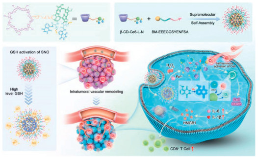
A prominent feature of the tumor microenvironment (TME) is that GSH concentrations are significantly higher than in normal tissues [67,68]. Capitalizing on this, researchers have designed a wide range of GSH-responsive NO donors [69,70]. Once encapsulated in nanocarriers, these NO donors enable efficient systemic delivery and accumulation within lesions. Then the high GSH concentration serves as a trigger to enable targeted release of NO at the lesion site, thereby opening new avenues for precision disease treatment. For instance, Li et al. developed a multifunctional liposome (Lip-DTI/NO), composed of near-infrared Ⅱ (NIR-Ⅱ) aggregation-induced emission (AIE) photosensitizer DTITBT with type-Ⅰ ROS generation and NO donor S-nitroso-N-acetyl-D-penicillamine (SNAP), which was applied for ONOO− induced collagen digestion to enhance drug delivery and pancreatic cancer therapy [71]. In this system, DTITBT possesses the characteristics of excellent NIR-Ⅱ fluorescence, O2•− production and photothermal conversion efficiency when triggered by 808 nm laser. Meanwhile, under the overexpressed GSH and 808 nm laser irradiation, SNAP was decomposed to release NO Simultaneously, the 808 nm laser-triggered O2•− generation by DTITBT and the GSH or heat-triggered NO release from SNAP lead to generate highly toxic and active ONOO− in the tumor region. This process promotes the expression of matrix metalloproteinases and the degradation of collagen in the extracellular matrix, as well as the enhancement of the penetration and accumulation of Lip-DTI/NO within the tumor. Taken together, the Lip-DTI/NO provides a promising strategy for NIR-Ⅱ fluorescence imaging guided ONOO−-enhanced synergistic photodynamic therapy (PDT) and photothermal therapy (PTT) in both subcutaneous and in situ pancreatic cancer models. Besides that, Zou et al. constructed the nanomedicine JSK@PEG-IR-820, incorporating a pH-responsive diisopropyl group, a photo-responsive IR820 molecule, and a NO donor (2-(2,4-dinitrophenyl) 1-[(4-ethoxycarbonyl)piperazin-1-yl]diazen-1-ium-1,2-diolate (JSK), which was applied for NO generation enhanced type Ⅰ PDT by enhancing O2•− generation while diminishing O2•− consumption based on the concept of “broadening sources” [30]. In this system, the diisopropyl group in PEG-IR820 was utilized as a pH-responsive sensor to induce nanoparticles disassembly and release JSK upon exposure to the low pH microenvironment of tumor, as well as facilitate the intermolecular electron transfer to IR820 for efficient O2•− generation. And JSK, serving as a NO pro-drug, released NO upon GSH induction. Besides, the highly cytotoxic ONOO− can be further produced through the reaction between O2•− and NO during this process, thereby facilitating the therapeutic efficacy of tumor regression. Meanwhile, JSK@PEG-IR820 NPs, with the excellent biocompatibility and passive tumor targeting ability, enable NIR-Ⅱ fluorescence-guided synergistic PDT/NO therapy. This study offers a promising NO therapy-sensitized type Ⅰ PDT approach for tumor treatment. Leveraging this characteristic, Xiong et al. reported a GSH-responsive polyurethane-based nanomicelles delivery system PEG-PNU-PEG@PDDPD (PNP), composed of an amphiphilic triblock polyurethane PEG-PNU-PEG and a photothermal agent PDDPD, which involved synergistic NO/PTT therapy for broad-spectrum antibacterial applications [72]. In this system, the PEG-PNU-PEG containing dinitrate NO donors were synthesized by combining a binary alcohol monomer, a L-lysine diisocyanate (LDI) monomer derived from natural amino acid and polyethylene glycol methyl ether (mPEG). In the overexpressed-GSH bacterial microenvironment, the nitroxy ester was reduced to form NO After being encapsulated, PDDPD showed excellent photothermal properties when irradiated by 808 nm laser, which can achieve effective PTT and accelerate the release of NO The animal experiments demonstrated that the synergistic PTT/NO therapy could enhance antibacterial efficacy as well as promote the healing of skin tissue, indicating great potential in the field of broad-spectrum antibacterial treatment based on NO gas therapy. Similarly, Dai et al. constructed a multifunctional supramolecular nanoparticle (T-NPCe6-L-N) by integrating chlorin e6 (Ce6), luminol and SNAP, amino-β-cyclodextrin (β-CD-NH2) with benzimidazole capped pancreatic cancer cell targeting peptide (BM-EEEGGSYENFSA), which was used for chemiluminescence resonance energy transfer (CRET)-activated PDT of pancreatic cancer (Fig. 2) [73]. Due to the high H2O2 and Fe3+ levels in cancer cells, chemiluminescence of luminol in T-NPCe6-L-N can be activated to induce the CRET-activated PDT by luminol and Ce6 without external light. Furthermore, the NO donor SNAP can release NO activated by GSH to normalize blood vessels, alleviate the hypoxia condition of tumors and deplete intracellular reductive GSH, thereby enhancing PDT efficacy. Additionally, this self-excited PDT system also triggered immunogenic cell death for enduring long-term inhibition of tumor growth. This approach addresses key drawbacks of traditional PDT, including light penetration depth and tumor hypoxia, enabling effective treatment of deep-seated and metastatic tumors.
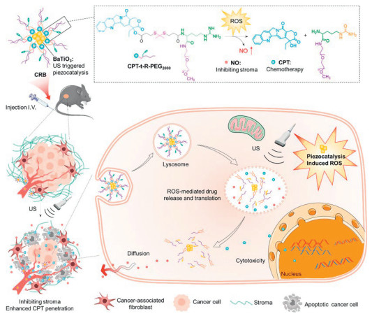
Leveraging the elevated levels of pathological ROS in tumor and inflammatory microenvironments, researchers have devised various ROS-responsive NO delivery systems [74,75]. These systems typically contain NO donors that are sensitive to specific ROS, especially H2O2 [76]. The abnormally elevated ROS within lesion site can specifically trigger the breakage of chemical bonds, resulting in the selective and controlled release of NO gas at the lesion site, thereby aiming to enhance therapeutic efficacy and reduce systemic toxicity [77]. For instance, Fu et al. fabricated the nanogels (NGs) through a Schiff base reaction between the aldehyde groups of the oxidized hyaluronic acid (OHA) and the two amine groups of L-Arg. Then, the NGs were loaded with gelatin and further coated with polydopamine (PDA) to construct a NG-based NO delivery microcapsules PDA@gelatin@nanogel (P@G@NG) for the inflammatory bowel disease treatment [78]. The P@G@NG microcapsules orally administered to mice with colitis were metabolized through the stomach, small intestine and colon, respectively. In the system, the PDA shell can protect the microcapsules for the delivery to the intestine. Once there, the PDA and gelatin were decomposed by the digestive enzymes and colonic microbes of the small intestine, leading to the release of the NGs. HA enables NGs to target and accumulate at colonic inflammation sites. In the inflammatory bowel disease microenvironment, the elevated levels of ROS and inducible NO synthase can lead to the production of NO through the catalytic action of L-Arg at the sites of colonic inflammation. Subsequently, NO gradually diffused into the microbial to inhibit the pathogenic bacteria and promote the beneficial bacteria, which in turn influenced the structure of the metabolite, promoted immune regulation and restored the intestinal barrier. This work offers the potential for applying gas therapy strategies based on NO delivery microcapsules in the clinical colitis management and intestinal flora treatment. Similarly, Wang et al. reported an US-triggered nanoprodrug CPT-t-R-PEG2000@BaTiO3 (CRB) for pancreatic cancer therapy by incorporating the nanomaterials BaTiO3 into amphiphilic prodrug molecules containing chemotherapeutic drug camptothecin (CPT), NO-donor L-Arg, and ROS-responsive thioketal bonds (Fig. 3) [79]. In this system, the piezoelectric material BaTiO3 can sustainably generate ROS through US-induced piezocatalysis and water splitting even in the tumor hypoxic microenvironment. Then, the thioketal bond was cleaved by ROS to release CPT, while L-Arg was oxidized to produce NO, enabling the spatiotemporal co-delivery of CPT and NO to the tumor site. Meanwhile, animal experiments demonstrated that the released NO can further facilitate the CRB NPs penetration in the deeper tumor and inhibit the extracellular matrix, thereby significantly improving the chemotherapeutic efficacy. Overall, the CRB nanoprodrug has achieved remarkable antitumor effects on highly malignant Panc02 tumors in mice by inhibiting the resistance of chemotherapy. It thus becomes a highly potential candidate for clinical translation in the field of combining NO gas therapy with cancer chemotherapy. To address this challenge, Li et al. engineered an innovative NO-driven bioorthogonal delivery platform (TTB-NH2@HA-PArg), which consisted of a photothermal precursor TTB-NH2, amphipathic polyarginine (PArg) and negative hyaluronic acid (HA) [80]. In this system, HA ensured the tumor-targeting properties of the nanosystem. PArg not only acted as a self-assembling nanocarrier for TTB-NH2 to form the NPs (TTB-NH2@PArg), but also served as a NO generator that reacted with the TME at high H2O2 levels, accompanied by the fluorescence conversion changes of TTB-NH2 to TTB-AZO from 620 nm to 850 nm. Meanwhile, TTB-AZO, with potent photothermal effect, induced real-time fluorescence monitoring of bioorthogonal activation and drug accumulation process, while simultaneously enhancing the photothermal therapy efficiency for highly precise tumor ablation. After in vivo administration, the bioorthogonal nanoassembly NP TTB-NH2@HA-PArg allows for efficient drug delivery, controlled release, and minimized side effects, demonstrating significant tumor inhibition and potential for clinical translation. This approach offers a promising strategy for achieving efficient and safe NO-mediated bioorthogonal-activated drug release in cancer treatment. In a notable study, Zhu et al. constructed a H2O2 self-catalyzed NO nanorod (Arg-ZnO2@siRNA@HA, AZOSH) with excellent biocompatibility and stability by anchoring ferritin heavy chain siRNA and HA to an arginine-stabilized zinc peroxide core, which induced the intracellular Fe2+ overload, depleted the GSH levels and triggered ferroptosis [81]. In this system, the AZO was reversibly decomposed to release the endogenous substances arginine and H2O2, allowing them to produce NO in the acidic tumor environment. In addition, the generated NO possessed multiple functions that it can be converted into ONOO− via free radical cascade oxidation, deplete GSH and suppress glutathione peroxidase 4 (GPX4) expression, thereby simultaneously triggering HMOX1 to mobilize the release of intracellular Fe2+ and inducing ferroptosis. More critically, the accumulation of NO can directly destabilize the endo/lysosomal membranes, facilitate siRNA escape to block iron storage and amplify the effects of gene silencing. Furthermore, the released Zn2+ from AZOSH induced mitochondrial oxidative stress to amplify ROS (H2O2 and O2•−) production and ferroptosis efficiency. Meanwhile, AZOSH was further effective in triggering the immunogenic cell death (ICD) and T cells infiltration on the basis of ferroptosis. When combined with anti-PD-1, AZOSH induced a potent anti-tumor immune response and produced significant therapeutic effects in vivo. This work provides a multifunctional strategy for NO-mediated iron apoptosis.
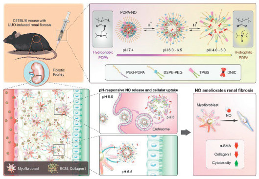
Tumor tissues produce large amounts of lactic acid due to vigorous glycolysis, resulting in a weakly acidic microenvironment (pH 6.5–6.8), while intracellular lysosomes and endosomes within cells have a lower pH environment (pH ~5.0). Taking advantage of this feature, NO donors containing acid-sensitive chemical bonds can be designed to cleave under acidic conditions, thus triggering the release of NO For example, Yu et al. proposed an acid-activated prodrug (P-NO—CA@Fe) to trigger intracellular cascade reactions for cinnamaldehyde (CA)/NO-enhanced chemodynamic therapy (CDT) against melanoma [82]. Hydrazine-terminated polyamidoamine (PAMAM) dendrimers functioned as nanoscaffolds to conjugate H2O2-boosting CA through Schiff-base reactions and chelate Fe3+ Fenton action initiator. Concurrently, arginine (Arg), a NO precursor, was grafted onto the PAMAM dendrimers. These nano-CDT reactors were embedded in water-soluble polyvinylpyrrolidone (PVP) matrix of microneedles (MNs) which served as a carrier for precise prodrug delivery, thereby minimizing systemic toxicity risks. In the acidic tumoral environment, triggered release of H2O2-enhancing components initiates a cascade of NO production and Fenton-like reaction. Hydroxyl radical (•OH) generation combined with the depletion of GSH by NO synergistically amplifies tumor oxidative stress, promoting both apoptosis and ferroptosis. The antitumor efficacy of the MN patch was validated using an A375 melanoma-bearing mouse model. This H2O2 boost-GSH depletion-Fenton killing strategy expands therapeutic options for superficial tumor via MN-enhanced CDT. Similarly, Lee et al. engineered a novel pH-responsive nanoparticle PDPA-NO to deliver NO to halt kidney fibrosis, which was assembled a NO donor dinitrosyl iron complex (DNIC; [Fe2(µ-SEt)2(NO)4]) into acid-responsive poly(2-(diisopropylamino)ethyl methacrylate) (PDPA) polymers (Fig. 4) [83]. The PDPA-NO remained stable at physiological pH 7.4 while disintegrated in acidic conditions (pH 4.0–6.0) via PDPA protonation, facilitating PDPA-NO release. Subsequently, dioxygen-induced decomposition of free DNIC then generated NO within acidic endosomes/lysosomes of fibrotic kidney cells. In unilateral ureteral ligation/obstruction (UUO)-induced fibrotic mice, these NPs demonstrated prolonged blood stability, accumulated in fibrotic kidneys, and significantly suppressed myofibroblast activation and collagen I/extracellular matrix (ECM) production, achieving a 70% reduction in fibrotic area, demonstrating potent antifibrotic efficacy with broad therapeutic applicability. This strategy presents a potent and targeted therapeutic platform for kidney fibrosis with broad applicability. For instance, Huang et al. developed a pH/light dual-activated NP (Rho-Arg) via self-assembly of a PEGylated iodinated rhodamine photosensitizer (Rho-1) and NO donor L-Arg, enabling acidic tumor-microenvironment-responsive and 808 nm NIR-laser-triggered co-delivery of ROS for PDT and NO for gas therapy [84]. Upon the acidic TME activation (pH ~5.5) followed by 808 nm irradiation, Rho-Arg generates ROS for PDT, which concurrently oxidizes L-Arg to NO for gas therapy. Subsequently, the reaction between ROS and NO yields cytotoxic ONOO−, amplifying therapeutic synergy through lipid peroxidation, mitochondrial damage, and DNA fragmentation. In vitro and in vivo validation demonstrated tumor-targeted accumulation of Rho-Arg via ex vivo organ/slice fluorescence imaging. Under the pH/light activation, the nanoplatform induced lysosomal damage, triggered significant apoptosis, and achieved potent tumor regression with excellent biocompatibility. This nanoplatform establishes a promising paradigm for synergistic gas-mediated cancer therapy. Leveraging this specificity, Yang et al. engineered a bacteria-responsive self-activating hydrogel CAOP/M/PL by integrating pH/enzyme-sensitive lactate oxidase-loaded micelles (PPEL) and H2O2-generating CuO2−MoS2 nanozymes (MSCO) within the self-healing hydrogels, constituted of L-Arg modified CS and phenylboronic acid modified oxidized dextran (ODP) [85]. In an acidic infection microenvironment, the hydrogel releases PPEL and MSCO through degradation that lactate oxidase (Lox) converts bacterial lactate to H2O2, which synergistically interacts with H2O2 produced by MSCO to drive CA-derived NO release and nanozyme-enhanced ROS generation via Cu2+-mediated Fenton reactions, thereby achieving synergistic bacterial elimination effects. After post-infection, the hydrogel remodels the regenerative microenvironment through Cu2+-mediated angiogenesis and multi-enzymatic oxidative stress relief. The hydrogel demonstrated potent MRSA biofilm eradication and accelerated the healing of infected wounds in motion-impaired models, achieving effective microenvironment remodeling by enhancing collagen deposition, promoting angiogenesis, and scavenging ROS. This intelligent hydrogel platform represents a transformative strategy for self-adaptive treatment of infected wounds through dynamically responsive microenvironment remodeling.
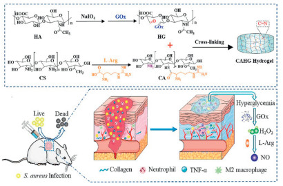
In tumors and diabetes, there is an abnormal elevation of glucose concentration in the microenvironment. This common metabolic characteristic provides an important basis for the design of targeted therapeutic strategies [86]. Researchers have developed a glucose-responsive NO delivery system, which typically consists of glucose oxidase (GOx) and NO precursors (such as L-Arg) [87]. In this system, GOx catalyzes the oxidation of glucose in a locally high-glucose environment and produces H2O2 [88]. And the generated H2O2 can further oxidize NO precursors, thereby achieving in situ release of NO at the lesion site, ultimately achieving gas therapy for tumors or diabetic wounds. For instance, Ma et al. developed a multienzyme hydrogel dressing (COH-GB) by incorporating a GOx and hemoglobin (Hb) nanoflower into a carboxymethyl CS (CMCS)/oxidized sodium alginate (OSA)/hydroxyurea (HU) hydrogel [89]. Hb can deliver O2 to alleviate hypoxia in the MRSA-infected diabetic wound, thereby promoting the oxidation of glucose catalyzed by GOx and generation of H2O2. This elevated H2O2 further promotes NO release, thus enhancing the regulation effect of COH-GB on microenvironment. Under hyperglycemia, the cascade reaction consumes glucose and produces H2O2. Hb, possessing peroxidase-mimicking activity, then catalyzes H2O2-driven NO production from HU. Furthermore, as blood glucose decreases to normal levels, HU releases trace amounts of NO to promote angiogenesis. COH-GB remodels the diabetic wound microenvironment by reducing glucose level and pH, alleviating hypoxia, scavenging ROS, and releasing glucose-responsive NO, thereby exerting antibacterial and pro-angiogenic effects. And COH-GB significantly accelerated the healing of MRSA-infected diabetic wounds by reducing infection/inflammation, promoting collagen deposition, angiogenesis and the migration and differentiation of fibroblasts and keratinocytes. Collectively, the COH-GB multienzyme nanoflower hydrogel provides an efficient and secure dressing, promoting diabetic wound healing through glucose-responsive NO release and microenvironment regulation. Similarly, Hu et al. developed a hyperglycemia-responsive antibacterial cryogel system (GOA@HG) that generates and releases H2O2 and NO for treating diabetic wound infections [90]. L-Arg and GO were conjugated with HA aldehyde methacryloyl (HAAMA) to form Arg-GO—HAAMA base on a Schiff reaction. Meanwhile, gelatin methacryloyl (GelMA) and Arg-GOHAAMA were copolymerized by an ice crystal template technology to fabricate GOA@HG cryogels. The cryogels have a highly interconnected porous structure, which accelerates wound healing by efficiently absorbing exudate and facilitating the exchange of oxygen/nutrients. The system catalyzes the conversion of glucose into H2O2 via GO, which subsequently oxidizes L-Arg to produce a sustained release of NO Besides, GOA@HG cryogels exhibit superior breathability, exudate management, and hemostasis in vitro. In vivo experiments demonstrate that GOA@HG can significantly accelerate the healing of infected diabetic wounds by inhibiting bacterial growth, promoting angiogenesis, and enhancing collagen deposition. Overall, GOA@HG cryogels exhibit synergistic antimicrobial effects and exceptional dressing functionality, establishing an efficient therapeutic strategy for infected diabetic wounds. Addressing this challenge, Zhou et al. developed a glucose-responsive NO-releasing hydrogel (CAHG) for treatment of infected diabetic wounds. This system was fabricated by in situ crosslinking between L-Arg-conjugated CS and GOx-modified HA via a Schiff-base reaction (Fig. 5) [29]. Under hyperglycemic conditions, this system enables sustained H2O2 and NO release via a glucose/L-Arg cascaded consumption. Specifically, glucose is catalytically oxidized by GOx to generate H2O2, which subsequently oxidizes L-Arg-conjugated CS, thereby inducing the release of NO. In vitro experiments confirm the potent antibacterial effects of CAHG hydrogel via H2O2/NO cascade. In diabetic mice, it accelerates wound healing through bacterial inhibition, pro-inflammatory factor downregulation, M2-macrophage elevation, collagen deposition and angiogenesis. Taken together, CAHG hydrogel with excellent biocompatibility and glucose-responsive NO release characteristic can serve as a highly efficient therapeutic strategy for diabetic wound treatment. Leveraging this characteristic, Shi et al. engineered a glucose-responsive NO-releasing orthopedic implant (SPG-GOx@Arg), which was consisted of polyetheretherketone (PEEK), GO, PDA, GOx, and L-Arg [91]. Under hyperglycemic conditions, immobilized GOx catalyzes glucose oxidation to generate H2O2, which reacts with L-Arg to release NO in situ, thereby promoting periprosthetic angiogenesis and vasodilation. Concurrently, NIR irradiation activates the photothermal properties of GO and PDA, providing synergistic antibacterial efficacy against Gram-positive and Gram-negative bacteria. The implant further enhances osteoblast activity, facilitating bone remodeling and osseointegration. The systemic in vivo studies confirm that the engineered implants can promote angiogenesis to facilitate diabetic osseointegration, demonstrating antibacterial activity and vasodilation in an infected diabetic mouse femur model. This work establishes a novel strategy for overcoming compromised diabetic osseointegration by employing hyperglycemic microenvironment-responsive NO gas-releasing orthopedic implants.
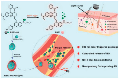
The light-responsive NO delivery system is an advanced therapeutic platform that utilizes light to trigger the NO release at a target site [92]. NIR light with its non-invasiveness, deep tissue penetration, and high controllability, enables precise regulation of NO release in terms of temporal, spatial, and dose-dependent manner [93,94]. The system relies on light-sensitive NO donors by triggering photolysis or photoinduced bond cleavage through NIR light irradiation, enabling the site-specific NO release. This light-controlled release mechanism provides precise spatial control of NO delivery to enable effective localized treatment, making it particularly suitable for applications in wound healing, cancer treatment, and microbial infection treatment [95]. Besides, the photothermal and photodynamic therapy generated accompanied by the light-triggered process can be further combined with NO gas therapy to amplify the therapeutic effect. To illustrate, Cheng et al. integrated the apurinic/apyrimidinic endonuclease 1 (APE1)-responsive DNAzyme, a NO-releasing prodrug SNAP, and upconversion NPs (UCNPs) to construct a nanoscale platform USD that synchronized NIR-light illumination with cascade reactions to achieve precise control of gene inhibition and synergistic gas-based cancer eradication [96]. In this system, the UCNPs convert light into the spectral window required to release NO from the prodrug upon NIR light exposure. In addition to its inherent cytotoxicity, the released NO can increase the abundance of APE1 in the cytoplasm, thereby accelerating the maturation of DNA enzymes and enabling spatially restricted gene knockdown. In the cellular and in vivo experiments, this two-stage amplification cycle combined NIR-controlled NO release with APE1-triggered gene disruption, significantly improving therapeutic efficacy. This strategy provides new insights into the quest for the precision and efficacy of next-generation cancer treatments. Additionally, Chai et al. engineered a NIR-light activatable platelet-mimetic NO nanomedicine (RBT3-NO-PEG@PM) that integrated a NIR-Ⅱ NO organic precursor (RBT3-NO), DSPE-mPEG5k and the platelet membrane for the diagnosis and therapy of atherosclerosis in mice (Fig. 6) [97]. In this system, RBT3-NO-PEG@PM can actively target to atherosclerotic plaques that was irradiated by an 808 nm laser to precisely release NO and synchronously turn on NIR-Ⅱ fluorescence, enabling high-resolution real-time imaging. In vivo experiments demonstrated that laser-controlled NO release can effectively reduce the deposition of plaque lipid, alleviate local inflammation, and stimulate endothelial cell migration, collectively retarding the plaque progression. This strategy thus provides a clinically applicable paradigm for the early and accurate diagnosis of atherosclerotic disease. Similarly, Yu et al. constructed NRh-pH-NO NPs, a NIR-light triggered rhodamine NO nanoprodrug, to achieve efficient gas therapy of tumors [98]. In this system, the rhodamine-N-nitrosamine conjugate (NRh-pH-NO) was integrated into liposomal carriers to form NRh-pH-NO NPs. Under 808 nm laser excitation, the nanoprodrug NRh-pH-NO NPs executed a programmed cascade reaction. The NRh-pH-NO NPs were photocleaved to release NO with concomitant generation of the photosensitizer agent NRh-pH, which further produced superoxide anion O2•−. Subsequently, the system combined superoxide anions with NO, thereby generating ONOO−. The reactive nitrogen species reduced the cell membrane potential (ΔΨm), activated caspase-3, and triggered immune cell apoptosis. This process can be tracked by real-time Frequency upconversion luminescence (FUCL) detection at a wavelength of 740 nm, enabling FUCL image-guided tumor ablation with extremely low systemic toxicity. Overall, this work provided a powerful strategy for establishing an all-in-one molecular nanoplatform for the next generation of NO based gas therapy. In a notable study, Ma et al. constructed an injectable NIR-light responsive antimicrobial hydrogel (DEPN) assembled by covalently cross-linking oxidized dextran (Dex), the cationic antimicrobial peptide ε-poly-L-lysine (EPL) and encapsulating photothermal poly(dopamine-co-glucosamine) (PDG) NPs with thermosensitive NO donors. This system enables controlled sequential release of NO to disrupt the biofilms, eliminate multidrug-resistant pathogens and promote wound repair [99]. In this system, DEPN can slowly release low concentrations of NO without the external stimulation, thereby destroying the protective biofilm and making the microorganisms inside the biofilm sensitive. Under 808 nm laser irradiation, the hydrogel DEPN can convert light into localized heat and trigger the NO release, which synergized with the mild hyperthermia and membrane disruption effects of ε-polylysine to eliminate the pathogenic microorganisms. In acute injuries, the hydrogel DEPN can sustainably release NO, promote endothelial cell migration, inhibit excessive inflammation, and enhance the collagen deposition. In a MRSA-infected wound model in diabetic mice, DEPN eliminated 99.96% of biofilm biomass, promoted angiogenesis, and significantly accelerated wound healing. This work provided the translational potential in addressing the biofilm-associated infections in chronic wounds.
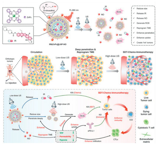
US, an established non-invasive therapeutic modality, enables deep tissue penetration (up to several centimeters) and precise focusing that can facilitate localized NO delivery and controlled release within target tissues, making US an ideal exogenous stimulus for site-specific NO therapy [100,101]. When US acts on NO nanocarriers, it induces cavitation effects that generate localized thermal effects to enhance local blood flow and promote repair. Alternatively, it produces abundant ROS to catalyze the conversion of the NO donors into NO Furthermore, the mechanical effects can disrupt force-sensitive chemical bonds within NO prodrugs, ultimately achieving localized, controlled NO release at the US-irradiated target site [102,103]. This exogenous stimulus-responsive approach offers the advantage of precise on-off control through US devices, overcoming the limitations of tumor heterogeneity for endogenous stimulation. In addition, US-triggered NO release can be synergistically combined with sonodynamic therapy to achieve multimodal treatment of deep lesions. However, this strategy still faces the challenges of fluctuating cavitation efficiency and precise tumor localization in vivo, which propels the development of imaging-guided US-responsive NO nanomedicines. For example, Fang et al. developed an US responsive lipid nanoplatform (IR&ZnPc@LNP-NO) that was encapsulated zinc phthalocyanine (ZnPc), irinotecan (IR), and NO prodrugs (SNO) in the biocompatible lipid NPs (LNPs) for eradicating orthotopic pancreatic tumors (Fig. 7) [104]. Under low-intensity US stimulation, the size of IR&ZnPc@LNP-NO was reduced from ~120 nm to ~40 nm, resulting in enhanced tumor penetration. In this system, the surface-modified SNO can release NO and remodel the TME to degrade the extracellular matrix, normalize blood vessels and alleviate hypoxia. Simultaneously, IR was released as a chemotherapeutic agent, causing DNA damage with single-strand break and the replication inhibition, thereby achieving US-induced chemotherapy. Subsequently, upon high-intensity US exposure, the tumor-retained IR&ZnPc@LNP-NO were induced to release the reactive oxygen species from the sonosensitizer ZnPc, along with the release of IR. In this process, the synergistic effects of ROS and chemotherapy can lead to potent death of immune cells and the activation of anti-tumor immunity. When integrated with PD-L1 checkpoint blockade, the survival time was significantly prolonged in orthotopic models, providing a clinically applicable strategy for overcoming the stromal and immune barriers of deep-seated pancreatic cancer. Besides, Fang et al. engineered an US responsive 4D bio-scaffold (BP-GSNO-BG) that was fabricated by several layers of black phosphorus (BP) embedded into a 3D-printed bioactive glass framework and NO donor (GSNO) for acoustic piezoelectric-gas synergistic therapy [105]. In this system, BP-GSNO-BG underwent a series of sequential treatment processes. Among them, the BP nanosheets, serving as an US-responsive piezoelectric blade, generated ROS by the piezoelectric effect. These ROS synergized with the NO gas generated from US-induced GSNO, enabling the destruction of bone-invasive tumors with high spatiotemporal specificity. In addition, BP nanosheets were biodegraded in situ and sustainably release low concentrations of NO, which promoted osteogenesis and bone integration. This programmable 4D biomaterial platform integrated US-responsive BP nanosheets with NO donors, setting a new standard for intelligent and patient-adaptive cancer treatments in the fields of tumor ablation and bone regeneration. Additionally, Zhang et al. constructed a US-stimulated nanovaccine (SB@CD86) by cloaking an organic sonosensitizer (SP), US-responsive NO donor N,N’-di-sec-butyl-N,N’-dinitroso-1,4-phenylenediamine (BNN6) and artificial dendritic cell membranes with the tumor-matched pMHC-I and CD86 [106]. In this system, the nanocore SP and BNN6 can release ROS and NO in response to the US stimulation, and further generate the cytotoxic ONOO− in situ that ignites the endoplasmic reticulum (ER) stress, protein S-nitrosylation, and mitochondrial collapse, ultimately leading to strongly immunogenic cell death. Furthermore, SB@CD86 are able to target tumor tissue and directly activate cytotoxic T lymphocytes without relying on resident dendritic cells. In mouse models, this US-activated ROS/NO-releasing nanovaccine can not only eradicate primary tumors, but also generate systemic long-lasting immunity against distal metastases, providing a versatile strategy to address issues such as tumor drug resistance and poor patient responsiveness. Moreover, Cheng et al. fabricated the bimetallic biligand frameworks (Cu[PBA-NO]) by multiple metal/ligand self-assembly using two Food and Drug Administration (FDA)-approved drug ligands (ferricyanide and nitroprusside), potassium ferricyanide and nitroglycerin, as well as copper and iron ions [107]. In this system, the Cu[PBA-NO] with the excellent US catalytic ability can simultaneously release NO and generate ROS under US irradiation, both of which were rapidly converted to highly cytotoxic peroxynitrite. In addition, the Cu[PBA-NO] can reduce intracellular GSH levels, thereby disrupting the intracellular redox balance, promoting the accumulation of ROS and triggering the immune cell death with the expression of adenosine triphosphate (ATP), high mobility group box 1 (HMGB1), and calreticulin. Besides, the Cu[PBA-NO] can propel the maturation of dendritic cells within draining lymph nodes, resulting in the generation of potent CD8+/CD4+ T cell initiation and systemic antitumor responses. This work provides a new approach for the efficient combination of US-driven NO gas therapy and cancer immunotherapy.
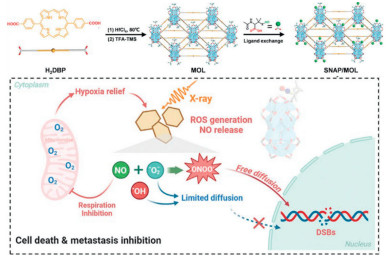
As a deep-tissue irradiation technique, X-rays are widely used in clinics due to their excellent non-invasiveness, limitless tissue penetration, and spatial and temporal controllability [108,109]. The High energy X-rays disrupt cellular homeostasis by damaging DNA structure, thereby killing cancer cells [110-112]. Furthermore, X-rays can cleave specific low-energy chemical bonds, directly destroying those in NO donors, such as the S-N bond in nitrosothiol (SNO) [113]. By utilizing this property, designing X-ray sensitive protective groups to cover the active sites of NO prodrugs can effectively reduce the systemic toxicity of the drug. In turn, the released NO acts as a highly effective radiosensitizer, significantly enhancing the killing effect of radiotherapy on tumor cells. For instance, Liu et al. developed an X-ray-responsive nanoradiosensitizer Hf-nIm@PEG (HNP) via the coordination of hafnium ions (Hf4+) with 2-nitroimidazole (2-nIm), encapsulated within PEGylated lipid bilayers for enhanced tumor targeting [33]. Under X-ray irradiation, HNP induces DNA damage by depositing radiation energy via Hf4+, simultaneously releasing NO from 2-nIm to alleviate hypoxia. And then NO reacts with superoxide anion to generate reactive nitrogen species (RNS), thereby inducing apoptosis. Synergistically, Hf4+ activates the cGAS-STING pathway to amplify dendritic cell maturation and activate potent anti-tumor immunity to against cancer progression, metastasis, and recurrence. In vivo experiments, HNP-mediated radiotherapy achieved complete ablation of the primary tumor, induced immunogenic cell death with antigen release, and elicited robust abscopal effects, effectively suppressing distant metastasis and establishing durable immune memory. Overall, the novel nanoplatform HNP NPs offers a new strategy for synergistic radioimmunotherapy, effectively addressing hypoxia tolerance and metastatic recurrence through targeted NO delivery and immune activation. Similarly, Xiong et al. engineered an X-ray-responsive nanoscale metal-organic layer (SNAP/MOL), based on Hf, which was constructed by conjugating the NO donor S-nitroso-N-acetylpenicillamine (SNAP) to the Hf12 secondary building units (SBUs) of Hf-5,5′-di-p-benzoatoporphyrin MOL, thereby utilizing the periodic high-Z metal arrays for enhanced X-ray energy deposition (Fig. 8) [113]. Under low-dose X-ray irradiation, SNAP/MOL achieves spatiotemporally synchronized O2•− generation via electron transfer, thereby promoting the release of NO from SNAP. These two species rapidly react to form long-acting cytotoxic ONOO−, which diffuses into the cell nucleus and causes DNA double-strand breaks. Concurrently, the sustained NO release alleviates tumor hypoxia to overcome radioresistance. The SNAP/MOL with low-dose irradiation suppressed primary tumor growth in the CT26 colorectal cancer model and 4T1 triple-negative breast cancer model, while simultaneously reducing lung metastasis and enhancing DNA damage through ONOO−-mediated nuclear targeting. Thus, this MOL nanoplatform has pioneered the spatiotemporal synchronization of highly cytotoxic RNS, providing a paradigm for metastatic cancer treatment. Additionally, Liu et al. engineered X-ray-responsive silica-based nanoscintillators (P/SMNO—C) by integrating a Ce-doped LiLuF4 core within dendritic mesoporous silica, functionalized with g-C3N4 quantum dots, S-nitrosothiol NO donors (SNO), and PEG for colon cancer radiotherapy [114]. Upon X-ray irradiation, the Ce-doped LiLuF4 scintillators as dual-role mediators can both deposit X-ray energy to directly damage DNA and convert X-ray photons into UV light to trigger NO release and O2•− generation, leading to in situ formation of highly cytotoxic ONOO− and activation of the cellular nitrosative stress. Additionally, the P/SMNO—C treated with X-ray irradiation exerts its effects on a dual DNA damage through both direct radiation-induced ionization and indirect inhibition of DNA repair via ONOO−-mediated inactivation of glutamine synthetase (GS). Meanwhile, ONOO− induces mitochondrial damage and promotes caspase-3-mediated apoptosis. Conversely, NO-induced vasodilation reduces hypoxia inducible factor-1α (HIF-1α) levels to overcome hypoxia-associated radiation resistance. This nitrosative stress-amplifying nanoplatform provides an open-source and reduce-expenditure DNA damage accumulation strategy for precision radiotherapy, enabling low-dose postoperative RT for colon cancer. Leveraging this property, Liu et al. constructed a two-dimensional nanoplatform (LRC) by integrating the NO donor Roussin’s red salt (RRS) with g-C3N4 nanosheets onto the Ce-doped LiYF4 scintillators, enabling X-ray-to-UV conversion via the scintillation effect of the high-Z material [115]. Under X-ray irradiation, the emitted UV simultaneously photoactivates RRS to release NO and g-C3N4 to generate O2•−. Their rapid reaction forms cytotoxic ONOO−, which induces mitochondrial and DNA damage and inactivates glutamine synthetase (GS) to block DNA repair. Consequently, LRC with X-ray irradiation significantly inhibits tumor growth in vivo and vitro by enhancing DNA damage accumulation and suppressing GS activity, while functioning as a CT contrast agent for imaging-guided therapy and alleviating hypoxic radioresistance. This scintillator-mediated platform establishes ONOO− overexpression as a combinatorial strategy for imaging-guided radio-immunotherapy, synergistically overcoming hypoxia and DNA repair limitations to enhance theranostic efficacy.
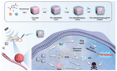
MW, as a localized tumor treatment technique, has garnered significant attention due to its advantages such as deep tissue penetration, minimally invasiveness, operational simplicity, and excellent clinical applicability [116-118]. Researchers have developed MW-responsive NO delivery systems by co-assembling NO precursors (e.g., L-Arg) with MW-sensitized nanomaterials. Under MW irradiation, the sensitized materials can generate ROS to oxidize the NO precursors, enabling the in-situ and controlled release of NO at the tumor site. Additionally, MW-induced hyperthermia can also facilitate the release of NO from the NO precursors. The high concentration of NO generated can not only induce mitochondrial dysfunction and enhance the sensitivity of tumor cells to MW hyperthermia, but also synergistically exert gas therapy effects with ROS and modulate the immunosuppressive microenvironment, thereby effectively inhibiting tumor metastasis and recurrence [119]. For instance, Guo et al. designed a mitochondria-targeted MW-immunosensitizer (GaMOF-Arg-TPP) by conjugating triphenylphosphine (TPP) to gallium-based MOFs loaded with L-Arg (GaMOF-Arg), enabling MW-triggered NO release [120]. Under MW irradiation, GAT generates ROS and releases L-Arg, which further reacts with ROS to produce NO to exert a triple therapeutic effect on inducing mitochondria damage to enhance thermal sensitivity, downregulating HIF-1α to alleviate tumor hypoxia, and triggering immunogenic cell death (ICD) via calreticulin (CRT) exposure, HMGB1 release, and T-cell proliferation. In 4T1 tumor-bearing mice, the GaMOF-Arg-TPP with MW irradiation achieved 100% primary tumor suppression and significantly increased intratumoral CD8+ T-cells, demonstrating potent abscopal effects with complete inhibition of metastasis/recurrence in dual-tumor models. This mitochondria-targeted NO-generation platform pioneers a triple-sensitization strategy for MW immunotherapy, offering a promising approach for clinical translation to overcome post-thermotherapy recurrence in cancer. Similarly, Wen et al. engineered a MW/TME-dual responsive nanocapsule (NPs@CDDP) via self-assembly of cisplatin (CDDP) with biodegradable mPEG-PCL-PArg triblock copolymers, integrating MW-absorbing L-Arg release for thermoacoustic imaging and NO generation [121]. In the TME, NPs@CDDP undergoes enzymatic degradation to release L-arginine and cisplatin. L-Arg serves as a thermoacoustic contrast agent for tumor delineation and elevates local temperature under ultralow-power MW irradiation. Concurrently, CDDP-induced H2O2 overproduction promotes the continuous oxidation of L-Arg to NO, which sensitizes therapy by enhancing lipid peroxidation through GPX4 inhibition and GSH depletion. NPs@CDDP enabled precise tumor ablation via thermoacoustic imaging, enhanced MW heating at ultralow power, and significantly suppressed hepatocellular carcinoma (HCC) growth in vivo by synergizing NO-sensitized thermo-chemotherapy with CDDP cytotoxicity. This theranostic platform established a novel paradigm for imaging-guided percutaneous MW therapy of HCC, simultaneously achieving precision guidance and tumor suppression through microenvironment-triggered NO sensitization. For instance, Wang et al. developed mitochondria-targeted nanoparticles (TiCuMOF@PEG@L-Arg@TPP, TCPAT) by conjugating TPP to TiCu MOFs loaded with L-Arg, where Cu2+ acts as MW co-catalyst to enhance ROS generation (Fig. 9) [122]. Under MW irradiation, TiCuMOF-generated ROS oxidize L-Arg to release NO for deep tumor penetration. Synergistically, ROS and NO can both upregulate p53 to inhibit glycolysis and increase tumor cell sensitivity to copper ions, thereby inducing self-sensitized cuproptosis. In triple-negative breast cancer models, TCPAT combined with MW irradiation significantly enhanced cuproptosis efficacy, demonstrating potent tumor growth suppression and metastasis inhibition through p53-mediated metabolic reprogramming and increased copper sensitivity. This platform has pioneered metabolic intervention-enhanced cuproptosis therapy, overcoming intrinsic copper resistance in breast cancer. In a notable study, Guo et al. constructed mitochondria-targeted nanoamplifiers (GCZMT) by encapsulating NO donor nitrocysteine (CSNO) and HSP70 activator teprenone (GGA) in ZrMOF-NH2, gated with L-menthol (LM) thermal-switches and conjugated to TPP [123]. Under MW irradiation, localized heating melts LM to release GGA and CSNO from GCZMT. CSNO generates NO, which disrupts mitochondrial energy metabolism and promotes apoptosis. Synergistically, NO, CGA, and MW radiation programmatically amplify HSP70 expression through direct thermal induction, NO-mediated upregulation, and GGA-induced heat shock factor (HSF-1) phosphorylation. This enhanced HSP70 expression promotes CD4+ and CD8+ T-cell responses, boosting immunogenic cell death. In breast cancer models, GCZMT-mediated therapy achieved 97% tumor inhibition with significant T-cell infiltration, validating its programmed immune amplification. This nanoamplifier platform offers a cascading immune enhancement strategy for MW immunotherapy, addressing the clinical challenge of tumor recurrence in post-thermotherapy of tumors.
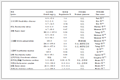
In summary, NO-based gas therapy has shown significant potential in disease treatment. This review highlights the potential and importance of NO in therapeutic applications, discusses various types of stimulus-responsive NO delivery systems and their critical roles in various diseases, and summarizes the latest research progress on NO nanodelivery systems (Table 1). To address issues such as its high diffusivity, short half-life, and low targeting efficiency, numerous NO-loaded nanodelivery systems have been developed by integrating NO donors with diverse nanomaterials through nanotechnology to improve the bioavailability and therapeutic efficacy of NO Concurrently, rationally designed stimulus-responsive NO nanodelivery systems could enable controlled release of NO at the lesion site, minimizing side effects. At present, NO-based nanodelivery systems are primarily employed in the treatment of cancer, cardiovascular disease, bacterial infections, wound healing, inflammatory enteritis and renal fibrosis. Despite some progress has been made in these fields, there is still room for improvement in delivery efficiency, in vivo monitoring, and pharmacodynamic evaluation. In addition, we have further sorted out these challenges and looked forward to the future development trends of this field, hoping that new breakthroughs can be achieved in the development of NO nanodelivery strategies.
 DownLoad:
CSV
| Nanosystem | NO donors | Stimulus | NO payload | NO release profile | Disease model | Ref. |
| Lip-DTI/NO | SNAP | GSH | NO (10 mg/kg); DTI (10 mg/kg) | >90% release | NIH3T3/PANC-1 tumor-bearing mice | [71] |
| JSK@PEG-IR-820 NPs | JSK | GSH | JSK (200 µg/mL) | ~80% release | U87MG tumor-bearing mice | [30] |
| PNP | Dinitrate NO donors | GSH | 40 µg/mL | 3.8 µmol/L | S. aureus-infected mice | [72] |
| T-NPCe6-L-N | SNAP | GSH | Ce6 (2 mg/kg); NO (0.5 mg/kg) | 42.3% release | L3.6 pl Pancreatic tumor-bearing mice | [73] |
| P@G@NG | L-Arg | ROS | / | ~3 µmol/L | Dextran sodium sulfate-induced mice | [78] |
| CRB | L-Arg | ROS | 8 µmol/L | ~3 µmol/L | Panc02 tumor-bearing mice | [79] |
| TTB-NH2@HA-PArg | PArg | ROS | PArg (50 µg/mL) | ~30 µmol/L | A549 tumor-bearing mice | [80] |
| AZOSH | Arg | ROS | 10 mg/kg | / | 4T1 tumor-bearing mice | [81] |
| P-NO-CA@Fe | Arg | pH | CA (75 µg/kg); NO (40 µg/kg); iron ions (25 µg/kg) | 33% release | A375 Melanoma tumor-bearing mice | [82] |
| PDPA-NO | [Fe2(µ-SEt)2(NO)4] | pH | 3 mg/kg | / | UUO-induced mice | [83] |
| Rho-Arg | L-Arg | pH | 100 µL, 500 µg/mL | / | 4T1 tumor-bearing mice | [84] |
| CAOP/M/PL | L-Arg | pH | / | 8.2 µmol/L/L | MRSA-infected mice | [85] |
| COH-GB | Hydroxyurea | Glucose | / | ~40 µmol/L | MRSA-infected mice | [89] |
| GOA@HG | L-Arg | Glucose | L-Arg (1%, w/v) | 9.35 ± 0.82 µmol/L | MRSA-infected mice | [90] |
| CAHG | L-Arg | Glucose | / | ~15 µmol/L | S. aureus-infected diabetic mouse | [29] |
| SPG-GOx@Arg | L-Arg | Glucose | Arg (10 g/L) | ~29 µmol/L | Diabetic rat models | [91] |
| USD | SNAP | NIR laser | 73.07% | ~25 µmol/L | HeLa tumor-bearing mice | [96] |
| RBT3-NO-PEG@PM | RBT3-NO | NIR laser | 50 µmol/L | 81.2% | Mouse models of atherosclerosis | [97] |
| NRh-pH-NO NPs | NRh-pH-NO | NIR laser | 100 µL, 5 mg/kg | / | 4T1 tumor-bearing mice | [98] |
| DEPN | Aldehyde substituted cupferron | NIR laser | PDG@NO (150 µg/mL) | 11.4 µmol/L | MRSA biofilm-infected diabetic wound | [99] |
| IR&ZnPc@LNP-NO | S-Nitrosothiol | US | 200 µL, IR (1.26 mmol/L); ZnPc (0.09 mmol/L); NO (3.6 mmol/L) | 23.7 ± 2.5 µmol/L | Pan02 tumor-bearing mice | [104] |
| BP-GSNO-BG | GSNO | US | BP (1.00 mg/mL); GSNO (0.40 mg/mL) | >1 µmol/L | MG63 tumor-bearing mice | [105] |
| SB@CD86 | BNN6 | US | 200 µL, 0.5 mg/mL | ~18 µmol/L | 4T1 tumor-bearing mice | [106] |
| Cu[PBA-NO] | Sodium nitroprusside (SNP) | US | 0.4 mg/mL | ~200 nmol/L | 4T1 tumor-bearing mice | [107] |
| HNP | 2-nIm | X-ray | 0.55 mmol/L | ~15 µmol/L | CT26 tumor-bearing mice | [33] |
| SNAP/MOL | SNAP | X-ray | SNAP (200 µmol/L) | 1.44-folds higher | 4T1 tumor-bearing mice | [113] |
| P/SMNO-C | SNO | X-ray | 20 mg/kg | ~25 µmol/L | CT26 tumor-bearing mice | [114] |
| LRC | RRS | X-ray | RSS:LiYF4:Ce- PEI (7.01 mg/mg) | ~2.4 µmol/L | U14 tumor-bearing mice | [115] |
| GaMOF-Arg-TPP | L-Arg | MW | 2 mg/mL | ~7 µmol/L | L929 and 4T1 tumor-bearing mice | [120] |
| NPs@CDDP | L-Arg | MW | ~1.2 mg/mL | ~4.8 µmol/L | SMMC-7721 tumor-bearing mice | [121] |
| TCPAT | L-Arg | MW | 2 mg/mL | ~3.5 µmol/L | 4T1 tumor-bearing mice | [122] |
| GCZMT | CSNO | MW | 3 mg/mL | ~150 µmol/L | 4T1 tumor-bearing mice | [123] |
To develop novel NO nanocarrier systems with enhanced delivery efficiency and superior stability, we can integrate organelle-targeting strategies to create organelle-targeted nanocarriers, enabling intelligent and on-demand release of NO at specific biological targets, thereby enhancing the precision and efficiency of treatment. Meanwhile, employing biomimetic strategies to construct biomimetic NO donors holds promise for achieving more precise site-specific delivery. For commonly used liposomal materials, precise regulation of NO release rates can be achieved by adjusting the chain saturation, chain length, and fluidity of the lipid bilayer. Besides, constructing diversified carriers by combining multiple polymer matrices (such as HA, polyethylene glycol) with NO donors can enhance water solubility, biocompatibility, and controlled release properties, improve the delivery capacity and avoid unnecessary side effects and long-term cumulative toxicity. Furthermore, the development of nanomedicines that are responsive to both endogenous and exogenous stimuli is a potential strategy. These nanomedicines can only release NO in the presence of dual triggers, both endogenous and exogenous stimuli, achieving more precise and controllable release of NO and more effectively avoiding the systemic toxicity of NO nanoprodrugs.
In terms of in vivo monitoring and pharmacodynamic assessment, the current major bottleneck is the lack of methods capable of achieving real-time, non-invasive tracking of NO release kinetics in living systems. It is difficult to establish a clear dose-response relationship and to confirm whether the nanocarriers function as expected under physiological conditions. To address these challenges, advanced non-invasive real-time detection technologies can be integrated to develop highly accurate NO-responsive probes. These probes, co-encapsulated with NO donors in nanoscale delivery systems, can facilitate efficient disease diagnosis and targeted treatment. Fluorescence imaging or multimodal imaging approaches may be employed to detect the dynamics of NO release and monitor its spatiotemporal distribution at specific sites, thereby visualizing the NO release process during therapeutic interventions. Furthermore, a novel NO prodrug system could be designed by integrating fluorescence probes with NO donors. The release of NO would then induce measurable changes in fluorescence signal, enabling quantitative correlation between NO liberation and optical output. This strategy offers a promising means to track the in vivo distribution and real-time release of NO with high spatial and temporal resolution. With the sustained innovation in technology and increasingly integrated interdisciplinary cooperation, NO delivery systems hold great promise for personalized and targeted disease treatment, potentially achieving the optimization of clinical outcomes.
Hui Yu: Writing – review & editing, Writing – original draft, Visualization, Supervision, Funding acquisition, Conceptualization. Ziyu He: Writing – review & editing, Writing – original draft. Bochuan Liu: Writing – original draft. Tianyou Du: Writing – original draft. Hai-Yan Wang: Supervision, Project administration. Yi Liu: Writing – review & editing, Supervision, Project administration, Funding acquisition.
The authors declare that they have no known competing financial interests or personal relationships that could have appeared to influence the work reported in this paper.
This study was supported by the National Natural Science Foundation of China (No. 82272148), and the Jiangsu Funding Program for Excellent Postdoctoral Talent (No. 2022ZB301).
L. Qiu, J. Liang, L. Xue, et al., Adv. Funct. Mater. 34 (2024) 2408189. doi: 10.1002/adfm.202408189
B. Chen, Y. Sun, H. Sun, et al., ACS Nano 18 (2024) 33232–33244. doi: 10.1021/acsnano.4c12676
T. Liu, X. Han, X. Sun, et al., Chin. Chem. Lett. 36 (2025) 110170. doi: 10.1016/j.cclet.2024.110170
H. Chen, T. Shi, Y. Wang, et al., J. Am. Chem. Soc. 143 (2021) 12025–12037. doi: 10.1021/jacs.1c03071
C. Won, S. Kim, D. Kwak, et al., Angew. Chem. Int. Ed. 63 (2024) e202411260. doi: 10.1002/anie.202411260
M. Wan, H. Chen, Q. Wang, et al., Nat. Commun. 10 (2019) 966. doi: 10.1038/s41467-019-08670-8
Z. Wang, N. Yang, Y. Hou, et al., Adv. Sci. 10 (2023) 2302123. doi: 10.1002/advs.202302123
X. Bao, S. Zheng, L. Zhang, et al., Angew. Chem. Int. Ed. 61 (2022) e202207250. doi: 10.1002/anie.202207250
Y. Wu, G. Deng, K. Jiang, et al., Biomaterials 268 (2021) 120588. doi: 10.1016/j.biomaterials.2020.120588
Z. Shen, W. Jiang, S. Zheng, et al., Angew. Chem. Int. Ed. 64 (2025) e202419939. doi: 10.1002/anie.202419939
C. Farah, L.Y.M. Michel, J.L. Balligand, Nat. Rev. Cardiol. 15 (2018) 292–316. doi: 10.1038/nrcardio.2017.224
Y. Peng, W. Feng, H. Huang, et al., Bioact. Mater. 48 (2025) 294–312.
L.A. Ridnour, D.D. Thomas, S. Donzelli, et al., Antioxid. Redox Signal. 8 (2006) 1329–1337. doi: 10.1089/ars.2006.8.1329
J.O. Lundberg, E. Weitzberg, Cell 185 (2022) 2853–2878. doi: 10.1016/j.cell.2022.06.010
D. Gao, S. Asghar, R. Hu, et al., Acta Pharm. Sin. B 13 (2023) 1498–1521. doi: 10.1016/j.apsb.2022.11.016
A.W. Carpenter, M.H. Schoenfisch, Chem. Soc. Rev. 41 (2012) 3742–3752. doi: 10.1039/c2cs15273h
J. An, Y. Hu, C. Li, et al., Biomaterials 230 (2020) 119636. doi: 10.1016/j.biomaterials.2019.119636
H. Cai, X. Wu, L. Jiang, et al., Chin. Chem. Lett. 35 (2024) 108946. doi: 10.1016/j.cclet.2023.108946
S. Xu, X. Xie, P. He, et al., ACS Nano 19 (2025) 6371–6387. doi: 10.1021/acsnano.4c16329
L. Chen, H. Ming, B. Li, et al., Small 20 (2024) 2403869. doi: 10.1002/smll.202403869
Z. Shen, S. Zheng, Y. Fang, et al., Angew. Chem. Int. Ed. 62 (2023) e202219153. doi: 10.1002/anie.202219153
S.M. Andrabi, N.S. Sharma, A. Karan, et al., Adv. Sci. 10 (2023) 2303259. doi: 10.1002/advs.202303259
T.A. Tabish, M.J. Crabtree, H.E. Townley, et al., JACC Basic Transl. Sci. 9 (2024) 691–709. doi: 10.1016/j.jacbts.2023.07.017
F. Lisi, A.N. Zelikin, R. Chandrawati, Adv. Sci. 8 (2021) 2003895. doi: 10.1002/advs.202003895
Y. Zhao, D. Shi, L. Guo, et al., J. Nanobiotechnol. 21 (2023) 35. doi: 10.1186/s12951-023-01776-8
K. Li, S. Hu, H. Li, et al., Nat. Commun. 16 (2025) 5677. doi: 10.1038/s41467-025-61256-5
W. Jiang, Z. Guo, Q. Wang, et al., Nat. Biomed. Eng. 9 (2025) 1486–1501. doi: 10.1038/s41551-025-01385-w
H. Cheng, G. Tian, H. Liu, et al., J. Colloid Interface Sci. 678 (2025) 560–571. doi: 10.1016/j.jcis.2024.08.210
X. Zhou, B. Zhao, L. Wang, et al., J. Control. Release 359 (2023) 147–160. doi: 10.1016/j.jconrel.2023.05.047
J. Zou, Z. Li, Y. Zhu, et al., Bioact. Mater. 34 (2024) 414–421.
H. Lu, B. Liang, A. Hu, et al., Adv. Mater. 37 (2025) 2412655. doi: 10.1002/adma.202412655
J. Hu, L. Yuan, Y. Zhang, et al., Angew. Chem. Int. Ed. 63 (2024) e202317578. doi: 10.1002/anie.202317578
N. Liu, J. Zhu, W. Zhu, et al., Adv. Mater. 35 (2023) 2302220. doi: 10.1002/adma.202302220
H. Xu, D. Jia, S. Guo, et al., J. Colloid Interface Sci. 679 (2025) 191–200. doi: 10.1016/j.jcis.2024.10.109
D. Fan, Y. Cao, M. Cao, et al., Sig. Transduct. Target. Ther. 8 (2023) 293. doi: 10.1038/s41392-023-01536-y
Z. Xu, Q. Luo, Y. He, et al., Adv. Funct. Mater. 34 (2024) 2314536. doi: 10.1002/adfm.202314536
R. Kumar, D.S. Dkhar, R. Kumari, et al., J. Drug Deliv. Sci. Technol. 74 (2022) 103526. doi: 10.1016/j.jddst.2022.103526
S. Zeng, Y. Chen, F. Zhou, et al., Adv. Drug Deliv. Rev. 199 (2023) 114965. doi: 10.1016/j.addr.2023.114965
D.E. Uti, E.U. Alum, I.J. Atangwho, et al., J. Nanobiotechnol. 23 (2025) 336. doi: 10.1186/s12951-025-03412-z
M.A. Elnaggar, R. Subbiah, D.K. Han, et al., Expert. Opin. Drug Del. 14 (2017) 1341–1353. doi: 10.1080/17425247.2017.1285904
H. Yang, Y. Guo, L. Pu, et al., Mol. Pharm. 20 (2023) 1404–1414. doi: 10.1021/acs.molpharmaceut.2c00973
R. Wu, Y. Wang, S. Sun, et al., Chem. Eng. J. 486 (2024) 150134. doi: 10.1016/j.cej.2024.150134
W.K. Rhim, J. Woo, J.Y. Kim, et al., J. Adv. Res. 69 (2025) 75–89. doi: 10.1016/j.jare.2024.03.018
H.V. de Almeida, L.C.E. da Silva, M.G. de Oliveira, Nitric Oxide 146 (2024) 48–57. doi: 10.24077/2024;5183207787815
M.T. Pelegrino, J.C. Pieretti, G. Nakazato, et al., Nitric Oxide 106 (2021) 24–34. doi: 10.1016/j.niox.2020.10.003
Z. Wang, M. Zhan, W. Li, et al., Angew. Chem. Int. Ed. 60 (2021) 4720–4731. doi: 10.1002/anie.202013301
M. Shi, J. Zhang, Y. Wang, et al., Biomaterials 283 (2022) 121448. doi: 10.1016/j.biomaterials.2022.121448
D. Jiang, L. Pan, X. Yang, et al., Acta Biomater. 159 (2023) 259–274. doi: 10.1016/j.actbio.2023.01.032
G. Li, K. Lv, Q. Cheng, et al., Adv. Sci. 10 (2023) 2206959. doi: 10.1002/advs.202206959
J. Kim, D.M. Francis, L.F. Sestito, et al., Nat. Commun. 13 (2022) 1479. doi: 10.3390/v14071479
H. Li, F. Meng, C. Hu, et al., Adv. Sci. 12 (2025) 2500833. doi: 10.1002/advs.202500833
J.A. Ankrum, O.R. Miranda, K.S. Ng, et al., Nat. Protoc. 9 (2014) 233–245. doi: 10.1038/nprot.2014.002
Z. Luo, G. Ng, Y. Zhou, et al., Small 19 (2023) 2200502. doi: 10.1002/smll.202200502
H. Yao, E.S. Turali Emre, Y. Fan, et al., Bioact. Mater. 48 (2025) 200–216.
T.A. Tabish, C.A. Lygate, Adv. Drug Deliv. Rev. 226 (2025) 115676. doi: 10.1016/j.addr.2025.115676
T.A. Tabish, R.J. Narayan, Acta Biomater. 129 (2021) 43–56. doi: 10.1016/j.actbio.2021.04.054
T.A. Tabish, M.Z. Hussain, S. Zervou, et al., Redox Biol. 72 (2024) 103144. doi: 10.1016/j.redox.2024.103144
J. Zheng, W. Wang, X. Gao, et al., Small 18 (2022) 2205252. doi: 10.1002/smll.202205252
T.A. Tabish, J. Xu, C.K. Campbell, et al., Nitric Oxide 147 (2024) 42–50. doi: 10.1016/j.niox.2024.04.008
Z. Pei, H. Lei, L. Cheng, Chem. Soc. Rev. 52 (2023) 2031–2081. doi: 10.1039/d2cs00352j
B.B. Mendes, Z. Zhang, J. Conniot, et al., Nat. Nanotechnol. 19 (2024) 867–878. doi: 10.1038/s41565-024-01673-7
C. Du, M. Zhou, F. Jia, et al., Biomaterials 269 (2021) 120642. doi: 10.1016/j.biomaterials.2020.120642
X. Hu, Z. Shen, H. Hao, et al., J. Colloid Interface Sci. 694 (2025) 137688. doi: 10.1016/j.jcis.2025.137688
H.B. Ji, S.N. Kim, C.R. Kim, et al., Biomater. Adv. 145 (2023) 213268. doi: 10.1016/j.bioadv.2022.213268
L. Du, H. He, Z. Xiao, et al., Small 18 (2022) 2107732. doi: 10.1002/smll.202107732
J. Mu, C. Li, Y. Shi, et al., Nat. Commun. 13 (2022) 2513. doi: 10.1038/s41467-022-29772-w
M. Xia, Y. Yan, H. Pu, et al., Chem. Eng. J. 442 (2022) 136295. doi: 10.1016/j.cej.2022.136295
Z. Shen, J. Xia, Q. Ma, et al., Theranostics 10 (2020) 9132–9152. doi: 10.7150/thno.46076
F. Xu, M. Wang, E. Dotse, et al., Angew. Chem. Int. Ed. 63 (2024) e202404561. doi: 10.1002/anie.202404561
X. Li, Y. Zhang, Q. An, et al., Adv. Funct. Mater. (2025) e08811.
D. Li, X. Chen, W. Dai, et al., Adv. Mater. 36 (2024) 2306476. doi: 10.1002/adma.202306476
Y. Xiong, S. Yang, Y. Di, et al., ACS Appl. Mater. Interfaces 17 (2025) 25097–25111. doi: 10.1021/acsami.5c03249
W. Dai, X. Zhou, J. Zhao, et al., Biomaterials 312 (2025) 122743. doi: 10.1016/j.biomaterials.2024.122743
X. Liu, Y. Li, J. Chen, et al., J. Colloid Interface Sci. 693 (2025) 137603. doi: 10.1016/j.jcis.2025.137603
J. Wang, J. Liu, Y. Sümbelli, et al., J. Control. Release 372 (2024) 59–68. doi: 10.1016/j.jconrel.2024.06.021
H. Wang, D. Bi, B. Yu, et al., J. Colloid Interface Sci. 682 (2025) 1185–1194. doi: 10.1016/j.jcis.2024.12.018
M. Ding, Q. Zong, D. Zhang, et al., ACS Nano 19 (2025) 662–679. doi: 10.1021/acsnano.4c11514
Y. Fu, X. Zhao, L. Wang, et al., Adv. Mater. 36 (2024) 2309972. doi: 10.1002/adma.202309972
Y. Wang, Q. Tang, R. Wu, et al., ACS Nano 17 (2023) 3557–3573. doi: 10.1021/acsnano.2c09948
B. Li, J. Tian, C. Wu, et al., Adv. Mater. 36 (2024) 2405502. doi: 10.1002/adma.202405502
L. Zhu, D. Leng, Z. Guo, et al., J. Control. Release 377 (2025) 524–539. doi: 10.1016/j.jconrel.2024.11.048
W. Yu, F. Jia, J. Fu, et al., ACS Nano 17 (2023) 15713–15723. doi: 10.1021/acsnano.3c02964
T.Y. Lee, H.H. Lu, H.T. Cheng, et al., J. Control. Release 354 (2023) 417–428. doi: 10.1016/j.jconrel.2022.12.059
X. Huang, W. Sun, Y. Zhu, et al., Sens. Actuators B Chem. 408 (2024) 135522. doi: 10.1016/j.snb.2024.135522
Y. Yang, J. Wang, S. Huang, et al., Nat. Sci. Rev. 11 (2024) nwae044. doi: 10.1093/nsr/nwae044
H. Liu, S. Qin, H. Zhang, et al., Adv. Funct. Mater. 35 (2025) 2404461. doi: 10.1002/adfm.202404461
Y. Shi, Y. Cao, J. Cheng, et al., Adv. Funct. Mater. 32 (2022) 2111148. doi: 10.1002/adfm.202111148
M. He, Z. Wang, H. Yang, et al., Adv. Sci. 10 (2023) 2300986. doi: 10.1002/advs.202300986
H. Ma, Y. Luo, Y. Wang, et al., Bioact. Mater. 49 (2025) 531–548.
J. Hu, K. Duan, Y. Zhao, et al., J. Control. Release 378 (2025) 912–931. doi: 10.1016/j.jconrel.2024.12.061
H. Shi, H. Yang, C. Wu, et al., Biomaterials 321 (2025) 123334. doi: 10.1016/j.biomaterials.2025.123334
H. He, T. He, Z. Zhang, et al., Chin. Chem. Lett. 29 (2018) 1497–1499. doi: 10.1016/j.cclet.2018.08.019
X. Zhao, S. Long, M. Li, et al., J. Am. Chem. Soc. 142 (2020) 1510–1517. doi: 10.1021/jacs.9b11800
R. Liu, Y. Peng, L. Lu, et al., J. Nanobiotechnol. 19 (2021) 443. doi: 10.1186/s12951-021-01078-x
H. Tian, J. Lin, F. Zhu, et al., Chin. Chem. Lett. 34 (2023) 107577. doi: 10.1016/j.cclet.2022.05.091
Y. Cheng, Y. Feng, J. Zhao, et al., Angew. Chem. Int. Ed. 64 (2025) e202422375. doi: 10.1002/anie.202422375
Y. Chai, L. Shangguan, H. Yu, et al., Adv. Sci. 11 (2024) 2304994. doi: 10.1002/advs.202304994
H. Yu, A. Tiemuer, X. Yao, et al., Acta Pharm. Sin. B 14 (2024) 378–391. doi: 10.1016/j.apsb.2023.08.019
H. Ma, T. Wang, G. Li, et al., Acta Biomater. 188 (2024) 169–183. doi: 10.1016/j.actbio.2024.09.017
Y. Bao, Y. Ge, M. Wu, et al., Adv. Sci. 10 (2023) 2302278. doi: 10.1002/advs.202302278
Y. Li, W. Chen, Y. Kang, et al., Nat. Commun. 14 (2023) 6973. doi: 10.1038/s41467-023-42509-7
Z. Jin, Y. Wen, Y. Hu, et al., Nanoscale 9 (2017) 3637–3645. doi: 10.1039/C7NR00231A
X. Xu, J. Cao, Y. Mu, et al., Adv. Sci. 12 (2025) e16709. doi: 10.1002/advs.202416709
L. Fang, W. Zeng, Y. Liu, et al., Angew. Chem. Int. Ed. 64 (2025) e202507388. doi: 10.1002/anie.202507388
H. Fang, D. Zhu, Y. Chen, et al., Adv. Sci. 12 (2025) 2417208. doi: 10.1002/advs.202417208
L. Zhang, X. Kang, T. Wang, et al., Angew. Chem. Int. Ed. 64 (2025) e202504684. doi: 10.1002/anie.202504684
Y. Cheng, W. Zhong, Y. Chen, et al., Adv. Mater. 36 (2024) 2408242. doi: 10.1002/adma.202408242
X. Ou, X. Chen, X. Xu, et al., Research 2021 (2021) 9892152.
Y. Zhang, Y. Liu, Z. Li, et al., Adv. Funct. Mater. 35 (2025) 2412165. doi: 10.1002/adfm.202412165
K. Qiao, Y. Pan, S. Zhang, et al., ACS Nano 18 (2024) 29689–29703. doi: 10.1021/acsnano.4c09021
J. Lin, Y. He, Y. Li, et al., Small 20 (2024) 2310957.
H. Li, J. Zeng, Q. You, et al., Biomaterials 316 (2025) 123031. doi: 10.1016/j.biomaterials.2024.123031
Y. Xiong, J. Li, X. Jiang, et al., Adv. Sci. 12 (2025) 2413518. doi: 10.1002/advs.202413518
S. Liu, W. Li, Y. Zhang, et al., Nano Lett. 22 (2022) 6409–6417. doi: 10.1021/acs.nanolett.2c02472
S. Liu, W. Li, H. Chen, et al., ACS Nano 16 (2022) 8939–8953. doi: 10.1021/acsnano.1c11422
S. Li, Z. Chen, L. Tan, et al., Biomaterials 283 (2022) 121472. doi: 10.1016/j.biomaterials.2022.121472
X. Ma, X. Ren, X. Guo, et al., Biomaterials 214 (2019) 119223. doi: 10.1016/j.biomaterials.2019.119223
H. Zhu, B. Li, X. Liu, et al., ACS Nano 16 (2022) 21098–21110. doi: 10.1021/acsnano.2c09036
H. Shi, T. Liu, C. Fu, et al., Biomaterials 44 (2015) 91–102. doi: 10.1016/j.biomaterials.2014.12.035
W. Guo, Z. Chen, Q. Wu, et al., Small 20 (2024) 2308055. doi: 10.1002/smll.202308055
L. Wen, H. Liu, C. Hu, et al., ACS Appl. Mater. Interfaces 15 (2023) 10477–10491. doi: 10.1021/acsami.2c22523
Y. Wang, Q. Wu, W. Guo, et al., J. Colloid Interface Sci. 691 (2025) 137421. doi: 10.1016/j.jcis.2025.137421
W. Guo, M. Niu, Z. Chen, et al., Adv. Healthc. Mater. 11 (2022) 2201441. doi: 10.1002/adhm.202201441

Figure 2 Schematic illustration of the GSH-triggered supramolecular nanoparticles T-NPCe6-L-N for tumor therapy. Copied with permission [73]. Copyright 2025, Elsevier.

Figure 3 Schematic illustration of the US-triggered nanoprodrug CPT-t-R-PEG2000@BaTiO3 for tumor therapy. Copied with permission [79]. Copyright 2023, American Chemical Society.

Figure 4 Schematic illustration of the pH-responsive nanoparticle PDPA-NO for renal fibrosis therapy. Copied with permission [83]. Copyright 2023, Elsevier.

Figure 5 Schematic illustration of the glucose-responsive CAHG hydrogel for S. aureus-infected diabetic wound treatment. Copied with permission [29]. Copyright 2023, Elsevier.

Figure 6 Schematic illustration of the NIR-triggered nano-prodrug RBT3-NO-PEG@PM for treatment of atherosclerosis. Copied with permission [97]. Copyright 2023, Wiley-VCH GmbH.

Figure 7 Schematic illustration of the US-responsive lipid nanosonosensitizers IR&ZnPc@LNP-NO for pancreatic cancer therapy. Copied with permission [104]. Copyright 2025, Wiley‐VCH GmbH.

Figure 8 Schematic illustration of the X-ray-responsive SNAP/MOL for tumor therapy. Copied with permission [113]. Copyright 2025, Wiley‐VCH GmbH.

Figure 9 Schematic illustration of the MW-responsive nanoparticles TCPAT for tumor therapy. Copied with permission [122]. Copyright 2025, Elsevier.
Table 1. The summary of NO delivery systems.
| Nanosystem | NO donors | Stimulus | NO payload | NO release profile | Disease model | Ref. |
| Lip-DTI/NO | SNAP | GSH | NO (10 mg/kg); DTI (10 mg/kg) | >90% release | NIH3T3/PANC-1 tumor-bearing mice | [71] |
| JSK@PEG-IR-820 NPs | JSK | GSH | JSK (200 µg/mL) | ~80% release | U87MG tumor-bearing mice | [30] |
| PNP | Dinitrate NO donors | GSH | 40 µg/mL | 3.8 µmol/L | S. aureus-infected mice | [72] |
| T-NPCe6-L-N | SNAP | GSH | Ce6 (2 mg/kg); NO (0.5 mg/kg) | 42.3% release | L3.6 pl Pancreatic tumor-bearing mice | [73] |
| P@G@NG | L-Arg | ROS | / | ~3 µmol/L | Dextran sodium sulfate-induced mice | [78] |
| CRB | L-Arg | ROS | 8 µmol/L | ~3 µmol/L | Panc02 tumor-bearing mice | [79] |
| TTB-NH2@HA-PArg | PArg | ROS | PArg (50 µg/mL) | ~30 µmol/L | A549 tumor-bearing mice | [80] |
| AZOSH | Arg | ROS | 10 mg/kg | / | 4T1 tumor-bearing mice | [81] |
| P-NO-CA@Fe | Arg | pH | CA (75 µg/kg); NO (40 µg/kg); iron ions (25 µg/kg) | 33% release | A375 Melanoma tumor-bearing mice | [82] |
| PDPA-NO | [Fe2(µ-SEt)2(NO)4] | pH | 3 mg/kg | / | UUO-induced mice | [83] |
| Rho-Arg | L-Arg | pH | 100 µL, 500 µg/mL | / | 4T1 tumor-bearing mice | [84] |
| CAOP/M/PL | L-Arg | pH | / | 8.2 µmol/L/L | MRSA-infected mice | [85] |
| COH-GB | Hydroxyurea | Glucose | / | ~40 µmol/L | MRSA-infected mice | [89] |
| GOA@HG | L-Arg | Glucose | L-Arg (1%, w/v) | 9.35 ± 0.82 µmol/L | MRSA-infected mice | [90] |
| CAHG | L-Arg | Glucose | / | ~15 µmol/L | S. aureus-infected diabetic mouse | [29] |
| SPG-GOx@Arg | L-Arg | Glucose | Arg (10 g/L) | ~29 µmol/L | Diabetic rat models | [91] |
| USD | SNAP | NIR laser | 73.07% | ~25 µmol/L | HeLa tumor-bearing mice | [96] |
| RBT3-NO-PEG@PM | RBT3-NO | NIR laser | 50 µmol/L | 81.2% | Mouse models of atherosclerosis | [97] |
| NRh-pH-NO NPs | NRh-pH-NO | NIR laser | 100 µL, 5 mg/kg | / | 4T1 tumor-bearing mice | [98] |
| DEPN | Aldehyde substituted cupferron | NIR laser | PDG@NO (150 µg/mL) | 11.4 µmol/L | MRSA biofilm-infected diabetic wound | [99] |
| IR&ZnPc@LNP-NO | S-Nitrosothiol | US | 200 µL, IR (1.26 mmol/L); ZnPc (0.09 mmol/L); NO (3.6 mmol/L) | 23.7 ± 2.5 µmol/L | Pan02 tumor-bearing mice | [104] |
| BP-GSNO-BG | GSNO | US | BP (1.00 mg/mL); GSNO (0.40 mg/mL) | >1 µmol/L | MG63 tumor-bearing mice | [105] |
| SB@CD86 | BNN6 | US | 200 µL, 0.5 mg/mL | ~18 µmol/L | 4T1 tumor-bearing mice | [106] |
| Cu[PBA-NO] | Sodium nitroprusside (SNP) | US | 0.4 mg/mL | ~200 nmol/L | 4T1 tumor-bearing mice | [107] |
| HNP | 2-nIm | X-ray | 0.55 mmol/L | ~15 µmol/L | CT26 tumor-bearing mice | [33] |
| SNAP/MOL | SNAP | X-ray | SNAP (200 µmol/L) | 1.44-folds higher | 4T1 tumor-bearing mice | [113] |
| P/SMNO-C | SNO | X-ray | 20 mg/kg | ~25 µmol/L | CT26 tumor-bearing mice | [114] |
| LRC | RRS | X-ray | RSS:LiYF4:Ce- PEI (7.01 mg/mg) | ~2.4 µmol/L | U14 tumor-bearing mice | [115] |
| GaMOF-Arg-TPP | L-Arg | MW | 2 mg/mL | ~7 µmol/L | L929 and 4T1 tumor-bearing mice | [120] |
| NPs@CDDP | L-Arg | MW | ~1.2 mg/mL | ~4.8 µmol/L | SMMC-7721 tumor-bearing mice | [121] |
| TCPAT | L-Arg | MW | 2 mg/mL | ~3.5 µmol/L | 4T1 tumor-bearing mice | [122] |
| GCZMT | CSNO | MW | 3 mg/mL | ~150 µmol/L | 4T1 tumor-bearing mice | [123] |
 下载: 导出CSV

 扫一扫看文章


扫一扫关注我们

 下载: