
Scheme 1.
Schematic principle of FRET-based polymer sensors for detection of cellular environments. D and A represent donor and acceptor, respectively, and r indicates the donor-acceptor distance.
FRET-based polymer materials for detection of cellular microenvironments
Chuang Weng , Nianqing Fan , Taoran Xu , Haodong Chen , Zifen Li , Yiwen Li , Hong Tan , Qiang Fu , Mingming Ding

Cells are the basic units of life. The physiological processes of cells and the conditions involved in these processes are the basis for unlocking the secrets of cell life. Therefore, understanding the physiological activities in cells is of great significance for early diagnosis and discovery of various diseases, as well as for the assessment of new drugs associated with apoptosis [1, 2]. Despite significant advancements that have been made in antibody, nucleic acid, and enzyme probes for cell analysis [3], collecting information from environments as small as cells remains a considerable challenge. Over the last decade, fluorescence technique has become a powerful tool for noninvasive visualization and optical imaging of diverse biological processes in vitro and in vivo by virtue of its high sensitivity and technical simplicity [4]. However, the conventional fluorescent biosensors are based mainly on the change of single emission intensity in response to target analytes, which are sometimes problematic for precise analysis under complicated biological conditions due to possible interferences from various environmental factors (pH, temperature, solvent, etc.), instrumental conditions, and the concentrations and locations of fluorescent probes inside the cells [5, 6]. To circumvent this problem, the simultaneous measurement of two or more fluorescence signals at different wavelengths enables ratiometric and specific imaging capacity with a greater precision [7, 8]. In particular, fluorescence (or Förster) resonance energy transfer (FRET) technology has recently emerged as a valuable method for real-time imaging of intracellular environments [9-16]. The principle of FRET was pioneeringly proposed by Förster in 1948 [17]. When two different chromophore molecules (donor and acceptor) have overlapping emission/absorption spectra, and the distance between the two fluorophores is appropriate (1-10 nm), a transfer of fluorescence energy from the donor to the acceptor will be observed [18, 19]. Hence, FRET is an effective and sensitive approach for probing very small changes in distance with high spatial resolution, such as molecular binding, bond cleavage, phase transition, and change in protein conformation, etc. [20-22]. These properties make FRET-based probes well suited for quantification of microenvironmental indicators in cells (Scheme 1) [23, 24]. On the other hand, polymer-based imaging systems possess higher thermodynamic and kinetic stability, increased dye payloads, and improved biodistribution compared with small molecular sensors. More importantly, they exhibit attractive signal amplification and high responsivity to external stimuli, allowing for an ultrasensitive detection at nanomolar-picomolar concentrations [25-27]. In recent years, FRET-based polymeric nanosystems have shown great promise in chemical and biological analysis including digitization of pH in cells, tracking of redox processes, and monitoring of changes in temperature and other biosignals (hydrogen sulfide, enzyme) in cells. Here, we outline the recent advances in various FRET probes constructed from polymer materials for exploring cellular microenvironments, highlighting the wide range of applications of FRET technology for understanding diverse biological activities and functions in cells.
The pH value is a vital physiological signal that plays a key role in maintaining cell and tissue homeostasis. Most of the cellular processes are related to the acidic environments in cells, including cell proliferation, apoptosis, signaling, and endocytosis [28, 29]. Abnormal pH values in organelles are recognized as a hallmark of cell dysfunction [30], which have been observed in Alzheimer's disease [31], cancer [32] and other common disease models. On the other hand, the acidic pathological microenvironments allows for the construction of pH-responsive nanosystems for specific delivery and triggered release of drugs and imaging agents in target sites [33-36]. Therefore, monitoring the changes of pH values inside living cells is not only important for better understanding of physiological and pathological processes, but also helpful in developing smart delivery systems for precise diagnosis and therapy of diseases. There have been many methods for studying cellular pH, including nuclear magnetic resonance (NMR) spectroscopy [37-39], surface-enhanced Raman spectroscopy (SERS) [40-44], microelectrodes and optical microscopy [45-50]. Among them, fluorescent probes have emerged as promising tools due to their high sensitivity and selectivity, low cost, simple operation and non-invasiveness [51-56].
Yan et al. [57] designed a ultra pH-responsive micellar system constructed from poly(oligo(ethylene glycol)methacrylate)-b-poly (benzyl-L-aspartic acid) (POEGMA-PBLA) copolymer. The side chains of PBLA block were aminolyzed by primary amine to introduce ionizable tertiary amines as pH-sensitive moieties, while the chain end was conjugated with a cyanine dye Cy5.5 and a quencher. The polymeric micelles are disassembled under weak acidic condition, leading to a high fluorescence activation ratio (> 20 times) for sensitive monitoring the pH values in cellular microenvironment.
Recently, semiconducting polymer dots (Pdots) have attracted great interest for using as ultrabright probes for pH sensing [58-60]. Chiu et al. [60] prepared a poly(2, 5-di(3', 7'-dimethyloctyl)phenyl-ene-1, 4-ethynylene) (PPE) dot. The Pdot was coupled with a pH-sensitive fluorescein (FITC) to construct a FRET-based ratiometric pH sensor. The probe shows a linear pH ratiometric measuring range of pH 5-8, which is favorable for detection of intracellular pH in different kinds of cells.
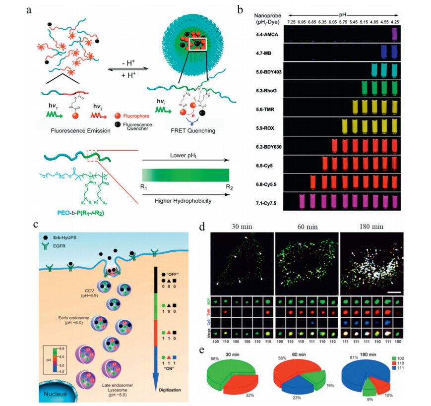
The above-mentioned pH sensors have limited ability to selectively image specific regions in cells, such as late endosomes/lysosomes (pH 4.0-5.5) [61]. Endosomes and lysosomes play critical role in cell physiology including protein/lipid metabolism, receptor recycling, nutrient senses, and cell death [62]. Unfortunately, there is no available method to image the dynamic maturation process of these organelles. To overcome this problem, Gao et al. developed a library of tunable multispectral ultra-pH-sensitive (UPS) nanosensors self-assembled from ionizable block copolymers to monitor the acidification process of endocytic organelles in live cells (Fig. 1a) [63-65]. The UPS library contains a set of nanoprobes with large fluorescent emissions (400-820 nm) in a wide pH range (4-7.4) (Fig. 1b) [65]. Lately, they incorporated three different fluorophore-encoded block copolymers in one nanoparticular system [66]. The mixed polymeric systems display an amplified (> 30 folds) and sharp response (ΔpH < 0.25) to microenvironments of clathrin (pH 6.9-7.2) [67], early endosomes (pH 6.0-6.5) [68], and late endosomes (pH 4.0-5.5) [61]. In particular, all the fluorescent polymer modules are "silent" at pH 7.4 due to the quenching of overall fluorescence by hetero-and homo-FRET effects. With decreasing of pH values, the polymers are sequentially lightened at predeter-mined pH values corresponding to different stages of organelle maturation. The UPS nanoprobe provides a smart tool to digitize the organelle pH and identify the cell signaling pathways involved in endosomal/lysosomal function regulation (Figs. 1c-e).
Redox homeostasis affects the fate of cells and participates in many pathological processes [69]. It has become a target for cancer chemotherapy [70-72], because the reduced susceptibility of cancer cells to stimuli that cause apoptosis has been closely associated with decreased redox potential [73]. Although selective redox regulation has potential benefits for cancer treatment, effective methods for testing the treatment remain largely unexplored since it is difficult to measure intracellular redox conditions over time [74]. To address this issue, Lin et al. [73] developed a novel genetic FRET system that undergoes a molecular conformation transition from an α-helix to a "clamped coil" state due to the formation of intermolecular disulfide bonds, which forces donor-enhanced yellow fluorescent protein (ECFP) and acceptor-enhanced blue-green fluorescent protein (EYFP) into a closer proximity for FRET detection (Fig. 2a). With the help of microfluidic device, this construct is capable of assessing intracellular redox homeostasis in real-time and monitoring the response of tumorigenic cells to glutathione (GSH). They found that mammalian cells can restore a reduced intracellular redox environment in few minutes after removal of an acute oxidative insult.

To further monitor the cleavage of disulfide bonds by living cells and determine the cytosolic transport of therapeutics, a variety of FRET-based imaging systems with disulfide-linked donors and acceptors have been developed [75-78]. For example, Gao et al. [75] reported a redox-activated sensor based on a cysteine-derivative hetero-FRET system, where tetramethylrhodamine (TMR) and cyanine dye (Cy5) as model fluorescence donor and acceptor were coupled with a disulfide bond. The fluorescence of the system is off through the endocytic pathway, and can be switched on by cytosolic GSH after endo-lysosomal disruption (Figs. 2b and c).
To avoid undesirable aggregation-caused quenching effect (ACQ) of traditional chromophores, Zhao et al. [79] recently incorporated a well-known aggregation induced emission (AIE) probe, tetraphenylethene (TPE) [80, 81], as a FRET donor, and curcumin (Cur) as a model drug and FRET receptor into a block copolymer (Fig. 2d). The fluorescence intensity of this system is relatively weak after self-assembly into intact micelles due to the ACQ effect of Cur and the energy transfer from TPE to Cur. Under a reducing condition, the cleavage of disulfide linkages induces the release of Cur and turns on both AIE signal and Cur fluorescence. This FRET-based polymer material can be potentially used for monitoring drug release and cell apoptosis in situ [82, 83].
Temperature is an important and fundamental parameter in a wide range of cellular functions and biological activities in living systems [84-87]. Many cell activities (e.g., division, metabolism, enzymatic reactions) are associated with temperature variation. In particular, some pathological phenomena including inflammation and cancers are generally accompanied by elevated temperature. In recent years, a variety of ingenious thermometers have been developed to real-time measure the cellular temperature at micro/nanoscale [88-90]. Nevertheless, most of the reported fluorescent thermometers are based on the change of fluorescence intensity at a single wavelength, which are impacted by many factors, such as probe distribution and concentration, excitation energy, and photobleaching [91].
To circumvent these unfavorable effects, researchers have constructed thermo-induced FRET systems for ratiometrically detecting and sensing cellular temperature [92, 93]. Yang et al. [94] covalently incorporated a fluorophore triarylboron compound (DPTB) into a poly(N-isopropylacrylamide) (PNIPAM) nanogel to fabricate a luminescent temperature probe (PNDP). The PNDP nanogel (donor) loading with NR (acceptor) could shrink to form an efficient ratiometric FRET colloidal nanoparticle with increase of temperature, resulting in significant fluorescence color change from red to green and blue with a high temperature resolution, good linear relationship and excellent reversibility at the single-cell level [94, 95].
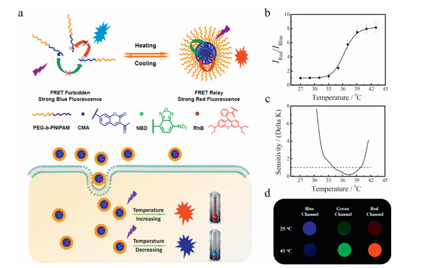
To realize long-range energy transfer for sensing complex biological environments in living cells, Hu and Liu et al. [96] designed a polymeric ratiometric fluorescent thermometer with a two-step cascade FRET property (Fig. 3a). They incorporated coumarinm (CMA, blue), 7-nitro-2, 1, 3-benzoxadiazole (NBD, green) and rhodamine B (RhBEA, red) separately into three PNIPAM-based thermoresponsive block copolymers. By facile mixing of the three fluorescent polymers in an optimized molar ratio, a two-step cascade FRET was achieved with NBD dye as a bridge for transferring energy from CMA to RhBEA. As a result, the system exhibits a ~8.4-fold change in intensity ratio in a broad temperature range of 20-44 ℃, with an imaging sensitivity of less than 1 ℃ or even 0.4 ℃ (Figs. 3b-d). This work demonstrates an excellent performance of intracellular cascade of FRET thermometers, which is helpful for understanding intracellular temperature-associated activities, such as signal transduction, cancer cell pathogenesis and inflammation.
H2S can be produced by enzymes in many organs (heart, liver, kidney, brain, ileum, uterus, etc.) and tissues in vivo [97]. Both endogenous and exogenous H2S are involved in a variety of physiological and pathological processes [97-99], including Alzheimer's disease, Down's syndrome, diabetes and cirrhosis [100]. Therefore, tracking and quantifying H2S in biological systems is critical to understanding the biology and pathology of H2S.
To fulfill this need, FRET systems with donors and acceptors that could be separated by the cleavage of NBD amine bonds in the presence of intracellular H2S have been widely researched [101-103]. Alternatively, Zhao et al. [104] encapsulated a semi-cyanine-BODIPY hybrid dye (BODInD-Cl) and its complementary energy donor (BODIPY1) into amphiphilic block copolymer micelles (Fig. 4a). The absorption spectrum of BODInD-Cl is red-shifted from 540 nm to 738 nm in response to H2S, thus switching off the FRET signal and recovering the donor fluorescence for in situ trapping of endogenous H2S generation. On the contrary, Ai et al. [105] lately designed a genetically encoded biosensor (hsFRET) with a FRET effect enhanced by H2S (Fig. 4b). They used a blue fluorescent protein EBFP2 as a fluorescent donor, and azido functionalized superfolder green fluorescent protein as a receptor. The azido group of hsFRET can react with both extracellularly added and endogenously generated H2S (1 mmol/L), leading to a significant change in FRET ratio (> 1.7 folds) for ratiometric imaging of H2S in live mammalian cells.

ROS are natural by-products of normal oxygen metabolism produced in a wide range of physiological process. While a low concentration of ROS is crucial to cell signal transduction and homeostasis, abnormal overproduction of ROS such as hydrogen peroxide (H2O2) may lead to oxidative damages to various biomolecules such as proteins, lipids and DNA, and cause cell apoptosis, which is closely related to the pathogenesis of cancer, diabetes and neurodegenerative diseases [106]. Therefore, ROS-responsive materials hold great potential as specific delivery systems for targeting oxidative microenvironments and diagnostic biosensors for imaging of ROS and cellular activities [107-109].
To understand the relationship between ROS and cell apoptosis, Yi et al. [110] designed a fluorescent probe containing a H2O2 reporter (NP1) and a Cy5 chromophore linked with a SGDEVDSG peptide (Fig. 5a). The absorption band of Cy5 overlaps with the emission portion of NP1 after reaction with H2O2, while the SGDEVDSG sequence can specifically recognized by a caspase 3 enzyme that activates apoptosis. As a result, FRET effect occurs in living cells resulting in detectable Cy5 fluorescence, while that is inhibited in apoptotic cells because peptide spacers are cleaved by specific proteases. Thus, this probe can differentiate H2O2 changes with different fluorescence wavelengths between living cells (red) and apoptotic cells (green).

In addition to the detection of endogenous ROS, FRET systems can also induce the production of ROS for tumor treatment under some conditions [111, 112]. For example, Zhao et al. [113] recently reported a semiconducting polymer nanoparticle (SPNs) containing fluorescent BODIPY (dipyrromethene boron difluoride) derivative as a donor and near-infrared (NIR) phosphorescent iridium(III) complex as an energy acceptor and photosensitizer (Fig. 5b). Upon irradiation, the energy is transferred from excited BODIPY to iridium(III) complexes via an efficient FRET process, then to the ground state 3O2 to obtain an excited state of 1O2 with a high quantum yield of 0.97. The SPN system can be used as a versatile theranostic platform for image-guided photodynamic therapy (PDT) of tumors.
Enzymes can exhibit abnormal activity in a variety of diseases [114]. For example, many kinds of enzyme proteins (e.g., proteases, phosphatases, lipases, oxidoreductases) are overexpressed in tumor microenvironments and show significant impact on cancer growth, angiogenesis, and metastasis [115]. As a result, enzymes could be exploited as promising targets for therapeutic and diagnostic applications, by virtue of their substrate specificity and biological catalysis ability. Hence, the detection of enzymatic activity is beneficial to early stage diagnosis of disease progression [116].
Jeong and Kim et al. [117] constructed a dual-channel fluorescent nanoprobe (DFNP) for highly-sensitive detecting of extracellular matrix metalloproteinase (MMP)-2 activity overexpressed in tumor metastasis tissues. They first designed an MMP-activatable probe integrating a cyanine fluorophore (Cy5.5) and a quencher (BK01) linked with an MMP-2, 9 degradable substrate peptide. Then the probe was hydrophobically associated with a Cy7-labeled polymer surfactant (F127-Cy7) to form self-assembled nanoparticles (Fig. 6a). The Cy5 fluorescence emission is activated by the cleavage of peptide in response to MMP-2, 9, allowing for precise imaging of tumor invasion and metastasis in lymph nodes, taking an "always-on" Cy7 fluorescence of polymer surfactant as an internal standard (Fig. 6b).

Since the distribution of a particular enzyme differs widely in various pathological situations, biosensors with simultaneous probing abilities for two or more target enzymes are highly desirable. Zhang et al. [118] designed a two-stage open fluorescent probe for sequential sensing of MMP-2 (a biomarker for tumor invasion and metastasis), and caspase-3 (a sign for early-stage apoptotic cells). The probe contains 5(6)-carboxyfluorescein (FAM) as a donor and two 4-{[4-(dimethylamino)-phenyl]-azo}-benzoic acid (Dabcyl) groups as quenchers (Fig. 6c). The donor fluorescence is first quenched by Dabcyl groups via a dual FRET processes under physiological conditions, then recovered in response to extracellular MMP-2 and further enhanced by caspase-3 after exposure to apoptosis stimuli, triggered by the cleavage of enzyme-responsive peptide sequences and break in corresponding FRET effect. This dual-FRET system renders real-time monitoring of DOX-and UV-induced cell apoptosis in situ with high sensitivity and selectivity.
The cellular microenvironment plays an extremely important role in cell life, and many methods have been used to study the microenvironment of cells. Conventional fluorescent sensors have the advantages of high sensitivity, noninvasive character, and ease of use. However, the accuracy of single-channel imaging can be influenced by various factors, such as the location of the sensor, and changes in the environments around the sensors. FRET technique enables simultaneously measurement of fluorescence intensities at multiple wavelengths and selective and ratiometric detection of cellular environments in a quantitative manner, which is more effective and sensitive than traditional biosensors. Therefore, a relatively large number of polymer-based FRET systems have been developed and widely used in specific quantification of cell indicators including pH, temperature, redox activity, and enzymatic reactions as mentioned above. With the help of these technologies, several mechanisms of cell endocytosis, signaling, apoptosis, as well as cancer cell pathogenesis and inflammation have been revealed. These studies are of great significance for understanding the secrets of cell life and developing smart materials for early diagnosis and effective therapy of a wide range of diseases.
In spite of significant progress in the development of FRET-equipped polymeric probes for bioanalysis, a more sophisticated design of polymer architecture should be involved with the advances in chemistry and materials science. The fine tuning of amphiphilicity, ionizable components and labile linkages in polymer structures may impart sharp transition and signal amplification of biosensors in response to diverse biological stimuli [25, 65, 119]. Moreover, FRET systems incorporating fluorescent dyes or chemiluminescent luminophores with NIR emission are particular promising for in vivo use, since the light with long wavelength shows greater capacity for deep tissue penetration and diminished phototoxicity [77, 120-122]. In addition, it is also imperative to understand the degradation, metabolism, and long-term toxicology profiles of fluorescent probes. Hence, further studies are still needed to developed novel kinds of biodegradable and biocompatible polymeric platforms for effective and safe bioimaging applications.
The authors declare that they have no known competing financial interests or personal relationships that could have appeared to influence the work reported in this paper.
The research was supported by the National Natural Science Foundation of China (Nos. 51873118, 21474064, 51203101); the National Science Fund for Distinguished Young Scholars of China (No. 51425305) and the Project of State Key Laboratory of Polymer Materials Engineering.
S. Lee, K.Y. Choi, H. Chung, et al., Bioconj. Chem. 22(2011) 125-131. doi: 10.1021/bc1004119
L. Yu, Y. Qiao, L. Miao, Y. He, Y. Zhou, Chin. Chem. Lett. 29(2018) 1545-1559. doi: 10.1016/j.cclet.2018.09.005
Q. Zhou, K. Son, Y. Liu, A. Revzin, Annu. Rev. Biomed. Eng. 17(2015) 165-190. doi: 10.1146/annurev-bioeng-071114-040525
H. Wang, E. Nakata, I. Hamachi, ChemBiochem 10(2009) 2560-2577. doi: 10.1002/cbic.200900249
Y. Kurishita, T. Kohira, A. Ojida, I.J. Hamachi, J. Am. Chem. Soc. 132(2010) 13290-13299. doi: 10.1021/ja103615z
J. Chen, Y. Tang, H. Wang, et al., J. Colloid Interface Sci. 484(2016) 298-307. doi: 10.1016/j.jcis.2016.09.009
E. Nakata, H. Wang, I. Hamachi, ChemBiochem 9(2008) 25-28. doi: 10.1002/cbic.200700364
K. Kikuchi, H. Takakusa, T. Nagano, Trends Analyt. Chem. 23(2004) 407-415. doi: 10.1016/S0165-9936(04)00608-9
A. Pietraszewska-bogiel, T.W.J. Gadella, J. Microsc. 241(2011) 111-118. doi: 10.1111/j.1365-2818.2010.03437.x
B. Hötzer, I.L. Medintz, N. Hildebrandt, Small 8(2012) 2297-2326. doi: 10.1002/smll.201200109
E.A. Jares-Erijman, T.M. Jovin, Nat. Biotechnol. 21(2003) 1387-1395. doi: 10.1038/nbt896
J. Garcia-Amorós, S. Tang, Y. Zhang, E.R. Thapaliya, F.M. Raymo, Selfassembling nanoparticles of amphiphilic polymers for in vitro and in vitro FRET imaging, in: S. Sortino (Ed.), Light-Responsive Nanostructured Systems for Applications in Nanomedicine, Springer International Publishing, Cham, 2016, pp. 29-59.
Y. Li, Y. Ban, R. Wang, et al., Chin. Chem. Lett. 31(2020) 443-446. doi: 10.1016/j.cclet.2019.07.047
J. Wen, P. Xia, Z. Zheng, et al., Chin. Chem. Lett. 28(2017) 2005-2008. doi: 10.1016/j.cclet.2017.09.014
H. Peng, J.A. Stolwijk, L. Sun, J. Wegener, O.S. Wolfbeis, Angew. Chem. Int. Ed. 49(2010) 4246-4249. doi: 10.1002/anie.200906926
F. Meng, H. Chai, X. Ma, Y. Tang, P. Miao, J. Mater. Chem. B 7(2019) 1926-1932. doi: 10.1039/C9TB00001A
T. Forster, Ann. Phys. 2(1948) 55-75.
G. Chen, F. Song, X. Xiong, X. Peng, Ind. Eng. Chem. Res. 52(2013) 11228-11245. doi: 10.1021/ie303485n
K.E. Sapsford, L. Berti, I.L. Medintz, Angew. Chem. Int. Ed. 45(2006) 4562-4589. doi: 10.1002/anie.200503873
D. Su, C.L. Teoh, S. Sahu, R.K. Das, Y. Chang, Biomaterials 35(2014) 6078-6085. doi: 10.1016/j.biomaterials.2014.04.035
L. Yu, Y. Liu, S. Chen, Y. Guan, Y. Wang, Chin. Chem. Lett. 25(2014) 389-396. doi: 10.1016/j.cclet.2013.12.014
S. Tsukiji, H. Wang, M. Miyagawa, et al., J. Am. Chem. Soc. 131(2009) 9046-9054. doi: 10.1021/ja902486c
C.E. Rowland, C.W. Brown, I.L. Medintz, J.B. Delehanty, Methods Appl. Fluoresc. 3(2015) 42006. doi: 10.1088/2050-6120/3/4/042006
K. Han, J. Zhu, S. Wang, et al., J. Mater. Chem. B 3(2015) 8065-8069. doi: 10.1039/C5TB01659B
Y. Wang, K. Zhou, G. Huang, et al., Nat. Mater. 13(2014) 204-212. doi: 10.1038/nmat3819
T. Tsuji, S. Yoshida, A. Yoshida, S. Uchiyama, Anal. Chem. 85(2013) 9815-9823. doi: 10.1021/ac402128f
K. Zhou, H. Liu, S. Zhang, et al., J. Am. Chem. Soc. 134(2012) 7803-7811. doi: 10.1021/ja300176w
W.B. Busa, R. Nuccitelli, Am. J. Physiol. 246(1984) R409-R438.
F.B. Loiselle, J.R. Casey, Measurement of intracellular pH, in: Q. Yan (Ed.), Membrane Transporters in Drug Discovery and Development: Methods and Protocols, Humana Press, Totowa, 2010, pp. 311-331.
J. Han, K. Burgess, Chem. Rev. 110(2010) 2709-2728. doi: 10.1021/cr900249z
T.A. Davies, R.E. Fine, R.J. Johnson, et al., Biochem. Biophys. Res. Commun.194(1993) 537-543. doi: 10.1006/bbrc.1993.1853
H. Izumi, T. Torigoe, H. Ishiguchi, et al., Cancer Treat. Rev. 29(2003) 541-549. doi: 10.1016/S0305-7372(03)00106-3
J. Wei, X. Shuai, R. Wang, et al., Biomaterials 145(2017) 138-153. doi: 10.1016/j.biomaterials.2017.08.005
M. Ding, N. Song, X. He, et al., ACS Nano 7(2013) 1918-1928. doi: 10.1021/nn4002769
X. Guo, L. Wang, K. Duval, et al., Adv. Mater. 30(2018) 1705436. doi: 10.1002/adma.201705436
J. Du, X. Du, C. Mao, J. Wang, J. Am. Chem. Soc. 133(2011) 17560-17563. doi: 10.1021/ja207150n
S.J.A. Hesse, G.J.G. Ruijter, C. Dijkema, J. Visser, J. Biotechnol. 77(2000) 5-15. doi: 10.1016/S0168-1656(99)00203-5
N. Cox, R. Kuemmerle, P. Millard, et al., Anal. Chem. 91(2019) 3959-3963. doi: 10.1021/acs.analchem.8b05147
J.K.M. Roberts, N. Wade-Jardetzky, O. Jardetsky, Biochemistry 20(1981) 5389-5394. doi: 10.1021/bi00522a006
Y. Wang, H. Kneipp, L.T. Perelman, et al., Phys. Rev. Lett. 78(1997) 1667-1670. doi: 10.1103/PhysRevLett.78.1667
Y. Zhang, X. Mi, X. Tan, R. Xiang, Theranostics 9(2019) 491-525. doi: 10.7150/thno.29875
C. Krafft, M. Schmitt, I.W. Schie, et al., Angew. Chem. Int. Ed. 56(2017) 4392-4430. doi: 10.1002/anie.201607604
S. Hennig, V. Mönkemöller, C. Böger, M. Müller, T. Huser, ACS Nano 9(2015) 6196-6205. doi: 10.1021/acsnano.5b01503
L. Bai, X. Wang, K. Zhang, et al., Chem. Commun. (2019) 1036-1039.
D. Ellis, R.C. Thomas, Nature 262(1976) 224-225. doi: 10.1038/262224a0
R.M. Wightman, Science 311(2006) 1570. doi: 10.1126/science.1120027
R. Sanders, A. Draaijer, H.C. Gerritsen, P.M. Houpt, Y.K. Levine, Anal. Biochem. 227(1995) 302-308. doi: 10.1006/abio.1995.1285
H.J. Park, C.S. Lim, E.S. Kim, et al., Angew. Chem. Int. Ed. 51(2012) 2673-2676. doi: 10.1002/anie.201109052
G.R. Bright, G.W. Fisher, J. Rogowska, D.L. Taylor, J. Cell Biol. 104(1987) 1019. doi: 10.1083/jcb.104.4.1019
D.S. Richardson, C. Gregor, F.R. Winter, et al., Nat. Commun. 8(2017) 577. doi: 10.1038/s41467-017-00606-4
X. Zhang, T. Zhang, S. Shen, J. Miao, B. Zhao, J. Mater. Chem. B 3(2015) 3260-3266. doi: 10.1039/C4TB02082K
J. Llopis, J.M. McCaffery, A. Miyawaki, M.G. Farquhar, R.Y. Tsien, Proc. Nat. Acad. Sci. U. S. A. 95(1998) 6803. doi: 10.1073/pnas.95.12.6803
Q. Fang, L. Yang, H. Xiong, et al., Chin. Chem. Lett. 31(2020) 129-132. doi: 10.1016/j.cclet.2019.04.021
K. Bi, R. Tan, R. Hao, et al., Chin. Chem. Lett. 30(2019) 545-548. doi: 10.1016/j.cclet.2018.11.020
Y. Yang, C. Zhang, R. Pan, et al., Chin. Chem. Lett. 31(2020) 125-128. doi: 10.1016/j.cclet.2019.04.037
D. Yue, M. Wang, F. Deng, et al., Chin. Chem. Lett. 29(2018) 648-656. doi: 10.1016/j.cclet.2018.01.046
L. Fu, P. Yuan, Z. Ruan, et al., Polym. Chem. 8(2017) 1028-1038. doi: 10.1039/C6PY01818A
W. Xu, S. Lu, M. Xu, et al., J. Mater. Chem. B 4(2016) 292-298. doi: 10.1039/C5TB02071A
A.M. Dennis, W.J. Rhee, D. Sotto, S.N. Dublin, G. Bao, ACS Nano 6(2012) 2917-2924. doi: 10.1021/nn2038077
Y. Chan, C. Wu, F. Ye, et al., Anal. Chem. 83(2011) 1448-1455. doi: 10.1021/ac103140x
J.R. Casey, S. Grinstein, J. Orlowski, Nat. Rev. Mol. Cell Bio. 11(2010) 50-61. doi: 10.1038/nrm2820
M. Schwake, B. Schröder, P. Saftig, Traffic 14(2013) 739-748. doi: 10.1111/tra.12056
K. Zhou, Y. Wang, X. Huang, et al., Angew. Chem. Int. Ed. 50(2011) 6109-6114. doi: 10.1002/anie.201100884
Y. Li, T. Zhao, C. Wang, et al., Nat. Commun. 7(2016) 13214. doi: 10.1038/ncomms13214
X. Ma, Y. Wang, T. Zhao, et al., J. Am. Chem. Soc. 136(2014) 11085-11092. doi: 10.1021/ja5053158
Y. Wang, C. Wang, Y. Li, et al., Adv. Mater. 29(2017) 1603794. doi: 10.1002/adma.201603794
S.L. Schmid, Annu. Rev. Biochem. 66(1997) 511-548. doi: 10.1146/annurev.biochem.66.1.511
F.R. Maxfield, D.J. Yamashiro, Endosome acidification and the pathways of receptor-mediated endocytosis, in: M.Z. Atassi (Ed.), Immunobiology of Proteins and Peptides IV: T-Cell Recognition and Antigen Presentation, Springer, Boston, 1987, pp. 189-198.
B. Li, Z. He, H. Zhou, H. Zhang, T. Cheng, Chin. Chem. Lett. 28(2017) 1929-1934. doi: 10.1016/j.cclet.2017.08.055
M. Ding, X. Zeng, X. He, et al., Biomacromolecules 15(2014) 2896-2906. doi: 10.1021/bm500506v
M. Ding, J. Li, X. He, et al., Adv. Mater. 24(2012) 3639-3645. doi: 10.1002/adma.201200954
F. Meng, W.E. Hennink, Z. Zhong, Biomaterials 30(2009) 2180-2198. doi: 10.1016/j.biomaterials.2009.01.026
C. Lin, V.L. Kolossov, G. Tsvid, et al., Integr. Biol. 3(2010) 208-217.
M. Oku, Y. Sakai, Antioxid. Redox Sign. 16(2011) 698-704.
Z. Wang, M. Luo, C. Mao, et al., Angew. Chem. 129(2017) 1339-1343. doi: 10.1002/ange.201610302
J. Yang, H. Chen, I.R. Vlahov, J. Cheng, P.S. Low, Proc. Nat. Acad. Sci. U. S. A.103(2006) 13872-13877. doi: 10.1073/pnas.0601455103
X. Wu, X. Sun, Z. Guo, et al., J. Am. Chem. Soc. 136(2014) 3579-3588. doi: 10.1021/ja412380j
M.H. Lee, Z. Yang, C.W. Lim, et al., Chem. Rev. 113(2013) 5071-5109. doi: 10.1021/cr300358b
X. Wang, J. Li, Q. Yan, et al., Macromol. Biosci. 18(2018) 1700339. doi: 10.1002/mabi.201700339
Y. Dong, J.W.Y. Lam, A. Qin, et al., Appl. Phys. Lett. 91(2007) 11111. doi: 10.1063/1.2753723
H. Tong, Y. Hong, Y. Dong, et al., Chem. Commun. (2006) 3705-3707.
J. Yu, Y. Chen, Y. Zhang, et al., Chem. Commun. 50(2014) 4699-4702. doi: 10.1039/c3cc49870k
S. Li, L. Liu, L. Rong, et al., Adv. Funct. Mater. 25(2015) 7317-7326. doi: 10.1002/adfm.201503262
R.S. Seymour, Biosci. Rep. 21(2001) 223-236. doi: 10.1023/A:1013608627084
X. Wang, O.S. Wolfbeis, R.J. Meier, Chem. Soc. Rev. 42(2013) 7834-7869. doi: 10.1039/c3cs60102a
A. Bahat, I. Tur-Kaspa, A. Gakamsky, et al., Nat. Med. 9(2003) 149-150. doi: 10.1038/nm0203-149
D.M. Wilkinson, Plant Ecol. Divers. 2(2009) 1-4. doi: 10.1080/17550870902722390
D. Zhao, X. Rao, J. Yu, et al., Inorg. Chem. 54(2015) 11193-11199. doi: 10.1021/acs.inorgchem.5b01623
Y. Cui, R. Song, J. Yu, et al., Adv. Mater. 27(2015) 1420-1425. doi: 10.1002/adma.201404700
E.J. McLaurin, L.R. Bradshaw, D.R. Gamelin, Chem. Mater. 25(2013) 1283-1292. doi: 10.1021/cm304034s
S.M. Borisov, I. Klimant, J. Fluoresc. 18(2008) 581-589. doi: 10.1007/s10895-007-0302-1
J.A. Molina-Bolívar, F. Galisteo-González, C.C. Ruiz, et al., Spectrochim. Acta Part A 214(2019) 161-169. doi: 10.1016/j.saa.2019.02.014
S.W. Hong, D.Y. Kim, J.U. Lee, W.H. Jo, Macromolecules 42(2009) 2756-2761. doi: 10.1021/ma802862h
J. Liu, X. Guo, R. Hu, et al., Anal. Chem. 87(2015) 3694-3698. doi: 10.1021/acs.analchem.5b00887
T. Bai, N. Gu, Small 12(2016) 4590-4610. doi: 10.1002/smll.201600665
X. Hu, Y. Li, T. Liu, G. Zhang, S. Liu, ACS. Appl. Mater. Inter. 7(2015) 15551-15560. doi: 10.1021/acsami.5b04025
B.L. Predmore, D.J. Lefer, G. Gojon, Antioxid. Redox Sign. 17(2012) 119-140. doi: 10.1089/ars.2012.4612
J. Zhang, R. Wang, Z. Zhu, L. Yi, Z. Xi, Tetrahedron 71(2015) 8572-8576. doi: 10.1016/j.tet.2015.09.028
G. Yang, L. Wu, B. Jiang, et al., Science 322(2008) 587. doi: 10.1126/science.1162667
S. Fiorucci, E. Antonelli, A. Mencarelli, et al., Hepatology 42(2005) 539-548. doi: 10.1002/hep.20817
L. Yi, Z. Xi, Org. Biomol. Chem. 15(2017) 3828-3839. doi: 10.1039/C7OB00332C
R. Wang, Z. Li, C. Zhang, et al., ChemBiochem 17(2016) 962-968. doi: 10.1002/cbic.201600060
F. Song, Z. Li, J. Li, et al., Org. Biomol. Chem. 14(2016) 11117-11124. doi: 10.1039/C6OB02354A
C. Zhao, X. Zhang, K. Li, et al., J. Am. Chem. Soc. 137(2015) 8490-8498. doi: 10.1021/jacs.5b03248
S. Youssef, S. Zhang, H. Ai, ACS Sens. 4(2019) 1626-1632. doi: 10.1021/acssensors.9b00400
Y. Wen, K. Liu, H. Yang, et al., Anal. Chem. 86(2014) 9970-9976. doi: 10.1021/ac502909c
H. Liu, R. Wang, J. Wei, et al., J. Am. Chem. Soc. 140(2018) 6604-6610. doi: 10.1021/jacs.8b01873
S. Lv, Y. Wu, K. Cai, et al., J. Am. Chem. Soc. 140(2018) 1235-1238. doi: 10.1021/jacs.7b12776
Q. Xu, C. He, C. Xiao, X. Chen, Macromol. Biosci. 16(2016) 635-646. doi: 10.1002/mabi.201500440
Y. Wen, F. Xue, H. Lan, et al., Biosens. Bioelectron. 91(2017) 115-121. doi: 10.1016/j.bios.2016.12.027
R. Timor, H. Weitman, N. Waiskopf, U. Banin, B. Ehrenberg, ACS Appl. Mater. Interface 7(2015) 21107-21114. doi: 10.1021/acsami.5b04318
S. Bhattacharyya, M.K. Barman, A. Baidya, A. Patra, J. Phys. Chem. C 118(2014) 9733-9740.
J. Jiang, Y. Qian, Z. Xu, et al., Chem. Sci. 10(2019) 5085-5094. doi: 10.1039/C8SC05501G
D. Chen, W. Qin, H. Fang, et al., Chin. Chem. Lett. 30(2019) 1738-1744. doi: 10.1016/j.cclet.2019.08.001
N. Poondla, A.P. Chandrasekaran, K. Kim, S. Ramakrishna, BMB Rep. 52(2019) 181-189. doi: 10.5483/BMBRep.2019.52.3.048
Z. Lukacs, M. Nickel, S. Murko, et al., Clin. Chim. Acta 492(2019) 69-71. doi: 10.1016/j.cca.2019.02.010
H. Cho, S. Lee, S. Park, et al., Colloids Surf. B 179(2019) 9-16. doi: 10.1016/j.colsurfb.2019.03.047
S. Li, L. Liu, H. Cheng, et al., Chem. Commun. 51(2015) 14520-14523. doi: 10.1039/C5CC04962H
P. Bawa, V. Pillay, Y.E. Choonara, L.C. du Toit, Biomed. Mater. 4(2009) 22001. doi: 10.1088/1748-6041/4/2/022001
X. Yue, Q. Zhang, Z. Dai, Adv. Drug Deliv. Rev. 115(2017) 155-170. doi: 10.1016/j.addr.2017.04.007
O. Green, S. Gnaim, R. Blau, et al., J. Am. Chem. Soc.139(2017) 13243-13248. doi: 10.1021/jacs.7b08446
J. Wan, Y. Qiao, X. Chen, et al., Adv. Funct. Mater. 28(2018) 1804229. doi: 10.1002/adfm.201804229

Scheme 1 Schematic principle of FRET-based polymer sensors for detection of cellular environments. D and A represent donor and acceptor, respectively, and r indicates the donor-acceptor distance.

Figure 1 (a) Schematic illustration and structural design of UPS nanoprobes. (b) The UPS library contains 10 nanoprobes with large fluorescent emissions (400-820 nm) in a wide pH range (4-7.4). Reprinted with permission [65]. Copyright 2014, American Chemical Society. (c) Schematic illustration of multispectral UPS probe for digitizing organelle maturation processes in tumor cells after receptor-mediated endocytosis. (d) Digitizing of endocytic organelles by UPS probe at different times in living tumor cells. Green, red and blue colors represent dipyrrometheneboron difluoride dyes (BDY), TMR and Cy5 fluorescence, respectively. (e) Quantitative results of distribution of endocytic organelles in different pH regions. The digital barcodes illustrate the heterogeneity of organelle maturation in tumor cells. Reprinted with permission [66]. Copyright 2016, John Wiley & Sons.

Figure 2 (a) Schematic illustration of conformation-modulated FRET effect in response to redox stimuli. The molecule undergoes a conformation transition from an α-helix to a "clamped coil" state due to the formation of intermolecular disulfide bonds under oxidative conditions, resulting in decreased donor-acceptor distance for FRET detection of redox response of tumorigenic cells to GSH. Reprinted with permission [73]. Copyright 2010, Oxford University Press. (b) A redox-activatable sensor based on a cysteine-derivative hetero-FRET system for detection of cytosolic delivery of biomacromolecules. (c) CLSM analysis of endo-lysosomal escape and cytosolic delivery of IgG labeled with redox-activatable sensor. Reprinted with permission [75]. Copyright 2016, John Wiley & Sons. (d) Schematic illustration of FRET systems integrating an AIE probe TPE for determination of intracellular drug release. Reprinted with permission [79]. Copyright 2018, John Wiley & Sons.

Figure 3 (a) Construction of polymeric ratiometric fluorescent thermometer with a two-step cascade FRET process for intracellular temperature imaging. (b) Normalized change in intensity ratio (Ired/Iblue) and (c) imaging sensitivity determined by CLSM analysis for live tumor cells after incubating at 37 ℃ for 40 min with the polymeric ratiometric fluorescent thermometer. (d) Fluorescence images obtained for the above sample at 25 and 41 ℃, respectively. Reprinted with permission [96]. Copyright 2015, American Chemical Society.

Figure 4 (a) Design of polymeric H2S sensor from BODInD-Cl and BODIPY1 encapsulated mPEG-DSPE micelles. Reprinted with permission [104]. Copyright 2015, American Chemical Society. (b) Illustration of a genetically encoded biosensor (hsFRET) based on FRET enhancement effect in the presence of H2S. Reprinted with permission [105]. Copyright 2019, American Chemical Society.

Figure 5 (a) Design of a ROS sensor containing NP1 and Cy5 linked with a caspase 3-cleavable peptide, which can differentiate H2O2 changes between living cells (red) and apoptotic cells (green). Reprinted with permission [110]. Copyright 2016, Elsevier. (b) Design of FRET system containing BODIPY as a donor and iridium (III) as an acceptor and photosensitizer for generation of 1O2 through FRET process. Reprinted with permission [113]. Copyright 2019, The Royal Society of Chemistry.

Figure 6 (a) Construction of DFNP for detection of MMP-2 activity overexpressed in tumor metastasis tissues. (b) in vitro dual-channel imaging property of DFNP after treatment with different amount of activated MMP-2. Reprinted with permission [117]. Copyright 2019, Elsevier. (c) Design of a two-stage open fluorescent probe for sequential sensing of MMP-2 and caspase-3. Reprinted with permission [118]. Copyright 2019, The Royal Society of Chemistry.

 扫一扫看文章

扫一扫关注我们
 DownLoad:
 下载:
下载:
 下载:
下载: