ywlin@usc.edu.cn (Y.-W. Lin). 1 These authors contributed equally to this work.
Received Date:
11 October 2025 Accepted Date:
05 December 2025 Revised Date:
04 December 2025 Available Online:
15 November 2026
Abstract:
Aggregation-induced emission (AIE)-based photosensitizers have emerged as promising candidates for image-guided antibacterial therapy. However, achieving efficient reactive oxygen species (ROS) generation under hypoxic infection conditions while maintaining broad-spectrum antimicrobial activity remains a significant challenge. Herein, we report two cationic triphenylamine-armed D–A–D’ type AIE photosensitizers, BTBH and BTBF, rationally designed to optimize their electronic structures and photophysical properties. Both photosensitizers exhibit strong selectivity toward bacteria and efficiently produce ROS through synergistic type Ⅰ and type Ⅱ photodynamic pathways. Notably, compared with the fluorine-free BTBH, the fluorinated analogue BTBF shows greater ROS-generating capacity. Moreover, BTBF demonstrates intrinsic dark toxicity by disrupting bacterial membranes, which, together with its photodynamic activity, enables a synergistic dual-mode antibacterial effect, resulting in superior antibacterial efficiency and broad-spectrum antimicrobial performance. This work highlights a fluorination strategy for sensitive bacterial detection and highly effective antibacterial therapy, providing a promising platform for theranostic applications against bacterial infections.
Bacterial infections, particularly those caused by multidrug-resistant strains, pose a serious threat to global public health, largely due to the overuse of antibiotics [1,2]. Therefore, there is an urgent need to develop antimicrobial materials that combine effective detection and treatment abilities. Photodynamic therapy (PDT), a non-invasive approach, has emerged as a promising solution because of its high pathogen-killing efficiency, spatiotemporal controllability, and minimal side effects [3–6]. PDT relies on photosensitizers (PSs) that, upon light irradiation, generate reactive oxygen species (ROS) to induce irreversible oxidative damage in bacterial cells [7–9]. This nonspecific mechanism reduces the likelihood of resistance development, highlighting the potential of PDT as a next-generation antibacterial strategy. However, conventional photosensitizers often face limitations, such as aggregation-caused quenching (ACQ), in which planar molecular structures and strong π–π stacking reduce fluorescence and ROS generation on bacterial surfaces [10–12].
Different from traditional PSs, aggregation-induced emission (AIE) photosensitizers exhibit minimal fluorescence in the molecular state and enhanced fluorescence in aggregates [13–16]. Moreover, AIE photosensitizers effectively mitigate non-radiative transitions by restricting intramolecular motion (RIM) when in their aggregated state [8,17]. This phenomenon not only enhances fluorescence but also promotes the generation of ROS [18]. Upon illumination, the PS is excited to a triplet state and produces ROS via two mechanisms: (1) Undergo electron transfer to generate free radicals such as hydroxyl radicals (•HO) or superoxide radicals (O2•−) as type Ⅰ ROS; and (2) undergo energy transfer to generate singlet oxygen (1O2) as type Ⅱ ROS. Most current photosensitizers rely on the type Ⅱ mechanism, which is contingent upon the concentration of O2. However, the formation of biofilms around bacteria-infected tissues can result in hypoxic environments, thereby significantly constraining the bactericidal efficacy [19,20]. In contrast, the type Ⅰ photosensitizers can efficiently generate radical-type ROS with stronger oxidative capacity and shorter diffusion distances even under hypoxic conditions, offering distinct advantages for overcoming the hypoxic microenvironments commonly found in infection sites [21–23]. Thus, type Ⅰ PDT offers a strategic advantage in overcoming hypoxia-induced inefficiency in conventional PDT.
Despite the promise of PDT in treating bacterial infections, most photosensitizers primarily target Gram-positive bacteria with limited efficacy against Gram-negative bacteria [24,25]. This limitation stems from structural differences between Gram-positive and Gram-negative bacteria, where Gram-negative bacteria have an outer membrane composed of tightly packed lipopolysaccharides in addition to phospholipid inner membranes [26–28]. To expand the antimicrobial spectrum of PDT, the coupling of antibacterial modules with good bacterial binding ability and photosensitizers is under exploration [29,30]. For instance, Tang et al. developed a bioconjugate that combines bacteriophage with photodynamic inactivation for targeted imaging and efficient bacterial killing [31]. Jiang et al. report an amphipathic peptide-photosensitizer conjugate that improves PDT efficacy against Gram-negative bacteria by disrupting their outer membrane [32]. These strategies have significantly enhanced the antibacterial coverage and the efficiency of photodynamic therapy. Additionally, incorporating intrinsic antibacterial activity in PSs can ensure effectiveness in deep tissues or regions inaccessible to light [33,34].
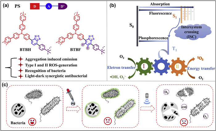
Cationization photosensitizers not only bind specifically to negatively charged bacterial membranes via electrostatic interactions, but their strong electron-withdrawing properties also promote intersystem crossing (ISC) in photosensitizers, significantly enhancing ROS generation efficiency [35–37]. Fluorination is one of the structural elements of antibacterial agents due to its exceptional electrical, physical, and biological characteristics and reactivity [38,39]. Moreover, C–F bonds can increase the molecular electron cloud density, strengthen the donor-acceptor interaction, and facilitate ROS production [40,41]. Based on these design principles, we developed two D–A–D’ AIE PSs, BTBH and BTBF, featuring cationic triphenylamine arms and a benzothiadiazole core (Fig. 1a). BTBF was further optimized via fluorination to enhance bacterial binding and intrinsic antibacterial activity. Both PSs exhibit good water solubility and selectively bind bacteria via electrostatic interactions. Under illumination, they efficiently generate type Ⅰ/Ⅱ ROS and exhibit potent antibacterial effects against both Staphylococcus aureus (S. aureus), Escherichia coli (E. coli) and Klebsiella pneumoniae (K. pneumoniae), with BTBF demonstrating superior overall performance, including significant non-light-dependent bactericidal activity (Figs. 1b and c). These results showed that combining cationization and fluorination to develop multifunctional AIE photosensitizers not only enhances bacterial detection and imaging but also provides dual-mode antibacterial therapy.
Figure 1
Figure 1.
Illustration of highly efficient free-radical ROS photosensitizers for killing bacteria. (a) Molecular structures of BTBH and BTBF. (b) ROS generation mechanisms of type Ⅰ and type Ⅱ. (c) Mechanism of action of photodynamic antibacterial using BTBH and BTBF.
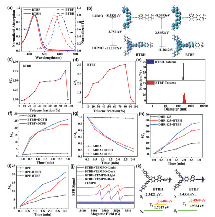
The photophysical properties of the synthesized BTBH and BTBF were first characterized through ultraviolet-visible (UV–vis) and fluorescence spectra (Fig. 2a). BTBH and BTBF exhibited maximum absorption peaks around 420 nm, with maximum emission wavelengths of 599 and 570 nm, respectively. Compared to BTBH, BTBF showed a blue shift of ~30 nm in emission spectra. The blue shift in BTBF is attributed to the electron-withdrawing fluorine atom, which lowers lowest unoccupied molecular orbital (LUMO) energy and increases torsional angles between aromatic rings, reducing π-conjugation and coupling efficiency between donor and acceptor units. Density functional theory (DFT) calculations further support these findings, indicating that BTBF has a larger highest occupied molecular orbital (HOMO)–LUMO energy gap than BTBH (Fig. 2b), which aligns with the experimentally observed blue-shift trend.
Figure 2
Figure 2.
(a) UV–vis and fluorescence spectra of BTBH and BTBF in DMSO. (b) HOMO and LUMO distributions of photosensitizers, calculated using DFT. (c, d) I/I0 plots of fluorescence intensity versus the toluene fraction of BTBH and BTBF in the DMSO/toluene mixture. (e) Size distributions of photosensitizers in 80% toluene. (f) Total ROS generation of photosensitizers upon light irradiation using DCFH indicator at different times. (g) Singlet oxygen generation of photosensitizers upon light irradiation using ABDA indicator for different times. (h) O2•− generation of photosensitizers upon light irradiation using DHR-123 indicator for different times. (i) •OH generation of photosensitizers upon light irradiation using HPF indicator for different times. (j) EPR signals of TEMPO after light irradiation for free-radical ROS characterization. (k) Calculated lowest excited singlet (S1) and triplet (T1) energy levels of photosensitizers.
The AIE characteristics of all photosensitizers were explored by monitoring their emission spectra in a DMSO/toluene mixed solvent. The intensities of both compounds were monitored by progressively increasing the volume fraction (fT) of the unfavorable solvent toluene (Fig. S1 in Supporting information). As fT increased, the photoluminescence intensities of both BTBH and BTBF rose significantly, peaking at fT = 90% and fT = 80%, respectively (Figs. 2c and d). This luminescence enhancement under high aggregation conditions aligns with the characteristic behavior of typical AIE property. To definitively confirm aggregate formation, dynamic light scattering (DLS) measurements were performed on samples with a high toluene ratio (fT = 80%). The results indicate that both BTBH and BTBF form nanoaggregates with narrow particle size distributions, exhibiting average diameters of ~350 and 370 nm, respectively (Fig. 2e), thereby providing reliable microstructural evidence for AIE behavior.
To investigate the photosensitization capacity of BTBH and BTBF, we first assessed their ability to generate ROS upon light exposure. 2′,7′-Dichlorodihydrofluorescein (DCFH) was selected to verify the total ROS generation of AIE photosensitizers under light irradiation (Fig. S2 in Supporting information). ROS can convert the nonfluorescent DCFH into the green fluorescent compound 2′,7′-dichlorofluorescein (DCF), resulting in a significant increase in green fluorescence. Both BTBH and BTBF markedly enhanced DCFH fluorescence intensity as the light exposure time increased, in contrast to the negligible changes observed in the DCFH-only control group (Fig. 2f). After 3 min of irradiation, the fluorescence intensity at 525 nm increased by 22- and 26-fold for BTBH and BTBF, respectively. These results demonstrate that both compounds act as efficient photosensitizers. To identify the specific ROS species generated, we employed dihydro-rhodamine 123 (DHR123), hydroxyphenyl fluorescein (HPF), and 9,10-anthraquinone-2,6-diyl-bis(methylene) dimalonic acid (ABDA) as selective probes for superoxide anion (O2•−), hydroxyl radicals (•HO), and singlet oxygen (1O2), respectively (Figs. S3-S5 in Supporting information). Upon illumination, the absorption of ABDA decreased by approximately 50% in the presence of both photosensitizers, indicating efficient 1O2 generation (Fig. 2g). Simultaneously, significant fluorescence enhancement with DHR123 and HPF confirmed concurrent type Ⅰ free-radical ROS production (Figs. 2h and i). BTBF consistently showed greater activity than BTBH in these assays. Thus, both compounds simultaneously produce type Ⅰ and type Ⅱ ROS upon photoactivation, with BTBF exhibiting slightly higher overall performance. Moreover, electron paramagnetic resonance (EPR) spectroscopy was employed to provide direct evidence of radical generation using 2,2,6,6-tetramethylpiperidine-1-yl-oxy (TEMPO) as a spin-trapping agent. No EPR signals were observed under dark conditions. However, upon illumination, mixing BTBH and BTBF with TEMPO yielded characteristic signals for 1O2 (Fig. 2j), confirming the generation of photoinduced ROS.
Further theoretical computational studies were conducted to investigate the inherent reasons for the difference in ROS generation efficiency. We calculated the singlet-triplet energy gap (ΔES1-T1) of both photosensitisers using the timedependent DFT (TD-DFT). BTBF exhibited a smaller ΔEST value (0.4948 eV) compared with BTBH (0.6406 eV) (Fig. 2k). A reduced ΔEST promotes intersystem crossing, thereby enhancing the triplet-state population and ROS generation efficiency. This theoretical result aligns with the experimental findings, confirming that fluorine substitution effectively strengthens donor–acceptor interactions and facilitates ROS production.
In consideration of their excellent luminescent properties in the aggregated state, the bacteria imaging capabilities for these photosensitizers were further explored. The fluorescence intensity of BTBH and BTBF was negligible in phosphate buffered saline (PBS) but increased significantly after incubation with eight bacterial strains (three Gram-positive and five Gram-negative bacteria, Fig. S6 in Supporting information). This increase is due to electrostatic interactions between the positively charged probes and negatively charged bacterial membranes, leading to aggregation and activation of AIE luminescence. To verify the selectivity of the photosensitizers, the fluorescence responses were further examined in the presence of various potential interfering substances (including metal cations, anions, amino acids, proteins and DNA). None of these interfering substances caused significant changes in the fluorescence signal (Fig. S7 in Supporting information), confirming that BTBH and BTBF have high specificity for bacterial detection. To quantitatively evaluate detection sensitivity, S. aureus (Gram-positive) and E. coli (Gram-negative) were used as model strains to examine the relationship between fluorescence intensity and bacterial concentration (102–108 CFU/mL). Both strains exhibited good linear response within the logarithmic concentration range (Fig. S8 in Supporting information). Calculated using the 3σ/s method, the limits of detection (LOD) for BTBH were 1.17 × 10³ CFU/mL for S. aureus and 1.14 × 10² CFU/mL for E. coli, while BTBF exhibited LODs of 4.42 × 10² and 8.80 × 101 CFU/mL for the same strains. These results consistently demonstrate that both probes exhibit excellent bacterial detection sensitivity.
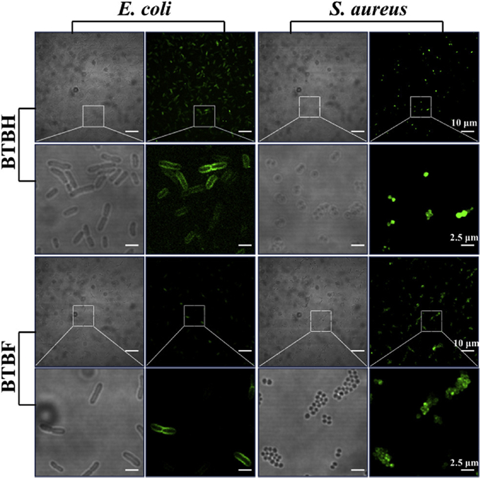
Encouraged by the remarkable AIE performance for bacteria, we selected S. aureus and E. coli as representatives to assess the bacteria staining capabilities of these photosensitizers preliminarily. Confocal laser scanning microscopy (CLSM) imaging (Fig. 3) revealed that BTBH and BTBF effectively labeled S. aureus and E. coli, resulting in bright fluorescence around bacterial cells. Notably, the probes predominantly localized to the membrane surface in E. coli, whereas some probes penetrated the membrane in S. aureus. This difference may relate to the denser outer membrane structure of Gram-negative bacteria. These findings visually confirm the probes’ effective binding to bacteria and rapid fluorescence response capability.
Figure 3
Figure 3.
Confocal images of E. coli and S. aureus incubated with BTBH and BTBF (10 µmol/L).
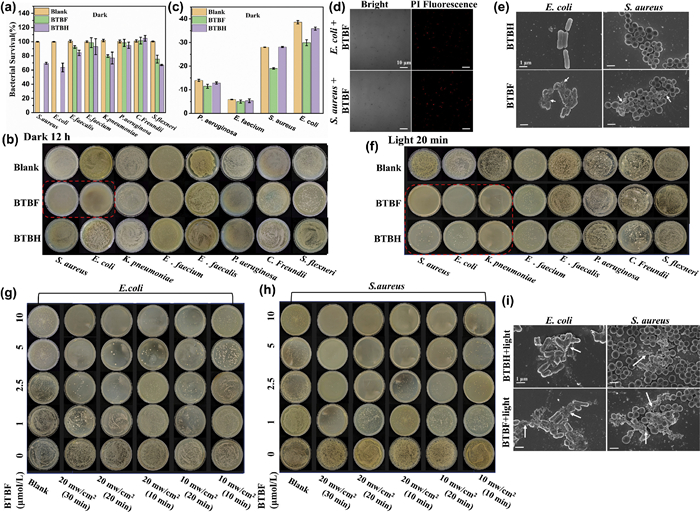
Then, the inherent antibacterial activity of BTBH and BTBF under dark conditions was evaluated. Using the standard microdilution method, the minimum inhibitory concentration (MIC) of BTBF against E. coli and S. aureus was determined to be 14 µmol/L (16 µg/mL), while the minimum bactericidal concentration (MBC) was 15 µmol/L (Fig. S9 in Supporting information), indicating its bactericidal properties. Further monitoring of OD600 growth curves for eight bacterial strains at 20 µmol/L concentration and calculating survival rates after 12 h revealed that BTBF completely inhibited the growth of E. coli and S. aureus, whereas BTBH only reduced the survival rate of these two bacteria to approximately 70% (Fig. 4a and Fig. S10 in Supporting information). Furthermore, the exceptional antibacterial efficacy of BTBF against E. coli and S. aureus was clearly illustrated by the number of bacterial colonies on agar plates subjected to various treatments (Fig. 4b). Consequently, BTBF itself possessed bactericidal activity, and demonstrated that fluorination can help enhance the antibacterial effect of ionized photosensitizers.
Figure 4
Figure 4.
(a) Survival rate of eight bacterial strains with PBS, BTBH, and BTBF treatment in dark. (b) Plate photographs of eight bacterial strains cocultured with PBS, BTBH, and BTBF treatment in dark for 12 h. (c) Zeta potentials of four bacterial strains pretreated with PBS, BTBH, and BTBF treatment in dark. (d) PI staining images of E. coli and S. aureus treated to BTBF treatments. Scale bar: 10 µm. (e) SEM images of E. coli and S. aureus subjected to BTBH and BTBF treatments in dark. Scale bar: 1 µm. (f) Plate photographs of eight bacterial strains cocultured with PBS, BTBH, and BTBF treatment under light irradiation (450 nm, 20 mW/cm2, 20 min). Plate photographs of (g) E. coli and (h) S. aureus with different concentrations of BTBF, light irradiation power and times. (i) SEM images of E. coli and S. aureus subjected to BTBH and BTBF treatments under light irradiation (450 nm, 20 mW/cm2, 20 min). Scale bar: 1 µm. Error bars represent SD (n = 3).
To elucidate the antibacterial mechanism, zeta potential analysis was employed to investigate electrostatic interactions between the compounds and bacterial surfaces. Testing on two Gram-positive and two Gram-negative bacteria, including E. coli and S. aureus, revealed that BTBH treatment did not cause significant changes in bacterial zeta potential, whereas BTBF markedly reduced the negative charge of S. aureus and E. coli (Fig. 4c). This confirms its ability to effectively adsorb onto negatively charged bacterial membranes via electrostatic interactions. To further validate whether BTBF exerts bactericidal effects by disrupting cell membranes, propidium iodide (PI) staining experiments were conducted. As a membrane-impermeable nucleic acid dye, PI enters cells and produces fluorescence only when membrane integrity is compromised. As shown in Fig. 4d, both S. aureus and E. coli treated BTBF exhibited strong fluorescent signals, indicating severe damage to the cell membranes leading to irreversible cell death. Scanning electron microscopy (SEM) showed that compared to relatively intact bacteria in the BTBH-treated group, BTBF-treated cells exhibited pronounced surface wrinkling, cellular collapse, and structural disruption for S. aureus and E. coli (Fig. 4e), consistent with its significant antimicrobial activity.
The intracellular ROS generation of BTBH and BTBF in bacteria was then systematically evaluated. Under illumination at 450 nm (20 mW/cm2 for 20 min), both compounds exhibited significantly enhanced antimicrobial effects. Notably, BTBF showed exceptional photodynamic bactericidal efficiency, achieving an inactivation rate exceeding 99.9% against S. aureus and E. coli, while also significantly inhibiting the growth of K. pneumoniae. Although BTBH also exhibited photodynamic activity and effectively inhibited the three aforementioned bacteria, its bactericidal efficiency was considerably lower than that of BTBF (Fig. 4f). The superior performance of BTBF can be attributed to its inherent dark toxicity, which facilitates the initial disruption of membrane structures and creates favorable conditions for subsequent attacks by ROS, resulting in a synergistic bactericidal effect. The influence of illumination duration on the bactericidal efficiency of BTBF was systematically examined. As shown in Figs. 4g and h, the survival rates of E. coli and S. aureus gradually decreased with prolonged exposure time. At 10 µmol/L BTBF and 20 mW/cm2 light intensity, 10 min of irradiation eliminated over 99% of S. aureus, whereas achieving comparable bactericidal efficacy for E. coli required 20 min. This discrepancy reflects the inherent resistance of Gram-negative bacteria to PDT due to the physical barrier function of their outer membrane structure. Morphological changes using SEM showed that both S. aureus and E. coli treated with BTBH or BTBF followed by 20 min of irradiation exhibited distinct damage features, including membrane surface wrinkling, leakage of contents, and structural collapse (Fig. 4i). Finally, the cytotoxicity of the photosensitizer for cells was evaluated. MTT assay results indicated that the survival rate of HeLa cells cultured with 10 µmol/L of the photosensitizers for 24 h in the dark remained as high as 90%, whereas it decreased under light conditions (Fig. S11 in Supporting information). These results visually confirm that photodynamic treatment causes severe physical damage to bacterial cell membranes, providing reliable morphological evidence for the highly effective antibacterial mechanism of BTBH and BTBF.
In summary, we have developed two cationic D–A–D’ photosensitizers, BTBH and BTBF, and systematically elucidated their structure–property relationships. Both compounds exhibit typical AIE behavior and efficiently generate ROS via dual type Ⅰ and type Ⅱ mechanisms. Fluorescence imaging experiments demonstrated selective bacterial binding and sensitive detection, enabling precise visualization of both Gram-positive and Gram-negative bacteria. Notably, the fluorinated derivative BTBF combines intrinsic dark toxicity with potent photodynamic activity, achieving synergistic dual-mode antibacterial effects. Mechanistic studies revealed that BTBF disrupts bacterial membranes through electrostatic interactions and ROS-mediated oxidative damage, resulting in highly effective, broad-spectrum bacterial inactivation. Therefore, BTBH and BTBF exemplify multifunctional photosensitizers that simultaneously enable sensitive bacterial detection, precise imaging, and efficient antibacterial therapy, offering promising platforms for combating drug-resistant infections and advancing clinical antibacterial photodynamic therapy.
CRediT authorship contribution statement
Shuang-Shuang Long: Writing – review & editing, Supervision, Investigation, Funding acquisition, Conceptualization. Wen-Xi Zhang: Writing – original draft, Investigation, Formal analysis, Data curation. Lu Ou: Investigation, Data curation. Wei Liu: Data curation. Fei Deng: Funding acquisition, Data curation. Ke Zeng: Funding acquisition, Data curation. Xu-Dong Jiang: Methodology, Funding acquisition. Ying-Wu Lin: Writing – review & editing, Supervision.
Declaration of competing interest
The authors declare that they have no known competing financial interests or personal relationships that could have appeared to influence the work reported in this paper.
Acknowledgments
This research was financially supported by the National Natural Science Foundation of China (Nos. 32301060, 22468023, 52303235). Natural Science Foundation of Hunan Province of China (Nos. 2023JJ40533, 2022JJ40354).
Supplementary materials
Supplementary material associated with this article can be found, in the online version, at doi:10.1016/j.cclet.2025.112226.
H. Mei, A.M. Remete, Y. Zou, et al., Chin. Chem. Lett. 31 (2020) 2401–2413. doi: 10.1016/j.cclet.2020.03.050
[40]
M. Tian, X. Zhao, S. Zhao, et al., Chem. Commun. 61 (2025) 10985–10988. doi: 10.1039/d5cc02234g
[41]
P. Zheng, Y. Ding, A. Abdurahman, et al., Sci. China Chem. 68 (2025) 3086–3099. doi: 10.1007/s11426-024-2487-4
Figure 1
Illustration of highly efficient free-radical ROS photosensitizers for killing bacteria. (a) Molecular structures of BTBH and BTBF. (b) ROS generation mechanisms of type Ⅰ and type Ⅱ. (c) Mechanism of action of photodynamic antibacterial using BTBH and BTBF.
Figure 2
(a) UV–vis and fluorescence spectra of BTBH and BTBF in DMSO. (b) HOMO and LUMO distributions of photosensitizers, calculated using DFT. (c, d) I/I0 plots of fluorescence intensity versus the toluene fraction of BTBH and BTBF in the DMSO/toluene mixture. (e) Size distributions of photosensitizers in 80% toluene. (f) Total ROS generation of photosensitizers upon light irradiation using DCFH indicator at different times. (g) Singlet oxygen generation of photosensitizers upon light irradiation using ABDA indicator for different times. (h) O2•− generation of photosensitizers upon light irradiation using DHR-123 indicator for different times. (i) •OH generation of photosensitizers upon light irradiation using HPF indicator for different times. (j) EPR signals of TEMPO after light irradiation for free-radical ROS characterization. (k) Calculated lowest excited singlet (S1) and triplet (T1) energy levels of photosensitizers.
Figure 4
(a) Survival rate of eight bacterial strains with PBS, BTBH, and BTBF treatment in dark. (b) Plate photographs of eight bacterial strains cocultured with PBS, BTBH, and BTBF treatment in dark for 12 h. (c) Zeta potentials of four bacterial strains pretreated with PBS, BTBH, and BTBF treatment in dark. (d) PI staining images of E. coli and S. aureus treated to BTBF treatments. Scale bar: 10 µm. (e) SEM images of E. coli and S. aureus subjected to BTBH and BTBF treatments in dark. Scale bar: 1 µm. (f) Plate photographs of eight bacterial strains cocultured with PBS, BTBH, and BTBF treatment under light irradiation (450 nm, 20 mW/cm2, 20 min). Plate photographs of (g) E. coli and (h) S. aureus with different concentrations of BTBF, light irradiation power and times. (i) SEM images of E. coli and S. aureus subjected to BTBH and BTBF treatments under light irradiation (450 nm, 20 mW/cm2, 20 min). Scale bar: 1 µm. Error bars represent SD (n = 3).


 DownLoad:

 下载:


 下载:

