Citation:
Yang Gao, Xiaocheng Wei, Jing Sun, Shaohu Ouyang. Adsorption, transformation, biodegradation and potential ecological toxicity of iron-based nanoparticles in the aqueous environment[J]. Chinese Chemical Letters,
2026, 37(4): 111600.
doi:
10.1016/j.cclet.2025.111600
Adsorption, transformation, biodegradation and potential ecological toxicity of iron-based nanoparticles in the aqueous environment
English
Adsorption, transformation, biodegradation and potential ecological toxicity of iron-based nanoparticles in the aqueous environment
School of Hydraulic and Ocean Engineering, Changsha University of Science and Technology, Changsha 410114, China
b.
Agro-Environmental Protection Institute, Ministry of Agriculture and Rural Affairs, Tianjin 300191, China
c.
Center of Eco-environmental Monitoring and Scientific Research, Administration of Ecology and Environment of Haihe River Basin and Beihai Sea Area, Ministry of Ecology and Environment of People’s Republic of China, Tianjin 300170, China
d.
Key Laboratory of Pollution Processes and Environmental Criteria (Ministry of Education)/Tianjin Key Laboratory of Environmental Remediation and Pollution Control, College of Environmental Science and Engineering, Nankai University, Tianjin 300350, China
Received Date:
21 February 2025 Accepted Date:
16 July 2025 Revised Date:
10 June 2025 Available Online:
15 April 2026
Abstract:
Iron-based nanoparticles (Fe-NPs) have wide environmental applications in various areas due to their excellent physicochemical properties, and these processes also increase their release into the water environment. However, the existing literature on environmental behavior fate (e.g., sorption and transformation) and potential ecotoxicity of Fe-NPs remains limited, which is vital for understanding the Fe-NPs environmental behavior and application as a multifunctional product. In this review, the green synthesis, characterization, and environmental application of Fe-NPs are summarized. We systematically examined the impacts of Fe-NPs physicochemical properties on its adsorption, transformation (e.g., aggregation dispersion, dissolution, oxidation), and biodegradation behavior in aqueous ecosystems. Moreover, we highlight the potential ecological toxicity of Fe-NPs to aquatic organisms. Upon exposure in water environments, Fe-NPs have potential ecological toxicity on aquatic organisms (e.g., microorganisms, plants, zooplankton, and fish). The common mechanisms of Fe-NPs ecotoxicity (e.g., bioaccumulation, oxidation stress, and DNA damage) at the cellular level are presented and the remaining unclear points on nano-toxic mechanisms (e.g., metabolic disturbance, genotoxicity) are discussed. Given the unresolved issues, the substantial gaps and the environmental risk assessment of Fe-NPs require further attention in the future. This paper will provide useful information for assessing the fate and potential ecological risks associated with Fe-NPs in aquatic environments.
Iron-based nanoparticles (Fe-NPs) encompass a diverse group of materials, including modified or uncoated pure iron (e.g., nano-zero-valent iron, nZVI), iron alloys, and iron oxide nanoparticles (e.g., α/γ-Fe2O3, α/γ-Fe3O4, Fe(OH)3, and γ-FeOOH) [1,2]. With the rapid advancement of nanotechnology, Fe-NPs have emerged as a promising class of green and intelligent materials due to their unique advantages over other nanoparticles, such as exceptional catalytic activity, mechanical strength, and magnetic properties [3-5]. To date, a wide array of synthesis methods, including sol-gel or hydrothermal synthesis, thermal decomposition, chemical coprecipitation, and microemulsion polymerization, have been rapidly developed to produce Fe-NPs [6,7]. However, with the escalating production and environmental applications of Fe-NPs in consumer products, the potential for their release into aquatic environments and subsequent exposure to aquatic organisms has become a growing concern. This underscores the critical need to elucidate the environmental fate and assess the potential ecological risks associated with Fe-NPs in aquatic systems [8,9].
Over the past few decades, the occurrence and characterization of naturally occurring environmental Fe-NPs have been extensively investigated [10-12]. However, research on the environmental behavior and fate of artificially synthesized Fe-NPs, particularly their sorption, transformation, and potential ecotoxicity, remains relatively limited. Notably, Fe-NPs can directly interact with various pollutants, such as inorganic and organic contaminants, in contaminated water, leading to modifications in their surface properties [13,14]. These interactions can significantly influence a range of physicochemical processes (e.g., dissolution and oxidation) and biological processes (e.g., biodegradation) of Fe-NPs, thereby altering their interactions with environmental sorbates, stability, and the fate of both nutrient elements and hazardous chemicals in aquatic ecosystems [15-17]. Additionally, the potential ecological toxicity of Fe-NPs to aquatic organisms, including microorganisms [18], plants [19], zooplankton [20], and fish [21], represents a critical concern that must be addressed before their large-scale environmental application in various fields. Despite this, few reviews have systematically explored the relationship between the evolution of modified Fe-NPs’ properties and their adsorption, transformation, and biodegradation behaviors, as well as their associated ecological risks [22,23]. Furthermore, most existing studies lack in-depth mechanistic insights, particularly at the cellular level, into the effects of Fe-NPs on aquatic organisms. Such mechanistic understanding is essential for accurately assessing and controlling the environmental risks of Fe-NPs as multifunctional products.
Therefore, this paper presents a comprehensive review of the adsorption, transformation, biodegradation, and potential ecological toxicity of Fe-NPs in aquatic environments. Given the widespread environmental applications of Fe-NPs, this review aims to: (1) Summarize the physical, chemical, and biological synthesis methods, characterization techniques, and environmental applications of Fe-NPs; (2) discuss the adsorption of various environmental contaminants (e.g., heavy metals and organic pollutants) onto Fe-NPs and analyze the underlying adsorption mechanisms in aqueous media; (3) elucidate the transformation behaviors (e.g., aggregation, dispersion, dissolution, and oxidation) and biodegradation processes (e.g., bio-oxidation and bio-reduction) of Fe-NPs in aquatic systems; and (4) provide a comprehensive assessment of the ecological risks posed by Fe-NPs by examining their toxicity to different aquatic organisms, such as microorganisms, plants, zooplankton, and fish. This includes a detailed exploration of cytotoxic mechanisms (e.g., bioaccumulation, oxidative stress, and DNA damage) at the cellular level, as well as a brief discussion of the factors influencing this toxicity. By addressing these aspects, this review aims to enhance the understanding of the environmental fate and ecotoxicological assessment of Fe-NPs in aquatic systems, thereby contributing to the safe and sustainable application of these nanomaterials.
2.
Synthesis, characterization, and environmental application of Fe-NPs
2.1
Synthesis of Fe-NPs
2.1.1
Physical method
Evaporation-condensation, ultrasound shot peening, and high-energy ball milling are the most important physical approaches [24,25]. There are numerous reports that physical ball milling or laser ablation can generate large, surface structure-limited, or defect-limited Fe-NPs for environmental applications [26]. Compared with the chemical method, the uniformity distribution and the absence of solvent contamination of nanoparticles are the advantages of physical synthesis methods [26]. However, this high energy consumption, and uncontrollable surface structure method has limited usefulness in practical environmental applications, which usually require a green low cost of nanomaterials [27].
2.1.2
Chemical method
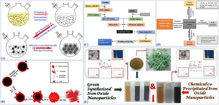
In contrast, chemical synthesis offers an excellent approach for preparing NPs with various functional groups, highly uniform morphology, and narrow size distribution. As shown in Fig. 1A, a variety of chemical methods have been reported for the synthesis of Fe-NPs, notable examples including microemulsion, thermal decomposition, co-precipitation, and liquid-phase reduction [28]. For example, nZVI can be commonly synthesized by the reduction of ferrous or ferric ions in aqueous solutions using reducing agents (e.g., sodium borohydride, polyphenolic compounds, and sorghum bran extracts) under inert conditions per reactions [29-31]. Moreover, nZVI is also synthesized through the aqueous-phase borohydride reduction approach [32], micro-emulsion-based methods [33], and sol-gel methods [34], etc. For Fe-NPs (Figs. 1B–E), co-precipitation is by far the most common synthesis for magnetic Fe-NPs as cheap and environmentally friendly precursors and simple experimental procedures [26,35]. In a recent study, a multistage flow reactor was invented to synthesize and stabilize stable Fe-NPs continuously with a residence time of <5 min by adding the appropriate quantity of a citric acid solution which yielded within minutes colloidally stable Fe-NPs solutions around a neutral pH value [36]. Other typical chemical methods (Fig. 1E) of Fe-NPs included green synthesis, thermal decomposition, sol-gel, solvothermal, and aqueous-phase synthesis [37,38]. Notably, non-stabilized Fe-NPs have been found more reactive and easier to aggregate in a lipid/solid environment than stabilized Fe-NPs, thus various surface modification or stabilization techniques have been investigated. During the agglomeration of metal nanoparticles, the inter-particle forces may include van der Waals forces, magnetic dipolar interactions, and electric dipolar interactions [39]. Currently, various stabilizers, including thiols, carboxylic acids, silica, surfactants, and polymers, have been found effective in stabilizing Fe-NPs [40]. However, the surface-bound organic solvents or surfactants on Fe-NPs need to be removed due to toxicity concerns and technical requirements in certain environmental applications.
Figure 1
Figure 1.
(A) Schematic illustration of the “heat-up” method for the synthesis of uniformly sized iron oxide nanoparticles. Copied with permission [28]. Copyright 2015, American Chemical Society. (B) Evolution of nanoscale zero-valent iron (nZVI) in water. Copied with permission [35]. Copyright 2015, Elsevier B.V. (C) Preparation scheme of magnetite (Fe3O4) nanoparticle via co-precipitation method. Copied with permission [26]. Copyright 2021, Elsevier B.V. (D) Processing steps of Fe3O4, α-Fe2O3, and γ-Fe2O3 nanoparticles. Copied with permission [26]. Copyright 2021, Elsevier B.V. (E) The co-precipitation method was used for the synthesis of iron oxide nanoparticles by using green and chemical-based materials. Copied with permission [38]. Copyright 2018, Springer Science Business Media.
Biogenic processes are important and green methods in the formation of nanoparticles generally known as biomineralization. Especially, microbial cells, including Fe(Ⅲ)-reducing bacteria, magnetotactic bacteria, and sulfate-reducing bacteria play a key role in the biosynthesis of Fe-NPs [27]. Biosynthesis of Fe-NPs using the natural abundance of functional groups in plant biomolecules also has aroused wide concern in pollution remediation as adsorbents [41]. Nowadays, biosynthesis of Fe-NPs has recently received potential application prospects due to its reliable eco-friendly and low cost, pressures, and neutral pH, which can be used in environmental remediation.
2.2
Characterization of Fe-NPs
Various intrinsic factors of Fe-NPs can affect their environmental fate and subsequent ecological health effects. Given the variety of available Fe-NPs, a description of the physicochemical properties must be provided in all environmental behavioral and toxicological studies. Thus, we provided an overview of selected techniques grouped by spectroscopic, spectrometric, microscopic, thermal, and labeling categories for Fe-NPs measurements (Fig. S1 in Supporting information) [28,35,38,42-45]. The morphology (Figs. S1A and B), dimensions (Figs. S1C and D), structural (Figs. S1E and F), chemical group (Fig. S1G), and component of the Fe-NPs are commonly characterized by spectrum, chromatography, and mass spectrum techniques [26]. For magnetic properties of the Fe-NPs. For the determination of the particle−particle and model surface interactions of superparamagnetic IONPs, time-resolved dynamic light scattering (DLS), zeta (ζ) potential, and real-time quartz crystal microbalance with dissipation monitoring (QCM-D) measurements (surface deposition and release) were performed [46].
2.3
Environmental application of Fe-NPs in aquatic environment
Global pollution of soil and water is a serious challenge for which environmental remediation is urgently needed. For challenging environmental applications, Fe-NPs have great vitality for environmental restoration as high-performance, green and multifunctional nanomaterials due to their interesting magnetic properties, biocompatibility, easy availability, and low cost. Moreover, Fe-NPs also to be an ideal material for the removal of pollutants (e.g., organic molecules and heavy ions) from contaminated water with excellent recyclability, sorption capability, and recyclability [24,27]. Generally, the environmental applications of Fe-NPs can be grouped into four aspects, namely the adsorption, transformation, detection, and recycling of environmental pollutants. Furthermore, iron oxides frequently play a passive role in the composites for environmental applications. They assembled the nanocomposite with a property to be magnetically separable once pollutants (e.g., organic or inorganic component) were adsorbed on the surface provided by the other nanoparticle counterparts (e.g., graphene and carbon nanotubes) [7]. If not magnetically functionalized, pollutants were difficult to separate from the aquatic environment, thus this was highly desirable in the case of magnetic nanocomposites acted as adsorbents for the removal of pollutants or toxic chemicals from water, due to their huge surface area, high reactivity, and strong ferromagnetism. Although there have been some reviews exist for the environmental applications (catalyst material, adsorbent, pigment) of Fe-NPs, we noted that a comprehensive overview of the application of Fe-NPs for environmental fate and ecological health risk assessment is still absent [24,26,27]. Fe-NPs were highly stable in aquatic environments, and could direct contact with aquatic organisms such as algae, plants, and fish. Most importantly, several studies have shown that iron nanoparticles could enter the cells, and cause damage to plants, animals, and humans once released into the environment [47]. This review summarized the environmental fate and behaviors of Fe-NPs and would focus mainly on the toxicity and mechanism of Fe-NPs to aquatic organisms.
3.
Fate and environmental behavior of Fe-NPs in the aquatic environment
3.1
Adsorption behavior of Fe-NPs in aquatic system
Adsorption is a critical physiochemical process for environmental applications of Fe-NPs as adsorbents due to their large surface-active properties. Moreover, adsorption plays an important role in the environmental applications for pollutants water treatment. During this process, the environmental fate and behavior of pollutants (e.g., organic molecule compounds, heavy metals) and Fe-NPs could be influenced. Moreover, the surface characteristics of Fe-NPs would be changed through the pollutant’s coating or modification. Adsorption mechanism identification is one of the most significant fundamental principles necessary to bridge the gap between adsorption investigations and environmental application security design. In Fig. 2, we outlined major interaction mechanisms reported in the current literature and discussed their generality under the structural properties of both Fe-NPs and pollutants [44,48-50].
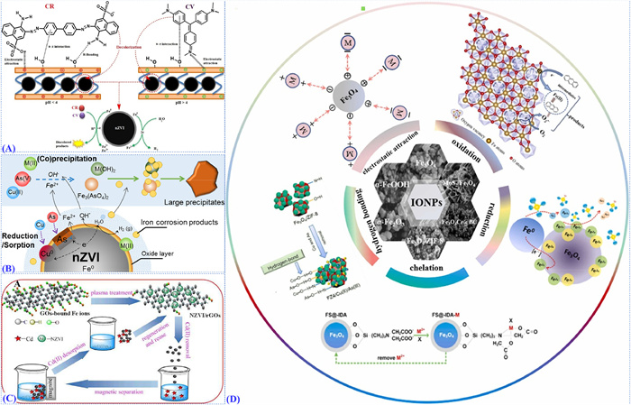
Figure 2
Figure 2.
(A) Removal mechanism of Congo red dye and crystal violet onto the nZVI@nBent–CMC composite. Copied with permission [44]. Copyright 2021, American Chemical Society. (B) Schematic of heavy metal removal using nZVI. Copied with permission [48]. Copyright 2016, Elsevier B.V. (C) Possible schematic mechanisms and reaction process of heavy metal ion removal by NZVI/rGOs and ZVI/ZVAl. Copied with permission [49]. Copyright 2016, American Chemical Society. (D) Proposed mechanisms of pollutant removal by IONPs. Copied with permission [50]. Copyright 2023, Elsevier B.V.
Eltaweil et al. synthesized an efficient nZVI-loaded nano bentonite intercalated carboxymethyl chitosan, which exhibited excellent reusability and adsorption performance, and applied in the removal of anionic and cationic dyes from wastewater successfully [44]. Liu et al. invent a new strategy to promote remediation of soil contaminated by pentachlorophenol and safe crop production in contaminated soil using nZVI [51]. Magnetic adsorption has great attention because of its high-adsorption capacity and recyclability, the superparamagnetic Fe3O4 provides an excellent surface for binding phosphonic acid and carboxylic acid to remove glyphosate and its major metabolite from water [52]. Subsequent removal of nanoparticles from wastewater can be strongly enhanced by using particles the core of which is magnetic Fe-NPs, providing their separation by sedimentation or filtration [53]. Recent research has focused on the surface modification of NPs, where different functional groups are modified by surface active agents, to enhance their performance. Functional NPs have great potential to improve the adsorption performance, as shown by Mahmood et al., where NPs are coated with octadecyl amine and dodecyl amine and exhibit high efficiency in the collection of oil spills [54]. Besides, magnetic nanoparticles are embedded into various materials, such as polymers, silica, and carbon, as high-performance nanocomposites to remove pollutants. Palchoudhury et al. separated MC252 oil from oil−water mixtures using PVP-coated Fe-NPs and achieved approximately 100% oil removal under optimum conditions [55]. Wang et al. modified Fe3O4 with tetraethyl orthosilicate and successfully synthesized Fe3O4@SiO2 nanospheres, which can efficiently remove Congo red from wastewater under laboratory conditions [45]. Moreover, FeOOH coated with g-C3N4 in the presence of H2O2 and visible light irradiation exhibits high performance in degrading antibiotic contaminants as heterogeneous Fenton-like catalysts [56]. As shown in Fig. 2A, Eltaweil et al. fabricated nZVI-loaded nanobentonite intercalated carboxymethyl chitosan for efficient removal of Congo red and cationic crystal violet [44]. Fe-nanocatalyst composed of oxide as a shell and carbide as a core was synthesized to accomplish catalytic dehydrogenation of feedstock chemicals as a kind of cheap and available catalyst [57].
3.1.2
Heavy ion adsorption
As shown in Figs. 2B and C, Fe-NPs (especially nZVI) have been widely investigated for the water treatment of heavy metal ions and received prosperous and tremendous potential environmental in situ and aggregation remediation of heavy metal ions in aquatic systems [48,49]. nZVI could be applied as a green and cost-effective adsorbent for removing heavy metal ions (e.g., Cu2+, Zn2+, Ni2+, and As5+) from wastewater due to the strong chemical reduction and sorption-precipitation [48]. Batch experiments results showed nZVI had extraordinarily high removal capacities (>96% after 60 min) and the maximum theoretical adsorption capacity of nZVI could reach 245 mg and 226 mg/g for As5+ and Cu2+, respectively [48]. Besides, nZVI was also potentially used as an adsorbent for the removal of other toxic heavy metal ions such as Hg2+(226 mg/g), Pb2+ (226 mg/g), Cd2+ (226 mg/g), Zn2+ (226 mg/g), Co2+(226 mg/g) [49]. Moreover, the heavy metal ions uptake capacity of pristine nZVI was lower than those of various surface modified (e.g., epoxide, hydroxyl, and carboxyl) nZVI, which could form strong complexes with heavy metal ions, and acted as adsorbents for heavy metal ion preconcentration and elimination [49].
The point of zero charge (PZC) of Fe-NPs (e.g., hematite and magnetite) was generally reached at pH around 8 [58]. Fe-NPs could hence be negatively or positively charged at naturally relevant pH. Since heavy metal ions are positively charged, only the negatively charged Fe-NPs were available for adsorption. Many studies have demonstrated that Fe-NPs also showed great performance in treatment by transforming heavy metals [49]. The adsorption capacity is one of the main factors driving the increasing application of Fe-NPs in various technologies, mainly obtained from the commonly used isotherm models (e.g., Langmuir and DA models) [59]. Notably, magnetic iron-based composite had recently received much attention for heavy ion removal in magnetic-based separation due to its specific surface area and high magnetism. For example, carbon-coated iron nanoparticles exhibit excellent removal efficiencies (>90%) for Cr, Ni, Cd, Pb, Co, and Mn in a pH range of 8–10, which was much higher than that of activated carbons [59].
3.1.3
Adsorption mechanisms
Adsorption mechanism identification is one of the most significant fundamental principles necessary to bridge the gap between adsorption investigations and environmental application security design. The mechanism of the interactions between nZVI and adsorbed heavy metal ions is summarized in Fig. 2D There have been great reports indicating remediation mechanisms depend on the adsorptive and reductive properties of the nZVI nanoparticles and the nature of the pollutants [60]. In general, the degradation of nZVI to heavy metal ions always contains two steps: first, the target pollutants will be adsorbed onto the nZVI’s surface, and second, relative reactions will occur to degrade those compounds to less harmful products [61]. Bare Fe-NPs were deemed poor performance in organic pollutants treatment because of their weak reactivity, but with some useful modifications and coatings such as TiO2, humic acid, and SiO2, environmental remediation could be achieved via the degradation of pollutants [62]. However, it was confirmed maghemite, magnetite, and their hybrid composites remove heavy metal pollutants mostly by adsorption. For instance, some researchers concluded that Cr(Ⅵ) could be adsorbed onto maghemite NPs by electrostatic interaction and ion exchange [63] and hematite was capable of binding tightly to Sb(Ⅲ) via inner-sphere complexation [64]. The single and combined use of X-ray absorption fine structure (XAFS) and density functional theory (DFT) could be more efficient in detecting and quantifying the interaction between nZVI and heavy metal ions rather than other traditional characterization techniques [65]. XAFS spectra were collected for the analysis of Fe/Cr species and Cr coordination information. DFT calculation was conducted for the coordination pattern of phosphate and chromium, and charge-density difference.
3.2
Transformation behavior of Fe-NPs in aquatic system
3.2.1
Aggregation dispersion
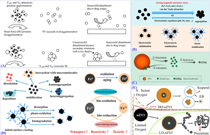
The adsorption kinetic process of as on bare hematite nanoparticles was investigated with regard to the aggregation of the nanoparticles [66]. It was probed that aggregation and sedimentation of hematite nanoparticles appear during the adsorption process, especially in the period of rapid arsenic adsorption. To study the effect of particle concentration, pH, and natural organic matter (NOM) on Fe-NPs, Baalousha et al. observed purified nanoparticle behavior in various factors [67]. This study shows that disaggregation is as important as the aggregation process and confirms that NOM might play a key role in the aggregation and disaggregation of NPs, and consequently, in controlling their fate and behavior in aquatic environments. Kelly compared nZVI, Fe2O3, and Fe3O4 in different aquatic systems and concluded the effect of the high ionic strength was more helpful than the effect of the organic matter on nanoparticles sediment, which contributes to a better dispersion efficiency [68]. A conceptual model of agglomeration (Fig. 3A) of modified nanoscale iron particles followed by deposition of clusters was used to explain the particle agglomeration and subsequent deposition in porous media [69]. In aquatic environments (Fig. 3B), the stability and transport of magnetic Fe-NPs are influenced by a combination of electrostatic and magnetic interactions [70,71].
Figure 3
Figure 3.
(A) Conceptual model of agglomeration of modified nanoscale iron particles followed by deposition of clusters. Copied with permission [69]. Copyright 2009, American Chemical Society. (B) Illustration of the aggregation behavior of iron-based nanoparticles. Copied with permission [71]. Copyright 2018, Elsevier Ltd. (C) Dissolution, adsorption, and hydrolysis in iron-based nanoparticles suspensions [72]. (D) Potential transformations of iron-based nanoparticles in the environment. Copied with permission [71]. Copyright 2018, Elsevier Ltd. (E) Oxidation of nanoscale zero-valent iron under sufficient and limited dissolved oxygen. Copied with permission [74]. Copyright 2015, Elsevier Ltd.
Most of the Fe-NPs are unlikely to persist in natural systems because they have higher surface reactivity than their regular-sized materials. As a result, most of them will go through chemical/biological transformation. Wang et al. tested the phytotoxicity of zero-valent iron nanoparticles in soil cultivated with Oryza sativa and concluded the “aging” nZVI exhibited lower toxicity compared with the fresh [72]. In the paper, it was shown the surface oxidation of the aged nZVI may have prevented full oxidation to maintain a higher toxicity than the control. Lei et al., investigated the potential transformations of Fe-NPs in the environment, modified with polymeric entrapment, such as the alginate, CMC, and PVA-alginate, which can effectively prevent Fe-NPs aging and passivation [71]. Similarly, Semerád et al. summarized the sulfidation nZVI particles were more stable than the bare particles, and exhibited higher toxicity owing to the increasing content of sulfur along with the aging of nanoparticles [73]. After entering the aquatic system, Wang et al., described the dissolution, adsorption, and hydrolysis in iron-based nanoparticles suspensions (Fig. 3C) [72], according to the reactions in Supplementary material.
3.2.3
Oxidation
As the above shows, upon contacting water and air, the surface of NZVI will be oxidized to iron oxides/iron hydroxide, which form a “shell-core” structure. Likely, nZVI can maintain its structure well under anoxic conditions but quickly corrode to γ-FeOOH under oxic conditions [35]. In the water environment (Fig. 3D), Fe-NPs undergo physical, chemical, and/or biological transformations as influenced by environmental factors such as pH, ions, dissolved oxygen, NOM, and biotas [71]. It was observed aging of nZVI was inhibited in the presence of microplastics. NOM may decrease the rates of Fe-NPs transformation and affect the composition and distribution of organic matter at the mineral–water interfaces. Oxidations of nZVI under aerobic and anaerobic conditions were simulated, and their influences on aggregation behaviors of nZVI were investigated and shown in Fig. 3E [74]. In a natural water system, a preliminary exposure to heterogeneous suspensions will accelerate the magnetic sedimentation of Fe-NPs [53].
3.3
Biodegradation behavior of Fe-NPs in aquatic system
3.3.1
Bio-oxidation and bio-reduction
Iron-oxidizing bacteria can utilize iron as an electron donor under both oxic and anoxic conditions, which can even successfully compete with abiotic Fe2+ oxidation in redox interfacial environments with low concentrations but high fluxes of both Fe2+ and oxygen [75]. Besides, In the existence of iron-oxidizing bacteria, nZVI corrosion will be enhanced by converting Fe2+ to Fe3+, which may shorten the age of the NPs. Similarly, iron-reducing bacteria can use organic carbon compounds or hydrogen as electron donors for Fe3+ reduction under anaerobic conditions. Some works found the dissolution of IONPs or transformation into solid-phase ferrous particles and mixed valence Fe2+/Fe3+ particles also resulted from the activity of iron-reducing bacteria [76].
3.3.2
Biodegradation
Generally, degradation of Fe-NPs, especially nZVI and Fe3O4, is a key process accompanied by the dissolution of Fe2+/Fe3+. Wang et al. summarized that NOM has a great influence on the dissolution and transformation of the form of metal oxide and zero-valent nanoparticles [77]. For instance, hydrolysis of Fe2+ and Fe3+ would be inhibited and thus transformed into IONPs because of the existence of humic acid and fulvic acid [78]. How to enhance the biodegradability of nanoparticles has gotten more and more attention. Bannerman invented a kind of polymeric Fe-NPs, which could be degraded over time [79]. Lenaic et al. applied high-resolution transmission electron microscopy to monitor the structural degradation of individual nanocubes with two different surface coatings (amphiphilic polymer shell and polyethylene glycol ligand molecules) [80]. Intracellular transformation was observed the polymer coating controls surface reactivity and access of chelating agents to the crystal surface governs the degradation rate. What is more, Ligand biodegradation of coated Fe-NPs will also alter the toxicity along with surface reconstruction [81].
4.
Ecological toxicities of Fe-NPs to aquatic organisms
4.1
Toxicity to different aquatic organisms
4.1.1
Microorganisms
Microorganisms are more susceptible to nanomaterials than plants [82]. The study of Krittanut et al. indicated that the level of bacterial cells inactivation corresponded to the concentration of nZVI and it was more resistant to nZVI in the lag and stationary phases compared to the exponential and decline phases [83]. To assess the ecotoxic potential of a new nZVI produced for the removal of chlorinated pollutants, Andreas et al. calculated that the obtained EC50 values of nZVI to crustaceans and fish were 163 mg/L and 458 mg/L respectively [84]. In the recent study done by Wang et al., the effects of Fe2O3/nZVI NPs on microorganism community shift in anaerobic granular sludge was investigated. It was observed low concentrations of nZVI and Fe2O3 NPs promoted the growth of microbes (Bacteria and Archaea) and activities of key enzymes [85]. The negligible effect of γ-Fe2O3 on Bacillus subtilis was investigated in natural river water samples compared to ZnO nanoparticles [86].
4.1.2
Algae and plant
Fe-NPs were also relatively considered highly toxic to algae. It was confirmed different kinds of algae were proven to possess different levels of sensitivity to Fe-NPs. Adeyemi et al. stated the coating of sulfide/silica-modified nZVI by algal exudates that strongly influence the toxicity of the nanoparticles [87]. Jara’s work showed a toxic response consisting of a decrease in metabolic activity, increased oxidative stress, and alterations in the mitochondrial membrane potential, which was caused by the microalga Chlamydomonas reinhardtii upon exposure to superparamagnetic IONPs [19]. Zdenka et al. surveyed the EC50 values of Fe-NPs produced by green tea extract cyanobacterium (Synechococcus nidulans), alga (Pseudokirchneriella subcapitata), and even invertebrate organisms (Daphnia magna) are 6.1 ± 0.5 (72 h), 7.4 ± 1.6 (72 h), and 21.9 ± 4.3 (24 h) mg/L, respectively [88]. Besides cyanobacteria were reported to be much more sensitive to nZVI than algae, and the type and dose of Fe-NPs have a great effect on their toxicity [89,90].
4.1.3
Zooplankton
Zooplankton regulates the mechanism through which pollutants and energy are transferred to the higher trophic level. The zooplankton community comprises organisms of different body sizes, taxonomy, and ecological roles. As the size of the microparticles decreased, the weight proportions of phosphorus, sulfur, and chlorine increased and thus altered the zooplankton functional diversity of drainage system reservoirs at an open-pit mine [91]. Edardo et al. assessed the effect of γ-Fe2O3 coated with meso‑2,3-dimercaptosuccinic acid (DMSA) stabilizer on the survival of aquatic snail Biomphalaria glabrata, and found that there is neither influence on fertility nor accumulation after 30 days exposure in clean water [92]. Although commonly, Fe2O3 were regarded as a kind of low-toxicity materials in a natural environment, Ji et al. investigated its bioaccumulation under natural conditions and compound toxicity with as using Ceriodaphnia dubia as the model organism [93]. It was concluded in the research the maximum accumulation was observed after 6 h of exposure and the toxicity of Fe2O3 would enhance due to its uptake.
4.1.4
Fish
In an aquatic ecosystem, fish are the top consumers in the food chain. The toxicity of Fe-NPs is different in the growth stage and tissues of types of fish. For instance, Pereira et al. showed that the early developmental toxicity of γ-Fe2O3 in zebrafish depends on the exposure system [94]. Nemi et al. assessed Fe3O4 exposure in adult zebrafish, and concluded that bare Fe3O4 exposure at high concentrations of 10 ppm may induce significant changes in aggressiveness, speed, and locomotion behavior in the zebrafish brains [95]. To assess the toxic effects of Fe-NPs on the fish, Chen et al. choose larvae of medaka fish (Oryzias latipes) as a model organism, carboxymethyl cellulose (CMC)-stabilized nZVI, aged nanoscale iron oxides or ferrous ion as representative nanoparticles [96]. In this study, it is confirmed the highest mortality in medaka larvae exposed to CMC-nZVI was caused by solution due to the combined effect of hypoxia, Fe2+ toxicity, and ROS-mediated oxidative damage. Madhubala et al. synthesized α-Fe2O3 nanoparticles by hydrothermal method and assessed their toxicity in zebrafish embryos [97]. The changes in hatching rate, survival percentage, heartbeat count, and body length of zebrafish were analyzed to study the effects of Hematite. It was summarized there was low toxicity at lower concentrations but some malformations at higher concentrations. Mohammad et al. assessed the toxicity of Fe-NPs and iron salts exposed to blackfish (Capoeta fusca). According to the Organization for Economic Co-operation and Development guidelines No. 203, Fe3O4 showed a higher level of uptake and lower acute toxicity compared with iron salts [21].
4.1.5
Animals in the sediment
In addition to microorganisms, fish, plankton plants, and animals, other aquatic organisms have also been studied to analyze the toxicity of Fe-NPs. Xu et al.’s study presented a detailed investigation on the effects of Fe2O3/Fe3O4 nanomaterials on bacterial community and function analysis. The results demonstrated that iron oxides promoted 4-nonylphenol degradation and enzyme activities in sediments and enhanced expressions of iron-regulated proteins [98]. In the presence of the Fe(Ⅲ) mineral nano-goethite, anoxic conditions were sustained in sediment systems via microbial processes including Fe(Ⅲ)-reduction and thus changed the sediment microbial community [99]. It was observed that Fe-NPs could stimulate tetrabromobisphenol A in situ anaerobic biodegradation in sediment and largely promoted the ecological abundance of Fe(Ⅲ) reducing and aromatics degrading bacteria [100]. Besides, Fe-NPs can influence the elements of sediment so that the change of sediment organisms. For example, it was reported that iron-based materials exhibit high efficiency for phosphorus immobilization due to their strong affinity with Phosphorus in water and sediment [101].
4.2
Toxic mechanism induces by Fe-NPs
4.2.1
Uptake and bioaccumulation
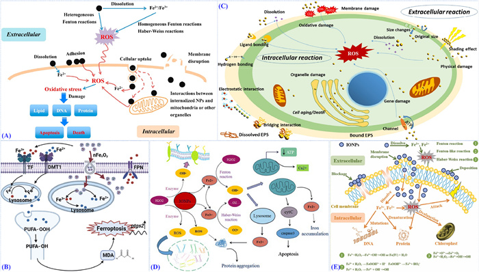
As shown in Fig. 4A, it was found that many nanoparticles can be adsorbed onto or reacted with cell membranes and then disrupt them [102]. As depicted in Fig. 4B, most nFexOy particles can be engulfed by larvae and then endocytosed by cells [8]. The small size of Fe-NPs results in good permeability to biological cell membranes and facilitates accumulation in organisms. Li et al. revealed uptake and translocation in different tissues of γ-Fe2O3 have respective concentrations and thus resulted in diverse toxicity level [103]. Cheng et al. tested the toxicity of sulfide-modified nZVI to Escherichia coli in aqueous solutions, in this work it was confirmed the membranes were destructed by Fe/S nanoparticles, which were observed in E. coli surface and cytoplasm by TEM images [104]. Risk assessment of Fe-NPs in an aquatic ecosystem has also been investigated by Caseta et al. [105]. It was reported that chronic exposure to Fe-NPs increased the behavioral impairments of snails and mortality and reduced fertility were observed only after the exposure to Fe-NPs at 15.6 mg/L compared to the control. Bioaccumulation is the increase in the level of contaminants in the body of an organism. Waterborne and food chains are two ways by which any pollutant enters the body [106]. It was found that average bioconcentration factors in the nematode exposed to waterborne and food-borne nZVI were ~50 and ~5 × 10⁻³, respectively, which was recognized as an important factor in fertility, locomotion, and development of organisms [107].
Figure 4
Figure 4.
(A) Possible toxic mechanisms for iron-based nanoparticles. Copied with permission [71]. Copyright 2018, Elsevier Ltd. (B) Scheme of iron overload triggering ferroptosis. Copied with permission [8]. Copyright 2023, Elsevier B.V. (C) Interaction and toxicity mechanisms of iron-based nanoparticles to green alga. Copied with permission [112]. Copyright 2022, Elsevier B.V. (D) The damage mechanism of iron oxide nanoparticles to the neural tissue. Copied with permission [124]. Copyright 2017, Zahra Yarjanli et al. (E) Proposed toxicity mechanisms of IONPs with soil organisms at the cellular level. Copied with permission [50]. Copyright 2023, Elsevier B.V.
The ions release of the metal oxide NPs in aquatic environment was complex, and correlated to both the dissolution and adsorption processes of the metal oxide NPs. Some studies showed that the toxicity of the nanoparticles originates from the dissolved iron ions rather than the particles themselves [108]. Likely, it was found oxidative stress induced by cells exposed to nanoparticles was equivalent to the cells exposed to Fe2+[109]. According to recent research, the aquatic toxicity of Fe2O3 was dominantly caused by itself [72]. The release of redox-active Fe2+, which resulted in the generation of reactive species, has been found to contribute to the cell inactivation of nZVI [110]. In oxygenated water, the iron dissolution generates OH radical via the Fenton reaction and Fe2+ by nZVI, which will enter the cells and induce oxidative stress [111]. Free radicals may initiate a way to damage the cell nucleus such as a sort of proapoptotic signals.
4.2.3
Oxidation stress and lipid peroxidization
According to the recent literature (Fig. 4C), reactive oxygen species (ROS) production has been reported to be the main mechanism of cell toxicity induced by Fe-NPs [112]. Increasing production of ROS has been investigated to result from the Fenton reaction decreased activity of antioxidant enzymes/inhibition of apoptotic mechanisms [113-115]. Currently, research on the toxicity of Fe-NPs shows potential risks associated with exposure, including inflammation, fibrosis, genotoxicity, and extra-pulmonary effects, all of which have been attributed to increased oxidative stress following exposure [116]. Released to the aquatic environment, the surface of Fe-NPs produces lots of hydroxyl radicals and the uptake of NPs will be unitized by organelles, including mitochondria, where the oxidize stress may be caused [117]. Shen et al. developed a microfluidic magnetophoretic device and accomplished the precise separation of cells into subpopulations according to their magnetic nanoparticle loading to assess the toxicity of Fe-NPs. It was found cells loaded with more iron might have higher ROS levels [118]. Zhu et al. investigated the potential toxicity of α-Fe2O3 using a unicellular eukaryote model, it was concluded that α-Fe2O3 were rapidly internalized in S. cerevisiae and induced mitochondrial impairment and oxidative stress resulted in cell apoptosis [119]. It was also confirmed iron oxide nanoparticles would inhibit the immune function of human T lymphocytes through mitochondrial damage and ROS production [120].
Lipid peroxidation, which could be monitored by secondary peroxidation products such as malondialdehyde (MDA), can lead to membrane leakage and integrity damage. Exposed to two kinds of coated-nZVI, the cell membrane damage was shown along with the increased MDA level at all kinetic time intervals [121]. Semerád et al. developed an assay that enables the direct determination of volatile oxidative damage products (aldehydes) of lipids and proteins in microbial cultures after exposure to commercial types of nZVI. It was confirmed that nZVI also caused oxidative damage to proteins in addition to lipids as a result of the presence of methional in all exposed organisms [122]. According to the toxicity test of Fe2O3 nanoparticles to mice, Fe2O3 administration increased the reactive oxygen species, lipid peroxidation, protein carbonyl content, glutathione peroxidase activity, and nitric oxide levels with a concomitant decrease in the levels of antioxidants-superoxide dismutase, catalase, glutathione, and vitamin C [123].
4.2.4
DNA damage
Fig. 4D shows the damage mechanism of iron oxide nanoparticles to the neural tissue [124]. Mazuel et al. suggest that Fe-NPs uptake and grade in tissue, along with expression of genes encoding ferritin light chains (iron loading) and iron transporters (iron exporting) [125]. Proposed toxicity mechanisms of Fe-NPs with soil organisms at the cellular level are shown in Fig. 4E [50]. DNA damage, which appeared correlated to the concentration of Fe in tissue, was observed and may reveal the toxicity mechanisms of nZVI to Eisenia fetida [126]. What is more, it was found that Fe-NPs may generate ROS by using DNA as an electron donor which might greatly enhance the oxidative stress to cells and even apoptosis [127]. As reported by Chrysa et al. on the toxicity assessment and comparison between two types of Fe-NPs in mussels, the increased ROS induced by nanoparticles, at all days of exposure, is positively correlated to DNA damage, lipid peroxidation, and protein carbonylation [128]. Similarly, exposed to Fe-NPs, DNA damage is caused in a vertebrate model such as zebrafish [129].
4.2.5
Other toxic molecular mechanisms
Fe-NPs are available for organisms, and their uptake can cause the change in metabolism and thus modulate the expression of relative genes. Renen et al. observed a significant effect on organisms’ metabolism though classical immobility or lethality parameters were not significant in acute experiments [130]. When Fe2O3 nanoparticles were uptake into the leaf, excessive hydroxyl radical generated via the Fenton-like reaction mediated by NPs in plants will cause the degradation of chlorophyll and inhabitation of photosynthesis, which will cause a decrease in biomass production [131]. Gao et al. identified the time-course metabolism of Fe-NPs in cells by using X-ray absorption near edge structure spectroscopy [132]. It was shown iron oxide transformed into ferritin and lipid peroxide began to accumulate when exposed to nanoparticles. Su et al. observed that the effect of Fe3O4 on vascular endothelial cells, including the Fe-NPs at high concentrations (>20 mg/kg in vivo) inhibited cell growth in vitro and damaged the endothelium in vivo, which was mediated by caveolin-1 and endothelial nitric oxide synthase [133].
The transcriptomic study identified differentially expressed genes exposed to Fe-NPs using nematode C. elegans as the model organisms [18]. Exposed to Fe-NPs may significantly promote Ferric reductase activity and reduce the chlorophyll leaf content of plants [134]. The effects of NPs can change in biological systems due to the effects of proteins and other compounds, which is called ‘protein corona’, a nontoxicity effect. The protein corona can reduce the influence of nanoparticles as a protective shield [135]. Alterations in iron metabolism are recognized as a main factor via inflammatory or oxidized stress [136]. Transcriptome sequencing analyses are applied in studying the toxicity mechanism of adult zebrafish exposed to iron sulfides nanoparticles. It was reported that exposure of iron sulfide NPs caused significant expression alterations in genes related to immune and inflammatory responses, detoxification, oxidative stress, and DNA damage/repair [137].
4.3
Factors of toxicity
Released to the natural environment, there are complex interactions between the properties of nanomaterials and the structures and functions of organisms [138]. Cheng et al. determined the effects of particle size by investigating toxicities of nZVI of different sizes to a green alga (Chlorella pyrenoidosa) [95]. According to Chen et al.’s paper, nZVI inhibited the higher toxicity compared with Fe3O4 by increasing hypoxia and ROS in medaka fish [139]. To reveal the interactions between the redox state of Fe-NPs and its cytotoxicity to E. coli, Auffan et al. compared their toxic effects on bacterium [140]. According to this paper, it was shown that nZVI has more significant toxicity than maghemite and magnetite. The response generated by the iron nanoparticles entirely depends upon its properties and that of the nearby surroundings. A great deal of evidence demonstrated that surface modification has a great influence on the toxicity of nZVI to organisms [141].
Nowadays, biologically synthesized Fe-NPs have been widely accepted in various applications. Many studies confirm the biocompatible property of bio-synthesized Fe-NPs. Pavla et al. concluded in their comparative study that iron and Fe-NPs synthesized with green tea extract all tested organisms, including bacteria, cyanobacteria, algae, plants, and crustaceans [142]. Two types of nZVI modified by sodium alginate and bentonite contain lower toxicities to E. coli and higher mobility compared with the bare [143]. The effect of functional Fe-NPs was observed on the growth of chlorella. After being coated with Pr6O11 and SiO2, Pr6O11/SiO2@Fe3O4 can promote the growth of chlorella and reduce the mortality and malformation rates of zebrafish embryos compared with the uncoated [144]. Zhang et al. contrasted the toxicity of dimercaptosuccinate-coated Fe3O4 with the bare nanoparticles towards aquatic organisms, including green algae (Raphidocelis subcapitata), duckweed (Lemna minor) and water fleas (Daphnia magna). According to the test, EC50 values of DMSA-Fe3O4 are higher than bare nanoparticles in algae, and significant toxicity was found when Daphnia was exposed to uncoated-NPs [145]. While nZVI was modified with the surface coatings, carboxymethyl cellulose (CMC), it was observed CMC could inhibit the toxicity of nZVI to E. coli largely compared with bare nanoparticles [47].
As the above said, many factors (such as NOM, pH, and background ions) in the environment can change the surface properties of Fe-NPs and thus affect the toxicity of NPs. The state of iron nanoparticles in an aqueous environment is different from the diversity in pH. Ionic strength and pH were proved to have a great influence on the hetero-agglomeration of oxide NPs, thereby helping to explain the cytotoxicity of NPs [146]. Ca2+ was reported to have two different influences on the toxicity of nZVI [47]. On the one hand, Ca2+ can promote the adhesion of nZVI to the bacteria and thus enhance its toxicity. On the other hand, Ca2+ may cause the aggregation and sedimentation of nZVI to lower its toxicity. Upon an aquatic environment, nanoparticles may be coated with NOM and thus alter their fate and toxicity [147]. In this case, NOM may serve as a kind of barrier to hinder the contact of NPs with organisms and as a result, the toxicity of NPs is decreased [148]. Similarly, Gonzalez-Moragas et al. investigated the influence of interaction between α-Fe2O3 nanoparticles and dissolved fulvic acid on the physiological responses of Synechococcus sp. PCC7942. It was observed fulvic acid bound with α-Fe2O3 NPs through carboxyl groups to decrease the binding sites of α-Fe2O3 NPs, which resulted in the decrease of oxidize stress and lower of NPs toxicity [18]. Furthermore, ROS generated by Fe-NPs may be eliminated by NOM in the natural environment and result in the decrease of Fe-NPs [87].
5.
Conclusion and future work
This article reviews the adsorption, transformation, biodegradation, and potential ecological toxicity of iron-based nanoparticles (Fe-NPs) in aquatic environments. Fe-NPs exhibit significant potential for the adsorption of heavy metal ions and organic molecules due to their high specific surface area, but their environmental behavior and ecological risks still require in-depth research. Currently, there are three major challenges:
First, different synthesis methods (physical, chemical, biological) result in varied physicochemical properties (morphology, size, structure, chemical groups, and composition) of Fe-NPs, which in turn affect their environmental behavior (adsorption, transformation, degradation) and toxicity (impact on microorganisms, plants, and animals). Therefore, further research is needed to understand the relationship between the physicochemical properties of Fe-NPs and their environmental behavior and toxicity.
Second, machine learning offers a new perspective for understanding the behavior and fate of Fe-NPs in aquatic environments. It can enhance the prediction of adsorption capacity through literature analysis and provide data support for the assessment of environmental behavior and fate, accelerating related research progress.
Third, most existing studies are based on single laboratory conditions, lacking effective assessment of the environmental risks of Fe-NPs in real water bodies. Future research needs to focus on how to reduce the toxicity of Fe-NPs through synthesis and modification, and pay attention to their forms of existence (types, concentrations, distribution) in natural environments and the biological effects of long-term low-dose exposure, in order to bridge the gap between laboratory research and actual environmental conditions.
Declaration of competing interest
The authors declare no competing financial interest and personal relationships with other people or organizations that can inappropriately influence our work. There is no professional or other personal interest of any nature or kind in any product, service and or company that could be construed as influencing the position presented in the manuscript entitled.
CRediT authorship contribution statement
Yang Gao: Writing – review & editing, Writing – original draft, Data curation, Conceptualization. Xiaocheng Wei: Writing – original draft, Data curation. Jing Sun: Writing – review & editing. Shaohu Ouyang: Writing – review & editing, Writing – original draft, Data curation, Conceptualization.
Acknowledgments
Financial supports from National Key Research and Development Program of China (No. 2023YFC3709000), National Natural Science Foundation of China (Nos. 42207337, 52200185, 42107306 and 52300191), Natural Science Foundation of Hunan Province (No. 2024JJ5013), Natural Science Foundation of Tianjin Province (No. 24JCYBJC01980) and Shanghai Tongji Gao Tingyao Environmental Science & Technology Development Foundation (STGEF) are acknowledged.
Supplementary materials
Supplementary material associated with this article can be found, in the online version, at doi:10.1016/j.cclet.2025.111600.
H. Deng, D. McShan, Y. Zhang, et al., Environ. Sci. Technol. 50 (2016) 8840–8848. doi: 10.1021/acs.est.6b00998
[146]
S. Ma, K. Zhou, K. Yang, et al., Environ. Sci. Technol. 49 (2015) 932–939. doi: 10.1021/es504730k
[147]
S.X. Li, S.Q. Wang, B. Yan, et al., ACS Sustain. Chem. Eng. 9 (2021) 13705–13716. doi: 10.1021/acssuschemeng.1c02795
[148]
K. Padrova, O. Mat’atkova, M. Sikova, et al., New Biotechnol. 33 (2015) 144–152.
Figure 1
(A) Schematic illustration of the “heat-up” method for the synthesis of uniformly sized iron oxide nanoparticles. Copied with permission [28]. Copyright 2015, American Chemical Society. (B) Evolution of nanoscale zero-valent iron (nZVI) in water. Copied with permission [35]. Copyright 2015, Elsevier B.V. (C) Preparation scheme of magnetite (Fe3O4) nanoparticle via co-precipitation method. Copied with permission [26]. Copyright 2021, Elsevier B.V. (D) Processing steps of Fe3O4, α-Fe2O3, and γ-Fe2O3 nanoparticles. Copied with permission [26]. Copyright 2021, Elsevier B.V. (E) The co-precipitation method was used for the synthesis of iron oxide nanoparticles by using green and chemical-based materials. Copied with permission [38]. Copyright 2018, Springer Science Business Media.
Figure 2
(A) Removal mechanism of Congo red dye and crystal violet onto the nZVI@nBent–CMC composite. Copied with permission [44]. Copyright 2021, American Chemical Society. (B) Schematic of heavy metal removal using nZVI. Copied with permission [48]. Copyright 2016, Elsevier B.V. (C) Possible schematic mechanisms and reaction process of heavy metal ion removal by NZVI/rGOs and ZVI/ZVAl. Copied with permission [49]. Copyright 2016, American Chemical Society. (D) Proposed mechanisms of pollutant removal by IONPs. Copied with permission [50]. Copyright 2023, Elsevier B.V.
Figure 3
(A) Conceptual model of agglomeration of modified nanoscale iron particles followed by deposition of clusters. Copied with permission [69]. Copyright 2009, American Chemical Society. (B) Illustration of the aggregation behavior of iron-based nanoparticles. Copied with permission [71]. Copyright 2018, Elsevier Ltd. (C) Dissolution, adsorption, and hydrolysis in iron-based nanoparticles suspensions [72]. (D) Potential transformations of iron-based nanoparticles in the environment. Copied with permission [71]. Copyright 2018, Elsevier Ltd. (E) Oxidation of nanoscale zero-valent iron under sufficient and limited dissolved oxygen. Copied with permission [74]. Copyright 2015, Elsevier Ltd.
Figure 4
(A) Possible toxic mechanisms for iron-based nanoparticles. Copied with permission [71]. Copyright 2018, Elsevier Ltd. (B) Scheme of iron overload triggering ferroptosis. Copied with permission [8]. Copyright 2023, Elsevier B.V. (C) Interaction and toxicity mechanisms of iron-based nanoparticles to green alga. Copied with permission [112]. Copyright 2022, Elsevier B.V. (D) The damage mechanism of iron oxide nanoparticles to the neural tissue. Copied with permission [124]. Copyright 2017, Zahra Yarjanli et al. (E) Proposed toxicity mechanisms of IONPs with soil organisms at the cellular level. Copied with permission [50]. Copyright 2023, Elsevier B.V.

 DownLoad:

 下载:
下载:


 下载:
下载:
