
Figure 1.
Appearance and shape of freeze-dry reagents. (A) Extraction reagents and RT-LAMP freeze-drying microspheres in the cassette liquid storage base. (B) RT-LAMP freeze-drying microspheres in the 0.2 mL 8-way PCR tube.
Preparation of norovirus GII loop mediated isothermal amplification freeze-drying microsphere reagents and its application in an on-site integrated rapid detection platform
Yanqi Wu , Yuhong Guan , Peilin Huang , Hui Chen , Liping Bai , Zhihong Jiang

Norovirus can cause non-bacterial gastroenteritis in humans [1–3]. Small, non-enveloped globular human norovirus (HuNoV) containing single-stranded RNA, from the cupaviridae family, and is the most common pathogen of foodborne illness [4]. The HuNoV genome is about 7.6 kb and has three open reading frames (ORFs) in it, of which ORF1 encodes non-structural viral proteins, consisting of RNA-dependent RNA polymerase (RdRp), ORF2 and ORF3, which, respectively, encode main (VP1) and minor (VP2) capsid proteins [5,6]. Noroviruses can be classified genetically into 10 genomes (G), of which the GI, GII and GIV genomes are associated with most human infections, while the GII genome is again the most prevalent [7,8]. HuNoV is characterized by low infectious dose, rapid onset and strong transmission capacity; therefore, rapid and sensitive detection of HuNoV GII type is essential to reduce the spread of gastroenteritis infection [9].
Currently, nucleic acid detection is the most effective method for identifying pathogens [10,11], among which, due to its excellent sensitivity and specificity, quantitative polymerase chain reaction (qPCR) is the "gold standard" for detection, but it is not suitable for on-site point-of-care detection due to complicated operations and high instrument needs [12,13]. Loop-mediated isothermal amplification (LAMP) technology is currently the most widely used isothermal amplification technology with low equipment requirements, and this method, in combination with Point-of-care testing (POCT) detection instruments, can detect pathogen nucleic acids quickly, accurately, and effectively [14,15]. In order to obtain accurate assay results, the quality of the reactive components of the assay reagents must be ensured and maintained. The majority of these reagent components have a limited shelf life and are sensitive to temperature changes, this might restrict their storage, use, and logistics [16]. These drawbacks can be overcome by removing water from reagents using freeze-drying procedures, which will increase product stability and shelf life, a low-temperature dehydration process used mainly to stabilize the biological activity of the substances contained in aqueous solutions [17–19]. Freeze-drying reagents can also be stored at room temperature without frozen transport, reducing reagent transport and storage costs. In addition, since freese-drying reagents are more stable than liquid formulations, test results are more accurate [20,21].
In this study, LAMP was used to detect the ORF1-ORF2 junction gene fragment of norovirus GII, and LAMP liquid reagent was prepared into freeze-drying microspheres after bead-dropping machine molding and lyophilization, and the performance tests of lyophilization process optimization, lyophilization reagent system optimization, sensitivity, specificity, repeatability and storage stability of freeze-drying microsphere reagent were performed. A commercial automatic nucleic acid analyzer (Shenzhen LemnisCare Medical Technology Co., Ltd., Shenzhen, China), an integrated on-site real-time detection platform based on closed cassettes, integrated reagents that can be stored at room temperature, and norovirus GII, an integrated detection device based on magnetic bead nucleic acid extraction and LAMP fluorescence detection, was built. Finally, we used the platform to test 28 simulated samples and contrasted the outcomes with real-time quantitative PCR data after conventional laboratory nucleic acid extraction to verify the reliability of the platform.
The fully automated nucleic acid analyzer, the instrument structure mainly includes integrated cassette and supporting automated instruments. The integrated cassette consists of a reservoir base for the reagents, a liquid suction and drainage assembly and a sealing cap. The reservoir base contains the magnetic bead-based nucleic acid extraction reagents and a well slot for 11 fluorescent LAMP detection reagents; since the well slot of the detection reagent has no well cap, we use paraffin oil to seal the LAMP detection reagents, which acts to prevent evaporation of the reagent during amplification and aerosol contamination. The reservoir base is fixed in the external metal heat trap device and rotates with the rotation of the external metal heat trap device, so that the suction head in the suction and drainage component can enter each reservoir.
The pre-encapsulation process of reagents in the cassette reservoir base is shown in Fig. S1 (Supporting information), and the specific operation process is as follows: firstly, the magnetic bead extraction reagents (Shenzhen LemnisCare Medical Technology Co., Ltd.) are added to the cassette reservoir base in the clean area, and the specific reagents and doses are as follows: 300 µL lysis solution, 500 µL cleaning solution 1,500 µL cleaning solution 2,500 µL cleaning solution 3,200 µL magnetic bead suspension, 150 µL eluent, and 300 µL paraffin oil. After that, the cillin vial containing the freeze-drying microsphere reagent and the cartridge storage base containing the nucleic acid extraction reagent by the magnetic bead method were transferred to a clean glove box together, and the air inside the glove box was discharged from the glove box exhaust port by using nitrogen gas through the air inlet of the closed glove box, so that the humidity inside the glove box reached below 10% and then the air inlet valve and exhaust valve were closed. At humidity of 10% or less and a temperature is 22 ℃, use forceps to transfer the freeze-drying microsphere reagents into the cartridge reservoir base. Next, turn on the semiautomatic microplate heat sealer (Hangzhou Aosheng Instruments Co., Ltd., Hangzhou, China) with the temperature set to 160 ℃ and the sealing time set to 3.8 s. When the temperature reaches the set temperature, the cassette reservoir base containing the extraction reagent and freeze-drying microsphere reagent will be put into the semiautomatic microplate heat sealer to seal the membrane, and the above is the whole process of reagent pre-encapsulation in the cassette reservoir base.
The process of integrated detection of norovirus GII freeze-drying reagents is shown in Fig. S2 (Supporting information). First, the cassette reservoir base with pre-encapsulated reagents is assembled with the pipette assembly, then 100 µL sample is added into the sample well, the lid is closed, and the reservoir base is rotated and unscrewed from the main body lid (with a sealing ring inside), the whole process of cassette loading and then assembly is completed, the cassette is embedded in the instrument for detection. The overall manual operation time is less than 1 min. The entire pathogen detection cassette can be used for the detection of Norovirus GII in an enclosed environment in a one-time, in an integrated and automated manner, mainly by moving the plunger rod and suction head in the suction and drainage assembly described by external control, in combination with the rotation of the reservoir base. The Supporting information includes the part of materials and method.
The extraction and detection of norovirus GII plasmid was performed using a fully automated nucleic acid analyzer. 106 copies/µL of norovirus GII plasmid 100 µL and 20 µL of proteinase K were added to the pre-encapsulated cassette's sample wells. Negative and positive controls were set in pre-encapsulated cartridges, and the whole-process experimental procedures were set as follows: nucleic acid extraction parameters were lysis temperature of 90 ℃, lysis and mixing for 5 min, washing time of 1 min, drying temperature of 55 ℃, elution temperature of 56 ℃, elution and mixing for 1 min; nucleic acid dispensing volume was 25 µL, and oil volume was 40 µL; temperature program parameters were 65 ℃, 45 cycles, and fluorescence was collected every minute from the FAM channel.
To test the feasibility of this platform, we prepared fecal suspension by adding 1 g of feces to 1 mL phosphate buffered saline (PBS), then randomly adding 20 µL of norovirus GII plasmid at high, medium and low concentrations (106, 104 and 102 copies/µL) to 180 µL of fecal suspension as positive simulated samples, and fecal suspension without norovirus infection as negative simulated samples, and nucleic acid extraction and detection were performed on all randomly prepared simulated samples. Nucleic acid extraction kit magnetic beads method was used with commercial extraction instrument MGX-3200 for extraction, and RT-qPCR detection reagents were placed into commercial qPCR instrument Gentier mini for detection. The experimental results were compared with the results of nucleic acid extraction by automatic nucleic acid analyzer and reverse transcription loop-mediated isothermal amplification (RT-LAMP) lyophilization reagent detection, and the optimization of freeze-drying process parameters is shown in the Supporting information.
The appearance of norovirus GII RT-LAMP freeze-drying reagent is shown in Fig. 1. The reagents are encapsulated in a clean environment with the temperature of 22 ℃ and the humidity of less than 10%. Fig. 1A shows the nucleic acid extraction reagent and freeze-drying reagent in the cartridge reservoir base, which can be directly added to the sample well for closed integrated detection. The freeze-drying reagent was encapsulated in 0.2 mL 8-strand PCR tubes as shown in Fig. 1B. In both packaging processes, the freeze-drying microspheres were clamped out from the vial using forceps and transferred into the cartridge reservoir base and 0.2 mL 8-row PCR tube, indicating that the transferability of the freeze-drying reagent was good; the appearance of the freeze-drying reagent remained full and round after the completion of transfer, indicating that the shape assignment effect of the freeze-drying protectant was good. Then ddH2O was used to reconstitute the freeze-drying reagent, and the freeze-drying microspheres were thawed in 1 s at a rapid dissolution rate, and the solution became clear and transparent after 3–5 s, with a better reconstitution effect.
When excessive moisture remains in the lyoprotectant, the molecular structure of bioactive components such as enzymes contained in the lyoprotectant will change during long-term storage, which is not conducive to the storage of lyoprotectant and leads to poor detection effect. Therefore, the residual moisture of the freeze-drying biological reagent should be controlled within 3% (mass fraction). Thirty norovirus GII RT-LAMP freeze-drying microsphere reagents were divided into one group and three replicate experiments were set for moisture determination. Table S2 (Supporting information) displays the results of the weight reduction method's determination of the residual moisture content of the freeze-drying reagent for three times, with an average moisture content of 0.97%, which is suitable for long-term storage of freeze-drying reagent at a later stage without changing the bioactive components in the reagent, thus ensuring the amplification effect of freeze-drying reagent, as shown in Fig. 2. Analysis of lyophilization reagent specificity test results and performance are shown in the Supporting information.

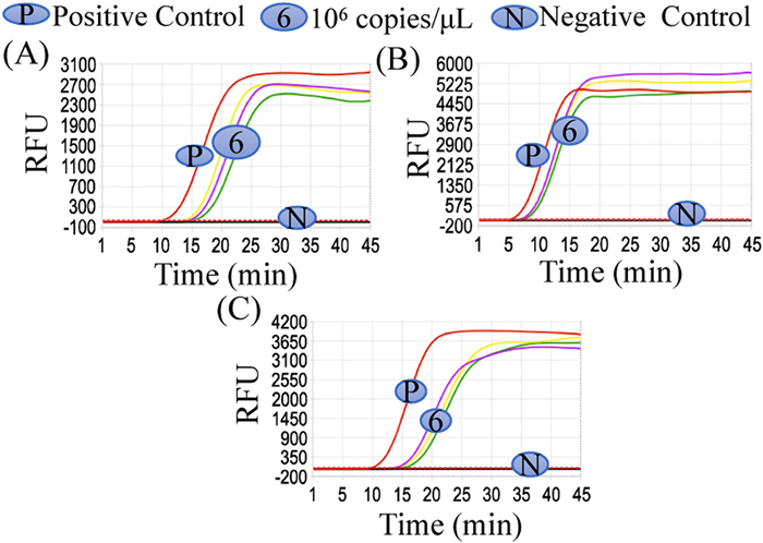
The freeze-drying microsphere reagents were stored at 37 ℃ at room temperature and amplified after re-solubilization on day 0 (Fig. 3A), day 7 (Fig. 3B) and day 14 (Fig. 3C) of lyophilization, and the liquid reagent was set as the control group. The outcomes of the trial revealed that the detection performance of the freeze-drying reagents was basically the same as that of the liquid reagents, whether on day 0, 7, or 14 after lyophilization, and the overall linearity of the time to threshold (Tt) values value with concentration change was good and the stability was fair under different template conditions, which laid the foundation for the long-term storage stability evaluation of subsequent freeze-drying reagents (such as 6 months, 1 year, 2 years).

Norovirus GII lyophilized reagent combined with magnetic beads nucleic acid extraction kit can stably extract and detect norovirus GII plasmid in automatic nucleic acid analysis, and the experimental results are shown in Fig. 4. Norovirus GII could be detected stably after three repeated tests, indicating that the automatic nucleic acid analyzer, extraction reagent and norovirus GII lyophilized reagent had good stability.
The platform was used to test 28 simulated samples and compared with the extraction instrument followed by RT-qPCR conventional laboratory testing method, and the results are shown in Table S4 (Supporting information). The two methods were compared and the experimental results were consistent, with the former being significantly faster than the latter, reducing the time required for clinical diagnosis and improving the efficiency of pathogen detection to a greater extent. The concentration (ratio) of reagents in the freeze-drying reagents was pre-formulated to avoid manual intervention, and the ease of experimental operation, together with the fact that the cassette was closed, also avoided the problems of experimental and environmental pollution.
In this study, the overall performance of the lyophilized reagents was good in terms of sensitivity, homogeneity, reproducibility, and storage stability. Jeon et al. [22] developed a one-step ring-mediated isothermal amplification (RT-LAMP) for detection of norovirus in oysters targeting the ORF1-ORF2 gene junction of norovirus. The sensitivity was 101 copies/µL, and the kit requires ready-to-use and low-temperature storage and transportation. Luo et al. [23] developed a visualized reverse transcription loop-mediated isothermal amplification for norovirus detection with a sensitivity of 103 copies/reaction, whereas the lyophilized assay kit developed in this study had a sensitivity of 101 copies/µL and was easy to operate, only need to add samples, and proteinase K can be put into the integrated system to detect, the length of the experiment is about 1 h. From the results of the storage stability of the lyophilized reagent, the lyophilized reagent kit can be stored and transported at room temperature, which saves the cost of storage and transportation to a certain extent [24]. However, the thermal stability test was only conducted for a short period of time, and the long-term thermal stability test is needed to confirm the validity of the lyophilized microsphere reagent for ambient storage in the future.
In summary, we successfully developed a norovirus GII lyophilized microsphere assay as well as an on-site norovirus rapid detection platform suitable for norovirus GII. The whole system is capable of rapid and effective detection of pathogens, and the reagents can be transported and kept at room temperature; the long-term storage stability of the reagents needs to be further validated. For the extraction and detection of norovirus, the process can be completed in 1 h. The results of the simulated samples are consistent with the qPCR results but the experimental time is greatly reduced, which is ideal for immediate on-site rapid detection.
The authors declare that they have no known competing financial interests or personal relationships that could have appeared to influence the work reported in this paper.
This research was financially funded by the Science and Technology Development Fund, Macau SAR (Nos. 0065/2020/A2, SKL-QRCM(MUST)-2020-2022), Shenzhen-Hong Kong-Macao Science and Technology Project (Grade c) (No. SGDX20210823104201010).
Supplementary material associated with this article can be found, in the online version, at doi:
A.N. Desai, JAMA 322 (2019) 2032. doi: 10.1001/jama.2019.15921
N. Dabilla, T.N. Vieira Almeida, A.C. Reboucas Oliveira, et al., J. Clin. Virol. 87 (2017) 60–66. doi: 10.1016/j.jcv.2016.12.009
G.A.M. Tarr, X.L. Pang, R. Zhuo, et al., J. Infect. Dis. 223 (2021) 452–461. doi: 10.1093/infdis/jiaa391
C. Dongqing, Z. Songyan, L. Ningbo, et al., J. Food Sci. 83 (2018) 393–400. doi: 10.1111/1750-3841.14022
P. Chhabra, H. Browne, T. Huynh, et al., J. Clin. Virol. 134 (2021) 104689. doi: 10.1016/j.jcv.2020.104689
S. Honjo, K. Kuronuma, Y. Fujiya, et al., Infect. Genet. Evol. 104 (2022) 105348. doi: 10.1016/j.meegid.2022.105348
M. Farahmand, M. Moghoofei, A. Dorost, et al., Rev. Med. Virol. 32 (2022) e2237. doi: 10.1002/rmv.2237
R.M. Callejon, M. Isabel Rodriguez-Naranjo, C. Ubeda, et al., Foodborne Pathog. Dis. 12 (2015) 32–38. doi: 10.1089/fpd.2014.1821
Z. Chen, T. Yang, H. Yang, et al., J. Biomed. Nanotechnol. 14 (2018) 198–205. doi: 10.1166/jbn.2018.2524
K. Leera, T. Anyarat, C. Suwat, et al., Food Environ. Virol. 8 (2016) 133–140. doi: 10.1007/s12560-016-9228-6
C. Tang, Z. He, H. Liu, et al., J. Nanobiotechnol. 18 (2020) 62. doi: 10.1186/s12951-020-00613-6
Y. Fang, H. Liu, Y. Wang, et al., J. Biomed. Nanotechnol. 17 (2021) 407–415. doi: 10.1166/jbn.2021.3028
Y. Yongxin, C. Hui, H. Linghao, et al., Appl. Environ. Microbiol. 81 (2015) 7615–7624. doi: 10.1128/AEM.01729-15
L. He, H. Yang, P. Xiao, et al., J. Biomed. Nanotechnol. 13 (2017) 1243–1252. doi: 10.1166/jbn.2017.2422
H. Chen, Y. Wu, Z. Chen, et al., J. Biomed. Nanotechnol. 13 (2017) 1619–1630. doi: 10.1166/jbn.2017.2478
N.O. Prado, L.A. Lalli, L. Blanes, D.L. Zanette, M.N. Aoki, Mini Rev. Med. Chem. 23 (2023) 480–496. doi: 10.2174/1389557522666220802144057
L.J.J. Hansen, R. Daoussi, C. Vervaet, J.P. Remon, T.R.M. De Beer, Vaccine 33 (2015) 5507–5519. doi: 10.1016/j.vaccine.2015.08.085
A. Ahlford, B. Kjeldsen, J. Reimers, et al., Analyst 135 (2010) 2377–2385. doi: 10.1039/c0an00321b
D. Fissore, T. McCoy, Front. Chem. 6 (2018) 622. doi: 10.3389/fchem.2018.00622
L. Thirion, A. Dubot-Peres, L. Pezzi, et al., Viruses 12 (2020) 159. doi: 10.3390/v12020159
X. Zhang, Y. Chen, Y. Pan, et al., Chin. Chem. Lett. 35 (2024) 108378. doi: 10.1016/j.cclet.2023.108378
S.B. Jeon, D.J. Seo, H. Oh, D.H. Kingsley, C. Choi, Food Control 73 (2017) 1002–1009. doi: 10.1016/j.foodcont.2016.10.005
X.F. Qian, A.L. Duan, R.X. Huang, et al., J. Clin. Lab. Anal. 35 (2021) e23919. doi: 10.1002/jcla.23919
Y. Pan, Y. Xiao, Y. Hao, et al., Chin. Chem. Lett. 33 (2022) 2486–2490. doi: 10.1016/j.cclet.2021.12.093

Figure 1 Appearance and shape of freeze-dry reagents. (A) Extraction reagents and RT-LAMP freeze-drying microspheres in the cassette liquid storage base. (B) RT-LAMP freeze-drying microspheres in the 0.2 mL 8-way PCR tube.

Figure 2 (A) Physical drawing of the drying vessel used in this experiment. (B) The discolored silica beads were blue when the humidity was below 20%. (C) The discolored silica beads were pink when the relative humidity was above 50%.

Figure 3 Long-term storage stability validation of freeze-drying microsphere reagents. (A) Sensitivity test on day 0 after lyophilization. (B) Sensitivity test on day 7 after lyophilization. (C) Sensitivity test on day 14 after lyophilization.

Figure 4 Verify the repeatability of extraction and RT-LAMP freeze-dried reagent detection in automated nucleic acid analyzer. (A) The first integrated rapid detection experiment. (B) The second integrated rapid detection experiment. (C) The third integrated rapid detection experiment.

 扫一扫看文章

扫一扫关注我们
 DownLoad:
 下载:
下载:
 下载:
下载: