Figure 1.
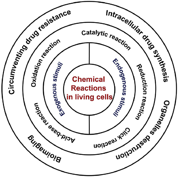
Schematic illustration of the relationship between intracellular chemical reactions and biological applications.
Chemical reactions in living cells for enhanced biological treatment
Yunfei Fu , Hui Li , Chengfei Liu , Wei Tian

Chemical reactions underpin the fundamental processes of nature, governing both the synthesis and utilization of substances in the biological world [1–5]. Living cells, with their highly complex chemical environments, have inspired chemists to extend synthetic reactions into the cellular microenvironment [6–10]. Recently, chemists have successfully transformed small molecules into functional biomaterials through a series of intricate and precise reactions within the complex intracellular environment [11,12]. The creation of entirely new molecules or structures within cells endow them with novel functions. For example, the synthesis of non-natural macromolecules can extend their retention time within cells, thereby enabling the regulation of a wide range of physiological processes [13,14]. These innovative strategies not only enhance our understanding of the complex reactions occurring within living cells but also enable the prediction of potential reaction pathways. More importantly, they have the potential to emulate natural cellular processes and facilitate the artificial manipulation of cellular functions, thereby opening promising avenues for the development of cellular-based therapies and biotechnological applications.
Depending on the underlying chemical mechanisms, intracellular chemical reactions can generally be categorized into five major types: Reduction reaction, oxidation reaction, catalytic reaction, acid-base reaction and click reaction. Generally, these reactions can be triggered by either endogenous or exogenous stimuli. On the basis of the endogenous stimuli, they typically include acidic pH, reductive environments, reactive oxygen species (ROS), and overexpressed enzymes originating from diseased or infected cells [15]. Exogenous stimuli, on the other hand, often involves electromagnetic radiation-such as ultraviolet (UV) light, near-infrared (NIR) light, and γ-ray radiation-as well as external triggers like ultrasound and magnetic fields [16]. These stimuli have demonstrated remarkable effectiveness and hold great potential for therapeutic applications in a wide range of diseases. For instance, emerging intracellular chemical reactions have been extensively applied to enable circumvent drug resistance, organelles destruction, bioimaging and intracellular drug synthesis (Fig. 1).
The rapid advancement of chemical reactions in living cells has sparked significant interest among researchers in both chemistry and biology. In recent reviews on intracellular chemical reactions, Chen's group reported in situ catalysis for therapeutic substance synthesis, focusing on the regulation of related chemical reactions through fundamental thermodynamics and kinetics [17]. Zhang and colleagues summarized recent progress in intra-tumoral stimuli-responsive in situ assembly of supramolecular self-assemblies using artificial small molecule nanomaterials, elucidating how and why these tumor microenvironment triggers can induce the self-assembly of artificial small molecules [18]. To provide a powerful approach for bridging the gap between synthetic materials and biological systems, a comprehensive review and comparison of intracellular chemical reactions based on different reaction mechanisms is essential. This review provides a comprehensive summary of the latest advancements and future trends in intracellular chemical reactions, organized by reaction types. It specifically addresses the design and synthesis of monomers, reaction mechanisms, responsive characteristics, and functional applications. Furthermore, we explore the relationship between various chemical designs and their corresponding reaction mechanisms, emphasizing their potential for biological applications. We hope this review can offer a comprehensive overview of current achievements and challenges, inspiring future directions in the field of intracellular chemical reactions.
The cellular environment is full of reactive species, generating enormous possibilities for intracellular chemical reactions of molecules [19]. Thus, moving chemical reactions from chemical laboratory into an intracellular microenvironment can be regarded as a powerful and versatile way to modulate cell activities, which provide a variety of possibilities for chemists and biologists to understand the cellular world as for the diversity and tunability of chemical reactions [20]. In this section, we discussed design strategies that have enabled small molecule chemistry to be conducted within cells and to establish monomer design principles and functional features.
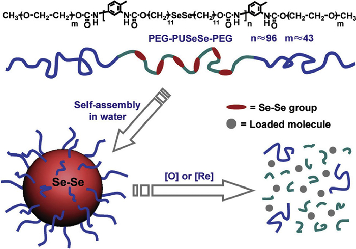
The overproduced glutathione (GSH) in cancer cells has abundant thiol with the necessary functions in regulating biological functions [21]. Compared with normal cells and tissues, tumor tissue has been found to be much higher GSH concentration than that in the normal cells, leading to reducing and hypoxic intracellular milieu [22]. For example, reduction-responsive materials with characteristic disulfide (S–S) bonds in the backbone, side chain or cross-linker have been developed to endow stimulus responsiveness to the intracellular reducing microenvironment [23]. The disulfide bonds can maintain stability in extracellular environments with low concentration of GSH and tends to be quickly degraded under reducing environments via the thiol-disulfide exchange reaction, leading to the disassembly of materials [24,25]. Yang's group synthesized nanodevice ssPPELap@Fe-TA for potentiating ferroptosis. The S–S bonds in ssPPE could deplete GSH via the thiol/disulfide exchange reaction to inactivate GPX4 [24]. More reactive diselenide (Se–Se) bonds have been modified into materials to acquire redox responsiveness [26–28]. The interesting example was reported by Zhang's group. They described a diselenide block copolymers (PEG-PUSeSe-PEG) that was self-assembled into micelles because of the amphiphilic property (Fig. 2) [29]. Se–Se bonds in PEG-PUSeSe-PEG could be oxidized into seleninic acid under oxidation conditions and reduced into selenol in the presence of reducing agent.
Reducing intracellular microenvironment can be also utilized to release active agents through reducing prodrugs inside cells. Gasser's group presented RuⅡ-PtIV conjugate with function of chemotherapy and photodynamic therapy (PDT) to circumvent cancer drug resistance [30]. Upon entering cancer cells, the PtIV-RuⅡ conjugate could be degraded to produce CDDP and RuⅡ compound to realize the aim of synergetic therapeutic effects of chemotherapy and PDT. Our group synthesized chlorambucil-oxoplatin and water-soluble pillar[6]arene to obtain supramolecular complexes on the basis of host-guest interaction [31]. The supramolecular complexes could self-assemble into nano self-assemblies (SDSAs). Upon reaching tumor cells, SDSAs could be reduced by cellular GSH to obtain the high toxicity chlorambucil and cisplatin, with the amplification of oxidative stress in cancer cells. Meanwell, we also formed amphiphilic host-guest complex based on pillar[6]arene and chlorambucil-arylboronic acid through host-guest interaction between chlorambucil and pillar[6]arene [32]. The arylboronic acid moiety of chlorambucil-arylboronic acid could not only consume GSH but also bind curcumin. Curcumin could inhibit the expression of thioredoxin reductase (TrxR) to disrupt GSH biosynthesis pathway, enhancing anticancer effect.
Compared to healthy cells, the relatively high concentration of ROS is an important biomarker for the complicated cancer microenvironment [33]. Cancer cells continuously generate ROS derived from by-products of aerobic metabolism as for oncogenic transformation, resulting in cellular macroenvironment with a highly oxidative state [34]. The ROS-responsive functional materials have been extensively exploited for site-specific stimulus response, mainly encompassing characteristic groups such as boronic ester, thioketal, selenium and ferrocene groups [35–38]. A novel strategy has been pioneered by Almutairi and coworkers. They presented a biocompatible polymeric capsule consisting of H2O2-sensitive polymers with a boronic ester to achieve the controlled release of cargo [36]. Xia's group established cationic polymer by polymerization of oligoamines with ROS-cleavable thioketal linkages [37]. The overexpressed ROS in cancer cells was used as cancer-related stimulus to achieve intracellular gene delivery. Yan and Huang et al. developed an ROS-sensitive hyperbranched polymer with selenide groups and phosphate segments in the dendritic backbone, mediating drug release in cells for combined chemotherapy under intracellular conditions of oxidation [38]. Besides, Se–Se bonds have been proposed in biomedical applications as for their unique properties [39,40,42]. For example, Xu's group constructed diselenide-pemetrexed complex, simultaneously combining cancer immunotherapy with radiotherapy and chemotherapy [41]. Se–Se bonds could be reacted into seleninic acid, inhibiting the expression of intracellular human leukocyte antigen E and inducing the immune response for NK cells.
The overproduced ROS in cancer cells can oxidize original hydrophobic ferrocene moiety into hydrophilic ferrocene, which leads to disaggregate host-guest interactions, leading to the controlled release of active agents inside cells [43]. Our group reported supramolecular complex formed by the supramolecular interactions between curcumin-bridged bis(β-cyclodextrin) and ferrocene-linked camptothecin (CPT) [44]. The overproduced intracellular ROS can easily destroy host-guest interactions, triggering the burst release of therapeutic reagents to circumvent multidrug resistance. Recently, we also synthesized supramolecular organometallic drug complex (SOMDC) with H2O2 self-provision constructed by GEM-(Fc)3 and β-CD-DOX [45]. Because of hydrophobic interaction, the amphiphilicity of SOMDC could self-assemble into supramolecular organometallic drug micelles (SOMDMs). Similarly, SOMDMs could be rapidly destroyed as for the endogenous ROS to release β-CD-DOX. The biological assessment showed that β-CD-DOX could substantially elevate the endogenous H2O2 level in cells and accelerate the Fenton-like intracellular autocatalysis to create hypertoxic •OH [45].
Intracellular enzymes can affect cellular behavior in the course of metabolism, proliferation, and invasion [46]. Alkaline phosphatase (ALP) is usually overproduced in cancer cells, which has been widely applied into construction of functional materials for modulating cell activities. For example, Xu's group designed intranuclear nanoribbons constructed upon dephosphorylation of leucine-rich L- or D-phosphopeptide catalyzed via ALP to inhibit the proliferation of osteosarcoma cells [47]. Upon dephosphorylation catalyzed by ALP, the peptides could be converted into micelles and then changed into nanoribbons. Yang's group showed a novel way to selectively modify cancer cell membranes through using an enzyme-induced peptide self-assembly strategy (Fig. 3a) [48]. They formed some self-assembling peptides (pY1, pY2, and pY3) constructed through NBD fluorophore, –GFF– core self-assembly motif and GE11. After treatment with ALP for a period of time, the light-scattering intensities of pY1, pY2, and pY3 were increased compared with their phosphorylated precursors.
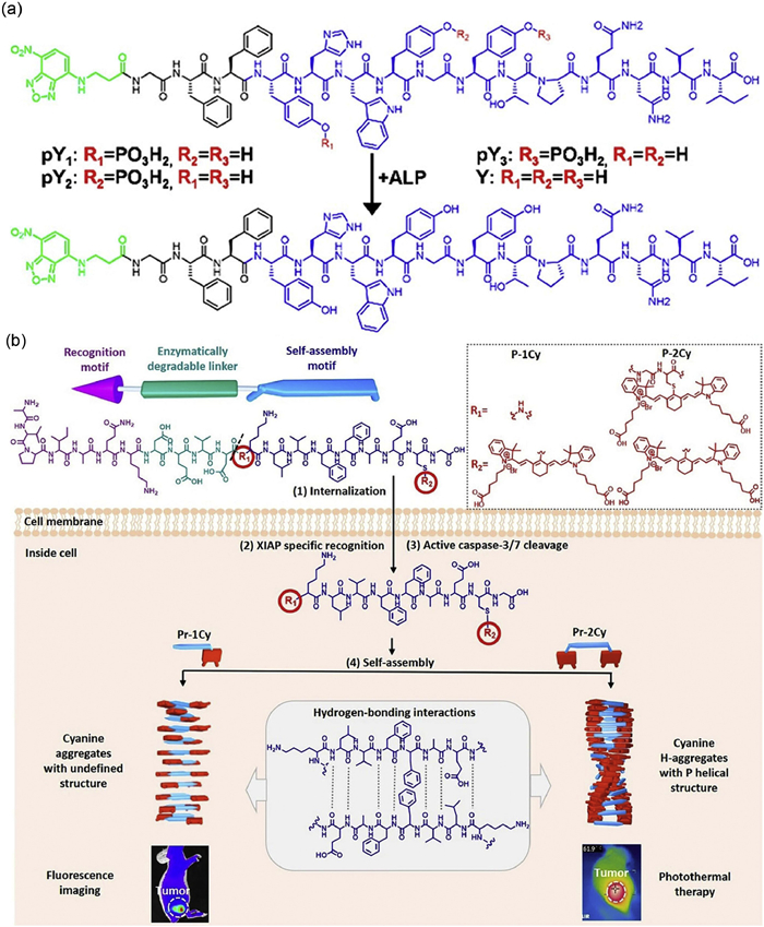
Other enzymes overexpressed in cancer cells also play an important impact on the regulation and metabolism of cellular activities. For example, Wang and coworkers exploited the gelatinase-responsive small molecule precursor (P18-PLGVRGRGD), which could self-assembled into nanofibers in cells and prolong retention time in tumors [49]. After accumulation around the tumor microenvironment, the overproduced gelatinase could cleave PLGVRG linker into small molecule precursors, which increased the hydrophobicity of the molecules in precursors and decrease steric hindrance, leading to the formation of the ordered fibrous nanotube structure [49]. Caspases as proteolytic enzymes could regulate the growth, differentiation, and apoptosis of eukaryotic cells. Wang's group synthesized peptide-cyanine conjugates (P-1Cy and P-2Cy) which could self-assemble into 1D columnar superstructures with orderly arrangement of cyanine inside living cells, showing distinct imaging or photothermal properties (Fig. 3b) [50]. After entering the cancer cells, the backbone of peptide-cyanine conjugates could be cleaved by caspase-3/7, inducing the intracellular self-assembly of Pr-1Cy and Pr-2Cy. Yu's group also designed the self-amplifying assembly of peptides precisely inside macrophages with expression of enzyme NQO1 for enhancing the anti-inflammatory effects of therapeutic reagents [51].
Cells, coined as micro laboratories, can perform various chemical reactions via enzymes [52]. Many enzymes feature metals at the active sites, and thus are viewed as metalloenzymes. Recently, there has been an impressive progress in the development of metalloenzymes [53]. For example, Mascarenas's group verified the intracellular metal carbene transfer reactions (Fig. 4A) [54]. They demonstrated that copper(Ⅱ) catalysts can cause the annulation of alpha-keto diazocarbenes with ortho-amino arylamines inside living cells, triggering through the N–H carbene insertion. Li and coworkers presented a Fe–N doped graphene (FeNGR) nanomaterial with the function of simulating NOX via catalyzing the conversion of NADPH into NADP+ and producing oxygen radicals [55]. Recently, performing abiotic metal catalysis inside cells has also been investigated. The intriguing example was reported by Bai's group, who presented the cell substrate selectivity of an enzyme-mimicking macromolecular catalyst on the basis of cationic dense-shell nanoparticle (DSNP) scaffold [56]. The generation of DSNP could use its densely packed lipo-cationic arms to protect the metal centers. Moreover, it also showed superior affinity for cell membranes to supply additional guarantee towards the binding sites and catalytic enters. In addition, some other transition-metal reagents that propargylic deprotections [57], cyclizations [58] and formal cycloadditions [59] have been studied in detail.
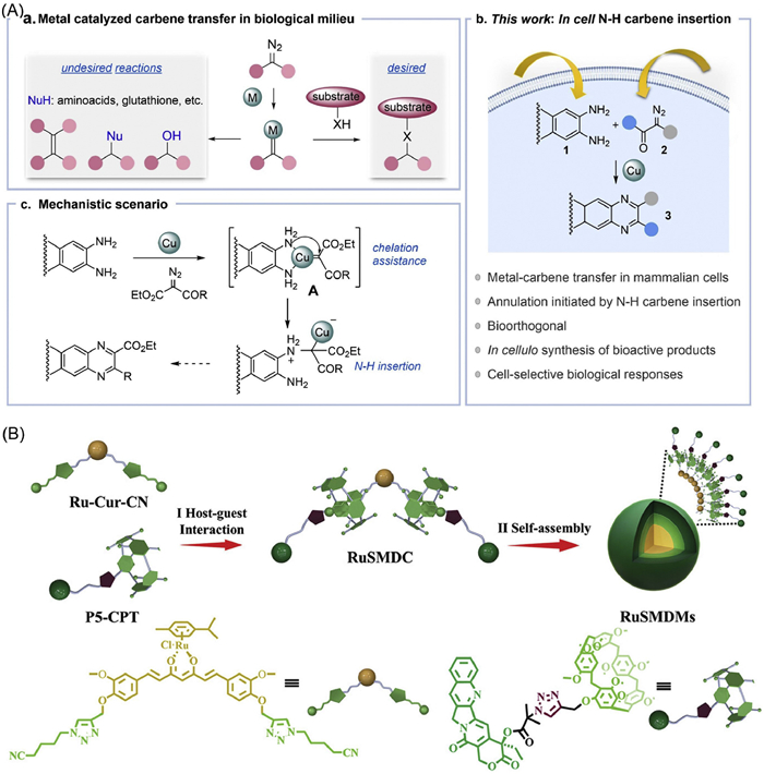
To alleviate hypoxic tumors, we designed and synthesized the novel RuSMDC with efficient O2 self-supply through host-guest interactions between P5-CPT and Ru-Cur-CN (Fig. 4B) [60]. The ruthenium of RuSMDC could catalyze the decomposition of H2O2 to generate O2 and achieve synergetic therapeutic effects of chemotherapy and PDT in hypoxic tumor environment. In recent years, we also synthesized ferrocene-cinnamaldehyde conjugates, in which the Fenton of conjugates threaded into the porous cavity of nano-sized porphyrin-based MOF [61]. Hyaluronic acid (HA) was coated on its surface to generate Fc-CA-PCN-HA. In cancer cells, cinnamaldehyde of Fc-CA-PCN-HA activated NADPH oxidase to generate more H2O2, supplying sufficient "fuel" for CDT. On the other hand, Fenton could catalyze intracellular H2O2 to produce ⋅OH for the improvement of CDT and generate more O2 production to relieve inherent tumor hypoxia for PDT.
Because of the limited catalytic activity and selectivity, traditional nano-catalysts are difficult to achieve efficient and excellent catalytic effects in a broad scope of biological applications [62]. Single-atom catalysts (SACs) represent emerging frontier of heterogeneous catalysts with high atom utilization efficiency, tunable reaction pathway and excellent performance [63]. Thus, SACs may perform well catalytic activity and selectivity in the field of natural biocatalytic systems, providing new prospects to modulate cell behavior. For example, Na and colleagues designed and synthesized C3N4-Mn SACs for O2-independent PDT (Fig. 5a) [64].
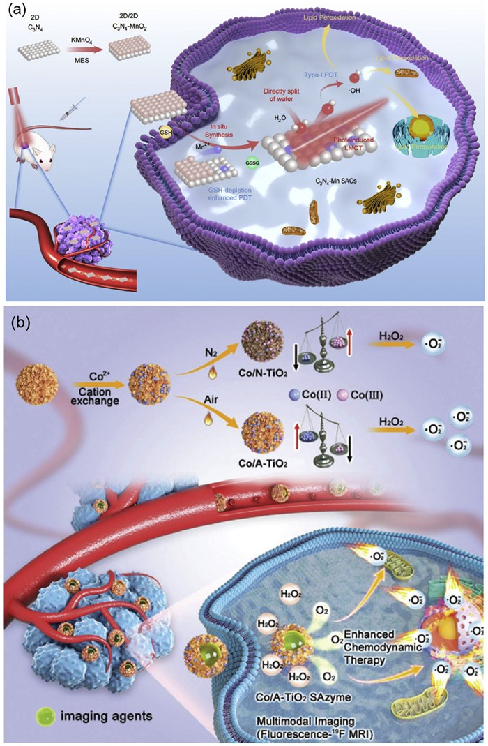
With irradiation at 660 nm, C3N4-Mn SACs caused water-splitting process to produce highly toxic ·OH for efficient cancer treatment [64]. Ruthenium based single-atom catalysts are also widely applied in the field of intracellular chemical reactions. Hu's group prepared Ru-Ala-C3N4 as biomimetic enzyme, which was formed through dispersing Ru atoms on the carbon nitride (C3N4) [65]. As for abundant and efficient active sites, the resultant Ru-Ala-C3N4 could effectively detect dopamine and uric acid with detection limits of 20 and 170 nmol/L, respectively. Xu et al. successfully synthesized carbon dots supported Fe-SAzyme, imitating the non-heme iron center of HppE [66]. The biological evaluation showed that Fe-CDs-based therapeutic agent exhibited an excellent peroxidase activity. In addition, some other noble metals, such as cobalt (Co), copper (Cu), and zinc (Zn) based single-atom catalysts have also been successfully explored for various biological functions [67]. Wang and coworkers reported Co/TiO2 SAzymes for multimodal image-guided synergistic therapy (Fig. 5b) [68]. Shi's group developed hollow N-doped carbon spheres doped with the single-atom copper species (Cu-HNCS) with high surface area for tumor parallel catalytic treatment [69].
In comparison with blood and healthy tissues (pH 7.4), intracellular pH of subcellular organelles decreases to 5.0–6.0 in the endosomes and 4.0–5.0 in the lysosomes [70,71]. Thus, pH-responsive functional materials were designed and synthesized developed through incorporating pH labile chemical bonds into the system, such as hydrazone and ester groups [70]. These bonds tend to maintain stability in neutral or alkaline conditions, but they may be hydrolyzed inevitably under acidic microenvironment. For example, El-Sayed and coworkers presented the novel drug delivery systems by conjugation of doxorubicin with pH-sensitive hydrazone linkage [72]. Anticancer drugs could be controlled release inside cells because of the degradation of hydrazone bond under the lysosomes' acidic pH. Our group constructed supramolecular drug-drug complex via supramolecular interaction between bis(pillar[5]arene)-amine-cisplatin and cyano-methylpropionyl-CPT, which could further aggregate into supramolecular vesicles [73]. The pH-responsive ester bond was modified into cyano-methylpropionyl-CPT for smart drug release upon the acidic lysosomal condition. In addition, DNA with i-motif sequences can also respond to the acidic lysosomal environment, causing transformation of nanoparticles inside living cells. For example, Yang' group synthesized the DNA-ceria nanocomplex (DCNC) to realize intracellular dynamic assembly [74]. Under the acidic microenvironments, i-motif sequences of DNA can drive DCNC to aggregate into stable microscale architecture to prolong retention time inside cells.
Some protonatable groups such as amino and carboxyl groups can be modified into synthetic materials, effectively modulating drug release through the variation of pH in the tumor microenvironment [75]. Once these groups become protonated below the acid dissociation constant, the system may be destabilized because of the charge repulsion. For example, Shen's group created γ-glutamyl transpeptidase-responsive CPT-polymer for deep intratumoral penetration and treatment [76]. The overexpressed γ-glutamyl transpeptidase on cell membrane could induce γ-glutamyl moieties of CPT-polymer into positively charged primary amines, actively infiltrating throughout the tumor to reach the inner cells. We also reported a new trimetallic supramolecular drug complex based on the cation-π interaction (Cπ-TMSDC), simultaneously combining the unique virtues of passive transport and active penetration [77]. Because of the acidic tumor microenvironment, cation-π interactions in Cπ-TMSDC could be smartly dissociated as for the acid-base reaction to increase tumor penetration and treatment effect.
In 2001, Sharpless and coworkers defined the concept of "click chemistry", which are highly specific and selective for rapid synthesis of functional molecules [78]. Click chemistry refers to the set of highly efficient, reliable, and stereoselective reactions that can be used to develop promising functional materials through using facile reaction conditions [79]. Because of the rapid and efficient formation of covalent bonds between two molecules, click reaction is regarded as an important and promising strategy for intracellular chemistry reaction, which can be widely performed in living cells, organisms, and animals [80]. In this section, we explored the strategies for Azide-alkyne cycloaddition, Diels-alder cycloaddition, and Thiol-Michael click reactions based on the different mechanisms.
Azide-alkyne cycloaddition is one of the most classic and common click reactions in organic synthetic chemistry, which can be triggered by Cu(Ⅰ) catalysts or Ru(Ⅱ) catalysts [81]. Over the past few decades, Azide-alkyne cycloaddition has aroused great interest among chemists and biologists, who have been dedicated to elucidating the underlying mechanisms and synthesis methods with the aim of efficiency and simplicity [82]. For example, Chen's group presented Cu(Ⅰ)-stabilizing ligands catalysts with biosafety and high efficiency to perform intracellular modification of proteins through azide-alkyne cycloaddition (Fig. 6a) [83].
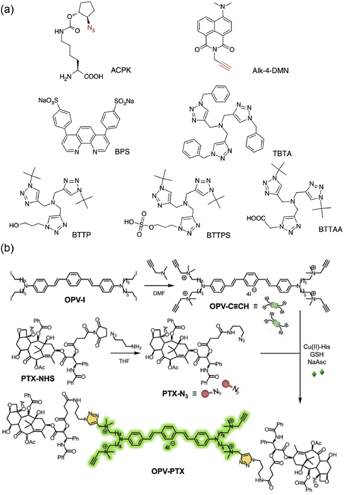
The formed Cu(Ⅰ)-stabilizing ligands catalysts could realize in situ labelling of azide-incorporated proteins in the cytoplasm of Escherichia coli. Wang et al. achieved intracellular click reaction between OPV-C≡CH with four terminal alkynyl groups and PTX-N3 consisting of one paclitaxel molecule (Fig. 6b) [84]. After cellular uptake of OPV-C≡CH, PTX-N3 and Cu(Ⅱ)-His, the Cu(Ⅱ)-His could be changed into an active Cu(Ⅰ) catalyst to achieve in situ Azide-alkyne click reaction on the surface of E. coli. Biological evaluation confirmed that bacteria-mediated intracellular Azide-alkyne click reaction realized enrichment of the PTX for inducing the apoptosis of drug-resistant cancer cells. The supramolecular organometallic drug complex (SOMDC) with H2O2 self-provision constructed by GEM-(Fc)3 and β-CD-DOX has been synthesized [45]. The GEM-(Fc)3 as guest molecule was prepared through Azide-alkyne click reaction between Fc-≡ and GEM-N3. The β-CD-DOX monomer was prepared through a Schiff base reaction between DOX and β-CD-hydrazone. In tumor cells, the obtained SOMDC dissociated efficiently because endogenous H2O2 rapidly disrupted the host-guest interactions. The released DOX prodrug significantly increased the endogenous H2O2 level and improved the Fenton-like intracellular autocatalysis, generating abundant •OH and thereby enhancing chemodynamic therapy efficacy.
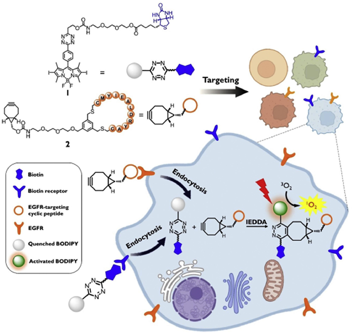
Diels-alder click reaction is the powerful and widely used methods in synthetic chemistry for stereospecific construction of carbon-carbon bonds [85–87]. Taking advantages of the unmatchable kinetics, excellent orthogonality and biocompatibility, Diels-alder click reaction has emerged as a promising strategy in the field of chemistry and biology [88]. Rao and coworkers presented a novel pre-targeting method through utilizing two bioorthogonal reactions-condensation reaction of aromatic nitriles and aminothiols, and the Diels-Alder reaction between tetrazine and trans-cyclooctene [89]. Ye et al. developed an innovative strategy of pre-targeted multimodality imaging through combing enzyme-mediated fluorogenic reaction and Diels-alder cyclization [90]. In this study, FMNPs-TCO with active groups of TCO could rapidly react with radiolabeled tetrazines (Tz-68Ga) through Diels-alder click reaction, generating FMNPs-DHP-68Ga nanoparticles with excellent functions of radiation, fluorescence imaging, and magnetism [90]. Ng's group reported a novel dual receptor-mediated bio-orthogonal activation approach based on the Diels-alder click reaction between biotinylated tetrazine-substituted BODIPY and EGFR-targeting peptide linked with a BCN dienophile for precise antitumoral PDT (Fig. 7) [91]. To control such ultrafast bioorthogonal reactions, Liu and coworkers presented a novel molecular-recognition strategy to control the reactivity of tetrazine through host-guest interaction between macrocyclic naphthotubes and phenyltetrazine [92]. Because of the low-micromolar and sub-micromolar binding affinities, the reactivity of phenyltetrazine group can be rapidly recovered in the presence of other competitive guests.
Due to the modular "click" nature, Thiol-Michael click reactions are considered as a prized tool in materials science, which allows for performing small molecule synthesis and polymer modifications with highly efficient and green features [93]. Thiol-Michael click reactions, including Thiol-ene and thiol‑yne reactions have been widely used for synthesizing various functional materials in biological systems [94]. For example, Kloxin's group presented functionalized cyclic peptides through thiol-ene and Azide-alkyne click reactions for biomaterial applications [95]. Visible-light has emerged as powerful synthetic tools for Thiol-Michael click reactions under mild reaction conditions. Ning's group reported novel LG-M(N-DOX)-PEG NPs with pH-responsiveness for anticancer drug delivery [96]. During the preparation of LG-M(N-DOX)-PEG NPs, thiol-ene click reaction was performed between LG-M and PEG-thiol to generate reactive intermediates (LG-M-PEG) under UV illumination. To overcome the limitation of successful protein bioconjugation, Hong and coworkers utilized fluorescent photosensitizer QPEG to activate the S–H bond of cysteine, performing Thiol-ene click reaction [97].
Although small molecules can be quickly absorbed by cells and effectively distributed within tissue, they can be easily recognized and removed outside the cells, which limited accumulation around the tumor cells and reduced the intracellular molecules levels, leading to the lower effect [1,4]. Thus, increasing monomer doses or prolonging circulation time may inevitably cause unwanted side effects. Changing small molecules into macromolecules or polymeric materials inside living cells can integrate the merits of both effective distribution and clearance of monomers and the retention of macromolecules. Developing intracellular polymerization may provide a unique opportunity to design new functional materials for regulation of cell activities.
The intriguing intracellular chemical reactions not only expands the realm of artificial synthetic chemistry but also brings new opportunities and possibilities for the efficacy of biomedical applications. Intracellular chemical reaction, as a sparkling frontier has been extensively applied in a wide range of biological applications, such as regulating cell activities, interference of organelles, circumventing drug resistance, bioimaging, drug synthesis and immunomodulation. The recent and important developments of intracellular chemical reactions and their features utilized as biomedical materials are briefly summarized (Table 1).
 DownLoad:
CSV
DownLoad:
CSV
| Empty Cell | Formulations | Chemical bonds | Type of stimuli | Application | Ref. |
| Endogenous stimuli | ssPPELap@Fe-TA | Disulfide bond | GSH | Antitumor immune | 24] |
| PEG-PUSeSe-PEG | Diselenide bond | ROS/GSH | Actinotherapy | 29] | |
| DDSC | Host-guest interaction | ROS | Drug release | 44] | |
| SOMDC | Host-guest interaction | H2O2 | Cancer chemotherapy | 45] | |
| Exogenous stimuli | OPV-PTX | Cu(Ⅱ)-His | Sodium ascorbate | Anticancer | 84] |
| Cyclic RGD peptide | Thiol-ene/azide-alkyne bond | Visible light | Cell culture | 95] | |
| DEVD-DLPA@C3 | Cyanine-derivative photosensitizer | Laser irradiation | Antitumor therapy | 102] | |
| PSeR/DOX | Diselenide bond | Radiation | Immunomodulatory | 121] |
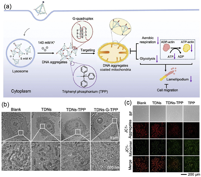
Organelles could grow, divide and fuse within cells [98]. These dynamic processes of organelles in cytoplasm could control the distribution and function of these cells, which is closely associated with the outer membrane of the organelles [99]. Based on these properties, Yang's group presented triphenylphosphine (TPP)-modified DNA tetrahedron (TDNs-G-TPP), containing TPP for mitochondrial targeting (Fig. 8a) [100]. After cell internalization, TDNs-G-TPP self-assembles could specially target mitochondria and served as a polyanionic barrier for substance and energy communication between mitochondria and the cytoplasm (Fig. 8b). Mitochondrial destruction could lead to the superior inhibitory effect for aerobic respiration function and glycolysis process, decreasing the production of adenosine triphosphate in cells (Fig. 8c). In vitro and in vivo investigation verified that TDNs-G-TPP treatment group exhibited a significant inhibitory effect on cell migration due to the lack of ATP. In continuation of their work, they also reported the proton-driven dynamic assembly of DNA nano-framework inside lysosome, performing lysosome interference. After internalization into lysosome, DNA nano-framework could aggregate in an acidic environment, increasing the retention time of nano-framework in lysosomes [101]. Recently, Zhu's group reported a tumor targeting and caspase-3 responsive nanoparticles, containing DEVD-DLPA and cyanine-derivative photosensitizer with function of mitochondria-targeting [102]. Upon laser irradiation, the produced ROS could lead to mitochondrial dysfunction and induce cell apoptotic process.
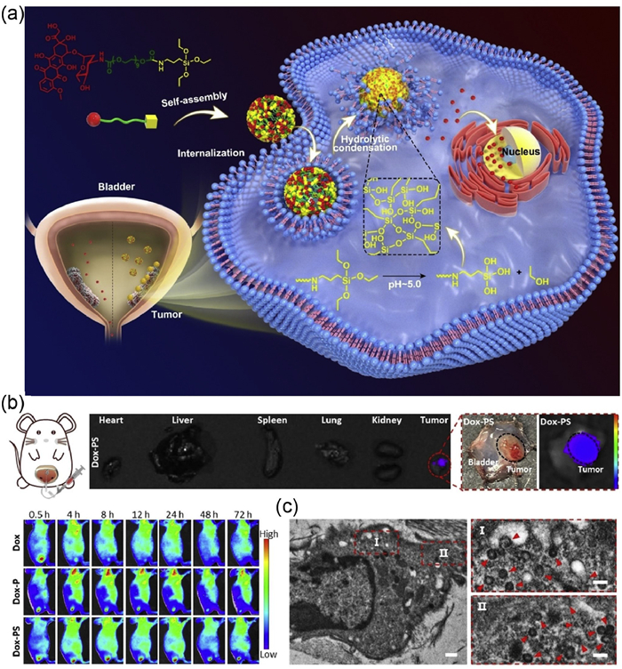
The in situ "small-to-large" intracellular polymerization can combine the rapid uptake of monomeric molecules with the strong retention capability of polymers, overcoming the problem of drug resistance [11]. For example, Xu's group successfully realized intracellular hyperbranched polymerization based on the organic telluride monomer B3-Te, consisting of three Te atoms as polymerization active sites [103]. After entering tumor cells, B3-Te could realize intracellular hyperbranched polymers under ROS oxidation. The generated Te-O based hyperbranched polymer could aggregate into branched nanostructures in cells, evading drug pumps well and ensuring persistent anticancer activity. Biological experiments exhibited that the designs of intracellular hyperbranched polymerization could realize selective anticancer efficacy and well biosafety. Xu and coworkers presented hydrolytic condensation in living cells to form long-term retentive nano-drug depots, which could ensure the sustained and effective drug release for inhibiting bladder cancer (Fig. 9a) [104]. In vivo imaging study showed that Dox-PS group exhibited the long duration from 4 h to 72 h (Fig. 9b). The formation of silicon particles was obviously found in the cytoplasm of cancer cells via Bio-TEM images (Fig. 9c). To overcome resistance issues, Hedrick and coworkers have synthesized novel macromolecular chemotherapeutic agents, combining the cationic block with positive charge to bind with negatively charged lipid membranes [105]. The amphiphilic macromolecular chemotherapeutic agents could self-assemble into micellar structures owing to hydrophobic interaction. In vitro study confirmed that cationic polymers demonstrated significant potency against drug-resistant cancer cells and could effectively prevent cancer cell migration.
Thanks to the intrinsic advantages of fluorescence imaging such as high sensitivity, noninvasiveness, direct and simple operation, bioimaging can be a powerful and versatile tool for detection and tracing of therapeutic effects [106–110]. Intracellular reactions can amplify imaging efficacy inside living biosystems through increasing imaging signals. For example, Tang's group reported the intracellular crystallization-assisted self-assembly to produce large-size nanoaggregates and realize stronger fluorescence output [111]. In this research, they designed and synthesized Au(Ⅰ)-disulfide nanosheets (NSs@TTVP), consisting of an aggregation-induced emission photosensitizer. The generated NSs@TTVP could achieve pH-responsive crystallization-driven self-assembly in acidic conditions, endowing NSs@TTVP with high fluorescence emission efficiency and efficient ROS production output. Biological experiments verified that microscale NPs obtained in cells could prolong retention inside tumors and achieve persistent treatment for cancer ablation, which may present new insights on fluorescence imaging guided tumor tracking and cancer theranostics.
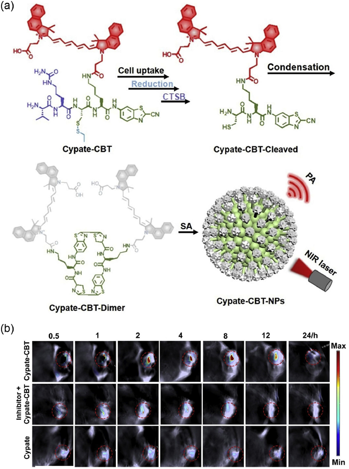
Photoacoustic imaging (PAI), as for its unique features, such as deep tissue-penetrating depth, noninvasive and high spatial resolution, has emerged as a promising imaging modality to probe specific biomarkers, which was widely used to diagnose some small-sized diseases at early stage [112,113]. Different from small molecules, large-scale nanoaggregates can greatly accumulate inside cells and extend their retention time, significantly enhancing imaging signals of nanoaggregates-based probes [114]. Based on the above consideration, Liang and coworkers designed CTSB-activatable PA probe (Cypate-CBT) (Fig. 10a) [115]. After Cypate-CBT were internalized into tumor cells, redox-responsive disulfide bonds were reduced by rich GSH and Val-Cit was cleaved via CTSB. Then, Cypate-CBT-Dimer could be formed through intermolecular CBT-Cys click reaction, which was further aggregated into nanoparticles driven by π-π stacking. Both cell and animal experiments displayed that the concentration and retention time of probe Cypate-CBT was increased effectively, enhancing the PA signal during the process of tumor imaging (Fig. 10b) [115]. Continuous research in this field, Liang's group presented near-infrared fluorescence probe (TK-CBT-NPs) for imaging staphylococcus aureus infection [116]. In vitro study verified that TK-CBT-NPs demonstrated about 6.1-fold fluorescence intensity for the macrophages infected by S. aureus. Meanwhile, 2.8-fold fluorescence imaging signal increase was acquired for mice experiment infected by S. aureus.
The development of methods for active drug synthesis in living systems is highly valuable for exploring and manipulating biological processes [117–119]. This innovative approach represents a fundamental extension of the biorthogonal chemistry concept and holds transformative potential for pioneering advances in therapeutic applications. Due to the rapid and efficient formation of covalent bonds between drug fragments, click reactions are considered an important approach for intracellular drug synthesis. For example, Taran et al. reported a pioneering approach that combines bioorthogonal and confined chemistry for intracellular drug synthesis [120]. They engineered nanoparticles specifically for synthesizing drugs inside cells, utilizing bioorthogonal reactions with cyclooctynes to produce the FDA-approved drug Sorafenib within cancer cells. In vitro experiments have shown that they synthesized and monitored Sorafenib in aqueous solution using HPLC. This innovative strategy not only offers significant prospects for advancing therapeutic approaches but also opens new avenues for exploring non-natural reactions in living systems.
Intracellular chemical reactions can regulate cytokine secretion and activate immune cells to control cell fate [121–123]. For example, Xu's group presented combination therapy to realize combination therapy of immunotherapy, chemotherapy, and radiotherapy to improve anticancer activity [121]. The obtained PSeR/DOX self-assemblies could transport the active reagent DOX into tumor sites. Radiation stimuli could facilitate release of active reagent DOX from self-assemblies because of the cleavage of the diselenide bonds. In addition, radiation could oxidize diselenide bonds into seleninic acid, realizing immunomodulatory activity via enhancing the NK cell function. Recently, they also proposed selenium-containing nanoparticles, which co-assembled by cetuximab, C5SeSeC5, and inhibitor LY345899 [42]. Upon entering the cancer cell, selenium-containing nanoparticles of LY345899 can suppress activity of methylene tetrahydrofolate dehydrogenase 2 and generate excessive ROS in cells, resulting in the generation of seleninic acid to reverse NK cell exhaustion. The chemo-immunotherapy holds great promise for treating advanced cancers [124]. However, therapeutic effects are often unsatisfactory because anticancer drugs have poor tumor accumulation and cause severe off-target side effects. Recently, Xie's group developed PSMT nanoparticles that co-deliver a paclitaxel prodrug and the IDO1 inhibitor 1-methyl-tryptophan (1MT) for tumor-specific combination of chemotherapy and immunotherapy [125]. After internalization of PSMT NPs into cancer cells, PSMT NPs could respond to endogenous redox conditions and release PTX and 1MT. The released PTX not only induced apoptosis by disrupting mitosis but also triggered immunogenic cell death. In addition, the 1MT can inhibit the IDO activity to achieve the purpose of regulating T cells. Biological assessment displayed that PSMT NPs displayed potentiated antitumor activity toward triple-negative breast cancer and good safety with respect to normal organs. The study showed that the unique advantage of PSMT NPs may have significant potential for application in the field of synergistic chemo-immunotherapy.
In summary, intracellular chemical reactions represent a transformative advancement in the evolving fields of chemistry. The precise design of chemical reactions within living biological systems enables efficient disease treatment. Particularly, innovative chemical transformations promise applications such as the intracellular synthesis of functionally active drugs. This review systematically summarizes the chemical reactions in living cells to provide guidance for the future design of intracellular chemical processes. Additionally, we highlight the current progress in disease treatment, covering areas such as regulation of cell activities, interference with organelles, overcoming drug resistance, and intracellular drug synthesis. We anticipate that intracellular chemical strategies will be effectively applied to regulate cellular behavior and achieve therapeutic efficacy in the near future, marking a significant milestone in the development of new therapeutic approaches.
Although various methodologies and monomers have been used to realize intracellular chemical reactions, it is still necessary to develop new controlled approaches to expand the kind of strategy. Particularly, intracellular drug synthesis is attracting increasing research interest owing to its enhanced biosafety and superior drug utilization efficiency. Importantly, developing new chemical strategies may play fundamental roles in designing the novel structures and conferring unique functionality to biological systems. Although chemical reactions in living cells demonstrate tremendous potential for biomedical applications, this field remains in its nascent stages, with several key challenges currently limiting the successful clinical translation of intracellular chemical reactions.
The field of intracellular chemical reactions has witnessed remarkable expansion, marking a significant shift in disease treatment strategies while establishing itself as a distinct discipline within biomedical science. However, for the development of intracellular chemical reactions in therapeutic applications, a meticulous design approach is required to navigate the complex microenvironment of diseases. While in vitro chemical reactions are well-characterized by defined mechanisms and processes, the translation of these reactions to in vivo settings introduces significant complexities due to the physiological environment. These diverse and dynamic biological environments act as a black box, obscuring the long-term toxicity of the accumulated reactants within the cells. This involves tailoring reactions to function under complex microenvironment to both enhance efficacy and minimize potential side effects. In addition, the toxic effect of these reactants has not been sufficiently studied, slowing the progress toward clinical trials. Based on the above considerations, there is still a long way to go to achieve the ideal biological applications of chemical reactions in living cells, requiring substantial investment from researchers to develop more advanced chemical and biological methods. Despite these challenges, we still believe that the advances of knowledge from different disciplines will gradually solve these issues, which may become a milestone in efficient disease treatment and would be challenging frontiers in biomedical science in the future.
The authors declare no conflict of interest.
Yunfei Fu: Writing – original draft, Software, Data curation. Hui Li: Writing – original draft, Software, Data curation. Chengfei Liu: Writing – review & editing, Writing – original draft, Software, Funding acquisition. Wei Tian: Writing – review & editing, Writing – original draft, Visualization, Validation, Funding acquisition.
This work was financially supported by the National Natural Science Foundation of China (Nos. 22405212 and 22471219), the Shaanxi Fundamental Science Research Project for Chemistry & Biology (No. 23JHZ002) and Innovation Capability Support Program of Shaanxi (No. 2025RS-CXTD-022), the Fundamental Research Funds for the Central Universities (No. G2025KY06148).
Z. Zhou, K. Maxeiner, D.Y.W. Ng, et al., Acc. Chem. Res. 55 (2022) 2998–3009. doi: 10.1021/acs.accounts.2c00420
Y. Dai, Z. Zhang, M. Daniels, et al., Chem 9 (2023) 2006. doi: 10.1016/j.chempr.2023.05.042
C. Liu, Y. Jin, J. Li, et al., Chin. J. Chem. 43 (2025), 2053–2068. doi: 10.1002/cjoc.70041
Y. Zhang, H. Yan, R. Yu, et al., Adv. Sci. 11 (2024) 2306350. doi: 10.1002/advs.202306350
Q. Shen, Y. Huang, H. Bai, et al., Acc. Mater. Res. 4 (2023) 57–70. doi: 10.1021/accountsmr.2c00194
Y. Dai, T. Li, Z. Zhang, et al., J. Am. Chem. Soc. 143 (2021) 10709. doi: 10.1021/jacs.1c04821
C. Liu, B. Xianyu, C. He, et al., CCS Chem. 7 (2025) 403–415. doi: 10.31635/ccschem.024.202404842
M. Pieszka, S. Han, C. Volkmann, et al., J. Am. Chem. Soc. 142 (2020) 15780–15789. doi: 10.1021/jacs.0c05261
M. Jia, Y. Liu, P. Wei, et al., Aggregate 5 (2024) e533. doi: 10.1002/agt2.533
F. Li, Y. Liu, Y. Dong, et al., J. Am. Chem. Soc. 144 (2022) 4667. doi: 10.1021/jacs.2c00823
M. Tang, Z. Yang, X. Tang, et al., J. Am. Chem. Soc. 147 (2025) 3488–3499. doi: 10.1021/jacs.4c14847
X. Liu, M. Li, J. Liu, et al., J. Am. Chem. Soc. 144 (2022) 9312–9323. doi: 10.1021/jacs.2c01025
J. Liu, B. Liu, Prog. Polym. Sci. 129 (2022) 101545. doi: 10.1016/j.progpolymsci.2022.101545
S. Pan, J. Yang, S. Ji, et al., CCS Chem. 2 (2020) 225–235. doi: 10.31635/ccschem.020.202000124
C. Liu, C. Liu, Y. Bai, et al., Adv. Healthcare Mater. 12 (2023) 2202769. doi: 10.1002/adhm.202202769
D. Schauenburg, T. Weil, Adv. Sci. 11 (2023) 2303396.
H. Wang, W. He, J. Liao, et al., Adv. Mater. 37 (2025) 2411967. doi: 10.1002/adma.202411967
X. Liang, Y. Zhang, J. Zhou, et al., Coordin. Chem. Rev. 473 (2022) 214824. doi: 10.1016/j.ccr.2022.214824
L. He, D. Lu, H. Liang, et al., J. Am. Chem. Soc. 140 (2018) 258. doi: 10.1021/jacs.7b09789
C. Liu, H. Ma, S. Yuan, et al., ACS Nano 19 (2025) 2047–2069. doi: 10.1021/acsnano.4c16669
Y. Chen, P. He, D. Jana, et al., Adv. Mater. 34 (2022) 2201706. doi: 10.1002/adma.202201706
X. Zhang, J. Wang, Y. Zhang, et al., Chem. Soc. Rev. 52 (2023) 8126. doi: 10.1039/d2cs00999d
W. Zhang, W. Lin, Q. Pei, et al., Chem. Mater. 28 (2016) 4440. doi: 10.1021/acs.chemmater.6b01641
X. Dai, Y. Zhu, M. Su, et al., Adv. Funct. Mater. 33 (2023) 2215022. doi: 10.1002/adfm.202215022
C. Liu, H. Li, P. Li, et al., Polym. Chem. 11 (2020) 5810. doi: 10.1039/d0py00862a
Y. Zhang, C. He, H. Xu, ACS Appl. Mater. Interfaces 17 (2025) 13324–13341. doi: 10.1021/acsami.4c22844
Y. Tan, H. Xu, Acc. Mater. Res. 5 (2024) 739. doi: 10.1021/accountsmr.4c00065
C. Liu, Y. Tan, H. Xu, Sci. China Mater. 65 (2022) 2017. doi: 10.1007/s40843-021-2018-y
N. Ma, Y. Li, H. Xu, et al., J. AM. Chem. Soc. 132 (2010) 442–443. doi: 10.1021/ja908124g
J. Karges, T. Yempala, M. Tharaud, et al., Angew. Chem. Int. Ed. 59 (2020) 7069. doi: 10.1002/anie.201916400
Y. Bai, C. Liu, J. Yang, et al., Colloids Surf. 217 (2022) 112606. doi: 10.1016/j.colsurfb.2022.112606
Y. Bai, X. Li, M. Li, et al., J. Mater. Chem. B 10 (2022) 4952. doi: 10.1039/d2tb00989g
C. Liu, Y. Jin, J. Ma, et al., Chin. J. Polym. Sci. 43 (2025) 1293–1310. doi: 10.1007/s10118-025-3341-7
L. Zhang, Y. Dai, S. Pan, et al., ACS Appl. Bio Mater. 5 (2022) 1794. doi: 10.1021/acsabm.2c00150
C. Sun, Y. Tan, H. Xu, ACS Mater. Lett. 2 (2020) 1173. doi: 10.1021/acsmaterialslett.0c00272
C. de Gracia Lux, S. Joshi-Barr, T. Nguyen, et al., J. AM. Chem. Soc. 134 (2012) 15758. doi: 10.1021/ja303372u
M.S. Shim, Y. Xia, Angew. Chem. Int. Ed. 52 (2013) 6926–6929. doi: 10.1002/anie.201209633
J. Liu, Y. Pang, Z. Zhu, et al., Biomacromolecules 14 (2013) 1627. doi: 10.1021/bm4002574
B. Xianyu, S. Pan, S. Gao, et al., Small 20 (2024) 2306225. doi: 10.1002/smll.202306225
L. Zhang, C. Sun, Y. Tan, et al., CCS Chem. 4 (2022) 2239–2248. doi: 10.31635/ccschem.021.202101203
T. Li, S. Pan, S. Gao, et al., Angew. Chem. Int. Ed. 59 (2020) 2700–2704. doi: 10.1002/anie.201914453
S. Pan, J. Guan, B. Xianyu, et al., Adv. Mater. 35 (2023) 2211370. doi: 10.1002/adma.202211370
B. Yang, Y. Chen, J. Shi, Chem. Rev. 119 (2019) 4881. doi: 10.1021/acs.chemrev.8b00626
M. Li, Y. Bai, C. Liu, et al., ACS Biomater. Sci. Eng. 7 (2021) 5515. doi: 10.1021/acsbiomaterials.1c01144
C. Liu, M. Li, C. Liu, et al., J. Mater. Chem. B. 10 (2022) 8981. doi: 10.1039/d2tb01834a
D. Wu, J. Lei, Z. Zhang, et al., Chem. Soc. Rev. 52 (2023) 2911. doi: 10.1039/d2cs00759b
S. Liu, Q. Zhang, H. He, et al., Angew. Chem. Int. Ed. 61 (2022) e202210568. doi: 10.1002/anie.202210568
Y. Ding, D. Zheng, L. Xie, et al., J. Am. Chemi. Soc. 145 (2023) 4366. doi: 10.1021/jacs.2c11823
D. Zhang, G.B. Qi, Y.X. Zhao, et al., Adv. Mater. 27 (2015) 6125–6130. doi: 10.1002/adma.201502598
R. Zheng, J. Yang, M. Mamuti, et al., Angew. Chem. Int. Ed. 60 (2021) 7809. doi: 10.1002/anie.202015126
Y. Song, M. Li, N. Song, et al., J. Am. Chem. Soc. 144 (2022) 6907. doi: 10.1021/jacs.2c01323
Y. Wang, X. Jia, S. An, et al., Adv. Mater. 36 (2024) 2301810. doi: 10.1002/adma.202301810
T. Vornholt, F. Christoffel, M.M. Pellizzoni, et al., Sci. Adv. 7 (2021) eabe4208. doi: 10.1126/sciadv.abe4208
S. Gutiérrez, M. Tomás-Gamasa, J.L. Mascareñas, Angew. Chem. Int. Ed. 60 (2021) 22017–22025. doi: 10.1002/anie.202108899
D. Wu, J. Li, S. Xu, et al., J. Am. Chem. Soc. 142 (2020) 19602. doi: 10.1021/jacs.0c08360
Y. Deng, T. Wu, X. Chen, et al., J. Am. Chem. Soc. 145 (2023) 1262. doi: 10.1021/jacs.2c11168
M. Martínez-Calvo, J.R. Couceiro, P. Destito, et al., ACS Catal. 8 (2018) 6055. doi: 10.1021/acscatal.8b01606
C. Vidal, M. Tomás-Gamasa, P. Destito, et al., Nat. Commun. 9 (2018) 1913. doi: 10.1038/s41467-018-04314-5
J. Miguel-Ávila, M. Tomás-Gamasa, J.L. Mascareñas, Angew. Chem. Int. Ed. 59 (2020) 17628. doi: 10.1002/anie.202006689
C. Liu, M. Li, P. Li, et al., Adv. Funct. Mater. 31 (2021) 2105837. doi: 10.1002/adfm.202105837
Y. Bai, R. Wang, X. Wang, et al., Int. J. Biol. Macromol. 245 (2023) 125523. doi: 10.1016/j.ijbiomac.2023.125523
B. Chang, L. Zhang, S. Wu, et al., Chem. Soc. Rev. 51 (2022) 3688. doi: 10.1039/d1cs00421b
Y. Pan, X. Wang, W. Zhang, et al., Nat. Commun. 13 (2022) 3063. doi: 10.1038/s41467-022-30766-x
Y. Yin, X. Ge, J. Ouyang, et al., Nat. Commun. 15 (2024) 2954. doi: 10.1038/s41467-024-46987-1
X. Xie, D.P. Wang, C. Guo, et al., Anal. Chem. 93 (2021) 4916. doi: 10.1021/acs.analchem.0c05191
Q. Yang, J. Liu, W. Cai, et al., Nano Lett. 23 (2023) 8585. doi: 10.1021/acs.nanolett.3c02406
J. Ye, W. Lv, C. Li, et al., Adv. Funct. Mater. 32 (2022) 2206157. doi: 10.1002/adfm.202206157
H. Wang, Y. Wang, L. Lu, et al., Adv. Funct. Mater. 32 (2022) 2200331. doi: 10.1002/adfm.202200331
X. Lu, S. Gao, H. Lin, et al., Adv. Mater. 32 (2020) 2002246. doi: 10.1002/adma.202002246
Z. Ge, S. Liu, Chem. Soc. Rev. 42 (2013) 7289. doi: 10.1039/c3cs60048c
Y. Dong, C. Yao, Y. Zhu, et al., Chem. Rev. 120 (2020) 9420. doi: 10.1021/acs.chemrev.0c00294
B. Kang, M.M. Afifi, L.A. Austin, et al., ACS Nano 7 (2013) 7420. doi: 10.1021/nn403351z
C. Liu, C. Li, C. Pang, et al., ACS Appl. Mater. Interfaces 12 (2020) 27940. doi: 10.1021/acsami.0c04565
C. Yao, Y. Xu, J. Tang, et al., Nat. Commun. 13 (2022) 7739. doi: 10.1038/s41467-022-35472-2
Q. Zhou, C.Y. Dong, W.F. Fan, et al., Biomaterials 240 (2020) 119902. doi: 10.1016/j.biomaterials.2020.119902
Q. Zhou, S. Shao, J. Wang, et al., Nat. Nanotechnol. 14 (2019) 799. doi: 10.1038/s41565-019-0485-z
C. Liu, M. Li, J. Sun, et al., Adv. Funct. Mater. 32 (2022) 2205043. doi: 10.1002/adfm.202205043
H.C. Kolb, M.G. Finn, K.B. Sharpless, Angew. Chem. Int. Ed. 40 (2001) 2004. doi: 10.1002/1521-3773(20010601)40:11<2004::AID-ANIE2004>3.0.CO;2-5
C. Li, J. Liu, Y. Hong, et al., Angew. Chem. Int. Ed. 61 (2022) e2022020.
B. Song, D. Lu, A. Qin, et al., J. Am. Chem. Soc. 144 (2022) 1672. doi: 10.1021/jacs.1c10612
X. Fu, A. Qin, B. Tang, Aggregate 4 (2023) e350. doi: 10.1002/agt2.350
Q. Jiang, W. Zhan, X. Liu, et al., Nat. Commun. 14 (2023) 3935. doi: 10.1038/s41467-023-39658-0
M. Yang, A. Jalloh, W. Wei, et al., Nat. Commun. 5 (2014) 4981. doi: 10.1038/ncomms5981
Z. Gao, E. Zhang, H. Zhao, et al., ACS Appl. Mater. Interfaces 14 (2022) 12106. doi: 10.1021/acsami.2c01493
C. Liu, M. Li, P. Li, et al., Biomacromolecules 22 (2021) 2382–2392. doi: 10.1021/acs.biomac.1c00173
C. Liu, Z. Yang, X. Song, et al., Supramol. Mater. 1 (2022) 100014.
L. Gao, C. Su, X. Du, et al., Nat. Chem. 12 (2020) 620. doi: 10.1038/s41557-020-0467-7
B.L. Oliveira, Z. Guo, G.J. L. Chem. Soc. Rev. 46 (2017) 4895. doi: 10.1039/C7CS00184C
Z. Chen, M. Chen, K. Zhou, et al., Angew. Chem. Int. Ed. 59 (2020) 7864. doi: 10.1002/anie.201916352
Y. Hu, J. Zhang, Y. Miao, et al., Angew. Chem. Int. Ed. 60 (2021) 18082. doi: 10.1002/anie.202103307
J.C.H. Chu, C.T.T. Wong, D.K.P. Ng, Angew. Chem. Int. Ed. 62 (2023) e202214473. doi: 10.1002/anie.202214473
W. Cao, H. Wang, M. Quan, et al., Chem 9 (2023) 2881. doi: 10.1016/j.chempr.2023.05.034
M. Laird, K. Matsumoto, Y. Higashi, et al., Nanoscale Adv. 5 (2023) 2537. doi: 10.1039/d2na00839d
Y. Chen, L. Yu, B. Zhang, et al., Biomacromolecules 20 (2019) 2230. doi: 10.1021/acs.biomac.9b00179
P.J. L eValley, E.M. Ovadia, C.A. Bresette, et al., Chem. Commun. 54 (2018) 6923. doi: 10.1039/c8cc03218a
Z. Yang, Z. Zhou, Y. Li, et al., ACS Sustain. Chem. Eng. 10 (2022) 10590. doi: 10.1021/acssuschemeng.2c02209
H. Choi, M. Kim, J. Jang, et al., Angew. Chem. Int. Ed. 59 (2020) 22514. doi: 10.1002/anie.202010217
M. Giacomello, A. Pyakurel, C. Glytsou, et al., Nat. Rev. Mol. Cell Biol. 21 (2020) 204. doi: 10.1038/s41580-020-0210-7
H. Zhu, B. Zhang, N. Zhu, et al., Chin. Chem. Lett. 32 (2021) 1220. doi: 10.1016/j.cclet.2020.09.003
F. Li, Y. Liu, Y. Dong, et al., J. Am. Chem. Soc. 144 (2022) 4667. doi: 10.1021/jacs.2c00823
Y. Dong, F. Li, Z. Lv, et al., Angew. Chem. Int. Ed. 61 (2022) e202207770. doi: 10.1002/anie.202207770
T. Ma, R. Chen, N. Lv, et al., Small 18 (2022) 2204759. doi: 10.1002/smll.202204759
C. Liu, B. Xianyu, Y. Dai, et al., ACS Nano 17 (2023) 11905. doi: 10.1021/acsnano.3c03512
D. Hou, N. Zhang, M. Wang, et al., Angew. Chem. Int. Ed. 61 (2022) e202116893. doi: 10.1002/anie.202116893
N. Park, W. Cheng, F. Lai, et al., J. Am. Chem. Soc. 140 (2018) 4244. doi: 10.1021/jacs.7b11468
D. Wang, B. Tang, Acc. Chem. Res. 52 (2019) 2559. doi: 10.1021/acs.accounts.9b00305
Y. Ren, Z. Zhou, I. Harley, et al., Nat. Synth. 4 (2025) 673–683. doi: 10.1038/s44160-025-00751-5
H. Wang, Y. Zheng, J. Zhang, et al., Angew. Chem. Int. Ed. 64 (2025) e202500998. doi: 10.1002/anie.202500998
S. Dai, Z. Xiao, F. Shen, et al., J. Am. Chem. Soc. 147 (2025) 2037–2048. doi: 10.1021/jacs.4c15644
A. Zhang, S. Zhao, J. Tyson, et al., Nat. Synth. 3 (2024) 943–957. doi: 10.1038/s44160-024-00560-2
J. Wang, J. Li, M. Li, et al., J. Am. Chem. Soc. 144 (2022) 14388. doi: 10.1021/jacs.2c06111
Q. Li, S. Li, S. He, et al., Angew. Chem. Int. Ed. 59 (2020) 7018. doi: 10.1002/anie.202000035
Y. Zhang, S. He, W. Chen, et al., Angew. Chem. Int. Ed. 60 (2021) 5921. doi: 10.1002/anie.202015116
Y. Yuan, J. Zhang, X. Qi, et al., Nat. Mater. 18 (2019) 1376. doi: 10.1038/s41563-019-0503-4
C. Wang, W. Du, C. Wu, et al., Angew. Chem. Int. Ed. 61 (2022) e202114766. doi: 10.1002/anie.202114766
L. Xu, W. Zhan, Y. Deng, et al., Adv. Healthcare Mater. 11 (2022) 2200453. doi: 10.1002/adhm.202200453
C. Liu, J. Si, M. Cao, et al., Adv. Sci. 10 (2023) 2304518. doi: 10.1002/advs.202304518
A. Usami, H. Kono, V. Austen, et al., Science 388 (2025) 1055–1061. doi: 10.1126/science.adp9384
B. Xianyu, H. Xu, Supramol. Mater. 3 (2024) 100070.
L. Madegard, M. Girard, E. Blochouse, et al., Angew. Chem. Int. Ed. 64 (2025) e202422627. doi: 10.1002/anie.202422627
S. Gao, T. Li, Y. Guo, et al., Adv. Mater. 32 (2020) 1907568. doi: 10.1002/adma.201907568
H. Ma, Y. Jin, J. Li, et al., Supramol. Mater. 4 (2025) 100113.
S. Pan, J. Guan, B. Xianyu, et al., Adv. Mater. 35 (2023) 2211370. doi: 10.1002/adma.202211370
W. Chen, X. Li, C. Liu, et al., Proc. Natl. Acad. Sci. U.S.A. 117 (2020), 30942. doi: 10.1073/pnas.2007798117
Z. Li, Q. Pei, Min Zhao, et al., Adv. Funct. Mater. 34 (2024), 2312500. doi: 10.1002/adfm.202312500

Figure 1 Schematic illustration of the relationship between intracellular chemical reactions and biological applications.

Figure 2 Chemical structures of PEG-PUSeSe-PEG. Copied with permission [29]. Copyright 2010, American Chemical Society.

Figure 3 (a) Chemical structural of pY1, pY2 and pY3 and their biological properties in the presence of ALP. Copied with permission [48]. Copyright 2023, American Chemical Society. (b) Demonstration of the peptide-cyanine conjugates self-assemble into 1D columnar superstructures in cells. Copied with permission [50]. Copyright 2021, Wiley-VCH GmbH.

Figure 4 (A) Schematic illustration of the metal carbene transfer reactions inside living cells. Reproduced with permission [54]. Copyright 2021, Wiley-VCH GmbH. (B) Construction of RuSMDC and its working mechanism for realization of O2 self-supply in cancer cells. Reproduced with permission [60]. Copyright 2021, Wiley-VCH GmbH.


Figure 7 The dual receptor-mediated bio-orthogonal activation approach based on the Diels-alder click reaction. Reproduced with permission [91]. Copyright 2023, Wiley-VCH GmbH.

Figure 8 (a) Schematic illustration of dynamic intracellular assembly of TDNs-G-TPP. (b) Study of the mitochondrial interference of TDNs-G-TPP through Bio-TEM. (c) Measurement of mitochondrial membrane potential. Reproduced with permission [100]. Copyright 2022, American Chemical Society.

Figure 9 (a) Schematic illustration of intracellular hydrolytic condensation inside living cells. (b) In vivo biodistribution studies of the Dox-PS. (c) Bio-TEM images of the silicon particles in cells. Reproduced with permission [104]. Copyright 2022, Wiley-VCH GmbH.

Figure 10 (a) Schematic illustration of CTSB-triggered self-assembly of Cypate-CBT-NPs for photoacoustic imaging. (b) Time-course PA images of nude mice bearing a MDA-MB-231 tumor. Reproduced with permission [115]. Copyright 2022, Wiley-VCH GmbH.
Table 1. The formulation, type of stimuli and biological application of intracellular chemical reaction.
| Empty Cell | Formulations | Chemical bonds | Type of stimuli | Application | Ref. |
| Endogenous stimuli | ssPPELap@Fe-TA | Disulfide bond | GSH | Antitumor immune | 24] |
| PEG-PUSeSe-PEG | Diselenide bond | ROS/GSH | Actinotherapy | 29] | |
| DDSC | Host-guest interaction | ROS | Drug release | 44] | |
| SOMDC | Host-guest interaction | H2O2 | Cancer chemotherapy | 45] | |
| Exogenous stimuli | OPV-PTX | Cu(Ⅱ)-His | Sodium ascorbate | Anticancer | 84] |
| Cyclic RGD peptide | Thiol-ene/azide-alkyne bond | Visible light | Cell culture | 95] | |
| DEVD-DLPA@C3 | Cyanine-derivative photosensitizer | Laser irradiation | Antitumor therapy | 102] | |
| PSeR/DOX | Diselenide bond | Radiation | Immunomodulatory | 121] |
 下载: 导出CSV

 扫一扫看文章

扫一扫关注我们
 下载:
下载: