
Scheme 1.
Synthesis route of complex 1

Metal-organic frameworks (MOFs) constructed by coordination bonds between metal ions and organic ligands have attracted much attention not only because of their diverse striking structural topologies, but also for their wide range of potential applications, such as gas adsorption and separation, catalysis, luminescence, magnetism, chemical sensing, drug delivery[1-5]. In recent years, novel photocatalytic materials based on MOFs have been extensively studied due to their adjustable porosity, high crystallinity and semiconductor properties, which are largely driven by the need for green degradation of organic pollutants[6-7]. These applications make MOFs important functional materials. More significantly, these applications can be combined and integrated into individual framework to form multifunctional MOFs[8-10].
Lanthanide metal-organic frameworks (Ln-MOFs) are well known for their combination of both photoluminescent centers and magnetic properties because of the characteristic of central cations, making them attractive candidates for the development of novel multifunctional materials[11-14]. Moreover, proper organic bridging linkers are also significant for assembling the required MOFs materials. As is well - known, multicarboxylate ligands are commonly used in the architectures of Ln-MOFs due to their powerful topological and structural diversity capabilities[15-17]. Moreover, multicarboxylate ligands are good candidates for H-bonded acceptors and donors which serve as an effective building block for the formation of supramolecular skeleton[18].
In recent years, biphenyl-3, 4′, 5-tricarboxylic acid (H3bpt), a planar rigid ligand with three carboxylate groups, has attracted extensive attention[19-24]. The tricarboxyl oxygen-donor linker with a variety of bridging/chelating functions could have diverse coordination modes to produce fascinating structures, while the coexistence of two benzene rings can make H3bpt a highly conjugated organic linker, resulting in the formed MOFs with potential optical properties[25-29]. We focused on the design and synthesis of MOFs containing different aromatic multicarboxylate ligands, and reported some novel MOFs with luminescent, magnetic and photocatalytic functions in our previous work[30-33]. In this study, a Nd-MOF (1) with the chemical formula {[Nd(bpt)(DMF)(H2O)]·2H2O}n (DMF=N, N-dimethylformamide) was synthesized from the Y-shaped aromatic tricarboxylic ligand H3bpt under solvothermal condition (Scheme 1). The complex was characterized by elemental analysis (EA), infrared (IR) spectroscopy, thermogravimetric (TG) and X - ray diffraction analyses. Crystal structure analysis shows that complex 1 consists of one-dimensional (1D) binuclear secondary building unit along a axis which is connected into a three-dimensional (3D) framework by bpt3- ligands. In addition, the fluorescence, magnetic properties and photo-catalytic degradation of dyes of 1 have also been explored.
H3bpt ligand was bought from Jinan Henghua Sci. & Technol. Co., Ltd., and used directly without further purification. All solvents and reagents were of standard commercial grade and used directly without further purification. The sample for EA was dried under vacuum and performed with a CHN-O-Rapid instrument. IR spectra were obtained on KBr pellet by a BRUKER TENSOR27 spectrometer. Powder X-ray diffractions (PXRD) patterns were collected on a Bruker D8 Advance X - ray diffractometer employing Cu Kα radiation (λ = 0.154 18 nm) with a 2θ range of 5°~50°. The operating voltage and current were 40 kV and 25 mA, respectively. TG analysis was performed on a Dupont thermal analyzer under a nitrogen atmosphere with a heating rate of 10 ℃ ·min-1. Luminescence analyses were performed on a Fluoromax-4 spectrofluorometer with a xenon arc lamp as the light source. UV-visible spectra were obtained with a JASCO V-570 spectrophotometer. Magnetic susceptibility measurements data were obtained with a SQUID magnetometer (QuantumMPMS) in a temperature range of 2.0~300 K by using an applied field of 1 000 Oe.
A mixture of H3bpt (21.5 mg, 0.075 mmol) and NdCl3·6H2O (71.7 mg, 0.2 mmol) in H2O/DMF (4 mL, 1 ∶1, V/V) was heated at 393 K for 72 h under autogenous pressure in a sealed 23 mL Teflon-lined stainless-steel vessel. Purple block - shaped crystals of 1 were obtained after cooling to room temperature. The crystalline samples were collected by filtration, washed with H2O and dried under vacuum overnight (Yield: 40%, based on H3bpt). Anal. Calcd. for C18H20NO10Nd(%): C 38.97, H 3.61, N 2.53; Found(%): C 39.01, H 3.69, N 2.56. IR (KBr, cm-1): 3 410w, 2 932w, 1 666s, 1 624s, 1 583s, 1 533s, 1 405s, 1 252m, 1 107m, 1 062m, 1 017w, 871m, 777s, 720s, 676s, 469w.
Single-crystal X-ray diffraction data for complex 1 were collected at 100(2) K in a Beijing Synchrotron Radiation Facility (BSRF) beam-line 3W1A, which was equipped with a MARCCD- 165 detector (λ=0.072 00 nm) with the storage ring working at 2.5×10⁹ eV. The data were collected by a MARCCD diffractometer and processed by using HKL 2000[34]. The structures were solved by the direct method and refined by the full - matrix least squares method on F2 using the SHELXTL[35]. All the non-H atoms were refined aniso-tropically. Hydrogen atoms attached to C and N atoms were placed geometrically and refined by using a riding model. Hydrogen atoms in hydroxyl and water molecules were located from difference Fourier maps and refined using their global Uiso value with the length of O—H being 0.082 nm. In the refinement of 1, the SQUEEZE routine of PLATON[36] was used to remove the contributions of disordered solvent molecules in the structure factors. EA and TGA results matched with the formula C18H20NO10Nd, corresponding to [Nd(bpt) (DMF)(H2O)]·2H2O. A summary of the crystallographic data as well as the data collection and refinement parameters for complex 1 is provided in Table 1. Selected bond lengths and angles for 1 are provided in Table 2.
 下载:
导出CSV
下载:
导出CSV
| Formula | C18H20NO10Nd | F(000) | 2 200 | |
| Crystal system | Monoclinic | Dc/(Mg·m-3) | 1.563 | |
| Space group | C2/c | μ/mm-1 | 2.251 | |
| a/nm | 2.489 2(5) | Reflection collected | 7 283 | |
| b/nm | 1.272 1(3) | Independent reflection | 6 421 | |
| c/nm | 1.497 5(3) | Rint | 0 | |
| β/(°) | 96.32(3) | GOF | 1.088 | |
| V/nm3 | 4.713 1(16) | R1, wR2 [I>2σ(I)] | 0.040 2, 0.111 7 | |
| T/K | 100 | R1, wR2 (all data) | 0.044 3, 0.114 0 | |
| Z | 8 |
下载:
导出CSV
| Nd1—O3i | 0.241 1(2) | Nd1—O2 | 0.243 4(2) | Nd1—O1ii | 0.244 5(2) |
| Nd1—O8 | 0.246 6(3) | Nd1—O4iii | 0.248 9(2) | Nd1—O7 | 0.2500(3) |
| Nd1—O5iv | 0.251 4(3) | Nd1—O6iv | 0.254 2(2) | Nd1—O3iii | 0.272 5(2) |
| O3i—Nd1—O2 | 73.95(8) | O3i—Nd1—O1ii | 79.91(8) | O2—Nd1—O1ii | 132.62(8) |
| O3i—Nd1—O8 | 85.04(12) | O2—Nd1—O8 | 140.51(11) | O1ii—Nd1—O8 | 73.14(11) |
| O3i—Nd1—O4iii | 122.57(8) | O2—Nd1—O4iii | 70.84(9) | O1ii—Nd1—O4iii | 92.21(9) |
| O8—Nd1—O4iii | 146.61(12) | O3i—Nd1—O7 | 151.77(11) | O2—Nd1—O7 | 133.63(12) |
| O1ii—Nd1—O7 | 75.38(10) | O8—Nd1—O7 | 74.96(15) | O4iii—Nd1—O7 | 72.31(13) |
| O3i—Nd1—O5iv | 129.54(8) | O2—Nd1—O5iv | 75.43(9) | O1ii—Nd1—O5iv | 147.72(9) |
| O8—Nd1—O5iv | 94.26(11) | O4iii—Nd1—O5iv | 82.27(9) | O7—Nd1—O5iv | 72.63(11) |
| O3i—Nd1—O6iv | 80.84(8) | O2—Nd1—O6iv | 72.12(9) | O1ii—Nd1—O6iv | 141.09(9) |
| O8—Nd1—O6iv | 71.76(11) | O4iii—Nd1—O6iv | 126.58(8) | O7—Nd1—O6iv | 110.57(11) |
| Continued Table 2 | |||||
| O5iv—Nd1—O6iv | 51.84(9) | O3i—Nd1—O3iii | 75.36(8) | O2—Nd1—O3iii | 67.58(7) |
| O1ii—Nd1—O3iii | 67.94(8) | O8—Nd1—O3iii | 138.74(10) | O4iii—Nd1—O3iii | 49.78(7) |
| O7—Nd1—O3iii | 107.02(11) | O5iv—Nd1—O3iii | 126.22(8) | O6iv—Nd1—O3iii | 137.37(8) |
| Symmetry codes: i x, -y+1, z-1/2; ii -x+1/2, -y+1/2, -z; iii -x+1/2, y-1/2, -z+1/2; iv -x, y, -z+1/2; v -x+1/2, y+1/2, -z+1/2; vix, -y+1, z+1/2. | |||||
CCDC: 2074972.
The photocatalytic activity of the sample was evaluated by the degradation of three organic dyes, methyl orange (MO), rhodamine B (RhB) and methylene blue (MB), respectively, in aqueous solution. The experimental operation was similar. Here we took MB as the representative to illustrate. An MB aqueous solution (17 μmol·L-1, 15 mL) was mixed with complex 1 (2.0 mg), and the mixture was stirred in the dark for 30 min to reach the adsorption-desorption equilibrium, then it was exposed to the illumination. Then, the samples were periodically removed from the reactor and imme- diately centrifuged to separate any suspended solids. The transparent solution was transferred to trace cuvette and analyzed by a UV-Vis spectrometer. A 300 W medium pressure mercury lamp served as a source of ultraviolet light. The distance between the light and the solution was about 30 cm.
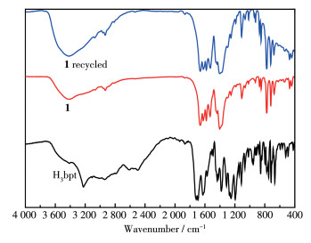
IR spectra of H3bpt and complex 1 were examined at room temperature (Fig. 1). The main characteristic absorption peaks present the typical stretching vibrations of COO- and O—H groups. The broad band in a region of 3 500~3 116 cm-1 indicates O—H stretching of the coordinated water molecule[37]. The strong bands at 1 107 and 1 666 cm-1 are attributed to the ester C—O and acyl C=O stretching vibrations, respectively[38]. The characteristic peaks of the asymmetric vibration and the symmetric stretching vibration of the carboxylate groups were at 1 624 and 1 405 cm-1, respectively. They were obviously shifted to lower wavenumbers relative to those of free H3bpt (1 720 and 1 410 cm-1), which suggests that the carboxylate groups in the complex are coordinated to the Nd3+ ions[39]. These structural features are in accord with the results of the X-ray diffraction analysis.
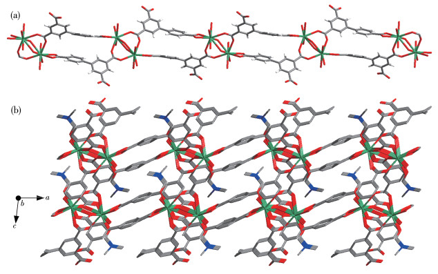
Complex 1 was well characterized by single crystal X-ray diffraction analysis. It crystallizes in the monoclinic system C2/c space group and displays a 3D structure. The asymmetric unit contains one Nd3+ cation, one bpt3- anionic ligand, one coordinated DMF, one coordinated water molecule, and two free water molecules. As shown in Fig. 2a, The Nd3+ cation adopts a nine-coordinated mode, coordinated by nine oxygen atoms from three monodentate (O2, O1i and O3ii) and two chelating (O3iii, O4iii, O5iv and O6iv) carboxyl groups from five different bpt3- ligands, one terminal water molecule (O7) and one terminal DMF molecule (O8) (Symmetry codes: i 0.5-x, 0.5-y, -z; ii x, 1-y, -0.5+z; iii 0.5-x, -0.5+y, 0.5-z; iv -x, y, 0.5-z). The coordination environment of Nd3+ cation is a slightly distorted tricapped trigonal prismatic. The Nd—O bond lengths vary from 0.241 1(2) to 0.272 5(2) nm, which correspond to those reported for other lanthanide-bpt3- complexes[40-42]. The bpt3- ligands adopt three different coordination modes: a bidentate bridging mode (mode Ⅰ), asymmetric chelating bridging mode (mode Ⅱ) and a chelating bidentate mode (mode Ⅲ)[20] (Fig. 2b).

Non-hydrogen atoms are represented by thermal ellipsoids drawn at the 30% probability level and coordinated DMF molecules are omitted except O for clarity; Symmetry codes: i 0.5-x, 0.5-y, -z; ii x, 1-y, -0.5+z; iii 0.5-x, -0.5+y, 0.5-z; iv -x, y, 0.5-z
Two adjacent Nd3+ cations are connected by two chelating/bridging and two bis(monodentate) bridging carboxyl groups, forming binuclear [Nd2(COO)6(H2O)2 (DMF)2] building units with the Nd1…Nd1iii separation of 0.407 0(1) nm, which is further extended into a 1D chain via the bpt3- ligand along a axis (Fig. 3a). These binuclear building units are further cross-linked by bpt3- ligands to form a 3D network with intersected channels[43] (Fig. 3b). The solvent accessible volume is 1.307 9 nm3 per 4.713 0 nm3 unit cell volume (27.8% of the total crystal volume) after the removal of the uncoordinated solvents calculated with PLATON.
H atoms are omitted for clarity
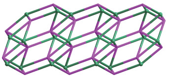
Topologically, if each Nd3+ cation and bpt3- ligand are considered as five - connected nodes, respectively, the structure can be considered as a 5, 5-connected net with the point symbol of the topology as (44·63·83) (48 62) (Fig. 4).
Nd: green, bpt3-: magenta
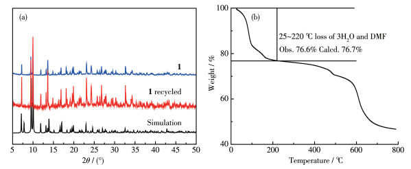
To verify the phase purity of the complex, PXRD was performed. The experimental PXRD pattern was in agreement with the calculated ones based on the X-ray single-crystal data, indicating the high phase purity of complex 1 (Fig. 5a). In order to estimate the thermal stabilities, TG analysis for 1 was performed on bulk samples in a range of 25~800 ℃ (Fig. 5b). As shown in Fig. 5b, the weight loss of 23.4% (Calcd. 23.3%) occurring between 25 and 220 ℃ corresponds to the removal of two free H2O molecules and coordinated H2O and DMF molecules. After taking off the solvent molecules, with the temperature further heating the skeleton of 1 decomposed gradually without displaying any distinct plateau.
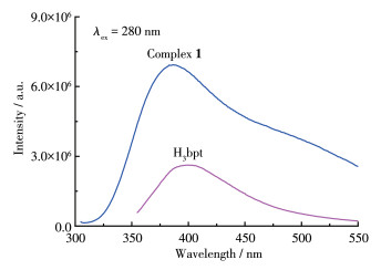
The solid-state photoluminescent properties of H3bpt ligand and complex 1 were investigated at room temperature. It was found that complex 1 showed significant fluorescence enhancement and the strong emission band was observed at 386 nm (λex=280 nm) as shown in Fig. 6. This band may be due to the emission of H3bpt ligand with a slight blue-shift of 12 nm since the free H3bpt ligand exhibited emission at 398 nm attributed to the π - π* transitions (λex=280 nm) [40, 44]. The observed much stronger emission intensity of 1 indicates that the formation of MOF enhances the rigidity of the aromatic backbone of the ligand and maximizes the intramolecular/intermolecular interactions among the organic ligands, which are conducive to energy transfer[26]. In addition, there was no obvious characteristic Nd - based emission in the region of 800~1 400 nm[45], indicating an inefficient energy transfer from ligand p-excited states to neodymium f-excited states.
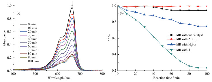
Studies have shown that lanthanide complexes may have good photocatalytic activity due to the diverse and stable valence states of lanthanide cations[6]. Although a variety of Ln-MOFs based on H3bpt have been reported[20, 22, 29, 40-43], their photocatalytic properties have hardly been studied. So, in this research, three organic dyes, MO, RhB and MB, were used as the model pollutant in aqueous media to evaluate the photocatalytic activity of 1. The results showed that complex 1 displayed good specific effect to degradation of MB but little effect to MO and RhB under ultraviolet light irradiation. As shown in Fig. 7a, the variation of UV - visible absorption spectra of MB dye solution in the presence of 1 was measured at each 10 min interval. The characteristic absorption (ca. 665 nm) of MB was selected to monitor the adsorption and photocatalytic degradation process. The photocatalytic activity of 1 was gradually enhanced with time increasing from 0 to 100 min, and nearly 77% of MB was degraded (Fig. 7a). As shown in Fig. 7b, the control experiments as MB without catalyst, and MB with NdCl3, H3bpt and 1, respectively, were carried out under the same conditions to ensure the results obtained from the photocatalytic experiments were consistent (where c0 is the initial concentration of MB solution at the beginning of photocatalytic degradation, and c is the concentration of MB solution at each min interval). Under the ultraviolet light, the degradation of MB in the absence of catalysts was negligible, implying that MB was relatively stable under illumination conditions. Furthermore, H3bpt ligand showed a certain catalytic degradation rate of MB (about 25%), which indicated that the ligand had certain optical activity. Complex 1 exhibited the best degradation performance for MB. The IR (Fig. 1) and PXRD patterns (Fig. 5a) of complex 1 after the photocatalytic experiment were almost the same as that of the as-prepared complex, which indicates that MB is degraded rather than adsorbed and as a photocatalyst, complex 1 has good stability during the heterogeneous catalytic reaction in the solution.
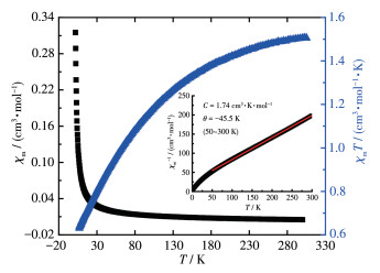
The magnetic behaviour of complex 1 was studied at a temperature range of 2.0~300 K under a 1 000 Oe direct current magnetic field. The magnetic susceptibil- ities and products χmT are presented as functions of the temperature in Fig. 8. When the temperature decreased, complex 1 exhibited a regular increase of χm and a usual decrease of χmT from 1.51 cm3·mol-1·K at 300 K to 0.63 cm3·mol-1·K at 2.0 K. The Curie constant derived from χm-1 vs T plots was 1.74 cm3·K·mol-1. Such behaviour is a typical isolated Nd3+ complex[46] that matches the structure of complex 1. Although there are binuclear building units in complex 1, the Nd…Nd separation is long. For Nd3+ cations, the spin- orbit coupling is very large; the free-ion ground state is 4I9/2 and the Zeeman factor gJ is equal to 8/11 which leads to χmT value of 1.64 cm3·mol-1·K[47]. At 300 K, the experimental χmT value of 1 (1.51 cm3·mol-1·K) was slightly smaller than the expected one for the free Nd3+ cation, which may be caused by the cumulative effects of crystal field variation, diamagnetic corrections or slight weighing errors. In a range of 50~300 K, the magnetic data obeyed the Curie-Weiss law (Inset in Fig. 8). The Weiss constant of -45.5 K agreed with previously reported values[46-47]. The susceptibility below 50 K did not conform to the Curie-Weiss law, which may be attributed to the effect of crystal field splitting of 4I9/2 ground state into five Kramers doublets[46].
In summary, one 3D Nd3+-MOF based on a Y-shaped tricarboxylic ligand (biphenyl-3, 4′, 5- tricarboxylic acid) was synthesized under solvothermal condition. The crystal structure shows that the frame- work possesses (5, 5)-connected topological network. The MOF exhibited strong emission in the solid-state at room temperature based on the ligand. The magnetic susceptibility displayed a typical isolated Nd3+ complex. In addition, the MOF showed high photocatalytic efficiency for the degradation of MB in aqueous solution. This study provides further insights into the rational design of MOF-based multifunctional materials.
刘志强, 黄永清, 孙为银. 无机化学学报, 2017, 33(11): 1959-1969 doi: 10.11862/CJIC.2017.244LIU Z Q, HUANG Y Q, SUN W Y. Chinese J. Inorg. Chem., 2017, 33(11): 1959-1969 doi: 10.11862/CJIC.2017.244
Zhou H C, Long J R, Yaghi O M. Chem. Rev., 2012, 112: 673-674 doi: 10.1021/cr300014x
Seoane B, Coronas J, Gascon I, Benavides M E, Karvan O, Caro J, Kapteijn F, Gascon J. Chem. Soc. Rev., 2015, 44(8): 2421-2454 doi: 10.1039/C4CS00437J
He C B, Liu D M, Lin W B. Chem. Rev., 2015, 115(19): 11079-11108 doi: 10.1021/acs.chemrev.5b00125
Wei Y S, Zhang M, Zou R, Xu Q. Chem. Rev., 2020, 120(21): 12089-12174 doi: 10.1021/acs.chemrev.9b00757
Bai Y T, Zhang S, Feng S S, Zhu M L, Ma S Q. Dalton Trans., 2020, 49: 10745-10754 doi: 10.1039/D0DT01648A
Rojas S, Horcajada P. Chem. Rev., 2020, 120(16): 8378-8415 doi: 10.1021/acs.chemrev.9b00797
Zheng M M, Wang Y X, Feng P Y. Catalysts, 2020, 10(3): 309 doi: 10.3390/catal10030309
Zhang L L, Guo B B, He H H, Zhang X R, Feng Y, Fan W D, Cao J L, Lu G, Chen Y H, Sun D F, Huang W. Inorg. Chem., 2020, 59(1): 695-704 doi: 10.1021/acs.inorgchem.9b02950
Li B, Jiang Y Y, Sun Y Y, Wang Y J, Han M L, Wu T P, Li D S. Dalton Trans., 2020, 49(42): 14854-14862 doi: 10.1039/D0DT03176C
Zhao J, Zhu G H, Xie L Q, Wu H L, Zhou A J, Wu Z Y, Wang J, Chen Y C, Tong L. Dalton Trans., 2015, 44(32): 14424-14435 doi: 10.1039/C5DT01894C
Larionov S V, Bryleva Y A, Glinskaya L A, Plyusnin V F, Kupryakov A S, Agafontsev A M, Tkachev A V, Bogomyakov A S, Piryazev D A, Korolkov I V. Dalton Trans., 2017, 46(34): 11440-11450 doi: 10.1039/C7DT01536D
Han M R, Li S D, Ma L, Yao B, Feng S S, Zhu M L. Acta Crystallogr. Sect. C, 2019, C75(9): 1220-1227
Han M R, Zhang H T, Wang J N, Feng S S, Lu L P. RSC Adv., 2019, 9(55): 32288-32295 doi: 10.1039/C9RA06920H
Shang K X, Jing S, Hu D C, Yao X Q, Zhi L H, Si C D, Liu J C. Cryst. Growth Des., 2018, 18(4): 2112-2120 doi: 10.1021/acs.cgd.7b01565
Ma Y L, Du L, Zhao Q H. Inorg. Chem. Commun., 2017, 77: 1-5 doi: 10.1016/j.inoche.2017.01.027
Wu J W, Zhang H B, Du S W. J. Mater. Chem. C, 2016, 4(16): 3364-3374 doi: 10.1039/C5TC04432D
Lu Y L, Zhao W J, Liu Y, Liu B, Feng S, Tan J T, Li X, Yang X W. J. Solid State Chem., 2012, 192: 144-152 doi: 10.1016/j.jssc.2012.04.003
Liu Y H, Lu L P, Zhu M L, Su F. Acta Crystallogr. Sect. C, 2016, C72: 358-362
Yan D, Duan Q. Inorg. Chem. Commun., 2013, 36: 188-191 doi: 10.1016/j.inoche.2013.09.005
Lin Z J, Xu B, Liu T F, Cao M N, Lü J, Cao R. Eur. J. Inorg. Chem., 2010, 24: 3842-3849
Han D, Yan X L, Liu J. Z. Anorg. Allg. Chem., 2019, 645: 422-427 doi: 10.1002/zaac.201800467
Wang H J, Cheng F J, Zou C C, Li Q Q, Hua Y Y, Duan J G, Jin W Q. CrystEngComm, 2016, 18(30): 5639-5646 doi: 10.1039/C6CE00960C
Hao Z M, Yang G C, Song X Z, Zhu M, Meng X, Zhao S N, Zhang H J. J. Mater. Chem. A, 2014, 2(1): 237-244 doi: 10.1039/C3TA13179C
Zhao J, Dong W W, Wu Y P, Wang Y N, Wang C, Li D S, Zhang Q C. J. Mater. Chem. A, 2015, 3(13): 6962-6969 doi: 10.1039/C4TA06537A
Li Y L, Zheng L P, Nie H, Wang Y F, Yao L H, Li J, Li J J, Zhou X L, Wang H F, Wang H Y. J. Mol. Struct., 2020, 1204: 127427 doi: 10.1016/j.molstruc.2019.127427
Li Y L, Zhao Y, Kang Y S, Liu X H, Sun W Y. Cryst. Growth Des., 2016, 16(12): 7112-7123 doi: 10.1021/acs.cgd.6b01352
Guo Z Y, Xu H, Su S Q, Cai J F, Dang S, Xiang S C, Qian G D, Zhang H J, O'Keeffe M, Chen B L. Chem. Commun., 2011, 47(19): 5551-5553 doi: 10.1039/C1CC10897B
Xing S H, Bing Q M, Song L F, Li G H, Liu J Y, Shi Z, Feng S H, Xu R R. Chem. Eur. J., 2016, 22(45): 16230-16235 doi: 10.1002/chem.201603102
Jia Y Q, Feng S S, Shen M L, Lu L P. CrystEngComm, 2016, 18(28): 5344-5352 doi: 10.1039/C6CE00308G
Zhang L Y, Lu L P, Zhu M L, Feng S S. CrystEngComm, 2017, 19(14): 1953-1964 doi: 10.1039/C7CE00149E
An Y Y, Lu L P, Feng S S, Zhu M L. CrystEngComm, 2018, 20(14): 2043-2052 doi: 10.1039/C8CE00008E
Yang D D, Lu L P, Feng S S, Zhu M L. Dalton Trans., 2020, 49(22): 7514-7524 doi: 10.1039/D0DT00938E
Otwinowski Z, Minor W. Methods Enzymol., 1997, 276: 307-326
Sheldrick G M. Acta Crystallogr. Sect. C: Cryst. Struct. Commun., 2015, C71: 3-8
Spek A L. Acta Crystallogr. Sect. C: Cryst. Struct. Commun., 2015, C71: 9-18
Li D S, Zhao J, Wu Y P, Liu B, Bai L, Zou K, Du M. Inorg. Chem., 2013, 52: 8091-8098 doi: 10.1021/ic4007718
Ma X L, Wang Z X, He X, Shao M, Li X M. Inorg. Chem. Commun., 2018, 92: 131-135 doi: 10.1016/j.inoche.2018.04.015
Li L N, Wang S Y, Chen T L, Sun Z H, Luo J H, Hong M C. Cryst. Growth Des., 2012, 12(8): 4109-4115 doi: 10.1021/cg300617h
Xing S H, Zeng G, Liu X M, Yang F, Hao Z Q, Gao W, Yang Y L. Wang X R, Li G H, Shi Zhan, Feng S H. Dalton Trans., 2015, 44(20): 9588-9595
Li X Y, Lin Z J, Yang Y Y, Cao R. CrystEngComm, 2014, 16(28): 6425-6432 doi: 10.1039/C4CE00158C
Chen Z F, Xue H B, Wu L R, Jin R F. J. Cluster Sci., 2018, 29: 1269-1274 doi: 10.1007/s10876-018-1445-8
Cui P P, Zhao Y, Zhang X D, Wang P, Sun W Y. Dyes Pigm., 2016, 124: 241-248 doi: 10.1016/j.dyepig.2015.09.024
Chen Y X, Tang K K, Wang X, Chen B Y, Qin G Q, Yang J H. Z. Anorg. Allg. Chem., 2014, 640: 2292-2295 doi: 10.1002/zaac.201400213
Niu W Y, Feng C, Fan N Y, Wang X Y, Yan P F, Sun J W, Li G M. Synth. Met., 2016, 221: 319-325 doi: 10.1016/j.synthmet.2016.09.008
Lhoste J, Campos P A, Henry N, Loiseau T, Rabu P, Abraham F. Dalton Trans., 2011, 40(36): 9136-9144 doi: 10.1039/c1dt10485c
Andruh M, Bakalbassis E, Kahn O, Trombe J C, Porcher P. Inorg. Chem., 1993, 32(9): 1616-1622 doi: 10.1021/ic00061a017

Figure 1 IR spectra of H3bpt, complex 1 and the complex after the photocatalytic reaction

Figure 2 (a) Coordination environment of central Nd3+ cation in 1; (b) Coordination modes for bpt3- in 1
Non-hydrogen atoms are represented by thermal ellipsoids drawn at the 30% probability level and coordinated DMF molecules are omitted except O for clarity; Symmetry codes: i 0.5-x, 0.5-y, -z; ii x, 1-y, -0.5+z; iii 0.5-x, -0.5+y, 0.5-z; iv -x, y, 0.5-z

Figure 3 (a) One-dimensional chain and (b) 3D network structure of complex 1
H atoms are omitted for clarity

Figure 4 Topological structure of complex 1 with Nd3+ cation and bpt3- ligand as 5-coonected nodes, respectively
Nd: green, bpt3-: magenta

Figure 5 (a) PXRD patterns of complex 1 at room temperature and (b) TG curve of complex 1

Figure 6 Luminescence spectra of H3bpt ligand and complex 1 at 298 K in the solid-state

Figure 7 (a) Variation in UV-Vis absorption spectra of MB solution in the presence of 1 irradiated by visible light; (b) Photocatalytic degradation rate of MB under ultraviolet light in the absence and presence of NdCl3, H3bpt and 1, respectively
Table 1. Crystal data and structure refinement for complex 1
| Formula | C18H20NO10Nd | F(000) | 2 200 | |
| Crystal system | Monoclinic | Dc/(Mg·m-3) | 1.563 | |
| Space group | C2/c | μ/mm-1 | 2.251 | |
| a/nm | 2.489 2(5) | Reflection collected | 7 283 | |
| b/nm | 1.272 1(3) | Independent reflection | 6 421 | |
| c/nm | 1.497 5(3) | Rint | 0 | |
| β/(°) | 96.32(3) | GOF | 1.088 | |
| V/nm3 | 4.713 1(16) | R1, wR2 [I>2σ(I)] | 0.040 2, 0.111 7 | |
| T/K | 100 | R1, wR2 (all data) | 0.044 3, 0.114 0 | |
| Z | 8 |
 下载: 导出CSV
Table 2. Selected bond lengths (nm) and angles (°) for 1
| Nd1—O3i | 0.241 1(2) | Nd1—O2 | 0.243 4(2) | Nd1—O1ii | 0.244 5(2) |
| Nd1—O8 | 0.246 6(3) | Nd1—O4iii | 0.248 9(2) | Nd1—O7 | 0.2500(3) |
| Nd1—O5iv | 0.251 4(3) | Nd1—O6iv | 0.254 2(2) | Nd1—O3iii | 0.272 5(2) |
| O3i—Nd1—O2 | 73.95(8) | O3i—Nd1—O1ii | 79.91(8) | O2—Nd1—O1ii | 132.62(8) |
| O3i—Nd1—O8 | 85.04(12) | O2—Nd1—O8 | 140.51(11) | O1ii—Nd1—O8 | 73.14(11) |
| O3i—Nd1—O4iii | 122.57(8) | O2—Nd1—O4iii | 70.84(9) | O1ii—Nd1—O4iii | 92.21(9) |
| O8—Nd1—O4iii | 146.61(12) | O3i—Nd1—O7 | 151.77(11) | O2—Nd1—O7 | 133.63(12) |
| O1ii—Nd1—O7 | 75.38(10) | O8—Nd1—O7 | 74.96(15) | O4iii—Nd1—O7 | 72.31(13) |
| O3i—Nd1—O5iv | 129.54(8) | O2—Nd1—O5iv | 75.43(9) | O1ii—Nd1—O5iv | 147.72(9) |
| O8—Nd1—O5iv | 94.26(11) | O4iii—Nd1—O5iv | 82.27(9) | O7—Nd1—O5iv | 72.63(11) |
| O3i—Nd1—O6iv | 80.84(8) | O2—Nd1—O6iv | 72.12(9) | O1ii—Nd1—O6iv | 141.09(9) |
| O8—Nd1—O6iv | 71.76(11) | O4iii—Nd1—O6iv | 126.58(8) | O7—Nd1—O6iv | 110.57(11) |
| Continued Table 2 | |||||
| O5iv—Nd1—O6iv | 51.84(9) | O3i—Nd1—O3iii | 75.36(8) | O2—Nd1—O3iii | 67.58(7) |
| O1ii—Nd1—O3iii | 67.94(8) | O8—Nd1—O3iii | 138.74(10) | O4iii—Nd1—O3iii | 49.78(7) |
| O7—Nd1—O3iii | 107.02(11) | O5iv—Nd1—O3iii | 126.22(8) | O6iv—Nd1—O3iii | 137.37(8) |
| Symmetry codes: i x, -y+1, z-1/2; ii -x+1/2, -y+1/2, -z; iii -x+1/2, y-1/2, -z+1/2; iv -x, y, -z+1/2; v -x+1/2, y+1/2, -z+1/2; vix, -y+1, z+1/2. | |||||
下载: 导出CSV

 扫一扫看文章

扫一扫关注我们


 下载:
下载: