Citation:
Lei Cao, Weidong Huang, Longlin Yin, Yu Liang, Wenhao Li, Congrui Liu, Ping Xie, Zhaonan Li, Tao Lu, Xueqin Huang, Shi Zhou, Wencheng Wu. Nanotechnology-driven innovations in transarterial chemoembolization for hepatocellular carcinoma: A focus on smart and flexible platforms[J]. Chinese Chemical Letters,
2026, 37(8): 112643.
doi:
10.1016/j.cclet.2026.112643
Nanotechnology-driven innovations in transarterial chemoembolization for hepatocellular carcinoma: A focus on smart and flexible platforms
English
Nanotechnology-driven innovations in transarterial chemoembolization for hepatocellular carcinoma: A focus on smart and flexible platforms
Central Laboratory and Department of Medical Ultrasound, Sichuan Academy of Medical Sciences, Sichuan Provincial People's Hospital, University of Electronic Science and Technology of China, Chengdu 610072, China
b.
Department of Radiology, Sichuan Academy of Medical Sciences, Sichuan Provincial People's Hospital, University of Electronic Science and Technology of China, Chengdu 610072, China
c.
Department of Interventional Radiology, Affiliated Cancer Hospital of Guizhou Medical University, Guiyang 550004, China
d.
Department of Ultrasound, Sichuan Academy of Medical Sciences, Sichuan Provincial People's Hospital, University of Electronic Science and Technology of China, Chengdu 610072, China
* Corresponding author. E-mail address: wuwencheng@uestc.edu.cn (W. Wu). 1 These authors contributed equally to this work.
Received Date:
12 January 2026 Accepted Date:
17 March 2026 Revised Date:
14 March 2026 Available Online:
15 August 2026
Abstract:
Transarterial chemoembolization (TACE) remains the standard of care for patients with unresectable hepatocellular carcinoma (HCC). However, clinical outcomes are frequently compromised by off-target toxicity, incomplete tumour necrosis, and the induction of a post-procedural immunosuppressive microenvironment. The integration of nanotechnology represents a paradigm shift designed to circumvent these biological and technical barriers. For instance, magnesium-enhanced TACE has demonstrated an objective response rate of ~93.3%, substantially surpassing that of conventional TACE therapies. This review evaluates nanotechnology's impact on TACE through four domains: Smart nanocarriers for stimuli-responsive delivery; theranostic platforms for real-time imaging; immune remodeling to boost immunotherapy; and next-generation embolic materials such as biodegradable polymers and liquid metals. Finally, we address the preclinical-to-clinical translation gap and outline a roadmap for personalized, high-precision interventional oncology.
Primary liver cancer, particularly hepatocellular carcinoma (HCC), poses a severe global health challenge, with its incidence and mortality being especially prominent in China [1,2]. Due to its insidious onset and rapid progression, the vast majority of patients are diagnosed at intermediate or advanced stages, precluding curative options such as surgical resection or liver transplantation [3,4]. In this clinical context, transarterial chemoembolization (TACE) has emerged as the primary standard of care for unresectable HCC. Its therapeutic mechanism involves the precise delivery of chemotherapeutic agents and embolic materials via an arterial catheter, which simultaneously blocks the tumor's blood supply and achieves high local drug concentrations to induce ischemic necrosis. This dual approach aims to maximize patient survival and quality of life while minimizing systemic side effects [5–8].
However, conventional TACE (cTACE) faces intrinsic limitations that constrain its long-term efficacy. First, the rapid establishment of collateral circulation post-embolization often leads to incomplete tumor necrosis and residual lesions, creating a high risk for recurrence. Second, conventional chemotherapeutic agents typically exhibit uncontrolled release kinetics and are rapidly cleared from the tumor site, making it difficult to sustain effective therapeutic concentrations. More critically, the procedure can trigger compensatory pathological changes, including the exacerbation of tumor hypoxia, upregulation of angiogenic factors, and the formation of an immunosuppressive tumor microenvironment (TME) [5–11]. Collectively, these adverse factors facilitate tumor recurrence and distant metastasis, highlighting the urgent need for improved therapeutic interventions.
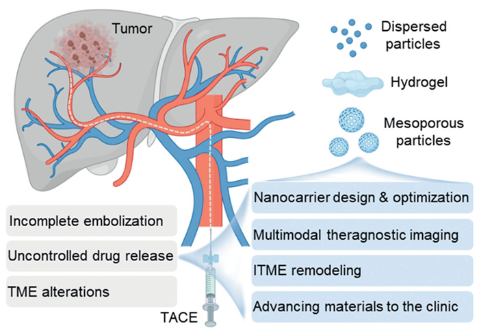
These challenges have spurred the development of next-generation, nanotechnology-driven TACE strategies (Fig. 1). To address existing bottlenecks, nanotechnology-driven solutions provide innovative avenues: Smart nanocarriers with pH-responsive or stimulus-triggered mechanisms enable controlled drug release and enhanced intratumoral accumulation; advanced embolic materials offer more durable vascular occlusion; nanoplatforms are designed for TME remodeling to reverse post-TACE immunosuppression; and theranostic integration achieves real-time visualization [12–14]. Thus, Fig. 1 delineates a comprehensive roadmap for the evolution of TACE toward precision, personalization, and multifunctional integration under the guidance of nanotechnology.
Figure 1
Figure 1.
Current limitations of cTACE and developmental directions of nanotechnology-driven therapies. This schematic highlights the key shortcomings of cTACE, including incomplete embolization, uncontrolled drug release, and TME alterations. In contrast, emerging strategies focus on smart nanocarriers, multimodal theranostic platforms, immune-modulating approaches, and advanced embolic materials. These innovations aim to enhance targeting, sustain drug release, and remodel the TME to improve therapeutic outcomes. Created with BioRender.com.
To address these obstacles, the rapid advancement of nanomedicine has invigorated interventional oncology. Due to their tunable structures, unique size effects, and excellent functional plasticity, nanomaterials offer immense potential for tumor diagnosis and therapy [15–17]. In the context of TACE, smart nanocarriers enable precise, controlled drug release via stimuli-responsive mechanisms, such as sensitivity to pH, temperature, or enzymes, while facilitating active accumulation in tumor regions through surface modification. Furthermore, nanoplatforms integrate multiple treatment modalities, including photothermal therapy and immunotherapy, with advanced imaging functions like magnetic resonance imaging (MRI) and computed tomography (CT), thereby achieving true theranostic integration [18–22]. Compared to systemic administration, transarterial delivery of nanodrugs ensures significantly higher local tumor concentrations while minimizing systemic exposure [16,17,23,24]. Additionally, "intelligent" embolic agents responsive to external stimuli allow for precise spatiotemporal control over the treatment process, enhancing antitumor efficacy and potentially reversing immunosuppression [25–29].
Currently, TACE-related nanostrategies are classified into two categories based on their pharmacokinetic and spatial distribution characteristics: (ⅰ) Nanomaterials serving as primary embolic agents (e.g., liquid metals and biodegradable hydrogels) with intrinsic vascular occlusion functions; and (ⅱ) nanocarriers suspended in traditional embolic agents like Lipiodol to enhance controlled drug release and targeting [30]. This classification facilitates a systematic analysis of the release behavior, distribution patterns, and therapeutic mechanisms of diverse nanoplatforms.
This review elucidates how nanomaterials fundamentally optimize TACE efficacy through four core dimensions. First, we explore construction and optimization strategies for smart nanocarriers to achieve targeted drug delivery. Second, we discuss the integration of multimodal imaging and theranostics for real-time monitoring and precision therapy. Third, we analyze TME remodeling strategies, detailing how nanotechnology reverses post-TACE immunosuppression and converts "cold" tumors into "hot" phenotypes. Finally, we examine the clinical translation of novel embolic materials, addressing the challenges of moving innovations from the laboratory to clinical practice. By synthesizing these advancements, we provide a theoretical basis for designing next-generation, personalized TACE strategies for patients with intermediate and advanced HCC.
2.
Construction and optimization strategies of smart nanocarriers
Although TACE is established as a primary intervention for local tumor control, the clinical utility of conventional chemotherapeutic formulations is frequently compromised by non-specific distribution and dose-limiting systemic toxicity. To address these pharmacokinetic limitations, smart nanocarriers have been engineered as a transformative solution. Unlike traditional passive vehicles, these platforms are designed to not only maximize intratumoral accumulation but also respond dynamically to microenvironmental cues for precise, on-demand drug release, thereby broadening the therapeutic window (Fig. S1 in Supporting information).
Strategies exploiting the physiological characteristics of the TME, particularly pH sensitivity, have shown substantial promise. For example, DOX@HmA nanoparticles utilize histidine-zinc ion coordination to achieve pH-responsive kinetics, triggering rapid doxorubicin release within the acidic tumor milieu [31]. When co-administered with Lipiodol, this system effectively blocks the epithelial-mesenchymal transition (EMT), inhibiting both local regeneration and metastasis. Similarly, leveraging temperature differentials, certain nanogels undergo a rapid sol-gel phase transition at body temperature (37 ℃) [32,33]. This thermal responsiveness enables smart nanocarriers to synergistically combine physical embolization with photothermal therapy, significantly augmenting the antitumor effect [34].
To further circumvent the limitations of passive diffusion, smart nanocarriers have been functionalized with active targeting ligands. Conjugating the integrin inhibitor GRGDSP onto superparamagnetic iron oxide nanoparticles (SPIONs) significantly enhanced embolization efficacy by actively binding to αvβ3 integrins overexpressed on tumor vascular endothelial cells [35]. In a parallel approach, AFGO-Dox nanoparticles demonstrated superior affinity for HCC cells and a reduced half-maximal inhibitory concentration (IC50) [36]. Additionally, magnetic nanocarrier platforms have achieved triple synergistic effects, combining chemotherapy, embolization, and hyperthermia, via magnetic field-directed self-assembly [28]. These data confirm that ligand-mediated and physical targeting strategies effectively enhance the retention and uptake of therapeutic agents at the tumor site.
Smart nanocarriers are also evolving into multifunctional platforms integrating diagnostic and therapeutic modalities [37]. For instance, photo-switchable microcapsules enable light-regulated drug release while permitting MRI visualization. From a materials perspective, liquid embolic agents like PAA-LDH@PEG200 rapidly absorb water to form stable hydrogels within 5 s, conferring dual functions of ultra-fast vascular occlusion and anti-angiogenesis without recanalization for 28 days [38]. Moreover, advanced systems have achieved distal vascular occlusion and induced macrophage polarization toward the antitumor M1 phenotype through the multidimensional synergy of physical occlusion and biological regulation [39].
Stimuli-responsive embolic systems can be categorized by their activation triggers, each offering distinct therapeutic advantages within the hepatic TME (Table S1 in Supporting information). These mechanisms exemplify how nanotechnology enables precise, dynamic control over drug release and embolic behavior, addressing key limitations of conventional TACE.
3.
Multimodal imaging and theragnostic integration
Precision interventional oncology necessitates real-time monitoring of therapeutic delivery and accurate post-procedural assessment. Conventional single-modality imaging often fails to adequately distinguish the distribution of therapeutic agents from tumor tissue, which limits the optimization of individualized treatment regimens. To address these limitations, multimodal theranostic nanoplatforms have been developed to combine diagnostic precision with therapeutic action, enhancing the controllability and predictability of interventional procedures (Fig. S2 in Supporting information).
In the domain of image-guided embolization, novel nanoprobes are engineered to differentiate drug pharmacokinetics from embolic deposition. For instance, the DOX-NPs-MB complex preserves ultrasound contrast capabilities while enabling the real-time visualization of drug diffusion from the lipiodol emulsion, resolving the discrepancy between embolic coverage and actual drug delivery [40]. Similarly, 131I-BaGdF5@PDA-CDDP nanoparticles incorporate single-photon emission computed tomography (SPECT)/CT/MRI trimodal imaging, facilitating comprehensive intraprocedural monitoring via131I radiolabeling [41]. Furthermore, the IF@Gel composite hydrogel utilizes dual-modality CT/MRI to evaluate dynamic changes in the TME, providing essential feedback for assessing the synergy between embolization and anti-programmed death-ligand 1 (anti-PD-L1) immunotherapy [42].
Beyond monitoring, theranostic nanoprobes effectuate the simultaneous execution of diagnosis and therapy within a single entity. The LG-PEG nanoprobe addresses the challenge of lipiodol artifacts by enabling virtual non-contrast (VNC) imaging while maintaining high-density contrast enhancement in tumor region [43]. In thermal therapies, magnetic microspheres achieve self-regulating hyperthermia at 50 ℃ under an alternating magnetic field, concurrently loaded with doxorubicin for dual-modal CT/MRI imaging [44]. Moreover, Fe@EGaIn/CA microspheres integrate photothermal and photodynamic therapies with vascular occlusion, resulting in complete tumor growth inhibition in vivo [45].
Innovations in radiosensitizing nanomaterials have also expanded the scope of TACE by facilitating synergy with radiotherapy. The DSeSeP-API(Lip) nanoradiosensitizer exploits an X-ray-responsive diselenide bond to trigger a phase transition in lipiodol, significantly potentiating the therapeutic effect of subsequent X-ray doses [46]. Similarly, ADM/Fe3O4−MS microspheres combine microwave thermal therapy with ferroptosis induction, demonstrating superior antitumor efficacy [47]. Additionally, upper critical solution temperature (UCST) microspheres achieved an 87.5% complete remission rate in VX2 tumor models through the synergistic application of photothermal therapy and chemotherapy [48]. These findings validate that the integration of radiosensitization and combination therapies can effectively overcome the resistance mechanisms associated with monotherapy.
4.
Immune regulation and tumor microenvironment remodeling
Conventional TACE often exacerbates the immunosuppressive TME due to treatment-induced ischemia. Emerging nanoplatforms aim to reverse this by converting immunologically "cold" tumors into "hot" phenotypes (Fig. S3 in Supporting information). Quantitatively, "hot" tumors feature high densities of cytotoxic CD8+ T cells and elevated CD8+/regulatory T cell (Treg) ratios, whereas "cold" tumors are dominated by myeloid-derived suppressor cells (MDSCs) and M2-type tumor-associated macrophages (TAMs). Key biomarkers for monitoring this transition include PD-L1 expression and interferon-γ (IFN-γ) signatures [49–51].
To trigger adaptive immunity, nanoplatforms induce immunogenic cell death (ICD) to release damage-associated molecular patterns (DAMPs). The coordinated release of ATP, high mobility group box 1 (HMGB1), and calreticulin (CRT) promotes dendritic cell (DC) maturation and subsequent CD8+ T cell activation [47]. For instance, Chen et al. developed SLNP-SCH/DOX@MS microspheres to co-deliver doxorubicin and an A2AR antagonist, alleviating adenosine-mediated T-cell exhaustion. Similarly, the Pt-P@PND nanogel designed by Shi et al. utilizes cisplatin-induced ROS to augment anti-programmed death-protein 1 (anti-PD-1) therapy, increasing the tumor suppression rate to 72% [52].
Metabolic reprogramming and cytokine delivery offer alternative pathways to restore immune surveillance. Wang et al. utilized L-Arg@CaPL emulsions to reduce PD-L1 expression by 50% through sustained L-arginine release [53]. Zhang et al. employed a CCaP-LPE Pickering emulsion to release CA4P, disrupting vasculature and activating purinergic signaling for DC maturation [54]. Additionally, the poly(N-isopropylacrylamide)-based (PNDS) hydrogel developed by Ren et al. facilitates the release of interleukin-12 (IL-12) and granulocyte-macrophage colony-stimulating factor (GM-CSF), increasing CD8+ T-cell infiltration six-fold [55].
Normalizing the physicochemical parameters of the TME is vital to maximize immunotherapy efficacy. The LHCa-LPE emulsion triples tumor oxygen partial pressure within 48 h via lipoxygenase (LOX) catalysis [56], while CCaP-LPE emulsions restore peripheral oxygen pressure to above 20 mmHg [57]. Synthetic biology approaches, such as clustered regularly interspaced short palindromic repeats and CRISPR-associated protein 9 (CRISPR-Cas9) mediated PHD gene knockout and nitric oxide-releasing AuNP@PNA/DOX nanogels, further alleviate hypoxia [58–61]. Regarding acidity, calcium phosphate-based neutralization raises the pH to 7.0 [56], while UCST microspheres maintain a physiological pH of 7.2–7.4, tripling the proportion of M1-type macrophages [48].
To circumvent therapeutic resistance, next-generation designs incorporate single-atom catalysts (SACs) for persistent ROS generation or leverage cuproptosis to bypass apoptosis resistance [62–65]. These multi-target interventions are essential for achieving durable responses in HCC.
5.
Clinical translation and development of novel embolic materials
The clinical translation of nanomedicine-enhanced TACE requires rigorous biosafety assessments. Gold nanoparticles (AuNPs) are primarily sequestered by the reticuloendothelial system (RES). Particles smaller than 6 nm undergo renal excretion, while larger ones may persist in RES organs, potentially inducing inflammation. Similarly, iron-based nanomaterials like SPIONs are degraded by macrophages into iron ions for hemoglobin synthesis. Liquid metals, such as gallium-indium alloys (EGaIn), represent emerging agents that oxidize in vivo into soluble ions for excretion.
6.
Clinical trial landscape and regulatory considerations
The translation of nanomedicine from laboratory to clinic is accelerating, fueled by the demand for high-performance embolic agents. Early clinical data are promising; for instance, Su et al. reported that TACE combined with the Ab-SFB-NP system achieved a 72.2% disease control rate with a favorable safety profile (n = 42) [66]. Furthermore, magnesium-enhanced TACE demonstrated an objective response rate (ORR) of approximately 93.3% in a pilot study of 15 patients, highlighting the potential of metal-ion modulating embolics [67]. Currently, ongoing trials focus on drug-eluting embolics under imaging guidance (e.g., ChiCTR2500097895), utilizing primary endpoints such as ORR (via mRECIST), progression-free survival (PFS), and safety profiles (via CTCAE).
These clinical successes stem from nanotechnology's ability to enhance TACE via immune and metabolic modulation. Examples include L-Arg@CaPL nanoparticles for microenvironment reprogramming and ZnCo-Fe3O4 microspheres for pyroptosis induction (Fig. S4 in Supporting information). However, widespread adoption faces hurdles in manufacturing scalability under good manufacturing practice (GMP) and the requirement for comprehensive toxicology data per ISO 10993 standards.
Precision oncology requires biomarker-driven stratification. "Hot" tumors with high PD-L1 expression or specific immune infiltrates may respond better to immunomodulatory agents, while hypoxic tumors are ideal candidates for oxygen-generating platforms. In preclinical models, the DOX-NPs-MB complex validated real-time drug diffusion monitoring [40], and LHCa-LPE emulsions significantly suppressed tumor progression via LOX-mediated ferroptosis [68].
To avoid the complications of permanent vascular occlusion, biodegradable agents such as lignin-based nanogels [69] and gelatin-based nanospheres [70] have been engineered to degrade post-treatment, facilitating tissue repair. Additionally, UiO-66/Bi2S3@DOX platforms integrate photodynamic therapy to induce extensive necrosis [71].
Novel materials like liquid metals are expanding the therapeutic repertoire. The Fe@EGaIn/CA microspheres combine dual-modality CT/MRI with photothermal therapy to achieve complete tumor inhibition in vivo [41]. Chemical interventions also show promise: CaO2 nanoparticles neutralize tumor acidity while releasing oxygen [72], and CCaP-LPE Pickering emulsions trigger ferroptosis [55]. Notably, magnesium-based microspheres (Mg MSs) neutralize the acidic TME to reverse CD8+ T cell exhaustion. In a pilot study, Mg MS-enhanced TACE achieved an ORR of 93.3% without additional safety concerns [67].
Finally, biomimetic strategies such as the ISBP agent (using Bletilla striata polysaccharide) [39], BSA-encapsulated TPZ nanoparticles [73], and rhein-based nanogels [74] offer superior biocompatibility and multi-target metabolic inhibition.
7.
Summary and prospects
In conclusion, nanotechnology-based delivery systems are fundamentally restructuring the TACE treatment paradigm for HCC. While cTACE remains the primary standard of care, its long-term utility is frequently compromised by uncontrolled drug release, inadequate intraprocedural imaging, and the post-embolic exacerbation of tumor hypoxia and immunosuppression. Nanomedicine provides robust solutions via four strategic dimensions: Engineering smart nanocarriers for microenvironmental responsiveness, developing multimodal imaging platforms, actively remodeling the TME to reverse immunosuppression, and translating novel embolic materials such as biodegradable agents and liquid metals. Collectively, these advancements transition TACE from a conventional catheter-based occlusion technique into a high-precision, integrated oncologic platform combining diagnosis, therapy, and immune modulation.
Despite these breakthroughs, the path to clinical application faces substantial hurdles, including systemic toxicity and patient intolerance. Next-generation systems must evolve into dynamic platforms capable of adapting to the spatial heterogeneity and temporal evolution of the tumor. Furthermore, establishing standardized criteria for efficacy and biosafety evaluation is essential to accelerate regulatory approval.
A structured translational roadmap is requisite to bridge the gap between preclinical success and clinical implementation. This process should begin with systematic pharmacotoxicology in large animal models, followed by biomarker-enriched Phase Ⅰ/Ⅱ trials and real-world evidence collection. Crucially, nanotechnology provides an optimal interface for synergizing TACE with complementary modalities, specifically radiotherapy, thermal ablation, and immunotherapy. With continued interdisciplinary innovation and rigorous clinical validation, these personalized interventional regimens are poised to significantly improve survival outcomes and bring new hope to patients with intermediate and advanced HCC.
Declaration of competing interest
The authors declare that they have no known competing financial interests or personal relationships that could have appeared to influence the work reported in this paper.
CRediT authorship contribution statement
Lei Cao: Writing – original draft, Visualization, Validation, Software, Methodology, Formal analysis, Data curation, Conceptualization. Weidong Huang: Writing – original draft, Software, Formal analysis, Data curation. Longlin Yin: Writing – original draft, Software, Formal analysis, Data curation, Conceptualization. Yu Liang: Writing – original draft, Conceptualization. Wenhao Li: Investigation, Data curation. Congrui Liu: Investigation, Data curation. Ping Xie: Investigation. Zhaonan Li: Investigation. Tao Lu: Investigation. Xueqin Huang: Investigation. Shi Zhou: Writing – review & editing, Supervision, Methodology, Conceptualization. Wencheng Wu: Writing – review & editing, Supervision, Project administration, Methodology, Conceptualization.
Acknowledgments
We greatly acknowledge the financial support from Sichuan Science and Technology Program (No. 2025ZNSFSC0237) and National Natural Science Foundation of China (No. 52402353).
Supplementary materials
Supplementary material associated with this article can be found, in the online version, at doi:10.1016/j.cclet.2026.112643.
[1]
S. Wang, R. Zheng, B. Han, et al., Chin. Cancer 33 (2024) 165–174.
[2]
R.L. Siegel, T.B. Kratzer, A.N. Giaquinto, et al., CA Cancer J. Clin. 75 (2025) 10–45. doi: 10.3322/caac.21871
X. Liang, H. Liu, H. Chen, et al., Aggregate 5 (2024) e552. doi: 10.1002/agt2.552
Figure 1
Current limitations of cTACE and developmental directions of nanotechnology-driven therapies. This schematic highlights the key shortcomings of cTACE, including incomplete embolization, uncontrolled drug release, and TME alterations. In contrast, emerging strategies focus on smart nanocarriers, multimodal theranostic platforms, immune-modulating approaches, and advanced embolic materials. These innovations aim to enhance targeting, sustain drug release, and remodel the TME to improve therapeutic outcomes. Created with BioRender.com.


 DownLoad:

 下载:
下载:


 下载:
下载:

