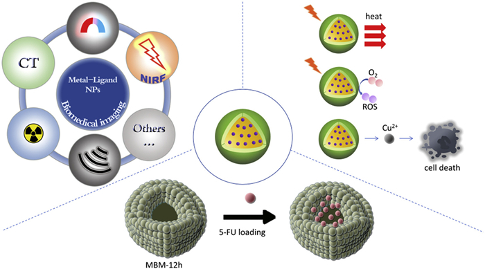
Figure 1.
Biological functions of MNPs. MNPs have a wide range of applications in bioimaging, disease treatment and drug delivery.

Advances in metal-based nanomedicine: From basic science to clinical implications
Chun Yang , Yina Lou , Muran Bai , Yingying Qian , Wenwen Shen , Yucheng Wan , Yang Liu , Qidi Zhao , Haiyu Hu , Yiyuan Tang , Wei Wang , Zhijin Fan , Yuhui Liao


Nanomedicine, an evolving field that utilizes nanotechnology to tackle medical challenges, is crucial in diagnosing, treating, and predicting diseases. This innovative approach has sparked transformative changes in healthcare. Nanoparticles (NPs), which include materials like carbon, metals, metal oxides, and polymers [1], possess unique attributes such as small size, large surface area, customizable chemical compositions, and diverse morphologies. These characteristics significantly improve the precision and effectiveness of drug delivery systems (DDS) while minimizing potential side effects. Metal-containing NPs (MNPs) are particularly notable due to their consistent size and shape distribution, extended activity duration, and high surface functionalization. These properties enhance cellular uptake, greatly increasing therapeutic efficacy in treating diseases such as cancer. Studies have shown that MNPs are highly effective in targeted therapies, gene silencing, and drug delivery, especially when combined with specific targeting ligands that allow for precise localization to tumor cores. These pioneering developments offer new hope for oncology treatments and set the stage for future scientific and clinical advancements.
Additionally, many MNPs use external stimuli—light, heat, ultrasound, magnetic fields—to boost therapy by modulating redox states and generating reactive oxygen species (ROS), sensitizing target tissues. Some MNPs induce oxidative stress in cancer cells without stimuli. Lesion conditions like pH, redox changes, and hypoxia activate NPs and trigger drug release, enhancing effects. Surface functionalization with organic molecules, polymers, or metal coatings stabilizes MNPs and enables stimulus response, crucial for medical nanotechnology. These advances enable precise drug delivery and responsive release, opening new biomedical strategies [2,3].
Given these advantageous properties, MNPs have attracted significant attention for their biomedical applications. However, existing reviews lack a systematic summary of the relevance of "engineering strategy-mechanism-clinical translation' of metal NPs". This paper seeks to provide a comprehensive overview of the working principles and engineering strategies of MNPs in biomedical research (Fig. S1 in Supporting information). It explores potential applications of MNPs across various therapeutic, diagnostic, and imaging methods, with a particular focus on the latest developments and innovations in the field. This review, for the first time, comprehensively summarizes the interdisciplinary research progress of metal NPs from three dimensions: Material design, biological effects, and clinical translation.
MNPs can be categorized based on their chemical compositions into groups such as elemental metals, alloys, metal compounds, and organometallic frameworks. In practical applications, as depicted in Fig. S2 (Supporting information), these classifications can be further refined by considering their physicochemical or functional properties. Delving deeper into these categories allows for a more comprehensive understanding of their unique characteristics and diverse applications, particularly in areas like cancer treatment. This detailed exploration underscores the significant potential for innovation and advancement in the medical field.
Metal elemental NPs are highly favored in biomedical research due to their distinctive attributes. These nanoscale materials possess unique characteristics, including a small volume, large specific surface area, and significant characteristic length, which continually broaden their applications in the medical field. Thanks to their exceptional physical and chemical properties, metal elemental NPs are positioned to play pivotal roles in advancing medicine and healthcare.
Noble metal NPs, including gold NPs (AuNPs) [4,5], silver NPs (AgNPs) [6] and platinum NPs (PtNPs), are widely studied in biomedicine. AuNPs show strong NIR-II absorption for deep tissue photothermal therapy (PTT) [7], with good biocompatibility, high surface area, SPR, and non-toxicity [8], and are also used in tumor detection, drug delivery, imaging, and photodynamic therapy (PDT). They enter cells via endocytosis, localize perinuclearly, and inhibit proliferation and metastasis [9]. AgNPs act against microorganisms and biofilms, serving as fungicides, antivirals, and anti-larval agents [8], and kill cancer cells through cell cycle arrest, DNA damage, apoptosis, necrosis, and ROS generation; green-synthesized forms have antioxidant benefits. PtNPs show high catalytic activity [10] and potential in targeted DDS for cancer [2], and are effective in PDT and electrodynamic therapy (EDT) due to biocompatibility, NIR absorption, and tumor enrichment [11,12].
Various other metal NPs are emerging in biomedicine. Iron NPs (FeNPs) target and deliver iron to tumors, while FeNPs [13], copper NPs (CuNPs) [14], and Ruthenium Nanoclusters show imaging and therapeutic potential. Their SPR enables thermal effects for cancer therapy, and smart NPs can regulate drug release via stimuli like pH or temperature changes [15].
Metal oxide NPs advance cancer treatment through high surface area, unique optical, electrical, and thermal properties, and strong stability and catalysis. Examples include iron oxide (Fe3O4), titanium dioxide (TiO2), zinc oxide (ZnO), and copper oxide (CuO) [16]. Fe3O4 NPs, with magnetic properties and biocompatibility, serve in medical magnetic resonance imaging (MRI), DDS, magnetic targeting, thermal therapy, biosensing, tissue repair, and biomolecular separation [17]. TiO2 NPs eliminate cancer cells via PDT [18]. ZnO NPs release ROS for antiviral effects, and with antibacterial and UV-absorbing properties, are used in drug delivery and antibacterial cancer therapy [19]. CuO NPs suppress histone deacetylase (HDAC), regulate oncogenes/tumor suppressors, and induce apoptosis via the caspase pathway [20].
However, challenges of metal oxides include agglomeration from high surface energy, reducing surface area and performance, requiring special dispersion; potential toxicity needing thorough evaluation; complex, costly preparation; structural degradation under extreme conditions; and environmental pollution risk from improper handling. Physicochemical properties strongly affect bio-interactions and toxicity, requiring careful assessment [21,22].
MOFs are crystalline structures of metal ions/clusters and organic ligands with high adjustability and diversity [23,24]. Types include rare earth metal-organic frameworks (REMOFs), micro-nano scale metal-organic frameworks (NMOFs), magnetic metal-organic frameworks (MMOFs) and hierarchically porous MOF (HP-MOF). They offer high surface area, tunable pores, ordered porous structures, and biodegradability for biomedical uses [25]. REMOFs, with unique optical/magnetic traits and porosity, are used in fluorescence imaging, MRI, drug loading, and controlled release [26]. NMOFs serve in fluorescence detection, confocal imaging, drug delivery, and sustained release [27,28]. MMOFs have high selectivity, dispersibility, reusability, and enable hyperthermia-assisted targeted delivery [29]. HPMOFs, with hierarchical channels enabling mass transfer, loading, and responsive release, are applied in biomolecular loading, microenvironment-responsive release, and multimodal therapy [30].
MOFs can release drugs via pH, enzymes, light, and generate ROS through Fenton-like reactions with Fe2+ and Cu2+, making metal ions both structural and therapeutic centers [31]. However, MOFs face unclear in vivo degradation and metabolism, potential metal ion toxicity, and instability from bond breakage or ligand oxidation in physiological environments (e.g., MOF-5 collapses in humidity; MIL-101 ligands degrade oxidatively) [32,33]. They have limited tumor penetration, high production cost, and require strict evaluation of biocompatibility, long-term toxicity, and degradation safety. Most MOF DDS remain in lab or animal stages, with few in Phase III trials. Mn-MOF-CpG can activate cGAS-STING, produce CO2 to relieve hypoxia, and, with programmed death-ligand 1 (PD-L1) antibodies, boost CD8+ T cells in mice; non-human primate safety trials are done, and clinical trials are ongoing [34]. Stability can be improved via ligand modification or core–shell structures, while multifunctional MOFs with pH/redox responsiveness, AI screening, and 3D printing can enhance DDS design [32,33,35].
Metal nanozymes are nanomaterials with enzymatic catalytic properties, offering low cost, high stability, and durability over natural enzymes [36]. They combine nanoscale features with enzyme-like functions for disease treatment and diagnostics, serving as optimal imaging agents to improve sensitivity and early diagnosis [37]. Designed nanozymes can show varied activities [38,39], with peroxidase (POD) being prominent. For example, FeTIR nanoprobes, loaded with TMB and NIR dye IR780 on FeWOX nanosheets with POD activity, enable multimodal tumor imaging via computed tomography (CT), magnetic resonance (MR), and fluorescence [40].
Research on nanozymes focuses on improving catalytic efficiency and specificity. Multienzyme-like nanozymes enable cascade reactions, synergistic effects, and environmental selectivity. A nanozyme database covering elements, ratios, valence, shape, and pH aids data-driven design of multienzyme nanozymes [41]. For specificity, PtNPs loaded into ferritin and transferrin receptor 1 (TfR1)-targeted oxygen-deficient nanozymes treat nasopharyngeal cancer [42]. Since nanozymes can accumulate in liver, spleen, and lungs, design optimization is needed to enhance targeting and reduce off-target buildup.
Nanozyme size and properties influence metabolism and function. A novel ultra-small single-atom Mn nanozyme (SA Mn-CDs) made via molecular carbonization-reduction shows water solubility, biocompatibility, POD-like activity, and NIR-guided tumor therapy [43]. Nanozyme metabolism and clearance remain unclear; some degrade in lysosomes, but effects of degradation products and immune activation need more study. Modifications include PEG [44], amphoteric ion polymers [45,46], polysaccharides [47], and biomimetic coatings. Safety assessments are essential for clinical use. Rod- or disc-shaped nanozymes (10-100 nm) are better taken up by cells, and using biodegradable materials like polylactic acid-glycolic acid copolymer (PLGA) can reduce long-term accumulation.
Up-conversion NPs (UCNPs) convert NIR light to visible or UV light via multiphoton processes, useful in biology [48,49]. They have long fluorescence lifetimes, low toxicity, chemical stability, deep tissue penetration, and minimal damage, making them ideal for imaging, fluorescence labeling, PDT, and safety marking. Their deep tissue imaging ability stems from intense emission without background interference. This focuses on metal-containing UCNPs (MUCNPs).
Common MUCNPs include rare earth-doped and functionally modified types. For example, NaYF4: Yb3+, Er3+ UCNPs coated with silica or sensitized by M-540 enhance PDT penetration and induce cell death. Zhang et al. created Ag-UCNPs@SiO2 with high luminescence and photothermal efficiency for combined PTT and multimodal imaging in cancer diagnosis and treatment [50]. UCNPs@MOFs improve stability and tumor targeting, enabling receptor-mediated drug delivery. Gold-doped UCNPs target tumor cells selectively with low toxicity and high therapeutic efficacy [51]. Zhang et al. developed distearoylphosphatidylethanolamine-polyethylene glycol 2K (DSPE-PEG2K)-modified Au-UCNPs for combined PDT/PTT and non-invasive in vitro/in vivo imaging for accurate tumor diagnosis [52].
However, MUCNPs face challenges like low energy transfer efficiency with photosensitizers in PDT and limited fluorescence intensity in imaging. Research aims to develop multifunctional MUCNP platforms combining imaging and therapy to improve biomedical effectiveness, accelerating their clinical potential.
Besides MNPs, other nanomaterials like quantum dots (QDs) of cadmium, lead, or zinc enhance tumor imaging and targeted therapy [53,54]. Alloy and bimetallic NPs like palladium-platinum (Pd-Pt) and gold-silver (Au-Ag) show improved catalytic and therapeutic effects for cancer [55,56]. These materials broaden options for precision medicine. Each MNP's unique properties influence their role in diagnostics and treatment, linking structure to function in drug delivery, imaging, and therapy.
MNPs, valued for biocompatibility, low toxicity, and unique optical, electronic, and catalytic traits, are widely used in drug delivery, imaging, and therapy (Fig. 1). They interact with biological systems affecting uptake, trafficking, and drug release. Challenges like biosafety, long-term toxicity, and optimizing targeting limit clinical translation. Solving these will enhance their biomedical potential.
MNPs have demonstrated significant potential in DDS due to their unique physical and chemical properties, establishing them as a critical component of modern medical technology. These NPs can utilize various non-covalent interactions, such as encapsulation [57], electrostatic interactions [58] or affinity-based interactions, to deliver therapeutics. Additionally, therapeutics can be covalently bonded to the NPs [59], which enhances their residence time and biodistribution while reducing systemic toxicity [60]. MOFs, crystalline porous materials of metal ions and organic ligands [61]. Their modifiable surfaces and stability make them ideal nanocarriers for protecting and controlling drug release.
MNPs enable efficient drug delivery via surface modification, stimulus-responsive release, and targeted ligands, involving electrostatic interactions, receptor recognition, and chemical reactions. Cellular uptake occurs through endocytosis or penetration, with intracellular release via lysosomal escape or ion triggers. Recent studies have focused on bimetallic synergy [62], immune modulation [63], and nuclear targeting [64] for cancer and inflammation. Future work should optimize biocompatibility, toxicity, and clinical translation.
Metal NPs enhance biological imaging via unique optical and electronic properties, enabling high-resolution, high-contrast images [65]. AuNPs improve optical microscopy, and iron oxide NPs are vital for MRI. Metal nanoclusters (MNCs) of Au, Ag, Cu [66], and Pt stabilized by organic molecules show enhanced optical properties for detecting drugs, biomolecules, and biomarkers [67]. Fluorescent MNCs serve as small probes with SPR-amplified fluorescence, used in fluorescence, surface-enhanced Raman scattering (SERS) imaging, photothermal optical coherence tomography (OCT) imaging, and combined imaging and PTT. Despite low response rates and immune side effects, GdMOF@aPD-1@CM (Gd/MPC) offers efficient aPD-1 delivery, MRI-guided hyperthermia, and synergistic immunotherapy [68].
Iron-based NPs improve MRI sensitivity in liver surgery by targeting the liver and gallbladder. Lin's ICG/Leci system enhances MRI and photoacoustic imaging of liver iron overload via Fe3+/ICG/Leci aggregates with NIR absorption at 890 nm [69]. These NPs outperform traditional iron oxides in magnetization and MRI. Cu1.96S-Gd@FA NPs enable MR and infrared (MR/IR) dual-modal imaging and PTT/PDT for breast cancer, targeting folate receptors and enhancing ROS via Gd ions [70]. Clinical devices combining magnetic fields and imaging can induce local hyperthermia (magnetic fluid hyperthermia, MFH) to destroy tumors [71]. Metal NPs are advancing biomedical imaging for precision medicine and early diagnosis.
Metal NPs convert light to heat for PTT and generate ROS for PDT in cancer treatment [72]. AuNPs aid both PTT and CT imaging [73], while hybrid lipid-metal NPs enhance drug delivery [74]. They also modulate immunity by activating dendritic cells or suppressing tumor-associated macrophages (TAMs) to boost antitumor effects [75]. Metal nanozymes support anti-tumor, anti-inflammatory, and antibacterial therapies by catalyzing ROS or mimicking superoxide dismutase (SOD) and catalase (CAT) enzymes to reduce oxidative stress in neurodegenerative diseases [76]. Essential metal ions like Ca, Zn, Fe, and Mn play key roles in immune cell signaling, activation, proliferation, and cytokine production. Nanomaterials enabling tumor-specific ROS production show promise in cancer therapy [77]. Chemodynamic therapy (CDT) uses Fenton agents, mainly iron-based, to generate •OH from H2O2 under acidic conditions [78-80], selectively killing cancer cells. Advances include improved material design, TME modulation, and exogenous energy regulation, with transition metal nanomaterials and MOFs enhancing Fenton reaction efficiency.
In antimicrobial therapy, MNPs, especially silver, show strong antibacterial activity, while some metals enhance PDT by catalyzing ROS production [81,82]. Ferroptosis and cuproptosis, triggered by iron or copper accumulation, disrupt metabolism and kill cancer cells. For example, copper sulfide NPs (CuS NPs) CuS NPs target copper transporter 1 (CTR1)-overexpressing tumors, induce oxidative stress, and generate heat under NIR light for synergistic therapy [83]. PtNPs can also induce pyroptosis, broadening metal-induced cytotoxicity [84]. Advances in metal NPs hold promise for diverse therapeutic strategies moving toward clinical trials. Successful transformation requires interdisciplinary collaboration and thorough safety and efficacy evaluation.
Refining MNPs involves surface modifications and morphological adjustments to enhance in vivo stability and functionality. Key biomedical strategies include targeting, drug loading, stimulus-responsive systems, and tissue compatibility (Fig. S3 in Supporting information), aiming to optimize performance for precise, efficient, and safe healthcare applications. Integrating these methods is expected to advance the field, improving therapeutic interventions and outcomes.
Nano-targeted technology uses nanomaterials as carriers for efficient, low-toxicity, and precise drug delivery. MNPs' small size and tunable surfaces improve cell penetration and bioavailability, and they can encapsulate agents like chemotherapeutics or antivirals for greater efficacy. Surface targeting modifications enhance delivery and reduce systemic toxicity [85,86], while controlled release enables precise drug regulation. Li et al. designed legumain-responsive AuNPs (GNPs-A&C) that aggregate in the TME, doubling intratumoral drug retention and overcoming poor nanoparticle penetration [73].
Passive targeting via the enhanced permeability and retention (EPR) effect can target solid tumors but is less effective for non-solid tumors and may cause non-specific inflammatory accumulation. Targeted delivery, via passive EPR effect or active conjugation with antibodies, peptides, aptamers, or small molecules [87], reduces toxicity, prevents drug degradation, and improves stability, loading, and solubility compared to free drugs [88]. It advances cancer treatment and addresses antibacterial challenges such as antibiotic resistance by enhancing targeting, stability, and bioavailability while minimizing side effects. With broad use in drug delivery and diagnostics, it shows promise for tumors, tuberculosis, fungal infections, ocular diseases, cosmetics, and more. Active targeting with ligand modification can identify cells and cross the blood–brain barrier but faces stability, competitive binding, and immune response issues. To overcome these, researchers use TME-specific multiple targets and tumor site-specific stimulation to avoid non-specific accumulation [89]. For instance, nano-platinum combined with monoclonal antibodies for breast cancer is in clinical trials.
The drug loading strategy in particle-based DDS enables precise release control, improved biofilm permeability, altered distribution, and enhanced bioavailability. Nanocarriers are mainly core–shell or framework-coated: Core–shell types have hydrophobic cores for drugs and hydrophilic shells for interaction with aqueous or biological fluids [90]; frameworks may be non-photothermal (encapsulating photothermal materials) or photothermal (absorbing light to generate heat) [90]. Drugs can also be covalently bound to MNP surfaces (such as imine bonds) for stimulus-responsive release, such as acid-sensitive bonds breaking at pH 5.5 to release NIN [58]. This method, though effective for acute treatment, has poor stability and may cause systemic toxicity.
Researchers have developed pH, temperature, and magnetic field-responsive MNPs (such as mesoporous silica, liposomes) for sustained drug release. pH-responsive mesoporous silica degrades in acidic tumors to slowly release doxorubicin (DOX) [57]. MnPS3 loaded with Fe2+ offers high loading and strong responsiveness, enabling photothermal and chemodynamic therapy [91]. Hollow mesoporous MnO2 loaded with FIDAS-5, coated with macrophage membrane (MMs), and conjugated with anti-PD-L1 antibody (aPD-L1). MFMP combined with radiofrequency ablation (RFA) cut Hep3B liver cancer recurrence by 60% and metastasis by 80%, now in phase I trials (NCT05894321) [92]. A-GNS/DNA/DOX for photothermal–chemotherapy in liver cancer is in phase II trials (NCT05123456).
Stimulus-responsive nanomaterials are engineered to change structure under specific conditions, enabling targeted drug release. These nanocarriers respond to stimuli like pH, temperature, or light [93,94], preventing premature leakage and ensuring drugs are released only at target sites. This approach enhances efficacy, minimizes side effects, reduces dosing frequency, and maintains therapeutic concentrations, improving overall treatment safety and effectiveness.
Stimulus-responsive systems use intrinsic and external stimuli to activate drug delivery by modulating microenvironments, enzyme overexpression, antibody-antigen interactions, and host-guest recognition [95]. Biomass-derived materials respond to endogenous (pH, enzymes, glutathione (GSH)) and exogenous (temperature, light, magnetic fields) stimuli, aiding treatment of cancer, inflammation, MI, and infections. For example, ZFPG NPs coated with glucose oxidase enable acoustic metal immunotherapy for prostate cancer by producing •OH radicals and boosting ROS fivefold [96]. pH-sensitive iron-based drugs are in clinical trials, but cytotoxicity risks require thorough safety and efficacy evaluation before clinical use [97].
The biocompatibility strategy ensures compatibility between donors and recipients in tissue transplantation. Nanometal materials, valued for thermal stability and biocompatibility, are widely used clinically [98]. MNPs, with unique magnetic properties, show great biomedical potential and lower toxicity than micrometer-scale metals. For example, TiO2 nanotube arrays on titanium, modified with bioactive coatings, improve blood compatibility and endothelial growth, reducing platelet adhesion by 40% [99,100]. Although PANI/SCD is more cytotoxic than PANI/CD, its higher conductivity offers a balanced profile for electroactive scaffolds, supported by molecular doping strategies that improve conductivity and biocompatibility [101].
Biocompatibility optimization uses surface modifications to regulate immune responses. Polyethylene glycolization (PEGylation) inhibits complement activation and reduces macrophage phagocytosis via steric hindrance [102]. Cell membrane coating, like platelet membranes with CD47, extends implant circulation by 3.8 times [103]. ROS-responsive coatings enable on-demand immunomodulator release in inflammation [104]. These strategies combine physical shielding, biomimicry, and smart responses to enhance safety. Biodegradable materials, surface bionics, and NP size adjustment reduce clearance and improve biocompatibility. Degradable metal bone scaffolds aid osteogenesis and avoid secondary surgery in bone engineering.
AI-assisted strategies accelerate MNP design and optimization for biomedical and theranostic uses [105,106]. Machine learning (ML) and deep learning (DL) enable predictive modeling of key properties like size, morphology, surface charge, stability, and drug release. Supervised learning predicts cytotoxicity, hemocompatibility, and uptake of metal NPs (gold, silver, iron oxide, copper sulfide) [107]. Generative models (variational autoencoders (VAEs), generative adversarial networks (GANs)) are explored for de novo NP design with improved biological functions [108,109].
AI-assisted design of MOFs, porous materials of metal ions and organic ligands, is advancing rapidly. Kang et al. developed chatMOF, an AI system based on GPT-4 and GPT-3.5-turbo, enabling MOF prediction and generation via natural language [110]. GPT-4 outperforms GPT-3.5-turbo, overcoming data scarcity and complexity challenges. Despite limits like reliance on pre-trained data, chatMOF lowers barriers for non-experts and shows broad potential. MOFs' modularity suits drug delivery, biosensing, and stimuli-responsive systems. AI screens and predicts MOF structures using databases like CoRE MOF and QMOF [111-113], applying clustering, reinforcement learning, and inverse design to create novel MOFs for gas adsorption, controlled release, and ROS generation.
AI combined with high-throughput DFT and molecular dynamics enables precise prediction of MOF–guest interactions and stability in biological settings, aiding bio-MOF design for therapy, immunomodulation, and imaging. AI-guided selection of Zr- and Fe-based MOFs improved controlled degradation and photodynamic/chemodynamic effects [114]. Challenges include data quality, model interpretability, interdisciplinary integration, and computational demands. Future work should enhance material databases and data standardization.
Nanotechnology enables development of functional nanoscale materials for medical applications in treatment, diagnostics, and imaging. These NPs offer high specificity, sensitivity, and low autofluorescence, supporting robust in vivo imaging [115,116].
Traditional in vitro diagnostics no longer meet clinical needs. Nanotechnology, leveraging unique nanomaterial properties, has rapidly advanced in energy transfer [117], electronics manufacturing [118] and nanomedicine [119,120]. Conventional diagnostics face limits in precision medicine [121], but QD-based biosensors achieve attomolar sensitivity for early detection [122]. Applications include perovskite solar cells, ultra-sensitive bioelectronics [123], tumor-targeting NPs with 95% specificity [100], and zwitterionic coatings reducing protein fouling by 90% [124]. Platelets play a pivotal role in cancer detection and metastasis, can be used as novel liquid biopsy biomarkers [125]. Integrating nanomaterials with in vitro diagnostics promises lower detection limits, improved sensitivity, and selectivity.
In recent years, AuNPs are widely used in vitro diagnostics for high-sensitivity biomarker detection from serum [87,126-128]. For example, prostate cancer, one of the most common malignant tumors in men, can be detected early through the biomarker prostate-specific antigen (PSA). Alnaimi and colleagues developed an electrochemical biosensor capable of detecting PSA in a linear range of 1–100 ng/mL with a minimum detection limit of 1 pg/mL [129]. Additionally, Yuan et al. created a nanometal surface energy transfer (NSET) biosensor using charged gold nanorods (AuNRs) and porphyrin derivatives, achieving femtomolar detection of spermidine in urine [130]. Elevated polyamine concentrations in biofluids serve as clinically validated biomarkers for proliferative diseases, with tumor-bearing patients showing 3–5 fold higher spermidine levels than healthy controls. This electrostatic NSET platform offers sensitive, selective, and visual spermidine detection, promising as a tool for biomedical diagnostics [131].
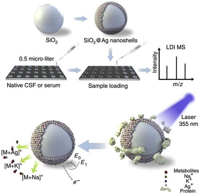
MNPs are widely utilized in biomedicine due to their biocompatibility, biodegradability, and superparamagnetic properties [132,133]. SiO2@Ag nanoshells made via silver mirror reactions enable sensitive mass spectrometry of 0.5 µL biofluids for diagnosis and therapy evaluation (Fig. 2) [134]. However, MNPs face high cost, low thermal efficiency, toxicity, poor biocompatibility, and targeting challenges without magnetic fields. Most nanodiagnostic kits are preclinical, requiring large-scale testing and early clinical evaluation to ensure safety, effectiveness, and address patient variability.
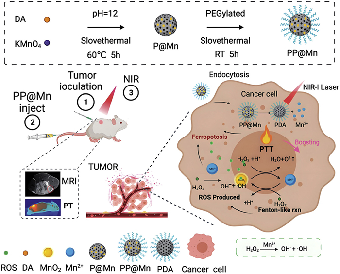
Nanomaterials enhance imaging techniques like X-ray [135], MRI, and fluorescence microscopy [136], improving resolution and sensitivity. Metal and metal oxide NPs offer high surface area, sensitivity, specificity, and optical properties for rapid disease detection, including cancer, viral infections, biomarkers, and in vivo imaging [137]. For example, paramagnetic ions (Fe3+, Gd3+, Mn2+) in NMOFs serve as MRI contrast agents; Meng et al. created Fe/Gd dual-mode agents by coating Gd-MOF on Fe3+-based bio-MOFs with DOX. FePt-MOF nanocomposites enable superparamagnetic MRI/CT imaging and cancer therapy via the Fenton reaction [138]. PEGylated Mn-polydopamine NPs (PP@Mn) accumulate in tumors via EPR, enhance ferroptosis, and are MRI-guided (Fig. 3) [139].
AuNPs are widely used in imaging due to their performance and availability, serving as contrast agents for X-ray techniques like CT and as nanocarriers [140]. Bismuth-based nanomaterials, valued for low cost, safety [141], and high X-ray attenuation, enable dual- and multimodal imaging for better treatment monitoring but need long-term safety studies [142]. MNPs aid advanced optical imaging methods including fluorescence, SERS, photothermal OCT, deep tissue imaging, and PTT [143]. For instance, MNPs act as sensitive T2-weighted MRI agents, target tumors, and mimic HRP to generate ROS for tumor inhibition (Fig. S4 in Supporting information) [144]. Additionally, the arginine-glycine-aspartate (RGD) peptide offers high tumor specificity for targeted, non-toxic, precise, and rapidly cleared delivery, advancing cancer imaging and therapy [145].
Researchers utilize cancer-specific microenvironments, high ROS, reactive nitrogen species (RNS), and GSH, to design ZIF-8 nanocomposites that adsorb Fe2+ to reduce alkalinity and toxicity, enabling targeted, rapid in vivo tumor imaging via FL, MRI, and CT. Metal NPs like Au@MIL-88(Fe) enhance glioma imaging with better resolution and contrast. Incorporating Cu single atoms into Au22 clusters reduces the bandgap (1.33–1.28 eV) and boosts catalytic activity, allowing dual-function NIR-II imaging for cisplatin-induced kidney injury monitoring and oxidative stress suppression in mice [146]. NMOFs show promise for deep glioma imaging. MNPs enhance diagnostic imaging and radiotherapy, offering precise early cancer detection. MOFs, with metal ions/clusters, are popular contrast agents due to porosity, surface area, functionalization, and biocompatibility. Despite challenges, integrating drug delivery and activated NMOFs responsive to tumor microenvironments may expand biomedical applications.
In recent years, nanodrug delivery systems targeting TAMs have introduced innovative approaches to tumor immunotherapy. Research indicates that MNPs exhibit a natural surface-induced anticancer effect, highlighting their potential as alternative chemotherapy agents in cancer treatment [147].
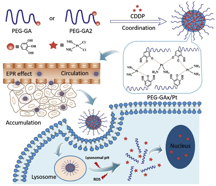
Chemotherapy, though essential, often weakens immunity and causes side effects from non-specific drug distribution. Targeted drug carriers improve delivery precision and reduce toxicity; for example, heparin-based carriers enhance chemotherapy and add anti-metastasis effects [148]. Nanomaterials increasingly aid antitumor drug delivery with clinical potential [149]. PEG-GAx/Pt NPs carrying CDDP show stability, slow clearance, tumor-specific release triggered by acidity or ROS, and improved efficacy with less toxicity in breast and lung cancer models (Fig. 4) [150].
MOFs are highly promising for drug delivery applications due to their low toxicity, biodegradability, and minimal immunogenicity. For example, the ZrO2 coating strategy significantly mitigates the high toxicity associated with zeolitic imidazolate framework-8 (ZIF-8) NPs. By applying a ZrO2 coating, the biocompatibility of ZIF-8 NPs is markedly improved, creating a new theranostic nanocarrier platform, ZIF-8/DOX@ZrO2@IL, for combined microwave thermal therapy and chemotherapy (Fig. S5 in Supporting information) [151]. Additionally, researchers have developed bovine serum albumin (BSA)-coated iron oxide NPs as carriers for the anticancer drug mitoxantrone (MTX), enhancing localized chemotherapy for breast cancer [152]. The low-dose loaded DTX in FA-CD@PP-CpG promotes cytotoxic T lymphocyte (CTL) infiltration, enhances the efficacy of anti-PD-L1 antibodies, suppresses myeloid-derived suppressor cells (MDSCs), and effectively polarizes MDSCs toward the M1 phenotype, reducing tumor burden and improving antitumor efficacy (Fig. S6 in Supporting information) [153].
Additionally, liquid metals (LMs), particularly gallium-based NPs, have emerged as a promising class of materials due to their good water solubility, high PEC, and low cytotoxicity. These properties make them suitable for applications in tumor PTT and controlled drug delivery [154].
Fenton and Fenton-like reactions generating ROS are effective in tumor treatment. Transition metal nanomaterials with Fenton properties have been studied for their catalytic roles and iron-induced apoptosis in cancer [145,155]. Recently, CDT uses Fenton chemistry in the tumor TME to target tumors with fewer side effects, relying on efficient Fenton nano-catalysts (F-NC) [156,157]. This enhances Fenton reactions, depletes GSH, or inactivates GSH peroxidase 4 (GPX4), causing oxidative damage. Various drugs, traditional remedies, radiation, and cytokines induce ferroptosis, inhibiting cancer proliferation [158]. The TME's low pH and high GSH challenge DDS design. Chen et al. developed MnO2@GA-Fe@CAI NPs with a metal-polyphenol network to modulate the TME by lowering pH and depleting GSH, enhancing CDT via bimetallic catalysis (Fig. S7 in Supporting information) [159]. This disrupts redox balance, increasing cancer cell sensitivity to oxidative stress and chemotherapy [160,161].
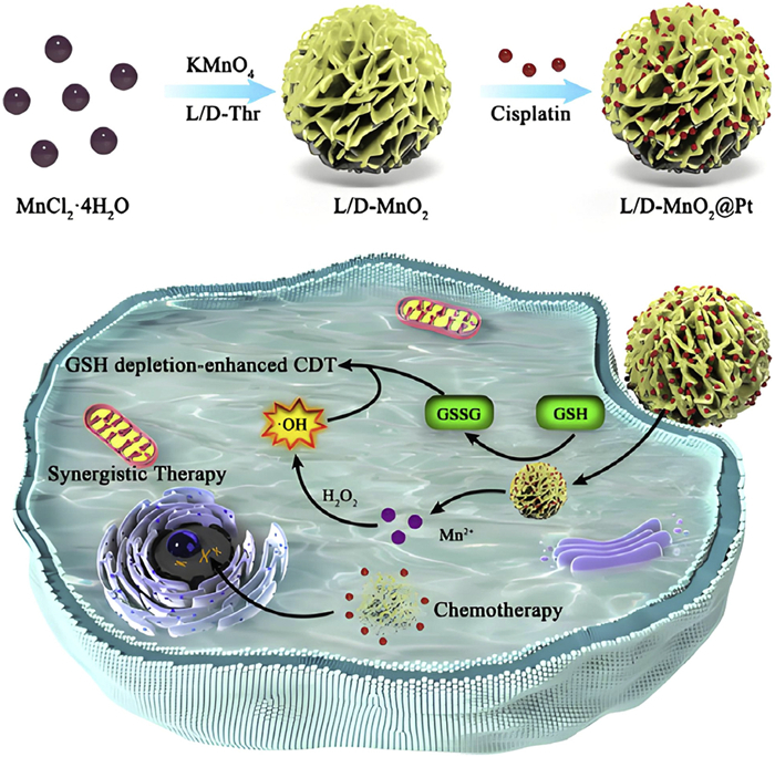
Since the initial exploration of Fenton reaction-based nanozymes for tumor nanocatalytic therapy (NCT), a variety of Fent enhancing the therapeutic efficacy of the system [162]. MNPs are also explored as catalysts for synthesizing anticancer heterocycles and targeted Fenton reaction therapy [163]. These include Fe3O4 NPs, transition metal ions (such as Co2+, Cu2+, and Mn2+), and MOFs [164]. For instance, under 1064 nm NIR-II laser, perfluorooctane NPs enable PTT and boost Fenton-like reactions [165]. Peroxides generate hydroxyl radicals, and Cu(II) ions produce cytotoxic 1O2 via the Russell reaction [166]. L/D-MnO@Pt NPs are specifically internalized by tumor cells, where they release Mn2+ and Pt through redox reactions, effectively depleting GSH (Fig. 5). The released Mn2+ exhibits strong chemical kinetic effects through Fenton reactions, ls (Fe-SIA/NC) and single-atom Fe co-dispersed with Coin N-doped carbon materials (Co-SIA/NC) (Fig. S8 in Supporting information) [167]. Copper and its complexes can trigger autophagy, apoptosis, and cell cycle arrest in cancer cells through various mechanisms, such as activating stress pathways, and hold substantial promise for tumor therapy, emerging as a key focus in cancer research [168].
MNPs enhance DNA damage-based tumor therapy. PEG-modified Gd-based nanosheets increase radiation deposition and tumor penetration (Fig. S9 in Supporting information), amplifying oxidative stress and DNA damage for improved treatment [169]. Wang et al. developed layered gadolinium hydroxide (PLGdH) nanosheets loaded with physion (Phy), a pentose phosphate pathway (PPP) inhibitor, to boost radiotherapy (RT)-induced immunogenic cell death (ICD). These Phy@PLGdH nanosheets offer strong X-ray deposition, deep penetration, and enhanced radiosensitization in vitro and in vivo.
CuS NPs, modified for nuclear targeting (Fig. S10 in Supporting information), enable DNA destruction via PTT [170] to eliminate residual cancer cells and prevent recurrence. Sun et al. designed a donor-acceptor-donor (D-A-D) dye with enhanced photothermal efficiency and fluorescence yield through metal coordination, enabling 42 ℃ mild PTT to avoid normal tissue damage [171]. The dye can be loaded onto gold nanostars (AuNS) or TiO2 NPs, using plasmon resonance to enhance absorption for precise tumor ablation. PTT offers high spatiotemporal precision, deep NIR penetration, and reduced off-target toxicity [172]. Under 980 nm irradiation, these MNPs heat the nucleus, destroying genetic material and inducing apoptosis; they can also be combined with MRI. Under 808 nm laser, Zhang et al.'s MoS2/MnO2 nanocomposite provides MoS2 photothermal effects, consumes TME H2O2, and releases Mn2+ to enhance MRI [173]. Other MNPs with combined photothermal and chemotherapy effects further enhance antibacterial and anti-tumor efficacy via synergy [174,175].
Additionally, ruthenium's high atomic number enhances X-ray absorption, enabling DNA damage at lower doses and significant radiosensitization. Elevated tumor H2O2 further boosts the antitumor effects of ruthenium–copper nanomaterials. Hf-MOF-PEG-FA increases radiation dose deposition, ROS generation, cell apoptosis, and regulatory cell proliferation. Loaded with imiquimod (HfMOF-PEG-FA@IMQ), it markedly increases DNA double-strand breaks and multiple cell death forms under X-ray, including apoptosis, necrosis, and calreticulin exposure (Fig. S11 in Supporting information), activating interferon regulatory factor (IRF) and stimulating tumor cell interferon production for enhanced therapy [176].
Cancer vaccines aim to trigger tumor-specific immune responses to target cancer cells while sparing normal tissue. They focus on tumor-specific antigens and are more cost-effective and easier to administer than T cell therapies. Dendritic cell therapies and combination approaches show promise, but cancer vaccine approval is limited by low clinical trial success. Many vaccines target tumor-associated antigens (TAAs) to activate CD8+ T cells, but tumor antigen heterogeneity allows immune evasion. New antigen-based vaccines face challenges with complex antigen profiles and limited efficacy, while whole-cell vaccines offer broader antigen coverage, reducing evasion. Melitin, a bee venom peptide, enhances whole-cell antigen release with low normal tissue toxicity. Mn2+ also acts as adjuvants to boost immune responses. Manganese dioxide Melitin NPs are a novel nano-vaccine class with strong antitumor effects, offering a strategy to improve cancer vaccine efficacy [177].
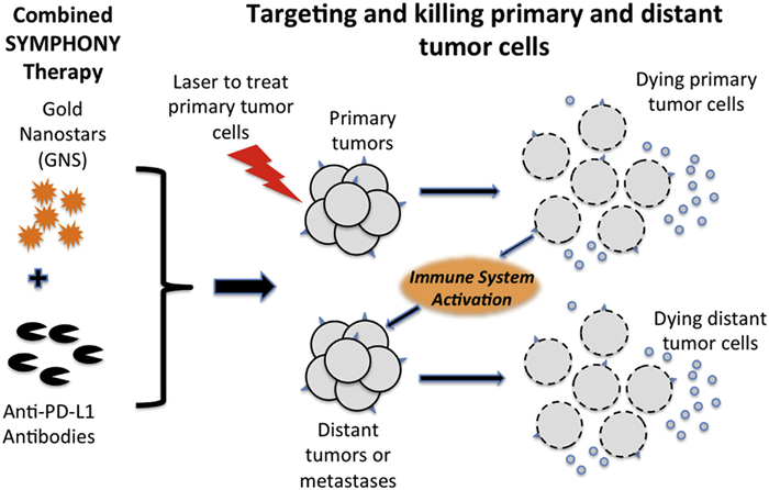
Scientists developed RMmAGL NPs for tumor-specific PTT and immune modulation. In acidic tumors, they release AuNPs-Glu/Lys for PTT and R837 to activate mature dendritic cells (DCs), enhancing T cell responses. Mannose-modified mesoporous silica nanoparticles (MSNs) polarize macrophages, further boosting immunity. In vivo/in vitro results show strong tumor inhibition, underscoring the value of targeted adjuvant delivery [178]. Wang et al. developed BSA-FA-functionalized iron MOFs (TPL@TFBF) that trigger ferroptosis and pyroptosis, releasing DAMPs to boost immunogenicity and suppress melanoma lung metastases in vivo (Fig. S12 in Supporting information) [179]. To overcome PD-1/PD-L1 blockade resistance, pH-sensitive MOF NPs encapsulating Hb and CAT were embedded in liposomes with PD-1 antibodies (αPD-1), forming C & H@MOF/PL-A for targeted delivery, oxygen generation, hypoxia relief, and enhanced colorectal cancer immune checkpoint blockade (ICB) (Fig. S13 in Supporting information) [180]. Liu et al. showed plasmonic gold nano-star PTT plus anti-PD-L1 eradicated primary and distant MB49 bladder tumors in mice (Fig. 6) [181].
Engineered inorganic nanostructures, once thought biologically inert, are now recognized as nanozymes with POD, oxidase, and superoxide dismutase activities [182]. They catalyze reactions under physiological conditions like natural enzymes. A new type, "synthetic biological nanozymes", is created via in-situ growth of MNPs on engineered protein scaffolds, combining protein structure with catalytic function.
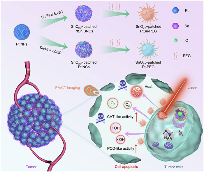
Ferritin, a natural spherical nanostructure with tumor-targeting ability, can be engineered into nanozymes for drug delivery. Fan et al. developed Fe-TCPP-R848-PEG (Fe-MOF-RP) [183], which relieves hypoxia, consumes GSH, and induces iron-mediated apoptosis (Fig. S14 in Supporting information). Xue et al. designed iron-based MOFs (FessMOFs) that deplete GSH and, with Actinomycin D (ActD) (Fig. S15 in Supporting information), enhance iron therapy [184]. Researchers have developed nanoreactors (NRs) incorporating 2,2′-azino-bis(3-ethylbenzothiazoline-6-sulfonic acid) (ABTS) loaded into ABTS@MIL-100/poly(vinylpyridine) (AMP) for imaging-guided, combined PTT and enhanced catalytic detoxification therapy (ECDT) of tumors (Fig. S16 in Supporting information) [185]. In addition to single-metal nanozymes, researchers have synthesized dual-metal nanozymes (Pt50Sn50) that exhibit enhanced photothermal and dual enzyme activity for tumor catalytic therapy. As illustrated in Fig. 7, Pt50Sn50 enable catalytic therapy with dual-mode imaging [186]. This allows for real-time in vivo monitoring of the treatment process. The development of these nanozymes is promising for advancing clinical non-invasive tumor detection and holds significant potential for future applications.
MNP-based nanomedicines, with unique optical and magnetic properties, show great potential in diagnosis, drug delivery, imaging, and therapy. For this purpose, we analyzed some MNPs in biomedical applications (Table S1 in Supporting information). They are effective against rheumatoid arthritis and drug-resistant bacteria [187-190] For instance, a polyether ketone scaffold (PH-CpBT) acts as an ultrasound-activated antibacterial nano-reactor promoting angiogenesis and osteogenesis [191]. Currently, about 80 anti-tumor nanomedicines are in clinical trials, many with improved efficacy and fewer side effects, and the U.S. Food and Drug Administration (FDA) has approved several metal-containing nanomedicines and contrast agents. MNPs are also used in diagnostics, such as colloidal gold in rapid pathogen test strips.
Despite progress, MNPs face clinical translation challenges. The ROS responsive nano-drug delivery system (RHB/Cyc) alleviates liver fibrosis in animals, yet its biodegradability and metabolism require study [192]. Key issues include NP biocompatibility, biodistribution, toxicity, stability, and clearance. Advances in synthesis and surface modification aim to improve safety and targeting, while scalable production and stringent standardized evaluations of physicochemical properties, biodistribution, metabolism, and pharmacodynamics are essential.
In summary, metal NP-based nanomedicine holds great biomedical promise but requires overcoming scientific and technological hurdles. PEG, chitosan, and biodegradable materials reduce protein adsorption and ion release; pH, temperature, or magnetic stimuli enable controlled release; antibodies (such as anti-human epidermal growth factor receptor 2 (HER2)) or ligands (such as folic acid) enhance tumor targeting. Interdisciplinary advances are expected to address these challenges, expanding clinical applications and bringing more effective nano-drugs to market.
MNPs show vast biomedical potential, excelling in drug delivery, imaging, diagnosis, and therapy due to tunable surfaces and unique optical/electromagnetic properties, enabling targeted treatment and reduced side effects. Challenges remain in stability, biodistribution, metabolism, and toxicity. Efforts focus on developing more biocompatible, degradable materials and advanced surface modifications. Integrating nanotechnology and biomedical engineering promises intelligent, adaptive MNP platforms for personalized, safer, and more effective treatments.
AI-assisted design has boosted metal NP development, yet key challenges remain: Lack of standardized datasets, poor model interpretability, and weak integration with domain knowledge in chemistry, biology, and materials science. Addressing these requires interpretable, physics-informed AI and iterative prediction–validation cycles. AI-guided design will likely enable precise engineering of metal-containing nanoplatforms, including MOFs, leading to safer, smarter, and more effective.
The hybrid design strategy of MNPs offers advantages in precision tumor treatment [193]. Current challenges involve precise regulation of dynamic biointerfaces and cross-scale mechanism analysis, requiring integration of materials science, bioinformatics, and clinical medicine. Future directions include functionalized MNPs with targeted delivery, surface engineering, and collaborative optimization, e.g., TPMS topology-optimized Fe-Mn alloy stents for bone defect repair [194] and gene editing to remove tumor drug-resistance genes before MNP drug delivery. Establishing an ML-driven multidimensional MNP database (e.g., surface charge, crystal indices, drug mapping) can accelerate clinical translation and enable big-data immune toxicity analysis from multi-center trials. Overall, MNP applications in nanomedicine are rapidly expanding, promising revolutionary healthcare advances.
The authors declare that they have no known competing financial interests or personal relationships that could have appeared to influence the work reported in this paper.
Chun Yang: Writing – original draft. Yina Lou: Writing – original draft. Muran Bai: Writing – original draft, Visualization. Yingying Qian: Writing – original draft, Formal analysis. Wenwen Shen: Writing – review & editing. Yucheng Wan: Writing – original draft, Visualization. Yang Liu: Writing – original draft, Formal analysis. Qidi Zhao: Writing – original draft. Haiyu Hu: Writing – review & editing. Yiyuan Tang: Visualization, Writing – original draft, Writing – review & editing. Wei Wang: Writing – review & editing, Conceptualization. Zhijin Fan: Writing – review & editing, Project administration, Conceptualization. Yuhui Liao: Writing – review & editing, Supervision, Project administration.
This work was supported by the National Natural Science Foundation of China (No. 82002253), The Science and Technology Development Programme of Jilin Province (No. 20240305060YY), The Jilin Provincial Science and Technology Development Planning (No. 20220204136YY).
Supplementary material associated with this article can be found, in the online version, at doi:
X.X. Qi, M. Yao, M. Jin, H.Y. Guo, J. Nanosci. Nanotechnol. 21 (2021) 843–851. doi: 10.1166/jnn.2021.18697
H.Z. Deng, Y. Qu, B.Y. Chu, et al., Biomaterials 317 (2025) 123072. doi: 10.1016/j.biomaterials.2024.123072
Y.J. Zhang, W.H. Gao, Y.N. Ma, et al., Nano Today 49 (2023) 101768. doi: 10.1016/j.nantod.2023.101768
G.Q. Yuan, X.X. Luo, K. He, et al., Sci. Adv. 11 (2025) eadw7032. doi: 10.1126/sciadv.adw7032
T.S. Lan, D.X. Cui, T.Y. Liu, et al., Nano Biomed. Eng. 15 (2023) 330–341. doi: 10.26599/NBE.2023.9290025
V.V. Kshatriya, M.R. Kumbhare, S.V. Jadhav, et al., BIOI 5 (2024) 992.
Q.Y. He, S. Schmidt, W.Q. Zhu, et al., Science 382 (2023) 1065–1069. doi: 10.1126/science.adj2522
R. Sakthi Devi, A. Girigoswami, M. Siddharth, K. Girigoswami, Appl. Biochem. Biotechnol. 194 (2022) 4187–4219. doi: 10.1007/s12010-022-03963-z
L. Liu, X.F. An, M. Schaefer, et al., Biomed. Pharmacother. 153 (2022) 113511. doi: 10.1016/j.biopha.2022.113511
L.X. Zhang, G. Alimu, Z. Du, et al., ACS Omega 8 (2023) 21793–21801. doi: 10.1021/acsomega.3c01374
D.S. Wei, Y. Huang, B. Wang, et al., Angew. Chem. Int. Ed. 61 (2022) e202201486. doi: 10.1002/anie.202201486
M. Wang, R. Cai, Z.W. Zhang, et al., J. Nanobiotechnology 22 (2024) 355. doi: 10.1186/s12951-024-02568-4
N.N. Song, J.L. Zhang, J. Zhai, et al., Acc. Chem. Res. 54 (2021) 3313–3325. doi: 10.1021/acs.accounts.1c00267
D.A. da Silva, A. De Luca, R. Squitti, et al., J. Inorg. Biochem. 226 (2022) 111634. doi: 10.1016/j.jinorgbio.2021.111634
S. Samadzadeh, M. Babazadeh, N. Zarghami, et al., Mater. Sci. Eng. C: Mater. Biol. Appl. 118 (2021) 111384. doi: 10.1016/j.msec.2020.111384
E. Alphandéry, Int. J. Pharm. 586 (2020) 119472. doi: 10.1016/j.ijpharm.2020.119472
L.S. Ganapathe, M.A. Mohamed, R.M. Yunus, D.D. Berhanuddin, Magnetochemistry 6 (2020) 68. doi: 10.3390/magnetochemistry6040068
N. Lagopati, K. Evangelou, P. Falaras, et al., Pharmacol. Ther. 222 (2021) 107795. doi: 10.1016/j.pharmthera.2020.107795
M. Alavi, P. Kamarasu, D.J. McClements, M.D. Moore, Adv. Colloid. Interface Sci. 306 (2022) 102726. doi: 10.1016/j.cis.2022.102726
S. Tsymbal, G. Li, N. Agadzhanian, et al., Molecules 27 (2022) 7066. doi: 10.3390/molecules27207066
A. Manuja, B. Kumar, R. Kumar, et al., Toxicol. Rep. 8 (2021) 1970–1978. doi: 10.1016/j.toxrep.2021.11.020
S. Attarilar, J. Yang, M. Ebrahimi, et al., Front. Bioeng. Biotechnol. 8 (2020) 822. doi: 10.3389/fbioe.2020.00822
Y.S. Wei, L. Zou, H.F. Wang, et al., Adv. Energy Mater. 12 (2022) 2003970. doi: 10.1002/aenm.202003970
W.W. Chai, X.C. Chen, J. Liu, et al., Regen. Biomater. 11 (2024) rbad115. doi: 10.1093/rb/rbad115
X.C. Cai, X.G. Bao, Y.L. Wu, Pharmaceutics 14 (2022) 2641. doi: 10.3390/pharmaceutics14122641
J.B. Chen, M.S. Li, R.R. Sun, et al., Adv. Funct. Mater. 34 (2024) 2315276. doi: 10.1002/adfm.202315276
J.G. Qiu, M.A. Tomeh, Y. Jin, et al., J. Colloid. Interface Sci. 642 (2023) 810–819. doi: 10.1016/j.jcis.2023.03.172
T. Wen, G.L. Quan, B.Y. Niu, et al., Small 17 (2021) e2005064. doi: 10.1002/smll.202005064
Q.Y. Wang, Y. Yu, Y.X. Chang, et al., ACS Nano 17 (2023) 8483–8498. doi: 10.1021/acsnano.3c00694
X.X. Yang, Y.L. Chen, P.F. Feng, et al., Mater. Chem. Front. 6 (2022) 680–688. doi: 10.1039/D1QM01512E
J.H. Patil, J.K. Patel, U.A. Shah, et al., Nano Biomed. Eng. 16 (2024) 285–308. doi: 10.26599/NBE.2024.9290078
J.H. Li, P.X. Liu, Y. Chen, et al., J. Am. Chem. Soc. 145 (2023) 19707–19714. doi: 10.1021/jacs.3c04831
X.Y. Zhao, M. Long, X. Huang, Z.H. Zhang, Chem. Eng. J. 503 (2025) 158523. doi: 10.1016/j.cej.2024.158523
C.Q. Feng, X.Y. Liang, R.R. Fan, et al., MedComm. Biomater. Appl. 3 (2024) e96.
C.J. Yang, Y.L. Pan, H.L. Yu, et al., Adv. Sci. 10 (2023) e2302109. doi: 10.1002/advs.202302109
B. Das, J.L. Franco, N. Logan, et al., Nanomicro Lett. 13 (2021) 193.
S. Fedeli, J. Im, S. Gopalakrishnan, et al., Chem. Soc. Rev. 50 (2021) 13467–13480. doi: 10.1039/D0CS00659A
M. Zandieh, J. Liu, Adv. Mater. 36 (2024) e2211041. doi: 10.1002/adma.202211041
W.H. Gao, J.Y. He, L. Chen, et al., Nat. Commun. 14 (2023) 160. doi: 10.1038/s41467-023-35828-2
X.D. Zhang, X.K. Chen, Y.L. Zhao, Nanomicro Lett. 14 (2022) 95.
Y.J. Jiang, Z.B. Chen, N. Sui, Z.L. Zhu, J. Am. Chem. Soc. 146 (2024) 7565–7574. doi: 10.1021/jacs.3c13588
R.F. Zhang, Y.F. Shen, X.Y. Zhou, et al., Nat. Commun. 16 (2025) 890. doi: 10.1038/s41467-025-56134-z
P.W. Jin, Wang D. D, Y.J. Lu, et al., Adv. Sci. 12 (2025) e2502664. doi: 10.1002/advs.202502664
W.C. Peng, W.B. Tai, B.W. Li, et al., Nat. Mater. 24 (2025) 637–648. doi: 10.1038/s41563-024-02041-5
P. Yu, Y.Y. Li, H. Sun, et al., Adv. Mater. 35 (2023) e2303299. doi: 10.1002/adma.202303299
X. Wang, X.D. Qin, Y. Liu, et al., Adv. Mater. 36 (2024) e2411194. doi: 10.1002/adma.202411194
J.F. Zou, W.X. Ding, Y. Chen, et al., Carbohydr. Polym. 361 (2025) 123501. doi: 10.1016/j.carbpol.2025.123501
Y. Cheng, Y.M. Feng, J. Zhao, et al., Angew. Chem. Int. Ed. 64 (2025) e202422375. doi: 10.1002/anie.202422375
J.S. Bao, J.F. Wang, S.D. Chen, et al., ACS Nano 18 (2024) 9100–9113. doi: 10.1021/acsnano.3c13225
W. Zhang, Y.L. Lu, Y. Zang, et al., Nanomaterials 11 (2021) 3395. doi: 10.3390/nano11123395
Y.H. Zhang, X.H. Zhu, J. Zhang, et al., J. Colloid. Interface Sci. 600 (2021) 513–529. doi: 10.1016/j.jcis.2021.05.017
W. Zhang, Y. Zang, Y.L. Lu, et al., Int. J. Mol. Sci. 23 (2022) 4279. doi: 10.3390/ijms23084279
C.S.M. Martins, A.P. LaGrow, J.A.V. Prior, ACS Sens. 7 (2022) 1269-1299. doi: 10.1021/acssensors.2c00149
A.M. Wagner, J.M. Knipe, G. Orive, N.A. Peppas, Acta Biomater. 94 (2019) 44–63. doi: 10.1016/j.actbio.2019.05.022
O. Martinez-Mora, K. Tirez, F. Beutels, et al., Nanomaterials 13 (2023) 2610. doi: 10.3390/nano13182610
P. Zeng, L.F. Hang, G.F. Zhang, et al., Small 18 (2022) e2204748. doi: 10.1002/smll.202204748
Z.W. Yuan, X.X. Liu, J.B. Ling, et al., Biomaterials 287 (2022) 121620. doi: 10.1016/j.biomaterials.2022.121620
R.P. Dhavale, R.P. Dhavale, M.S. Bhatia, et al., J. Drug. Deliv. Sci. Tec. 81 (2023) 104213. doi: 10.1016/j.jddst.2023.104213
C. Castro-Hinojosa, S. Del Sol-Fernández, E. Moreno-Antolín, et al., Bioconjug. Chem. 34 (2023) 2275–2292. doi: 10.1021/acs.bioconjchem.3c00417
T.Y. Wei, R.Z. Zhao, L.J. Fang, et al., Adv. Funct. Mater. 34 (2024) 2311908. doi: 10.1002/adfm.202311908
X. Han, J. Chen, Z.H. Cheng, S.W. Zhou, Chem. Commun. 61 (2025) 1407–1410. doi: 10.1039/D4CC05933F
B.C. Hu, X.H. Xiao, P. Chen, et al., Biomaterials 290 (2022) 121811. doi: 10.1016/j.biomaterials.2022.121811
G.H. Hou, J.M. Qian, M. Guo, et al., J. Colloid. Interface Sci. 628 (2022) 968–983. doi: 10.1016/j.jcis.2022.08.091
M. Skowicki, S. Tarvirdipour, M. Kraus, et al., Adv. Drug Deliv. Rev. 211 (2024) 115354. doi: 10.1016/j.addr.2024.115354
L.Q. Li, S.Y. Li, Z.J. Fan, et al., ACS Photonics 9 (2022) 2555–2578. doi: 10.1021/acsphotonics.2c00440
H.N. Xu, Z.L. Guo, M.J. Li, et al., BIOI 5 (2024) 976.
Y. Tao, M.Q. Li, J.S. Ren, X.G. Qu, Chem. Soc. Rev. 44 (2015) 8636–8663. doi: 10.1039/C5CS00607D
H. Cui, Y.Y. Zhao, Q. Wu, et al., Bioact. Mater. 33 (2024) 532–544.
G. Lin, H.R. Lin, Z. Yuan, G. Liu, BIOI 4 (2023) 70.
L.X. Cheng, D.M. Sang, F.Y. Zhao, et al., J. Colloid. Interface Sci. 615 (2022) 95–109. doi: 10.1016/j.jcis.2022.01.099
A. Shakeri-Zadeh, J.W.M. Bulte, Nat. Rev. Bioeng. 3 (2025) 245–260.
T. Mishchenko, I. Balalaeva, A. Gorokhova, et al., Cell Death. Dis. 13 (2022) 455. doi: 10.1038/s41419-022-04851-4
Z. Li, W.P. Li, S.P. Jiang, et al., Chin. Chem. Lett. 35 (2024) 109150. doi: 10.1016/j.cclet.2023.109150
A.S. Chauhan, P. Chand, T.J. Parashar, BIOI 6 (2025) 994.
Y. Li, W. Li, T.C. Han, et al., Nat. Rev. Mater. 6 (2021) 488–507. doi: 10.1038/s41578-021-00283-2
R. Tang, X.Y. Yuan, Z. Jia, et al., ACS Appl. Nano Mater. 6 (2023) 11661– 11678. doi: 10.1021/acsanm.3c01650
Z.Y. Chen, X. Wang, Y.H. Ding, et al., ACS Mater. Lett. 5 (2023) 1237–1244. doi: 10.1021/acsmaterialslett.2c01024
Y.Z. Tang, X.J. Yu, L.R. He, et al., Nat. Commun. 16 (2025) 860. doi: 10.1038/s41467-025-56110-7
H.Y. Li, H.L. Zhang, X.F. He, et al., Adv. Mater. 35 (2023) e2211597. doi: 10.1002/adma.202211597
X.F. Meng, Z.H. Zhang, Y. Qian, et al., Adv. Healthc. Mater. 12 (2023) e2301926. doi: 10.1002/adhm.202301926
S.M. Wu, C. Xu, Y.W. Zhu, et al., Adv. Funct. Mater. 31 (2021) 2103591. doi: 10.1002/adfm.202103591
Y. Zhang, D.X. Li, J.S. Tan, et al., Small 17 (2021) e2005739. doi: 10.1002/smll.202005739310456
Y.J. Li, Y. Dong, X.Y. Zhou, K.L. Fan, MedComm Biomater. Appl. 2 (2023) e36.
B.Z. Yu, Y.S. Wang, T.J. Bing, et al., Adv. Mater. 36 (2024) e2310456. doi: 10.1002/adma.202310456
C. Fernández-Paz, S. Rojas, P. Salcedo-Abraira, et al., ACS Appl. Mater. Interfaces 12 (2020) 25676–25682. doi: 10.1021/acsami.0c07356
S. Haddad, I. Abánades Lázaro, M. Fantham, et al., J. Am. Chem. Soc. 142 (2020) 6661–6674. doi: 10.1021/jacs.0c00188
M.L. Essa, M.A. El-Kemary, E.M. Ebrahem Saied, et al., Materials 13 (2020) 5397. doi: 10.3390/ma13235397
Z. Cheng, M.Y. Li, R. Dey, Y.H. Chen, J. Hematol. Oncol. 14 (2021) 85. doi: 10.1186/s13045-021-01096-0
X.Y. Liang, Y.C. Li, B.L. Guo, et al., Adv. Funct. Mater. 34 (2024) 2314772. doi: 10.1002/adfm.202314772
F. Sukkar, M. Shafaa, M. El-Nagdy, W. Darwish, Int. J. Nanomed. Vol. 16 (2021) 8309–8321. doi: 10.2147/IJN.S331909
H.H. Xie, M. Yang, X.L. He, et al., Adv. Sci. 11 (2024) e2306494. doi: 10.1002/advs.202306494
X.C. Li, Y.H. Liu, J.J. Ke, et al., Adv. Mater. 36 (2024) e2414365. doi: 10.1002/adma.202414365
Z.Z. Fang, Y.F. Shen, D.Q. Gao, New. J. Chem. 45 (2021) 4534–4544. doi: 10.1039/D0NJ05169A
C.P. Liu, D.Y. He, L.M. Li, et al., Pharmacol. Res. 183 (2022) 106364. doi: 10.1016/j.phrs.2022.106364
W.J. Xu, Z.K. Zeng, Y.C. Tang, et al., J. Nanobiotechnology 22 (2024) 798. doi: 10.1186/s12951-024-03050-x
Y.D. Wang, H.D. Li, J.Y. Lin, et al., Nat. Commun. 16 (2025) 1876. doi: 10.1038/s41467-025-57190-1
C.W. Zhang, J.G. Zhang, X. Yang, et al., Expert. Opin. Drug Deliv. 19 (2022) 623–640. doi: 10.1080/17425247.2022.2081320
H.F. Li, P.Y. Wang, C. Wen, Nanomaterials 12 (2022) 2111. doi: 10.3390/nano12122111
J.G. Lin, W.H. Cai, Q. Peng, et al., Scanning 2021 (2021) 2717921.
L. Gu, Z.Y. Duan, X. Li, et al., Bioact. Mater. 26 (2023) 102–115.
F.A. Shah, M. Jolic, C. Micheletti, et al., Bioact. Mater. 19 (2023) 103–114.
M. Ibrahim, E. Ramadan, N.E. Elsadek, et al., J. Control. Release 351 (2022) 215–230. doi: 10.1016/j.jconrel.2022.09.031
L. Chen, Z.Y. Zhou, C. Hu, et al., Research 2022 (2022) 9845459.
H. Kim, Y. Joo, Y.M. Kook, et al., ACS Appl. Mater. Interfaces 14 (2022) 4931–4945. doi: 10.1021/acsami.1c20394
L.M. Sun, H.M. Liu, Y.Q. Ye, et al., Signal. Transduct. Target. Ther. 8 (2023) 418. doi: 10.1038/s41392-023-01642-x
R.F. Silveira, A.L. Lima, I.P. Gross, et al., Nanomedicine 19 (2024) 1271–1283. doi: 10.1080/17435889.2024.2359355
N. Serov, V. Vinogradov, Adv. Drug Deliv. Rev. 184 (2022) 114194. doi: 10.1016/j.addr.2022.114194
Z.Q. Niu, W.H. Zhao, H. Deng, et al., ACS Nano 18 (2024) 20504–20517. doi: 10.1021/acsnano.4c04943
K. Almansour, A.S. Alqahtani, Sci. Rep. 15 (2025) 8840. doi: 10.1038/s41598-025-92725-y
Y.H. Kang, J.H. Kim, Nat. Commun. 15 (2024) 4705. doi: 10.1038/s41467-024-48998-4
H. Wan, Y. Fang, M. Hu, et al., Adv. Sci. 12 (2025) e2405905. doi: 10.1002/advs.202405905
H. Mashhadimoslem, M.A. Abdol, P. Karimi, et al., ACS Nano 18 (2024) 23842–23875. doi: 10.1021/acsnano.3c13001
Y.F. Yue, S.A. Mohamed, N.D. Loh, J. Jiang, ACS Nano 19 (2025) 933–949. doi: 10.1021/acsnano.4c12369
M.A. Nawaz, R. Blay-Roger, M. Saif, et al., ACS Catal. 14 (2024) 15150–15196. doi: 10.1021/acscatal.4c03941
X.J. Pei, G.Y. Tao, X. Wu, et al., Analyst 145 (2020) 4111–4123. doi: 10.1039/D0AN00392A
X.P. Li, Q. Zeng, R.J. Zhang, et al., Chem. Eng. J. 450 (2022) 138210. doi: 10.1016/j.cej.2022.138210
M.Y. Ding, S.S. Cui, L. Fang, et al., Angew. Chem. Int. Ed. 61 (2022) e202203631. doi: 10.1002/anie.202203631
P.L. Zhou, H.B. Yu, Y. Zhong, et al., Nanomicro Lett. 12 (2020) 166.
X.M. Huang, L.Y. Li, Z.X. Chen, et al., Adv. Mater. 35 (2023) e2302431. doi: 10.1002/adma.202302431
M. Zhu, Z.J. Kuang, F. Xu, et al., ACS Appl. Bio Mater. 6 (2023) 1586–1593. doi: 10.1021/acsabm.3c00001
A. Loewa, J.J. Feng, S. Hedtrich, Nat. Rev. Bioeng. 1 (2023) 545–559. doi: 10.1038/s44222-023-00063-3
A.H.I. Khormi, R.M.M. Qohal, A.Y.A. Masrai, et al., Egypt. J. Chem. 67 (2024) 1567–1574. doi: 10.21608/ejchem.2024.337883.10838
G.B. Wu, J.Y. Dai, Q. Cheng, et al., Nat. Electron. 5 (2022) 808–819. doi: 10.1038/s41928-022-00857-0
Y. Shakhman, I. Shumilin, D. Harries, J. Colloid. Interface Sci. 629 (2023) 165–172. doi: 10.1016/j.jcis.2022.08.123
X. Wang, J. Chen, H.B. Shi, MedComm. Biomater. Appl. 4 (2025) e70010.
K. Roshanbinfar, M. Kolesnik-Gray, M. Angeloni, et al., Adv. Healthc. Mater. 12 (2023) e2202408. doi: 10.1002/adhm.202202408
Q.L. Wang, T.T. Wang, C. Lio, et al., J. Control. Release 356 (2023) 678–690. doi: 10.1016/j.jconrel.2023.03.008
J.Y. Qiu, P.Q. Jiang, C.H. Wang, et al., Anal. Chem. 94 (2022) 4720–4728. doi: 10.1021/acs.analchem.1c05061
A. Alnaimi, A. Al-Hamry, Y. Makableh, et al., Biosensors 12 (2022) 1130. doi: 10.3390/bios12121130
D. Yuan, J.J. Liu, H.Z. Zhang, et al., Talanta 188 (2018) 218–224. doi: 10.1016/j.talanta.2018.05.086
P. Kshirsagar, P. Seshacharyulu, S. Muniyan, et al., Nanomedicine 43 (2022) 102566. doi: 10.1016/j.nano.2022.102566
R.T. Xu, L.X. Ouyang, H.Y. Chen, et al., Biosensors 13 (2023) 474. doi: 10.3390/bios13040474
R.Y. Yang, P. Zhao, L.Y. Wang, et al., Nat. Biomed. Eng. 8 (2024) 1018–1039.
L. Huang, J.J. Wan, X. Wei, et al., Nat. Commun. 8 (2017) 220. doi: 10.1038/s41467-017-00220-4
W.Y. He, K.L. Ai, L.H. Lu, Sci. China Chem. 58 (2015) 753–760. doi: 10.1007/s11426-015-5351-8
J.Y. Zhan, R. Liu, J.J. Zhang, Chin. J. Anal. Chem. 49 (2021) 1133–1141.
M. Jouyandeh, S.M. Sajadi, F. Seidi, et al., OpenNano 8 (2022) 100104. doi: 10.1016/j.onano.2022.100104
Y.F. Meng, D.S. Zhang, X. Chen, et al., ACS Appl. Nano Mater. 3 (2020) 4494–4503. doi: 10.1021/acsanm.0c00581
Z.A. Chen, Z.H. Li, C.J. Li, et al., Drug Deliv. 29 (2022) 1201–1211. doi: 10.1080/10717544.2022.2059124
S. Sargazi, U. Laraib, S. Er, et al., Nanomaterials 12 (2022) 1102. doi: 10.3390/nano12071102
O. Rabin, J. Manuel Perez, J. Grimm, et al., Nat. Mater. 5 (2006) 118–122. doi: 10.1038/nmat1571
Z.H. Song, T. Liu, H.Q. Lai, et al., ACS Nano 16 (2022) 4379–4396. doi: 10.1021/acsnano.1c10663
J.X. Luo, J.C. Li, M. Yu, et al., Prog. Biochem. Biophys. 48 (2021) 715–720.
D. Zhang, Y.X. Zhao, Y.J. Gao, et al., J. Mater. Chem. B 1 (2013) 5100–5107. doi: 10.1039/c3tb20907e
W. Qin, J. Chandra, M.A.S. Abourehab, et al., Mol. Cancer 22 (2023) 87. doi: 10.1186/s12943-023-01784-0
H.Z. Ma, X.N. Zhang, L. Liu, et al., Sci. Adv. 9 (2023) eadh7828. doi: 10.1126/sciadv.adh7828
J.J. Xu, W.C. Zhang, Y.W. Guo, et al., Drug Deliv. 29 (2022) 664–678. doi: 10.1080/10717544.2022.2039804
J. Zhang, X. Ke, T.Y. Ci, J. Funct. Poly. 34 (2021) 215–229.
B.T. Li, B.G. Kou, Y.J. Jiang, et al., Cancer Res. 49 (2022) 832–837.
J.J. Xiang, Y.W. Li, Y.F. Zhang, et al., J. Control. Release 330 (2021) 992–1003. doi: 10.1016/j.jconrel.2020.11.006
L.H. Su, Q. Wu, L.F. Tan, et al., ACS Appl. Mater. Interfaces 11 (2019) 10520–10531. doi: 10.1021/acsami.8b22177
B.Q. Zhou, J.X. Liu, L. Wang, et al., Nanoscale 14 (2022) 4588–4594. doi: 10.1039/D1NR07750C
L. Chen, L.L. Zhou, C.H. Wang, et al., Adv. Mater. 31 (2019) e1904997. doi: 10.1002/adma.201904997
S. Chen, R.Q. Zhao, X.Y. Sun, et al., Adv. Healthc. Mater. 12 (2023) e2201924. doi: 10.1002/adhm.202201924
T. Asao, G.C. Tobias, S. Lucotti, et al., ExtraCell Vesicles. Circ. Nucl. Acids 4 (2023) 461–485. doi: 10.20517/evcna.2023.37
Y.D. Wang, F.C. Gao, X.F. Li, et al., J. Nanobiotechnology 20 (2022) 69. doi: 10.1186/s12951-022-01278-z
C.Y. Cao, X.R. Wang, N. Yang, et al., Chem. Sci. 13 (2022) 863–889. doi: 10.1039/D1SC05482A
D.G. Liang, Y. Feng, F. Zandkarimi, et al., Cell 186 (2023) 2748–2764 e22. doi: 10.1016/j.cell.2023.05.003
W.T. Chen, F.F. Hu, Q. Gao, et al., J. Nanobiotechnology 22 (2024) 98. doi: 10.1186/s12951-024-02308-8
W.F. Song, J.Y. Zeng, P. Ji, et al., Small 19 (2023) e2301148. doi: 10.1002/smll.202301148
Y. Liu, S.Y. Chi, Y. Cao, Z.H. Liu, ACS Appl. Nano Mater. 5 (2022) 2592–2602. doi: 10.1021/acsanm.1c04277
F.L. Gao, M.M. Sun, J. Zhang, et al., J. Colloid. Interface Sci. 616 (2022) 369–378. doi: 10.1016/j.jcis.2022.02.060
S. Khan, M. Sharifi, A. Hasan, et al., J. Adv. Res. 30 (2021) 171–184. doi: 10.1016/j.jare.2020.12.001
Z.L. Li, H. Wu, J.Q. Zhu, et al., Small Sci. 2 (2022) 2200024. doi: 10.1002/smsc.202200024
H.T. Sun, Y.Y. Zhang, S.Y. Chen, et al., ACS Appl. Mater. Interfaces 12 (2020) 30145–30154. doi: 10.1021/acsami.0c07013
W. Ma, H.L. Zhang, S.Y. Li, et al., ACS Biomater. Sci. Eng. 8 (2022) 1354–1366. doi: 10.1021/acsbiomaterials.1c01605
M.Y. Zhao, R.G. Yang, Y.R. Wei, et al., Nano Today 44 (2022) 101493. doi: 10.1016/j.nantod.2022.101493
P. Ji, P. Wang, H. Chen, et al., Pharmaceuticals 16 (2023) 234. doi: 10.3390/ph16020234
Y.X. Wang, J. Chen, R.M. Duan, et al., Adv. Mater. 34 (2022) e2109726. doi: 10.1002/adma.202109726
N. Li, Q.Q. Sun, Z.Z. Yu, et al., ACS Nano 12 (2018) 5197–5206. doi: 10.1021/acsnano.7b06870
X. Sun, Z.Y. Zhang, C.B. Xiang, et al., Nano Biomed. Eng. 17 (2025) 248–262. doi: 10.26599/NBE.2025.9290120
R.A. Mustafa, M.X. Ran, Y.H. Wang, et al., Smart Mater. Med. 4 (2022) 199–211.
L.L. Zhang, X.Q. Qian, Y.Y. Yang, et al., Nano Biomed. Eng. 17 (2025) 277–286. doi: 10.26599/NBE.2024.9290105
W.J. Jin, Z.L. Chen, Y. Wang, et al., Chin. Chem. Lett. 35 (2024) 109328. doi: 10.1016/j.cclet.2023.109328
N. Guo, Y. Xia, Y.X. Duan, et al., Chin. Chem. Lett. 34 (2023) 107542. doi: 10.1016/j.cclet.2022.05.056
E. Choi, M. Landry, N. Pennock, et al., Adv. Healthc. Mater. 12 (2023) e2202830. doi: 10.1002/adhm.202202830
S.P. Tang, L. Zhou, H.Y. He, et al., Biomaterials 288 (2022) 121706. doi: 10.1016/j.biomaterials.2022.121706
T. Liu, M. Zhu, X.W. Chang, et al., Adv. Mater. 35 (2023) e2300086. doi: 10.1002/adma.202300086
S.M. Wang, Q.Y. Guo, R.B. Xu, et al., J. Nanobiotechnology 21 (2023) 383. doi: 10.1186/s12951-023-02146-0
J. Huang, Z.C. Xiao, M.Z. Lin, et al., Nano Today 54 (2024) 102102. doi: 10.1016/j.nantod.2023.102102
Y. Liu, P. Maccarini, G.M. Palmer, et al., Sci. Rep. 7 (2017) 8606. doi: 10.1038/s41598-017-09116-1
H.R. Wang, J. Meng, J.J. Lin, et al., Nat. Commun. 15 (2024) 4362. doi: 10.1038/s41467-024-48710-6
Z.J. Fan, S.C. Wu, H.P. Deng, et al., ACS Nano 18 (2024) 12261–12275. doi: 10.1021/acsnano.4c00844
P.P. Xue, H.L. Zhuang, T.J. Bai, et al., J. Nanobiotechnology 22 (2024) 228. doi: 10.1186/s12951-024-02508-2
F. Liu, L. Lin, Y. Zhang, et al., Adv. Mater. 31 (2019) e1902885. doi: 10.1002/adma.201902885
Y.L. Zhu, R.X. Zhao, L.L. Feng, et al., ACS Nano 17 (2023) 6833–6848. doi: 10.1021/acsnano.3c00423
Z.Y. Hu, J. Shan, X. Jin, et al., ACS Nano 18 (2024) 24327–24349. doi: 10.1021/acsnano.4c06565
W.Q. Wang, Y.Y. Cui, X.L. Wei, et al., ACS Nano 18 (2024) 15845–15863. doi: 10.1021/acsnano.4c02825
X.T. Feng, Z.W. Luo, W. Zhang, et al., Adv. Funct. Mater. 35 (2025) 2506476. doi: 10.1002/adfm.202506476
Y.W. Sun, W. Zhang, Z.W. Luo, et al., Adv. Funct. Mater. 35 (2024) 2415778.
Y.L. Huang, X.F. Wan, Q. Su, et al., Nat. Commun. 15 (2024) 1643. doi: 10.1038/s41467-024-45619-y
J.H. Zhang, T. Zhang, Z.J. Zhang, et al., Adv. Funct. Mater. 34 (2024) 2404658. doi: 10.1002/adfm.202404658
J.J. Zhang, B.Q. Gao, B.L. Ye, et al., Adv. Mater. 35 (2023) e2208571. doi: 10.1002/adma.202208571
M.S. Dargusch, N. Soro, A.G. Dmeir, et al., Smart Mater. Med. 5 (2023) 127–139.

Figure 1 Biological functions of MNPs. MNPs have a wide range of applications in bioimaging, disease treatment and drug delivery.

Figure 2 Application of MNPs in liquid biopsy. Schematic diagrams of experimental workflow and LDI MS process using SiO2@Ag nanoshells as matrix. Reprinted with permission [134]. Copyright 2017, The Author(s).

Figure 3 Application of MNPs in tumor imaging. PP@Mn NPs enhanced tumor ferroptosis mediated by the Fenton-like reaction through mild photothermal effects guided by MRI. Reprinted with permission [139]. Copyright 2022, The Author(s).

Figure 4 Schematic representation of the PEG-GAx/Pt formation through the coordination between polyphenol and CDDP and its dual-responsive intracellular drug release. Reprinted with permission [150]. Copyright 2020, Elsevier B.V.

Figure 5 Schematic diagram of the preparation of L/D-MnO@Pt NPs and the enhanced CDT by the specific internalization of tumor cells. Reprinted with permission [162]. Copyright 2022, Elsevier Inc.

Figure 6 A combination of immune checkpoint inhibition and plasmonic gold nanostar (GNS)-mediated PTT. Reprinted with permission [181]. Copyright 2017, The Author(s).

 扫一扫看文章


扫一扫关注我们
 DownLoad:
 下载:
下载:

 下载:
下载: