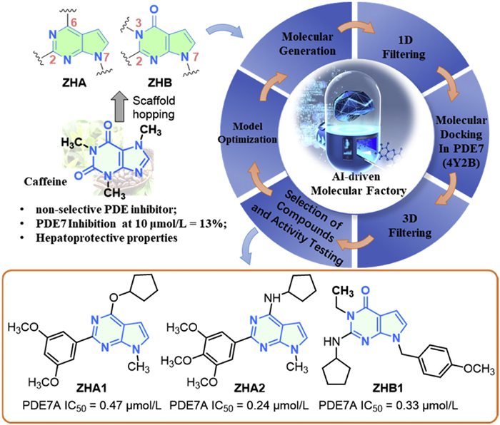
Figure 1.
The discovery of pyrrolopyrimidine PDE7 inhibitors ZHA1, ZHA2, and ZHB1 as lead compounds.

Discovery of novel pyrrolopyrimidine-based PDE7 inhibitors against alcoholic liver injury
Jia-Xi Huang , Chuang Xia , Le-Shen Li , Huichang Bi , Zhong-Zhen Zhou


Alcohol-associated liver disease is a significant global health issue affecting countries worldwide. Excessive alcohol consumption is a leading cause of liver disease and contributes to considerable morbidity and mortality worldwide [1]. In both China and the United States, the prevalence of alcohol-associated liver diseases, including alcoholic fatty liver, alcoholic hepatitis, and alcoholic cirrhosis, has been on the rise [2,3]. Currently, there is no approved specific pharmacotherapy for alcoholic liver injury [4]. Many drugs used in clinical trials for alcoholic liver injury are repurposed from other medical uses or are investigational compounds [5]. Aimed at identifying new therapeutic approaches to prevent, manage, and potentially reverse liver damage, drug development for alcoholic liver injury has become an active area of research.
Several strategies are being investigated to address different aspects of alcoholic liver injury, including anti-inflammatory agents, antioxidants, anti-fibrotic agents, metabolic modulators, and microbiome-targeted therapies [4]. It is essential to recognize that the cyclic adenosine monophosphate (cAMP) signaling pathway plays a pivotal role in regulating inflammation, oxidative stress, and fibrogenesis [6–11]. Targeting cAMP signaling pathways may offer a therapeutic approach for alcoholic liver injury by addressing inflammation, oxidative stress, and fibrogenesis [7]. In cAMP-specific hydrolases, both phosphodiesterase 7 (PDE7) and PDE4 are present in the liver; however, PDE7 inhibitors do not cause the vomiting side effect associated with PDE4 inhibitors [12,13]. Moreover, PDE7 inhibitors, such as BRL-50481 [14] and SUN11817 [15], have shown promise in preclinical studies for reducing liver inflammation and ameliorating liver injury. The exact role of PDE7 in alcoholic liver injury and its potential therapeutic implications are still under investigation [16]. So, what is the role of PDE7 inhibitors in alcoholic liver disease (ALD)?
Currently, there are no Food and Drug Administration (FDA)-approved PDE7A inhibitors [17], underscoring the need for novel compounds with distinct chemical structures to further advance the development of PDE7A inhibitors. Our ongoing research program involves the development of novel nitrogen-heterocyclic derivatives to discover PDE inhibitors (such as PDE4 and PDE7) for treating ALD. Recently, our previous studies have reported that 5-azaindazole derivative ZL40, as a PDE4 inhibitor, displays the potential to treat alcoholic liver injury and inflammation in the National Institute for Alcohol Abuse and Alcoholism (NIAAA) mice model. In this work, we described the identification of 7-deazapurines as PDE7 inhibitors against ALD and successfully discovered 3,7-dihydro-4H-pyrrolo[2,3-d] pyrimidin-4-one ZHB12 as a potent and selective PDE7 inhibitor (50% inhibitory concentration (IC50) against PDE7A = 0.2 µmol/L, PDE selectivity > 60-fold) with good oral bioavailability (F = 24.8%) and satisfactory safety against ALD.
Caffeine, a nonspecific PDE inhibitor structurally similar to adenosine, is known for its unique aroma and psychostimulant effects. It prevents the hydrolysis of cAMP and increases intracellular cAMP levels. Caffeine displayed weak inhibitory activity against PDE7 but exhibited anti-fibrotic properties on hepatic stellate cells and hepatocytes [18]. Based on the scaffold hopping strategy, novel scaffolds of pyrrolopyrimidine, such as 7H-pyrrolo[2,3-d]pyrimidine and 3,7-dihydro-4H-pyrrolo[2,3-d]pyrimidin-4-one (Fig. 1), were transitioned from caffeine. Pyrrolopyrimidines are present in natural alkaloids (such as cadeguomycin, tubercidin, and toyocamycin) and FDA-approved drugs (such as pemetrexed, ruxolitinib, tofacitinib, baricitinib, and ribociclib) [19,20]. Some pyrrolopyrimidine derivatives [21,22] displayed good anti-inflammatory activity. How can we develop PDE7 inhibitors using 7H-pyrrolo[2,3-d]pyrimidine and 3,7-dihydro-4H-pyrrolo[2,3-d]pyrimidin-4-one?
Artificial intelligence (AI)-assisted drug design has remarkably affected contemporary drug discovery. The DrugFlow platform (
Following the protein-ligand interaction observed in the PDE7 crystal structure (PDB ID: 4Y2B), the molecular factory generated derivatives using a side-chain modification strategy. These compounds exhibit significant structural differences from the reference molecules, demonstrating high novelty and diversity. To efficiently filter a large number of molecules generated by generative models, we employed an automated filtration process that includes both 1D and 3D screening, including molecular weight, topological polar surface area (TPSA), lipid-water partition coefficient (logP), water solubility index (logS), the root mean square deviation (RMSD), binding free energy prediction based on a continuum model, an AI-driven protein-ligand scoring function known as RTMScore, and interaction with key amino acid residues. This process enables us to eliminate molecules with poor druggability quickly.
Based on the 1D screening stage, 3D filtration, and key amino acid residues (such as the hydrogen bond with Gln413 and π-π interactions with Phe416), the highest-ranked compounds were selected for activity testing against PDE7A. Compared with the positive drug BRL-50481 (IC50 = 0.64 µmol/L), ZHA1, ZHA2, and ZHB1 displayed higher activity against PDE7A, with IC50 values of 0.47, 0.24, and 0.32 µmol/L, respectively. These results encourage us to optimize the structures of ZHA1, ZHA2, and ZHB1 to improve their structure-activity relationships (SARs) and to develop more potent pyrrolopyrimidine derivatives as PDE7 inhibitors.
The synthetic routes for the novel target derivatives ZHA1-ZHA12 and ZHB1-ZHB20 are illustrated in Schemes S1–S4 (Supporting information).
As shown in Table 1, compound ZHA2, bearing a cyclopentylamine group and a 3,4,5-trimethoxyphenyl group, displayed higher PDE7 inhibitory activity than compound ZHA1, bearing a cyclopentyloxy group and a 3,5-dimethoxyphenyl group. However, replacing the 3,4,5-trimethoxyphenyl group of ZHA2 or replacing the 3,5-dimethoxyphenyl group of ZHA1 with another polymethoxyphenyl group (such as ZHA3–8) resulted in a decrease in activity against PDE7. There was no apparent structure-activity relationship in compounds ZHA1–8. Based on these results, we initiated an investigation into the effect of substituents at the N-7 position on the activity, using ZHA2 as the lead compound. Unfortunately, compared with ZHA2, substituting the methyl group with larger groups, such as isopropyl (ZHA9), cyclopentyl (ZHA10), tetrahydro-2H-pyran-4-yl (ZHA11), and 4-fluorobenzyl (ZHA12), resulted in reduced PDE7A inhibitory activity.
 DownLoad:
CSV
 |
|||
| Compound | Ar or R1 | Inhibition rates (%)b at 1.0 µmol/L | IC50 (µmol/L)a |
| ZHA1 | 3,5-Dimethoxyphenyl | 75% | 0.47 ± 0.08 |
| ZHA2 | 3,4,5-Trimethoxyphenyl | 79% | 0.24 ± 0.05 |
| ZHA3 | 3,4,5-Trimethoxyphenyl | 4% | NAc |
| ZHA4 | 3,4-Dimethoxyphenyl | 9% | NAc |
| ZHA5 | 2,4-Dimethoxyphenyl | 9% | NAc |
| ZHA6 | 3,5-Dimethoxyphenyl | 7% | NAc |
| ZHA7 | 3,4-Dimethoxyphenyl | 3% | NAc |
| ZHA8 | 2,4-Dimethoxyphenyl | 50% | NTd |
| ZHA9 | Isopropyl | 11% | NAc |
| ZHA10 | Cyclopentyl | 74% | 0.32 ± 0.04 |
| ZHA11 | Tetrahydro-2H-pyran-4-yl | 42% | NAc |
| ZHA12 | 4-Fluorobenzyl | 75% | 0.33 ± 0.04 |
| BRL-50481 | - | 56% | 0.64 ± 0.02 |
| a Obtained IC50 values (mean ± SD) were the average values of triplicate independent experiments. b Inhibition rates (%) at 1 µmol/L were the average values of triplicate independent experiments. c NA, no activity. The inhibition rate (%) at 1 µmol/L against PDE7A is < 20%. d NT, no test. |
|||
While the initial optimization of activities did not produce compounds with improved efficacy, the PDE7A inhibitory activity of compounds ZHA1, ZHA2, ZHA10, and ZHA12 was higher than that of the reference BRL-50481. However, no distinct SARs were identified in compounds ZHA1–12. In addition, the ADMET prediction (Table S1 in Supporting information) and the test of solubility and lipid-water partition coefficient (Table S2 in Supporting information) indicated that ZHA1, ZHA2, ZHA10, and ZHA12 exhibited low solubility and unreasonable lipid-water partition coefficients. This finding is consistent with reports on 5H-pyrrolo[2,3-d]pyrimidine [24] and thienopyrimidin-4-amine analogs [25] bearing polymethoxyphenyl groups, which demonstrate poor druggability. Due to the unsatisfactory druggability of 7H-pyrrolopyrimidines, we explored alternative scaffolds by focusing on the structural optimization of 4H-pyrrolopyrimidin-4-one derivatives.
Based on the lead compound ZHB1 simulated by the Molecular Factory of DrugFlow, replacing the 4-methoxybenzyl group at the N-7 position with a small volume substituent resulted in the Discovery of ZHB2 (IC50 = 0.27 µmol/L, Table 2) and ZHB3 (IC50 = 0.24 µmol/L, Table 2), which demonstrated higher PDE7 inhibitory activity than ZHB1 and reference compound BRL-50481. These results prompted us to investigate the impact of N-7 substituents on PDE7 inhibitory activity. As shown in Table 2, the cyclopentyl group at the N-7 position, as in ZHB4, did not exhibit PDE7 inhibitory activity. In contrast, the long-side chain at the N-7 position of the 4H-pyrrolopyrimidin-4-one, such as ZHB5–10, could maintain PDE7 inhibitory activity compared to ZHB3 and exhibited higher inhibitory activity than the reference compound BRL-50481.
DownLoad:
CSV
 |
|||
| Compound | R1 or R2 | Inhibition rates (%)b at 1.0 µmol/L | IC50 (µmol/L)a |
| ZHB1 | 4-Methoxybenzyl | 77% | 0.32 ± 0.07 |
| ZHB2 | Allyl | 77% | 0.27 ± 0.05 |
| ZHB3 | Isopropyl | 78% | 0.24 ± 0.06 |
| ZHB4 | Cyclopentyl | 6% | NAc |
| ZHB5 | Pyridin-3-ylmethyl | 79% | 0.26 ± 0.08 |
| ZHB6 | 1-Allylpiperidin-4-yl | 82% | 0.29 ± 0.02 |
| ZHB7 | 1-Benzylpiperidin-4-yl | 76% | 0.31 ± 0.04 |
| ZHB8 |  |
74% | 0.36 ± 0.04 |
| ZHB9 | 1-Acetylpiperidin-4-yl | 72% | 0.35 ± 0.05 |
| ZHB10 | 1-Propionylpiperidin-4-yl | 80% | 0.28 ± 0.03 |
| ZHB11 | Cyclopropyl | 25% | NTd |
| ZHB12 | Isopropyl | 84% | 0.20 ± 0.02 |
| ZHB13 | 4,4-Difluorocyclohexyl | 9% | NAc |
| ZHB14 | Phenyl | 63% | NTd |
| ZHB15 | Thiophen-3-yl | 22% | NTd |
| ZHB16 | Cyclopropyl | 74% b | NTd |
| ZHB17 | -CH2CH2COOH | 66% b | NTd |
| ZHB18 | -CH2CH2CH2COOH | 36% b | NTd |
| ZHB19 |  |
27% b | NTd |
| ZHB20 |  |
66% b | NTd |
| a Obtained IC50 values (mean ± SD) were the average values of triplicate independent experiments. b Inhibition rates (%) at 1.0 µmol/L were the average values of triplicate independent experiments. c NA, no activity. The inhibition rate (%) at 1 µmol/L against PDE7A is < 20%. d NT, no test. |
|||
Based on our findings with ZHB3, we further explored the influence of amino substituents at the C-2 position of 4H-pyrrolopyrimidin-4-one on PDE7 inhibitor activity. As shown in Table 2, larger amino substituents, such as those in compounds ZHB13, ZHB14, and ZHB15, decreased inhibitor activity. More interestingly, substituting the cyclopentyl amino group with the isopropyl amino group resulted in the identification of compound ZHB12 (IC50 = 206 ± 25 nmol/L), which exhibited greater potency than ZHB3. Additionally, introducing a cyclopropanyl amino group and a cyclopropanyl group at the C-2 and N-7 positions resulted in compound ZHB16 (Table 2), which exhibited slightly lower inhibitor activity than ZHB12. However, attempts to enhance PDE7 inhibitory activity by introducing long alkyl chains with hydrophilic groups at their termini, as in compounds ZHB17–20, did not yield improved results; these modifications resulted in decreased activity.
To further clarify the structure-activity relationship, we conducted molecular docking studies on compounds ZHB3 and ZHB12. As shown in Fig. S1 (Supporting information), the 4H-pyrrolopyrimidin-4-one ring formed a hydrogen bond with glutamine (Gln413), a hydrogen bond bridge of water molecules contacting Asn365, and π-π interactions with conserved phenylalanines (Phe416 and Phe384) of PDE7. Regarding the C-2 position, the amino (-NH-) group interacts with Tyr211, forming an intermolecular hydrogen bond bridge through a conserved water molecule. The alkyl amino groups (-NHR2, such as isopropyl and cyclopentyl amino) at the C-2 position extend toward the metal. The isopropyl group at the N-7 position extended to the hydrophobic region.
As shown in Table S2 (Supporting information), ZHB12 demonstrated good solubility (S (pH 7.0) = 0.016 mg/mL) in pure water and a lipid-water partition coefficient (logP) of 3.02, indicating its favorable druggability. Thus, ZHB12 was chosen for further evaluation of its selectivity for PDE7, as detailed in Table 3. Compound ZHB12 exhibited fragile inhibitory activities against several PDEs, including PDE1A, PDE2A, PDE3B, PDE5A, PDE8A1, and PDE9A2 at 10 µmol/L. Additionally, the inhibition activities of ZHB12 on PDE4B and PDE4D were 12.41 and 16.53 µmol/L, respectively. This indicates that the selectivity against PDE7A reaches 60- and 80-fold compared to PDE4B and PDE4D, respectively. These results classify ZHB12 as a selective PDE7 inhibitor.
DownLoad:
CSV
| PDEs | IC50 (µmol/L) a | Selectivity index (PDE7/PDEs) |
| PDE1A | 21% b | > 80 |
| PDE2A | 25% b | > 80 |
| PDE3B | 31% b | > 80 |
| PDE4B | 12.41 ± 0.42 a | 62 |
| PDE4D | 16.53 ± 0.33 a | 80 |
| PDE5A | 27% b | > 80 |
| PDE7A | 0.20 ± 0.02 a | 1 |
| PDE8A1 | 17% b | > 80 |
| PDE9A2 | 9% b | > 80 |
| a Obtained IC50 values (mean ± SD) were the average values of triplicate independent experiments. b Inhibition rates (%) at 10 µmol/L were the average values of triplicate independent experiments. |
||
In subsequent experiments, we tested the pharmacokinetic parameters, acute toxicity, and efficacy of ZHB12 in mice. The animal experiment was approved by the Institutional Animal Care and Use Committee of Southern Medical University (No. SMUL202406021), and adhered strictly to the guidelines outlined in the "Guidelines for the Care and Use of Laboratory Animals" (National Institutes of Health, 1996). To investigate the pharmacokinetics of ZHB12, experiments were conducted in Sprague-Dawley rats using intragastric and intravenous administration. The mean plasma concentration-time curves for ZHB12 following these different drug delivery methods are illustrated in Fig. S2 (Supporting information). Additionally, pharmacokinetic parameters were calculated using non-compartmental analysis, as detailed in Table S4 (Supporting information).
After administering ZHB12 intravenously (i.v.) at 2 mg/kg, the area under the curve (AUC0−∞) was measured at 327.767 h ng mL−1. The peak plasma concentration (C0) reached 413.17 ng/mL, and the half-life (t1/2) was 0.42 h. This short half-life indicates that the drug is rapidly metabolized in the body. When ZHB12 was administered orally at 20 mg/kg, it demonstrated favorable pharmacokinetic properties. Specifically, the AUC0−∞ was recorded at 811.57 h ng mL−1, the maximum plasma concentration was 197.933 ng/mL, and the bioavailability was 24.8%. However, ZHB12 at a dose of 20 mg/kg displayed a short half-life (1.17 h) and a longer time to reach maximum concentration (Tmax). When the administration dose was increased from 20 mg/kg to 50 mg/kg, Tmax decreased to 0.5 h, but t1/2 increased to 2.58 h. Meanwhile, both clearance (Clz/F) and the apparent volume of distribution (Vz/F) showed improvements. The experimental results indicate that a dose of 50 mg/kg may demonstrate improved efficacy.
The significance of acute toxicity in clinical development prompted us to assess ZHB12′s acute toxicity by administering a single high dose (0.5 g/kg). Over a two-week observation period, the control and 0.5 g/kg dosage groups of mice remained alive with a slight upward trend in their weight and a favorable mental state and active condition (Fig. S3 in Supporting information). As illustrated in Fig. S4 (Supporting information), hematoxylin and eosin staining demonstrated that the tissue morphology of the heart, liver, spleen, lungs, and kidneys in the 0.5 g/kg dosage group did not differ significantly from that of the control group. These results indicate that ZHB12 is non-toxic and well-tolerated in mice at a dose of 0.5 g/kg. However, raising the dose to 0.75 g/kg resulted in the death of half of the mice (two females and one male) by the second day. The surviving mice exhibited reduced energy but began to recover by the third day. These results indicate that the mid-lethal dose of ZHB12 may be approximately 0.75 g/kg.
Previous studies [26,27] have demonstrated that chronic exposure to ethanol, combined with a single binge, in mice, referred to as the NIAAA model, results in liver injury, inflammation, and fatty liver. To evaluate the therapeutic effect of ZHB12 on ALD, we administered different doses (25 and 50 mg/kg) of ZHB12 to NIAAA model C57BL/6 mice (Fig. 2). Metadoxine (Met) is approved in Europe for treating alcohol intoxication and has shown promise in preclinical studies as a potential treatment for alcohol-induced liver damage when administered at higher oral doses (500–2000 mg kg−1 d−1) [28–30]. Thus, we used metadoxine (50 mg/kg) as the positive control in this model.
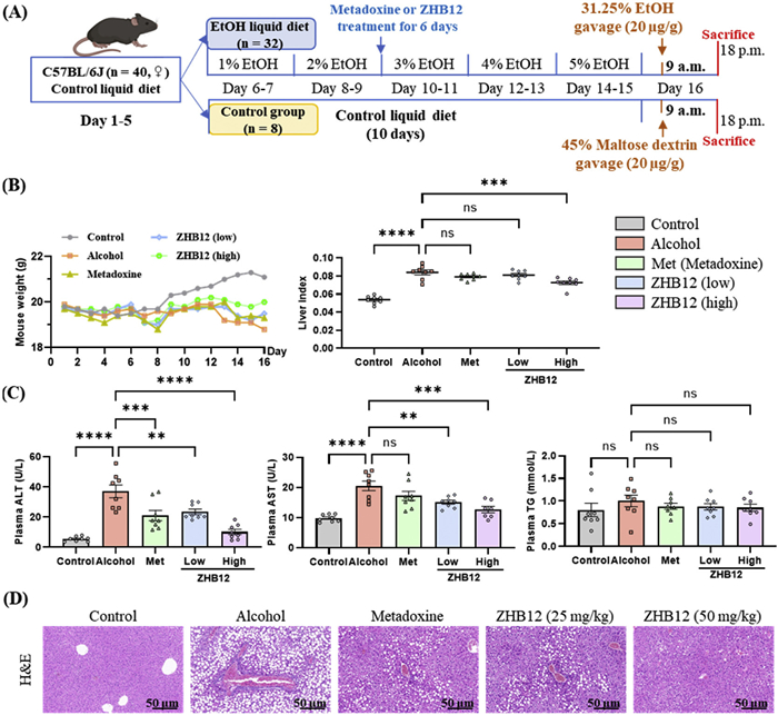
As illustrated in Fig. 2B, the alcohol-feeding groups showed a declining trend in weight; however, this decline was mitigated to varying degrees in the administration groups. Notably, the high-dose group administered in ZHB12 (50 mg/kg) successfully reversed the adverse effects associated with alcohol consumption. The levels of transaminases, specifically alanine transaminase (ALT) and aspartate aminotransferase (AST), are critical parameters for evaluating liver function. In the alcohol-feeding group, plasma levels of ALT and AST were significantly elevated compared to the control group. However, these levels decreased in a concentration-dependent manner in the ZHB12 groups.
More interestingly, compared with the metadoxine group, ZHB12 demonstrated a more potent effect in reducing plasma ALT and AST levels. To further investigate the extent of lipidation, we measured plasma triglyceride (TG) levels, as illustrated in Fig. 2C. The results indicated that TG levels in the alcohol-feeding group were slightly higher than those in the control group. We also observed minor decreases in other treatment groups, although these differences were not statistically significant.
Furthermore, histopathological examinations of liver sections stained with hematoxylin and eosin (H&E) showed significant improvements in alcoholic liver injury and steatosis after treatment with both metadoxine and ZHB12 (Fig. 2D). Remarkably, the liver tissue in the high-dose ZHB12 group (50 mg/kg) closely resembled that of the control group and exhibited greater effectiveness than the metadoxine group at the exact dosage (50 mg/kg). These findings suggest that ZHB12 effectively reduces liver injury and alleviates inflammation caused by alcohol in mice.
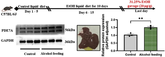
To better understand whether the anti-ALD efficacy of compound ZHB12 is associated with PDE7, we first assessed the impact of ethanol on PDE7 expression in the livers of NIAAA mouse models. More interestingly, we have discovered that the levels of PDE7A in the livers of ALD-model mice are higher than those of the standard mouse (Fig. 3). These results led us to explore further how compound ZHB12 regulates cAMP levels in liver tissue.
The cAMP signaling pathway plays a crucial role in various cellular programs and regulates numerous physiological activities, including cell proliferation, metabolism, migration, and adhesion [7,31]. Numerous studies have shown that hepatic cAMP levels are significantly lower in ethanol-fed rats compared to pair-fed controls [31,32]. Additionally, cAMP response element-binding protein (CREB) plays a crucial role in regulating fatty acid synthesis and oxidation in ethanol-fed ALD model rats [33]. As illustrated in Fig. 4A, hepatic cAMP levels are lower in ethanol-fed mice compared to the control group. However, the group receiving ZHB12 at either the low (25 mg/kg) or high (50 mg/kg) dose showed increased cAMP levels. These results suggest that the anti-alcoholic liver injury effect of ZHB12 is due to the upregulation of the cAMP signaling pathway.
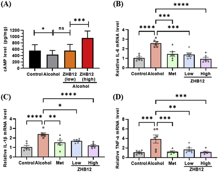
Inflammation is a key feature of alcoholic hepatitis [34,35]. The cAMP signaling pathway plays a crucial role in the progression of ALD, particularly in regulating metabolism and inflammation [7]. To assess the impact of ZHB12 on alcohol-induced liver inflammation, we used real-time quantitative fluorescence PCR (qRT-PCR) to evaluate the expression levels of the inflammatory cytokines interleukin-6 (IL-6), IL-1β, and tumor necrosis factor-alpha (TNF-α) in the NIAAA model mice. As shown in Figs. 4B–D, the alcohol-feeding group exhibited a notable upregulation of these cytokines (IL-6, IL-1β, and TNF-α) compared to the control group. However, with ZHB12 intervention, the levels of inflammatory cytokines decreased in a concentration-dependent manner. Notably, the high-dose ZHB12 group (50 mg/kg) demonstrated a more substantial effect against liver inflammation induced by alcohol.
Previous studies have shown that chronic ethanol feeding combined with carbon tetrachloride could establish an ALF model, resulting in a rise in liver inflammation, fibrosis, and fatty liver. In our study, we investigated the therapeutic effect of compound ZHB12 on the ALF model using C57BL/6 mice, administered at 50 mg/kg, with metadoxine (50 mg/kg) serving as a positive control (Fig. 5A).
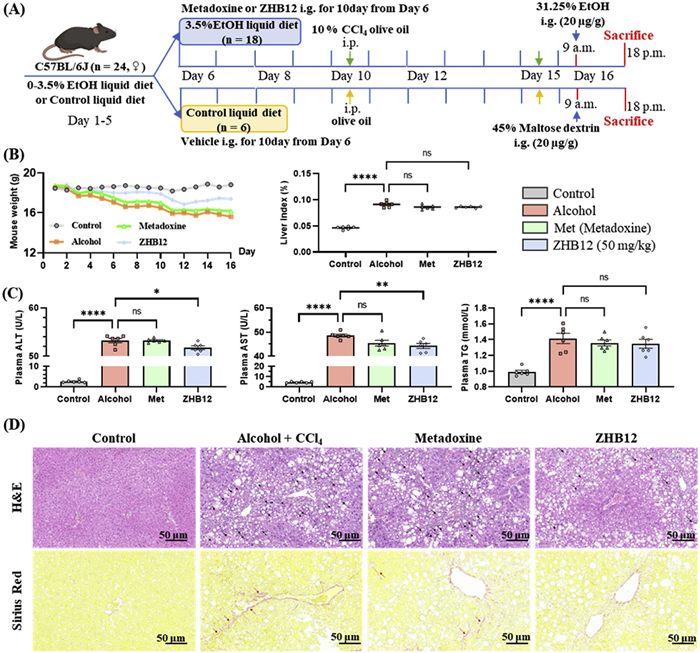
As shown in Fig. 5B, the alcohol-feeding groups showed a downward trend in body weight, whereas intervention with compound ZHB12 resulted in a modest improvement. The levels of ALT, AST, and TG in the plasma of ALF model mice were monitored to assess the model's response to ZHB12 treatment. Fig. 5C illustrates that ALT, AST, and TG levels were significantly elevated in the alcohol-feeding groups compared to the control group. The elevated ALT and AST levels in the ZHB12 group were reduced to a statistically significant extent, whereas no significant change was observed in the metadoxine group. Additionally, there was no significant difference in TG levels between the model and administration groups.
The H&E-stained liver sections exhibited a degree of neutrophil infiltration, and there was a noticeable increase in the number of oil droplets and scars in the alcohol-feeding groups compared to the control group (Fig. 5D). However, the ZHB12 intervention mildly suppressed these effects. Additionally, analysis of the Sirius red-stained liver sections indicated that both metadoxine and ZHB12 could slightly inhibit the expansion of the chicken-wire pattern caused by alcohol.
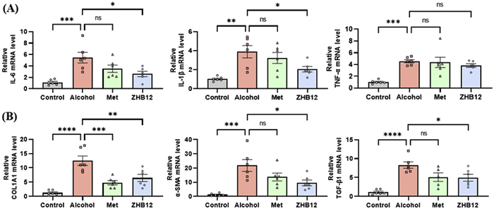
The aggravation of the inflammatory response promotes the pathological process of alcoholic liver fibrosis, which is often accompanied by further inflammatory response [36]. As shown in Fig. 6, we assessed the expression levels of inflammatory cytokines (IL-6, IL-1β, and TNF-α) and fibrosis-related factors (collagen type I alpha 1 (COL1A1), α-smooth muscle actin (α-SMA), and transforming growth factor-β1 (TGF-β1)) using qRT-PCR to evaluate the impact of ZHB12 on liver inflammation and fibrosis. The results demonstrated that these factors were unconventionally increased in the alcohol-feeding groups compared to the control group (Figs. 6A and B). However, the ZHB12 group showed downregulation of genes encoding inflammatory and fibrosis-related factors (IL-6, IL-1β, COL1A1, α-SMA, and TGF-β1) induced by alcohol. In contrast, the metadoxine group effectively increased COL1A1 expression but did not significantly affect the other factors. These findings suggest that ZHB12 may help reduce liver inflammation and fibrosis in the ALF model mice.
In conclusion, we describe the discovery of novel pyrropyrimidine derivatives, ZHB12 (PDE7 IC50 = 0.2 µmol/L), as potential PDE7 inhibitors derived from the natural product caffeine, utilizing a side-chain evolution strategy in the "molecular factory" module of the AI-driven Drug Flow platform. ZHB12 also showed impressive selectivity (PDE7/PDEs > 60), reasonable bioavailability (F = 24.8%), and a non-toxic dose of 500 mg/kg, indicating its safety. Specifically, ZHB12 significantly reduced the abnormal increase in alcohol-induced aminotransferase levels and inflammatory gene expression in the NIAAA model. Histological examination using H&E staining demonstrated that ZHB12 effectively improved liver injury caused by alcohol at a dosage of 50 mg/kg. Moreover, in the ALF model, ZHB12 successfully downregulated the abnormal expression of aminotransferase, inflammatory, and fibrosis-related genes, indicating that ZHB12 effectively alleviated ALF symptoms. Consequently, ZHB12 stands out as a promising PDE7 inhibitor for future drug development aimed at treating alcohol-associated liver disease.
Jia-Xi Huang: Writing – original draft, Validation. Chuang Xia: Writing – original draft, Validation, Data curation. Le-Shen Li: Writing – original draft, Visualization. Huichang Bi: Writing – review & editing, Funding acquisition. Zhong-Zhen Zhou: Writing – review & editing, Funding acquisition.
The authors declare that they have no known competing financial interests or personal relationships that could have appeared to influence the work reported in this paper.
This work was financially supported by the National Natural Science Foundation of China (Nos. U23A20535 and 82574227) and the Natural Science Foundation of Guangdong Province (No. 2024A1515011307).
Supplementary material associated with this article can be found, in the online version, at doi:
R. Ramkissoon, Alcohol Res. 42 (2022) 13.
Y.M. Li, J.G. Fan, J. Dig. Dis. 20 (2018) 174–180.
N.W. Habash, T.S. Sehrawat, V.H. Shah, S. Cao, JHEP Rep. 4 (2022) 100466. doi: 10.1016/j.jhepr.2022.100466
J. Yan, Y. Nie, M. Luo, et al., Front. Pharmacol. 12 (2021) 694475. doi: 10.3389/fphar.2021.694475
A. Wakil, M. Niazi, M.A. Meybodi, N.T. Pyrsopoulos, J. Clin. Exp. Hepatol. 13 (2023) 116–126. doi: 10.1016/j.jceh.2022.06.012
P. Mukhopadhyay, S.A. El Awdan, R.F. Abdel Rahman, et al., PLoS One 14 (2019) e0216301. doi: 10.1371/journal.pone.0216301
M. Elnagdy, S. Barve, C. McClain, L. Gobejishvili, Biomolecules 10 (2020) 1433. doi: 10.3390/biom10101433
Q. Zhang, L. Sun, L. Wu, et al., Chin. Chem. Lett. 36 (2025) 110795. doi: 10.1016/j.cclet.2024.110795
Z.P. Sang, T. Xue, Q.R. Xing, et al., Chin. Chem. Lett. 37 (2026) 111318. doi: 10.1016/j.cclet.2025.111318
Y.Y. Huang, X. Luo, K. Zhang, et al., Chin. Chem. Lett. 36 (2025) 110586. doi: 10.1016/j.cclet.2024.110586
Z. Chen, Y. Shi, F. Zhong, et al., Chin. Chem. Lett. 36 (2025) 109956. doi: 10.1016/j.cclet.2024.109956
V. Lakics, E.H. Karran, F.G. Boess, Neuropharmacology 59 (2010) 367–374. doi: 10.1016/j.neuropharm.2010.05.004
P. Wang, P. Wu, R.W. Egan, M.M. Billah, Biochem. Biophys. Res. Commun. 276 (2000) 1271–1277. doi: 10.1006/bbrc.2000.3613
A. Świerczek, B. Pomierny, E. Wyska, W.J. Jusko, J. Pharmacol. Exp. Ther. 381 (2022) 151–163. doi: 10.1124/jpet.121.001004
M. Goto, Y. Tanaka, M. Murakawa, et al., Int. Immunopharmacol. 9 (2009) 1347–1351. doi: 10.1016/j.intimp.2009.08.002
J.X. Huang, B.L. Zhu, J.P. Xu, Z.Z. Zhou, Eur. J. Med. Chem. 250 (2023) 115194. doi: 10.1016/j.ejmech.2023.115194
R. Bhanot, A.S. Grewal, A. Devi, Recent Adv. Inflamm. Allergy. Drug Discov. 20 (2026) 139–155. doi: 10.2174/0127722708362767250410094814
D. Di Pietrantonio, V. Pace Palitti, A. Cichelli, S. Tacconelli, Foods 13 (2024) 2280. doi: 10.3390/foods13142280
S. Pathania, R.K. Rawal, Eur. J. Med. Chem. 157 (2018) 503–526. doi: 10.1016/j.ejmech.2018.08.023
L.M. De Coen, T.S.A. Heugebaert, D. García, C.V. Stevens, Chem. Rev. 116 (2015) 80–139.
M.S. Mohamed, R. Kamel, S.S. Fatahala, Eur. J. Med. Chem. 45 (2010) 2994–3004. doi: 10.1016/j.ejmech.2010.03.028
K.M.H. Hilmy, H.G. Abdul-Wahab, D.H. Soliman, et al., Med. Chem. Res. 24 (2014) 2097–2110.
C. Shen, J. Song, C.Y. Hsieh, et al., J. Chem. Inf. Model. 64 (2024) 5381–5391. doi: 10.1021/acs.jcim.4c00621
J. Kempson, W.J. Pitts, J. Barbosa, et al., Bioorg. Med. Chem. Lett. 15 (2005) 1829–1833. doi: 10.1016/j.bmcl.2005.02.025
A.I. Sanchez, R. Meneses, J.M. Minguez, et al., Org. Biomol. Chem. 12 (2014) 4233–4242. doi: 10.1039/C4OB00175C
L. Zheng, Z. Aimaiti, L. Long, et al., J. Med. Chem. 67 (2023) 728–753.
A. Bertola, S. Mathews, S.H. Ki, et al., Nat. Protoc. 8 (2013) 627–637. doi: 10.1038/nprot.2013.032
F.H. Tijera, A.I. Servín-Caamaño, J. Cruz-Herrera, et al., Ann. Hepatol. 13 (2014) 343–352. doi: 10.1016/S1665-2681(19)30863-4
F.H. Tijera, World J. Gastroenterol. 21 (2015) 4975.
L. Leggio, G.A. Kenna, A. Ferrulli, et al., Hum. Psychopharmacol. 26 (2011) 554–559. doi: 10.1002/hup.1244
J. Ma, D.W. Staler, R.I. Mahato, Med. Chem. Res. 33 (2024) 1339–1353. doi: 10.1007/s00044-024-03267-3
R. Sun, M. Han, Y. Lin, et al., Int. J. Neuropsychopharmacol. 28 (2025) pyaf009. doi: 10.1093/ijnp/pyaf009
A.R. Aroor, D.E. Jackson, S.D. Shukla, Eur. J. Pharmacol. 679 (2012) 101–108. doi: 10.1016/j.ejphar.2011.12.045
L. Zikela, H. Zhu, Z. Yu, D. Wang, J. Li, et al., Eur. J. Nutr. 64 (2025) 36. doi: 10.1007/s00394-024-03549-x
V.T. Enciu, P.M. Ologeanu, A. Constantinescu, C. Fierbinteanu-Braticevici, Rom J. Intern. Med. 63 (2025) 97–106. doi: 10.2478/rjim-2025-0007
S.L. Friedman, Gastroenterology 134 (2008) 1655–1669. doi: 10.1053/j.gastro.2008.03.003

Figure 1 The discovery of pyrrolopyrimidine PDE7 inhibitors ZHA1, ZHA2, and ZHB1 as lead compounds.

Figure 2 Therapeutic effects of ZHB12 on the NIAAA mice model of ALD at low (25 mg/kg) and high (50 mg/kg) doses, using metadoxine (50 mg/kg) as the positive control. (A) Schedules of experimental procedures and drug treatments. (B) Body weight and liver index. (C) The plasma levels of ALT, AST, and TG. (D) Representative H&E-stained liver sections. Scale bar: 50 µm. The statistical analysis was an ordinary one-way ANOVA. **P < 0.01, ***P < 0.001, ****P < 0.0001. Data are presented as mean ± standard error of mean (SEM) (n = 8). ns, no significance.

Figure 3 Alcohol upregulated PDE7A levels in the liver of the NIAAA model mice (each group, n = 3). The statistical analysis was an ordinary one-way ANOVA. **P < 0.01. Data are presented as mean ± SEM.

Figure 4 In the liver of the NIAAA model mice, ZHB12 increased cAMP levels (A) in the livers of NIAAA model mice, and down-regulated the mRNA levels of the inflammatory cytokines ((B) IL-6, (C) IL-1β, and (D) TNF-α) at both low (25 mg/kg) and high (50 mg/kg) doses. The statistical analysis was an ordinary one-way ANOVA. *P < 0.05, **P < 0.01, ***P < 0.001, ****P < 0.0001. Data are presented as mean ± SEM (n = 6–8).

Figure 5 Therapeutic effects of ZHB12 on the ALF mice model of ALD at a dose of 50 mg/kg, using metadoxine (50 mg/kg) as a positive control. (A) Schedules of experimental procedures and drug treatments. (B) Body weight and liver index. (C) The plasma levels of ALT, AST, and TG. (D) Representative H&E and Sirius red-stained liver sections. The black arrow points to the scar, and the red arrow points to the chicken-wire pattern. Scale bar: 50 µm. The statistical analysis was an ordinary one-way ANOVA. *P < 0.05, **P < 0.01, ****P < 0.0001. Data are presented as mean ± SEM (n = 6).

Figure 6 PDE7 inhibitor ZHB12 downregulated the mRNA levels of inflammatory cytokines and fibrosis-related factors in the liver of ALF model mice at a dose of 50 mg/kg. (A) The mRNA levels of IL-6, IL-1β, and TNF-α in the liver. (B) The mRNA levels of COL1A1, α-SMA, and TGF-β1 in the liver. The statistical analysis was an ordinary one-way ANOVA. *P < 0.05, **P < 0.01, ***P < 0.001, ****P < 0.0001. Data are presented as mean ± SEM (n = 6).
Table 1. PDE7A inhibitory activities of 7H-pyrrolopyrimidines ZHA1–12.
|
|||
| Compound | Ar or R1 | Inhibition rates (%)b at 1.0 µmol/L | IC50 (µmol/L)a |
| ZHA1 | 3,5-Dimethoxyphenyl | 75% | 0.47 ± 0.08 |
| ZHA2 | 3,4,5-Trimethoxyphenyl | 79% | 0.24 ± 0.05 |
| ZHA3 | 3,4,5-Trimethoxyphenyl | 4% | NAc |
| ZHA4 | 3,4-Dimethoxyphenyl | 9% | NAc |
| ZHA5 | 2,4-Dimethoxyphenyl | 9% | NAc |
| ZHA6 | 3,5-Dimethoxyphenyl | 7% | NAc |
| ZHA7 | 3,4-Dimethoxyphenyl | 3% | NAc |
| ZHA8 | 2,4-Dimethoxyphenyl | 50% | NTd |
| ZHA9 | Isopropyl | 11% | NAc |
| ZHA10 | Cyclopentyl | 74% | 0.32 ± 0.04 |
| ZHA11 | Tetrahydro-2H-pyran-4-yl | 42% | NAc |
| ZHA12 | 4-Fluorobenzyl | 75% | 0.33 ± 0.04 |
| BRL-50481 | - | 56% | 0.64 ± 0.02 |
| a Obtained IC50 values (mean ± SD) were the average values of triplicate independent experiments. b Inhibition rates (%) at 1 µmol/L were the average values of triplicate independent experiments. c NA, no activity. The inhibition rate (%) at 1 µmol/L against PDE7A is < 20%. d NT, no test. |
|||
 下载: 导出CSV
Table 2. PDE7A inhibitory activities of 7H-pyrrolopyrimidines ZHB1–20.
|
|||
| Compound | R1 or R2 | Inhibition rates (%)b at 1.0 µmol/L | IC50 (µmol/L)a |
| ZHB1 | 4-Methoxybenzyl | 77% | 0.32 ± 0.07 |
| ZHB2 | Allyl | 77% | 0.27 ± 0.05 |
| ZHB3 | Isopropyl | 78% | 0.24 ± 0.06 |
| ZHB4 | Cyclopentyl | 6% | NAc |
| ZHB5 | Pyridin-3-ylmethyl | 79% | 0.26 ± 0.08 |
| ZHB6 | 1-Allylpiperidin-4-yl | 82% | 0.29 ± 0.02 |
| ZHB7 | 1-Benzylpiperidin-4-yl | 76% | 0.31 ± 0.04 |
| ZHB8 | |
74% | 0.36 ± 0.04 |
| ZHB9 | 1-Acetylpiperidin-4-yl | 72% | 0.35 ± 0.05 |
| ZHB10 | 1-Propionylpiperidin-4-yl | 80% | 0.28 ± 0.03 |
| ZHB11 | Cyclopropyl | 25% | NTd |
| ZHB12 | Isopropyl | 84% | 0.20 ± 0.02 |
| ZHB13 | 4,4-Difluorocyclohexyl | 9% | NAc |
| ZHB14 | Phenyl | 63% | NTd |
| ZHB15 | Thiophen-3-yl | 22% | NTd |
| ZHB16 | Cyclopropyl | 74% b | NTd |
| ZHB17 | -CH2CH2COOH | 66% b | NTd |
| ZHB18 | -CH2CH2CH2COOH | 36% b | NTd |
| ZHB19 | |
27% b | NTd |
| ZHB20 | |
66% b | NTd |
| a Obtained IC50 values (mean ± SD) were the average values of triplicate independent experiments. b Inhibition rates (%) at 1.0 µmol/L were the average values of triplicate independent experiments. c NA, no activity. The inhibition rate (%) at 1 µmol/L against PDE7A is < 20%. d NT, no test. |
|||
下载: 导出CSV
Table 3. Selectivity of ZHB12 across PDEs.
| PDEs | IC50 (µmol/L) a | Selectivity index (PDE7/PDEs) |
| PDE1A | 21% b | > 80 |
| PDE2A | 25% b | > 80 |
| PDE3B | 31% b | > 80 |
| PDE4B | 12.41 ± 0.42 a | 62 |
| PDE4D | 16.53 ± 0.33 a | 80 |
| PDE5A | 27% b | > 80 |
| PDE7A | 0.20 ± 0.02 a | 1 |
| PDE8A1 | 17% b | > 80 |
| PDE9A2 | 9% b | > 80 |
| a Obtained IC50 values (mean ± SD) were the average values of triplicate independent experiments. b Inhibition rates (%) at 10 µmol/L were the average values of triplicate independent experiments. |
||
下载: 导出CSV

 扫一扫看文章


扫一扫关注我们
 下载: