Table 1.
Crystal Data for the X-ray Structure Analyses of Complexes 1~3
Citation:
Shu-Ping DING, Zong-Yao ZHANG, Guo-Jun ZHOU, Rui CAO. Synthesis, Structure and Luminescence Properties of Dumbbell-like Silver Clusters[J]. Chinese Journal of Structural Chemistry,
2020, 39(10): 1824-1834.
doi:
10.14102/j.cnki.0254–5861.2011–2702

Synthesis, Structure and Luminescence Properties of Dumbbell-like Silver Clusters
English
Synthesis, Structure and Luminescence Properties of Dumbbell-like Silver Clusters
Abstract:
In this work, three stable dumbbell-shaped silver clusters 1~3, formed by a novel bifunctional ligand Ph2P-C6H4-3-C≡CH, are presented. All three complexes have cage structures constructed with thirty-six silver atoms, which are templated by chloride anions. During the self-assembly process of silver acetylides, argentophilic interactions and Ag(I)-ethynide interactions are important for the formation of silver cores to yield a diversity of silver clusters. Weak molecular interactions (π-π, C−H···π, C−H···F, C−H···O) are also found in these complexes, which are crucial to stabilizing these silver clusters. Compared with simple alkynyl ligands (RC≡C−), the introduction of phosphine groups into alkynyl ligand can effectively control the undesired infinite growth of silver acetylides. The photophysical behaviors of complexes 1~3 are studied, showing intense orange room-temperature luminescence in the solid state upon the exposure to UV light. The luminescence mainly arises from ligand-to-metal charge transfer (LMCT) and metal-cluster-lefted transitions (MC) within the silver clusters.
-
1. INTRODUCTION
The past few decades have witnessed amazing progress in exploring the structure-property relationship of silver clusters. Because of their peculiar structures[1-5] and potential applications in catalysis, luminescent material, biology, and other interesting fields[6-11], silver clusters have received sustained attention. Among all kinds of silver clusters, silver ethynide clusters have benefited from argentophilic interactions and Ag(Ⅰ)-ethynide interactions[12-14]. In this family of silver clusters, there are various silver acetylide cages with anions acting as templates, because [RC≡C]− is an anionic group with potential σ and π bonding interactions with silver atoms[15, 16]. In addition, silver acetylide clusters may contain phosphine, amine, and thiolate ligands[17-21], in which phosphine ligands are generally used as bridging ligands. However, silver acetylide clusters are usually polymers, which have very poor solubility in common solvents. Up to now, most silver acetylides have been obtained by using a simple alkynyl ligand (RC≡C)− or mixed alkynyl/phosphine ligands, but few of them use both modified alkynyl and phosphine groups in a ligand. Therefore, under mild experimental conditions, we try to synthesize silver acetylide complexes with the use of ligands bearing both phosphine and alkynyl groups. The solubility and nucleation number of silver acetylides can be improved by the introduction of phosphine ligands. On the other hand, we hope that some progress will be made in the luminescent properties of silver acetylide clusters.
In our previous studies, a bifunctional ligand, Ph2P-C6H4-4-C≡CH (L1) bearing both phosphine and alkynyl groups and its ability to assemble high-nuclearity silver clusters were reported. Phosphine ligands can limit the uncontrolled growth and increase the solubility of silver clusters[3, 22, 23]. The combination of two donor groups makes it possible for silver clusters to construct 2D and 3D frameworks, based on the high-nuclearity clusters as building blocks. The size and shape of silver clusters can also be adjusted under different reaction and crystallization conditions. These structures are attrac-tive[24]. In order to further explore the possible emissive properties, we designed the ligand Ph2P-C6H4-3-C≡CH (L), based on which we obtained three silver clusters withsupramolecular structures, (CH3CH2)3NH[Ag36L24Cl3(ClO4)6-(CH3CN)8](ClO4)4 (1), [Ag36L24Cl3(CO3)2(OH) (Py)4-(CH3CN)4](BF4)4 (2), and (CH3CH2)3NH[Ag36L24Cl3-(CH3CN)10](SbF6)10 (3). They can be prepared by systematic methods and characterized by single-crystal X-ray diffraction method. The cage structures of complexes 1~3 are templated with chloride anions, and are formed by Ag(Ⅰ)−Ag(Ⅰ) and Ag(Ⅰ)-ethynide interactions, which are further stabilized by weak molecular interactions (π-π, C−H···π, C−H···F, C−H··· O) to build the supramolecular structures.
2. EXPERIMENTAL
2.1 General methods and materials
All reactions were operated under nitrogen atmosphere using standard Schlenk line techniques. All reagents were commercially purchased and used as received. All solvents were treated by distillation and degassing in advance. Ligand Ph2P-C6H4-3-C≡CH was synthesized according to previously reported methods[25, 26]. Elemental analyses of C and H were recorded on a Vario EL-Ⅲ elemental analyzer. UV-vis absorption spectra were obtained using a Shimadzu UV-3600 spectrometer. The emission and excitation spectra were measured on the Hitachi F-7000 fluorescence spectrophotometer. The emission lifetime in the solid state was measured on an Edinburgh FIS1000 transient state fluorescence spectrometer. The absolute emission quantum yield in the solid state was determined by the Hamamatsu C9920-02G quantum efficiency measurement systems.
2.2 Synthesis of complex 1
Ph2P-C6H4-3-C≡CH (57.36 mg, 0.20 mmol) reacted with 1.1 equivalent of AgClO4 (45.61 mg, 0.22 mmol) in acetonitrile (5 mL) and dichloromethane (1 mL) solvent. Then, 1 mL of triethylamine was added. The reaction mixture was stirred overnight at room temperature. The solvent was removed on a rotary evaporator under reduced pressure. Recrystallization from anhydrous ether gave the crude product as a bright yellow powder. The crude product was further washed with ether, dried, and was then redissolved in 6 mL of acetonitrile and filtered. After filtration, slow vapor diffusion of hexane into the filtrate afforded pink crystalline plates of complex 1 in about 86% yield. Anal. Calcd. for C502H376Ag36Cl13P24N9O40: C, 49.17; H, 3.09%. Found: C, 49.07; H, 3.16%.
2.3 Synthesis of complex 2
Ph2P-C6H4-3-C≡CH (80.17 mg, 0.28 mmol) and AgBF4 (65.29 mg, 0.34 mmol) were added to a mixture of acetonitrile (5 mL) and dichloromethane (1 mL) under stirring. Then, 1 mL of triethylamine was added to the mixture. During addition, colorless solution immediately turned yellow. The solution was stirred overnight at room temperature. The solvent was removed on a rotary evaporator under reduced pressure. The crude product was further washed with ether, dried in vacuo, redissolved in 9 mL of acetonitrile and filtered. To the filtrate, a few drops of pyridine were added. After filtration, slow vapor diffusion from acetone/hexane (V/V = 1/1) afforded pink crystalline block of complexes (62 mg, 68% yield). Anal. Calcd. for C510H369Ag36Cl3P24B4F16N8O7: C, 51.90; H, 3.15%. Found: C, 51.90; H, 3.35%.
2.4 Synthesis of complex 3
Ph2P-C6H4-3-C≡CH (40 mg, 0.14 mmol) and triethylamine (1 mL) were dissolved in dichloromethane (1 mL). The mixture was added dropwise to the acetonitrile solution (8 mL) of AgSbF6 (58 mg, 0.28 mmol). The reaction mixture was stirred for two days at ambient temperature. The solvent was removed on a rotary evaporator under reduced pressure. The crude product was further washed with ether, dried under vacuum, redissolved in 5 mL of acetonitrile and filtered. Slow evaporation of the solution afforded complex 3 as pink crystalline plates (65% yield). Anal. Calcd. for C506H382Ag36Cl3P24N11Sb10F60: C, 44.34; H, 2.81%. Found: C, 44.12; H, 2.89%.
2.5 X-ray crystallography
Data were collected on a Bruker D8 QUEST X-ray diffractometer. The crystals of complexes 1, 2, and 3 were immersed in Paratone-N oil, mounted in a small fiber loop, and placed in a cooled nitrogen gas stream. Diffraction intensities were measured with graphite-monochromated MoKα radiation (λ = 0.71073 Å) at 153 K by using a φ-ω scan mode. The APEX2 program package was used for data collection, indexing, data reduction, and final unit cell refinements. Absorption corrections were applied to all data using the program SADABS. All structures were solved by direct methods using SHELXS[27] and refined against F2 on all data by full-matrix least-squares with SHELXL, following the established refinement strategies[28-30].
In all structures, non-hydrogen atoms were refined anisotropically. All hydrogen atoms binding to carbon were included in the model at geometrically calculated positions and refined using a riding model. The isotropic displacement parameters of all hydrogen atoms were fixed to 1.2 times the U value of the atoms they are linked to (1.5 times for methyl groups). In the crystal of 2, one hydroxyl anion was refined with the occupancy of 0.5. A series of restraints were applied to the solvent molecule and also to some phenyl rings of the main molecule. Though there are some large unresolved q-peaks in all structures, they are all found close to Ag(Ⅰ) cores. Some of the solvents were highly disordered in the crystal lattice of complexes 1~3, so they are treated by a SQUEEZE routine of the PLATON[31]. The contribution of the missing solvent was not taken into account in the unit cell content. Details of the data quality and a summary of the residual values of the refinements are listed in Table 1.
Table 1

 DownLoad:
CSV
DownLoad:
CSV
Complex 1 2 3 C496H360Ag36Cl13 C510H369Ag36B4Cl3 C500H366Ag36Cl3F60 N8O40P24 F16N8O7P24 N10P24Sb10 Fw 12159.37 11801.32 13604.48 Temperature/K 162(2) 153(2) 153(2) Crystal system Triclinic Triclinic Triclinic Space group P $ \overline 1 $ P $ \overline 1 $ P $ \overline 1 $ a/Å 23.3165(9) 24.601(8) 25.195(6) b/Å 23.7671(9) 25.851(8) 25.654(6) c/Å 23.9931(9) 26.209(9) 25.783(6) α/° 102.4590(10) 72.691(11) 73.276(7) β/° 110.4480(10) 65.506(10) 75.208(7) γ/° 95.7040(10) 66.505(10) 67.960(8) Volume/Å3 11941(8) 13735(8) 14589(6) Z 1 1 1 ρcalcd/g·m−3 1.691 1.427 1.548 μ/mm−1 1.653 1.387 1.773 F(000) 5985 5808 6589 Crystal size/mm3 0.2 × 0.2 × 0.2 0.2 × 0.2 × 0.1 0.2 × 0.2 × 0.1 θ range/° 2.13 to 25 2.13 to 25 2.10 to 25 Reflections collected 165344 297520 286523 Independent reflections 41994 48355 51183 (Rint = 0.0761) (Rint = 0.0696) (Rint = 0.0768) Completeness 99.8% 99.9% 99.7% GOOF 1.007 1.093 1.191 Final R indices (I > 2σ(I)) Ra = 0.0669 Ra = 0.0733 Ra = 0.1194 wRb = 0.1385 wRb = 0.2107 wRb = 0.3689 R indices (all data) Ra = 0.0966 Ra = 0.0967 Ra = 0.1358 wRb = 0.1531 wRb = 0.2313 wRb = 0.3935 Largest diff. peak and hole/Å–3 2.360 and −2.197 4.303 and −1.458 7.181 and −5.161 aR = Σ||Fo| − |Fc||/|Fo|, bwR = {Σ[w(Fo2 − Fc2)2]/Σ[w(Fo2)2]}0.5 3. RESULTS AND DISCUSSION
3.1 Structural analysis of 1
Single-crystal structural analysis reveals that complex 1 crystallizes in triclinic space group P
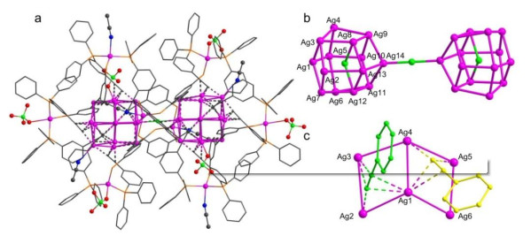
$ \overline 1 $ with Z = 1, which contains 36 Ag atoms, 24 Ph2P-C6H4-3-C≡C− ligands, 6 aceto-nitrile molecules, and 6 ClO4− anions (Fig. 1a). The dumbbell-like Ag28 cluster can be regarded as formed by two Ag14 subcomponents linked by a$ \overline 1 $ rotoinversion left (Fig. 1b). In the dodecahedral Ag14 cage, the Ag(Ⅰ)−Ag(Ⅰ) distances ranged from 2.8652(9) to 3.2745(9) Å with an average value of 3.0342(9) Å, much shorter than the sum of Van der Waals radii of two silver atoms (3.44 Å)[12, 32]. This indicates that strong argentophilic interactions are present in the crystal structure of complex 1. Furthermore, such interactions make the Ag14 cage tightly assembled. The Ag14 cage is capped by twelve Ph2P-C6H4-3-C≡C− ligands, each binding three Ag atoms through both σ and π bonds of the alkynyl moiety. There are three Ph2P-C6H4-3-C≡C− ligands taking μ3-η1, η2, η2 coordination mode and nine Ph2P-C6H4-3-C≡C− ligands adopting μ3-η1, η1, η2 coordination mode (Fig. 1c). The Ag(Ⅰ)−C bond distances fall in the 2.048(9)~2.696(8) Å range averaged by 2.348(8) Å.Figure 1
 Figure 1. (a) Molecular structure of the cationic cluster. (b) Dumbbell-like Ag28 core structure. (c) Two coordination models of ligand L in 1. Yellow: μ3-η1, η1, η2 coordination mode; Bright blue: μ3-η1, η2, η2 coordination mode. Color code: pink, Ag; orange, P; blue, N; red, O; green, Cl; gray, C. Partial hydrogen atoms and Ph2P- moieties are omitted for clarity
Inside the Ag14 cage, the Cl(4) atom is coordinated to Ag(1), Ag(8) and Ag(12) atoms, the Ag(Ⅰ)−Cl bond distances are 2.839(2), 2.848(2) and 2.818(2) Å, respectively. According to the previous reports, the Cl– anion could come from the decomposition of dichloromethane without the designed addition of Cl– anion. The Ag(3) atom is coordinated to an O(6) atom of ClO4− anion, with the Ag(3)−O(6) bond to be 2.592(11) Å. The O(3) and O(9) atoms in ClO4− anion are coordinated to Ag(16) and Ag(18) outside the Ag14 cage. The Ag(16)−O(3) bond distance is 2.456(9) Å and the Ag(18)−O(9) bond distance is 2.559(9) Å. The N(1) and N(2) atoms of acetonitrile molecules are coordinated to Ag(15) and Ag(17). The Ag(15)−N(2) bond is 2.386(9) Å and Ag(17)−N(1) 2.426(9) Å. The Ag(Ⅰ)−P bond distances are in the range of 2.489(2)~2.533(3) Å with the average of 2.499(2) Å. Another two uncoordinated acetonitrile molecules and four ClO4− counter anions were found in the X-ray structure of complex 1. According to structural analysis, the formula of complex 1 is determined to be (CH3CH2)3NH[Ag36L24Cl3(ClO4)6(CH3CN)8]4+, and the posi-tive charges were balanced by four uncoordinated ClO4− anions (Fig. S1). (CH3CH2)3NH+ might be formed by the addition of excessive triethylamine protonation in our previous synthesis.
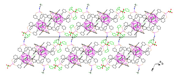
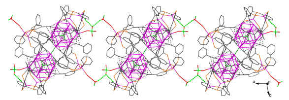
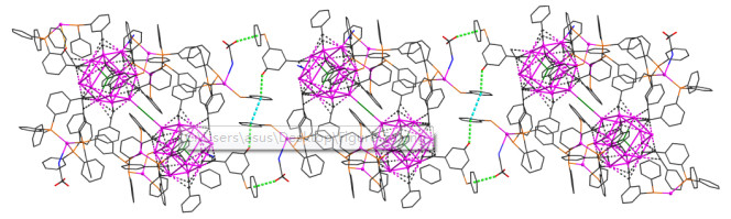
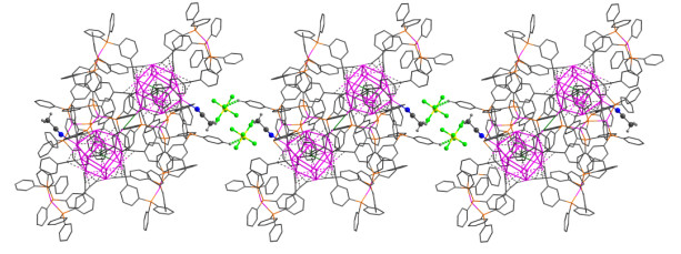
As shown in Fig. S2, the face-to-face π-π stacking interactions[33-35] (centroid-to-centroid distances of 3.712 Å) and C−H···π interactions[36, 37] (H···centroid 2.789 Å with a C(42)−H(42)···centroid angle of 159°, H···centroid 2.836 Å with a C(92)−H(92)···centroid angle of 154°, and H···centroid 2.983 Å with a C(96)−H(96)···centroid angle of131°, respectively) were found in complex 1. Crystallographic studies showed that the adjacent Ag28 cages are linked by ClO4− anions and acetonitrile molecules to form a two-dimensional coordination network in the solid state[38, 39], which is depicted in Fig. 2. A linear silver chain is linked by C(242)−H(24B)···O(5) and C(23)−H(23)···O(5) hydrogen bonds between the ClO4− anions and acetonitrile molecules viewed along the c axis (Fig. 3).
Figure 2
 Figure 2. 2D coordination network structures are linked by the C−H···O interactions in complex 1. Partial hydrogen atoms and benzene rings are omitted for clarity. Bond lengths: C(244)–H(24E)···O(9) H(24E)–O(9) 2.577 Å, C(29)–H(29)···O(12) H(29)–O(12) 2.576 Å, C(66)–H(66)···O(9) H(66)–O(9) 2.509 Å, C(86)–H(86)···O(12) H(86)–O(12) 2.531 Å
Figure 3
 Figure 3. Silver chains linked by C(242)−H(24B)···O(5) and C(23)−H(23)···O(5) interactions viewed along the c axis between the acetonitrile molecules and ClO4− anions in complex 1
3.2 Structural analysis of 2
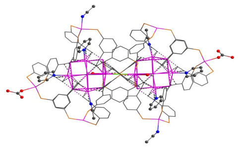
Complex 2 crystallized in the triclinic space group
$ P\stackrel{-}{1} $ with Z = 1, which contains 36 Ag atoms that were surrounded by 24 Ph2P-C6H4-3-C≡C− ligands, four pyridines(Py), four acetonitrile molecules, three Cl− anions, one hydroxyl anion, and two CO32− anions that were generated from the fixation of carbon dioxide in the air (Fig. 4). In the Ag14 cage, the Ag(Ⅰ)−Ag(Ⅰ) distances ranged from 2.8740(12) to 3.3490(12) Å with an average value of 3.0438(12) Å, indicating the interactions between Ag(Ⅰ)−Ag(Ⅰ) atoms. The Ag14 cage is coordinated to twelve Ph2P-C6H4-3-C≡C− ligands. Eight of them are adopting μ3-η1, η1, η2 ligation modes while the rest four are adopting μ3-η1, η2, η2 ligation modes, the ligation mode is clearly presented in Fig. S3a. The Ag(Ⅰ)−C bond distances are between 2.051(7) and 2.703(8) Å with an average value of 2.288(2) Å.Figure 4
 Figure 4. Crystal structure of complex 2. Color code: pink, Ag; orange, P; right green, Cl; red, O; blue, N; gray, C. Partial hydrogen atoms and benzene rings are omitted for clarity
Inside each Ag14 cage, the Cl(1) atom is coordinated to Ag(1), Ag(12), and Ag(13) atoms. The Ag(Ⅰ)−Cl bond distances are 2.884(19), 2.862(2), and 2.863(2) Å, respec-tively, which lead to a slight distortion of the dodecahedral Ag14 cage. The Cl atom might come from the decomposition of the dichloromethane solvent. The pyridyl N(1) and N(2) bind to Ag(6) and Ag(2) atoms. The Ag(Ⅰ)−N bond distances are 2.376(5) and 2.400(7) Å. The N(3) and N(4) atoms in the acetonitrile molecule are coordinated to Ag(16) and Ag(4) atoms, respectively. The Ag(Ⅰ)−N bond distances are 2.596(8) and 2.458(8) Å. Apart from the cationic Ag14 cage, there are other four Ag atoms lying outside the cage. Among those four Ag atoms, Ag(16) and Ag(18) are bound to three P atoms of the Ph2P-C6H4-3-C≡C− ligand, while Ag(15) and Ag(17) are bound to three P atoms and one additional oxygen. The Ag(Ⅰ)−P bond distances are in the range of 2.468(2)−2.530(2) Å, with an average value of 2.501(3) Å. Interestingly, Ag(15) and Ag(17) have different coordination environment. The Ag(15) atom is coordinated by carbonate through the O atom. Different from Ag(15), the Ag(17) atom is coordinated to an O atom from a half occupied hydroxyl anion. The introduction of OH− and CO32− anions terminates the growing of the structure, preventing it from assembling into coordination polymers. Based on the detailed analysis, complex 2 is positively charged with a formula of [Ag36L24Cl3(CO3)2(OH)(Py)4(CH3CN)4]4+, the positive charges are balanced by four uncoordinated BF4− anions established in the crystal lattice (Fig. S4).
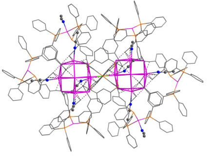
As shown in Fig. S5, weak intramolecular interactions are found in complex 2. The distances of the centroid to centroid between two parallel benzene rings are 3.631 and 3.795 Å, respectively, indicating the presence of π-π stacking interactions. The distances of H···centroid in non-classical C−H···π interactions are 2.514, 2.657, and 2.897 Å (C−H···centroid angles of 154°, 135°, 150°, respectively), and the distances of H···centroid are shorter than the normal C−H···π interaction of 3.050 Å[37, 40]. Weak molecular interactions play a key role in supramolecular chemistry, a variety of weak molecular interactions are beneficial for the construction of structures and control the crystal structure packing[36, 38]. A silver chain is formed by coordinated acetonitrile molecules between the adjacent silver clusters through C(253)−H(25A)···π interaction (H(25A)···centroid 2.761 Å with a C(253)−H(25A)···centroid angle of 156°) on the bc plane, which was also reinforced by weak face to face π-π stacking interaction (centroid to centroid distance 4.078 Å) and C(117)−H(117)···π stacking interaction (H(117)···centroid 2.749 Å with a C(117)−H(117)···centroid angle of 146°) (Fig. 5).
Figure 5
 Figure 5. A silver chain linked by a weak face-to-face π-π interaction (centroid-to-centroid distance 4.078 Å), C(253)−H(25A)···π interaction and C(117)−H(117)···π interaction
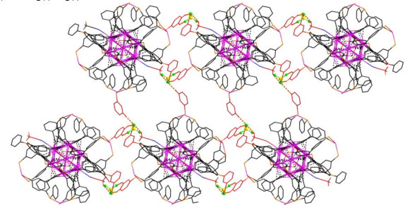
As shown in Fig. 6 and Fig. S6, a two-dimensional layer structure is linked by two kinds of hydrogen bonds between the adjacent four silver clusters. The first is linked by free BF4− anions and coordinated acetonitrile molecules via C(255)−H(25E)···F(8S) and C(255)−H(25E)···F(7S) hydro-gen bonds)[41, 42]. The second is linked by free BF4− anion and benzene ring of ligand L through the C(25)−H(25)···F(6S), C(24)−H(24)···F(8S), C(143)−H(143)···F(5S), and C(223)− H(223)···F(8S) hydrogen bonds along the a direction. The free BF4− anions linked another silver chain by C(69)− H(69)···F(1S) and C(205)−H(205)···F(1S) interactions (Fig. S7).
Figure 6
 Figure 6. 2D coordination structure linked by C−H···F interactions in complex 2. Partial hydrogen atoms, acetonitrile molecules, and BF4− anions are omitted for clarity
3.3 Structural analysis of 3
The basic structure and shape of complex 3 are similar to 1 and 2. As shown in Fig. 7, complex 3 crystallizes in triclinic space group P
$ \overline 1 $ with Z = 1. Each asymmetric unit of complex 3 contains an Ag14 cage surrounded by 12 Ph2P-C6H4-3-C≡C− ligands and 5 acetonitrile molecules. In complex 3, five Ph2P-C6H4-3-C≡C− ligands have μ3-η1, η2, η2 bridging mode and seven Ph2P-C6H4-3-C≡C− ligands have μ3-η1, η1, η2 bridging mode (Fig. S3b). The Ag(Ⅰ)−Ag(Ⅰ) bond distances are between 2.8750(14) and 3.3537(14) Å with an average value of 3.0260(9) Å, clearly showing the existence of Ag(Ⅰ)−Ag(Ⅰ) interactions.Figure 7
 Figure 7. Crystal structures of complex 3. Color code: pink, Ag; orange, P; bright green, Cl; red, O; blue, N; gray, C. Hydrogen atoms and partial benzene rings are omitted for clarity
Inside the Ag14 cage, the Ag(Ⅰ)−C bond distances ranged from 2.039(13) to 2.688(13) Å averaged by 2.342(12) Å. The Ag(3), Ag(5), and Ag(7) atoms are coordinated to N donors of acetonitrile molecules, with the Ag(Ⅰ)−N bond distances to be 2.493(19), 2.428(19), and 2.499(14) Å, respectively. Outside the Ag14 cage, the bond length between Ag(15) and acetonitrile N(2) atom is 2.424(12) Å. The bond length between Ag(18) and acetonitrile N(4) atom is 2.444(14) Å. All solvent acetonitrile molecules are coordinated to Ag atoms. However, the SbF6− counter anions are uncoordinated, established in the crystal lattice and balanced the positive charge of the cationic host. The formula of complex 3 can be determined to be (CH3CH2)3NH[Ag36L24Cl3(CH3CN)10]-(SbF6)10. The thermal ellipsoid plot of the X-ray structure of 3 is shown in Fig. S8.
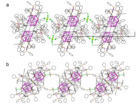
Weak intramolecular forces are found in the structure of complex 3, forming the supramolecular structure and stabilizing the rigid structure. Fig. S9 shows the C−H···π interac-tions in each asymmetric unit of complex 3 (H···centroid 2.936 Å with a C(126)−H(126)···centroid angle of 147°, H···centroid 2.849 Å with a C(111)−H(111)···centroid angle of 151°, and H···centroid 2.551 Å with a C(222)− H(222)···centroid angle of149°, respectively). The short dis-tances between the free SbF6− anions and coordinated CH3CN molecules indicate the existence of interaction. A silver chain is linked by C(164)−H(164)···F(21) and C(9)−H(9)···F(24) interactions along the b direction, which is further linked by offset face-to-face π-π interaction (centroid-to-centroid distance 3.797 Å), C(70)−H(70)···π interaction, and C(11)− H(11)···π interaction along the a direction (Fig. 8). Another silver chain is constructed by C(84)−H(84)···F(30) and C(246)−H(24I)···F(30) interactions between the free SbF6− anions and CH3CN molecules in 3 (Fig. 9).
Figure 8
 Figure 8. (a) Silver chain linked by C(164)−H(164)···F(21) and C(9)−H(9)···F(24) interactions. (b) Silver chain is formed by offset face-to-face π-π interaction, C(70)−H(70)···π interaction, and C(11)−H(11)···π interaction in complex 3
Figure 9
 Figure 9. Silver chain formed by C(84)−H(84)···F(30) and C(246)−H(24I)···F(30) interactions in complex 3
3.4 Photochromic properties
Upon excitation at λ ≥ 365 nm, complexes 1~3 emit intense orange room-temperature luminescence in the solid state. The photophysical data of complexes 1~3 are summarized in Table S1, and the luminescence properties were measured in the solid state at room temperature. As shown in Fig. S10, the UV-vis absorption spectra of complexes 1~3 in the solid state are acquired by a broad high-energy band at 270~360 nm and a low-energy band at ca. 490 nm. The high-energy absorption is typical for metal-perturbed intraligand (alkynyl and phosphine ligands) transitions and the low energy absorption band lefted at 490 nm shall be attributed to the characteristic transition of ligand-to-metal charge transfer (LMCT), mixed with a metal-cluster lefted character modified by argentophilic interaction[43-46].
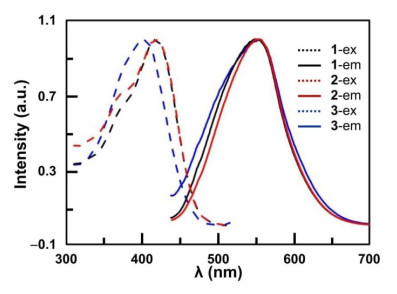
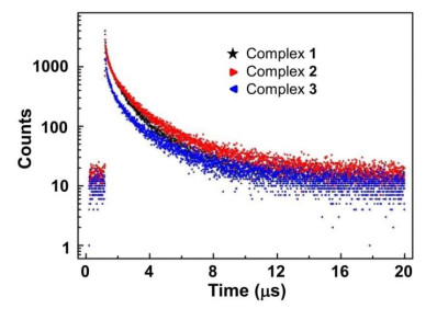
The solid-state excitation and emission spectra are shown in Fig. 10. Complexes 1 and 2 showed the maximum emission at about 550 nm with the absolute luminescence quantum yield is 15.6% and 14.2%, respectively. Complex 3 has a maximum emission at about 545 nm with an absolute luminescence quantum yield of 6.40%. The lifetime of complexes 1, 2 and 3 was determined to be 1.31, 1.82, and 1.73 μs, respectively (Fig. 11). The microsecond lifetime, in accordance with large Stokes shifts between the excitation and emission spectra, shows that the emission origin is triplet excited states[47-50].
Figure 10
 Figure 10. Excitation (left) and emission (right) spectra of complexes 1~3 at 298 K.Complexes 1 (black), 2 (red) and 3 (blue)
Figure 11
 Figure 11. Luminescence decay of complexes 1~3 (λex = 380 nm, 298 K)
4. CONCLUSION
In summary, we report a bi-functional ligand Ph2P-C6H4-3-C≡CH and three silver cluster complexes. As a family of cage complexes, complexes 1~3 are synthesized by the direct reaction between silver salts with alkynyl ligands and isolated by crystallization. Alkynyl ligands can bind silver atoms through δ and π bond to stabilize the cationic cages. Phosphine ligands increase the solubility and stability of silver clusters, structurally limit the uncontrolled growth of the silver cluster, and can be seen as a capping reagent. In the crystal structures of 1~3, the anions such as BF4−, ClO4− and SbF6− are more than counter anions, and together with solvent acetonitrile and benzene rings of the ligand L, they provide sites for intramolecular interactions which improve the rigidity and stability of the crystal structures 1~3.
We are grateful for support from the "Thousand Talents Program" of China -
-
[1]
Chai, J.; Yang, S.; Lv, Y.; Chen, T.; Wang, S.; Yu, H.; Zhu, M. A unique pair: Ag40 and Ag46 nanoclusters with the same surface but different cores for structure-property correlation. J. Am. Chem. Soc. 2018, 140, 15582–15585. doi: 10.1021/jacs.8b09162
-
[2]
Zhang, R.; Hao, X.; Li, X.; Zhou, Z.; Sun, J.; Cao, R. Soluble silver acetylide for the construction and structural conversion of all-alkynyl-stabilized high-nuclearity homoleptic silver clusters. Cryst. Growth Des. 2015, 15, 2505–2513. doi: 10.1021/acs.cgd.5b00286
-
[3]
Koshevoy, I. O.; Karttunen, A. J.; Shakirova, J. R.; Melnikov, A. S.; Haukka, M.; Tunik, S. P.; Pakkanen, T. A. Halide-directed assembly of multicomponent systems: highly ordered Au(Ⅰ)-Ag(Ⅰ) molecular aggregates. Angew. Chem. Int. Ed. 2010, 49, 8864–8866. doi: 10.1002/anie.201004386
-
[4]
Niihori, Y.; Hashimoto, S.; Koyama, Y.; Hossain, S.; Kurashige, W.; Negishi, Y. Dynamic behavior of thiolate-protected gold-silver 38-atom alloy clusters in solution. J. Phys. Chem. C 2019, 123, 13324–13329. doi: 10.1021/acs.jpcc.9b02644
-
[5]
Nan, Z. A.; Xiao, Y.; Liu, X. Y.; Wang, T.; Cheng, X. L.; Yang, Y.; Lei, Z.; Wang, Q. M. Monitoring the growth of Ag-S clusters through crystallization of intermediate clusters. Chem. Commun. 2019, 55, 6771–6774. doi: 10.1039/C9CC03533H
-
[6]
Sgibnev, Y.; Cattaruzza, E.; Dubrovin, V.; Vasilyev, V.; Nikonorov, N. Photo-thermo-refractive glasses doped with silver molecular clusters as luminescence downshifting material for photovoltaic applications. Part. Part. Syst. Charact. 2018, 35, 1800141. doi: 10.1002/ppsc.201800141
-
[7]
Novikov, S. M.; Popok, V. N.; Evlyukhin, A. B.; Hanif, M.; Morgen, P.; Fiutowski, J.; Beermann, J.; Rubahn, H. G.; Bozhevolnyi, S. I. Highly stable monocrystalline silver clusters for plasmonic applications. Langmuir 2017, 33, 6062–6070. doi: 10.1021/acs.langmuir.7b00772
-
[8]
Swasey, S. M.; Copp, S. M.; Nicholson, H. C.; Gorovits, A.; Bogdanov, P.; Gwinn, E. G. High throughput near infrared screening discovers DNA-templated silver clusters with peak fluorescence beyond 950 nm. Nanoscale 2018, 10, 19701–19705. doi: 10.1039/C8NR05781H
-
[9]
Wu, Z.; Yao, Q.; Zang, S.; Xie, J. Directed self-assembly of ultrasmall metal nanoclusters. ACS Materials Letters 2019, 1, 237–248. doi: 10.1021/acsmaterialslett.9b00136
-
[10]
Pérez Mariño, Á. M.; Blanco, M. C.; Buceta, D.; López Quintela, M. A. Using silver nanoclusters as a new tool in nanotechnology: synthesis and photocorrosion of different shapes of gold nanoparticles. J. Chem. Educ. 2019, 96, 558–564. doi: 10.1021/acs.jchemed.8b00573
-
[11]
Wang, Z. Y.; Wang, M. Q.; Li, Y. L.; Luo, P.; Jia, T. T.; Huang, R. W.; Zang, S. Q.; Mak, T. C. W. Atomically precise site-specific tailoring and directional assembly of superatomic silver nanoclusters. J. Am. Chem. Soc. 2018, 140, 1069–1076. doi: 10.1021/jacs.7b11338
-
[12]
Barreiro, E.; Casas, J. S.; Couce, M. D.; Laguna, A.; Lopez de Luzuriaga, J. M.; Monge, M.; Sanchez, A.; Sordo, J.; Vazquez Lopez, E. M. A novel hexanuclear silver(Ⅰ) cluster containing a regular Ag6 ring with short Ag–Ag distances and an argentophilic interaction. Dalton Trans. 2013, 42, 5916–5923. doi: 10.1039/c3dt33003f
-
[13]
Schmidbaur, H.; Schier, A. Argentophilic interactions. Angew. Chem. Int. Ed. 2015, 54, 746–784. doi: 10.1002/anie.201405936
-
[14]
Cheng, P. S.; Marivel, S.; Zang, S. Q.; Gao, G. G.; Mak, T. C. W. Argentophilic infinite chain, column, and layer structures assembled with the multinuclear silver(Ⅰ)-phenylethynide supramolecular synthon. Cryst. Growth Des. 2012, 12, 4519–4529. doi: 10.1021/cg300691n
-
[15]
Xie, Y. P.; Mak, T. C. Silver(Ⅰ)-ethynide clusters constructed with phosphonate-functionized polyoxovanadates. J. Am. Chem. Soc. 2011, 133, 3760–3763. doi: 10.1021/ja1112035
-
[16]
Zhang, R.; Zhang, Z.; Liang, Z.; Han, Y.; Ai, X.; Cao, R. Syntheses and structural characterizations of mononuclear and dinuclear platinum(Ⅱ) terpyridyl acetylide complexes. Chem. Select. 2017, 2, 5436–5443.
-
[17]
Koshevoy, I. O.; Karttunen, A. J.; Tunik, S. P.; Haukka, M.; Selivanov, S. I.; Melnikov, A. S.; Serdobintsev, P. Y.; Pakkanen, T. A. Synthesis, characterization, photophysical, and theoretical studies of supramolecular gold(Ⅰ)-silver(Ⅰ) alkynyl-phosphine complexes. Organometallics 2009, 28, 1369–1376. doi: 10.1021/om8010036
-
[18]
Koshevoy, I. O.; Koskinen, L.; Haukka, M.; Tunik, S. P.; Serdobintsev, P. Y.; Melnikov, A. S.; Pakkanen, T. A. Self-assembly of supramolecular luminescent Au(Ⅰ)-Cu(Ⅰ) complexes: "Wrapping" an Au6Cu6 cluster in a [Au3(diphosphine)3]3+ "belt". Angew. Chem. Int. Ed. 2008, 47, 3942–3945. doi: 10.1002/anie.200800452
-
[19]
Huang, R. W.; Dong, X. Y.; Yan, B. J.; Du, X. S.; Wei, D. H.; Zang, S. Q.; Mak, T. C. W. Tandem silver clusterisomerism and mixed linkers to modulate the photoluminescence of cluster-assembled materials. Angew. Chem. Int. Ed. 2018, 57, 8560–8566. doi: 10.1002/anie.201804059
-
[20]
Lu, T.; Wang, J. Y.; Shi, L. X.; Chen, Z. N.; Chen, X. T.; Xue, Z. L. Synthesis, structures and luminescence properties of amine-bis(N-heterocyclic carbene) copper(Ⅰ) and silver(Ⅰ) complexes. Dalton Trans. 2018, 47, 6742–6753. doi: 10.1039/C8DT00599K
-
[21]
Li, G.; Lei, Z.; Wang, Q. M. Luminescent molecular Ag−S nanocluster [Ag62S13(SBut)32](BF4)4. J. Am. Chem. Soc. 2010, 50, 17678–17679.
-
[22]
Blanco, M. C.; Camara, J.; Gimeno, M. C.; Laguna, A.; James, S. L.; Lagunas, M. C.; Villacampa, M. D. Synthesis of gold-silver luminescent honeycomb aggregates by both solvent-based and solvent-free methods. Angew. Chem. Int. Ed. 2012, 51, 9777–9779. doi: 10.1002/anie.201204859
-
[23]
Wang, J. Q.; Guan, Z. J.; Liu, W. D.; Yang, Y.; Wang, Q. M. Chiroptical activity enhancement via structural control: the chiral synthesis and reversible interconversion of two intrinsically chiral gold nanoclusters. J. Am. Chem. Soc. 2019, 141, 2384–2390. doi: 10.1021/jacs.8b11096
-
[24]
Zhang, S.; Zhang, Z.; Cao, R. Two- and three-dimensional silver acetylide frameworks with high-nuclearity silver cluster building blocks assembled using a bifunctional (4-ethynylphenyl)diphenyl phosphine ligand. Inorg. Chim. Acta 2017, 461, 57–63. doi: 10.1016/j.ica.2017.01.030
-
[25]
Lucas, N. T.; Cifuentes, M. P.; Nguyen, L. T.; Humphrey, M. G. Ruthenium cluster chemistry with Ph2PC6H4-4-C≡CH. J. Cluster Sci. 2001, 12, 201–221. doi: 10.1023/A:1016683331367
-
[26]
Grelaud, G.; Argouarch, G.; Paul, F. Synthesis of new triphenylphosphines with pending ethynyl substituents. Tetrahedron Lett. 2010, 51, 3786–3788. doi: 10.1016/j.tetlet.2010.05.055
-
[27]
Sheldrick, G. M. Phase annealing in SHELX-90: direct methods for larger structures. Acta Cryst. 1990, 46, 467–473. doi: 10.1107/S0108767390000277
-
[28]
Zhang, Z.; Yang, Y.; Sun, H.; Cao, R. Syntheses, structures and anion exchange properties of accommodative silver chains using a positively charged and flexible ligand. Inorg. Chim. Acta 2015, 434, 158–171. doi: 10.1016/j.ica.2015.05.021
-
[29]
Liu, X.; Du, P.; Cao, R. Trinuclear zinc complexes for biologically relevant μ3-oxoanion binding and carbon dioxide fixation. Nat. Commun. 2013, 4, 2375. doi: 10.1038/ncomms3375
-
[30]
Sheldrick, G. M. A short history of SHELX. Acta Crystallogr., Sect. A 2008, 64, 112–122. doi: 10.1107/S0108767307043930
-
[31]
Spek, A. PLATON, an integrated tool for the analysis of the results of a single crystal structure determination. Acta Crystallogr., Sect. A 1990, 46, c34.
-
[32]
Hau, S. C.; Mak, T. C. Synthesis of unstable carbides Ag2C2n (n = 3, 4) and characterization via crystallographic analysis of their double salts with silver(Ⅰ) trifluoroacetate. J. Am. Chem. Soc. 2014, 136, 902–905. doi: 10.1021/ja412095y
-
[33]
Zang, S. Q.; Mak, T. C. W. Assembly of silver(Ⅰ)-organic networks from flexible supramolecular synthons with pendant ethynide arms attached to a naphthyl skeleton. Inorg. Chem. 2008, 47, 7094–7105. doi: 10.1021/ic800576b
-
[34]
Zhang, T.; Song, H.; Dai, X.; Meng, X. One, two and three-dimensional organometallic networks constructed from silver(Ⅰ) pyridyldiethynide complexes. Dalton Trans. 2009, 7688–7694.
-
[35]
Yang, C.; Elbjeirami, O.; Gamage, C. S.; Dias, H. V.; Omary, M. A. Luminescence enhancement and tuning via multiple cooperative supramolecular interactions in an ion paired multinuclear complex. Chem. Commun. 2011, 47, 7434–7436. doi: 10.1039/c1cc11161b
-
[36]
Nishio, M. CH/π hydrogen bonds in crystals. CrystEngComm. 2004, 6, 130–158. doi: 10.1039/b313104a
-
[37]
Ramírez Gualito, K.; Alonso Ríos, R.; Quiroz García, B.; Rojas Aguilar, A.; Díaz, D.; Jiménez Barbero, J.; Cuevas, G. Enthalpic nature of the CH/π interaction involved in the recognition of carbohydrates by aromatic compounds, confirmed by a novel interplay of NMR, calorimetry, and theoretical calculations. J. Am. Chem. Soc. 2009, 131, 18129–18138. doi: 10.1021/ja903950t
-
[38]
Shukla, R.; Lindeman, S. V.; Rathore, R. Binding of an acetonitrile molecule inside the ethereal cavity of a hexaarylbenzene-based receptor via a synergy of C–H···O/C–H···π interactions. Chem. Commun. 2007, 3717–3719.
-
[39]
Hau, S. C. K.; Cheng, P. S.; Mak, T. C. W. Ligand-induced assembly of coordination chains and columns containing high-nuclearity silver(Ⅰ) ethynide cluster units. Organometallics 2014, 33, 3231–3234. doi: 10.1021/om5003733
-
[40]
Higaki, T.; Liu, C.; Zhou, M.; Luo, T. Y.; Rosi, N. L.; Jin, R. Tailoring the structure of 58-electron gold nanoclusters: Au103S2(S-Nap)41 and its implications. J. Am. Chem. Soc. 2017, 139, 9994–10001. doi: 10.1021/jacs.7b04678
-
[41]
Lee, H.; Knobler, C. B.; Hawthorne, M. F. Supramolecular self-assembly directed by carborane C–H···F interactions. Chem. Commun. 2000, 2485–2486.
-
[42]
Spada, L.; Gou, Q.; Vallejo Lopez, M.; Lesarri, A.; Cocinero, E. J.; Caminati, W. Weak C–H···N and C–H···hydrogen bonds and internal rotation in pyridine-CH3F. Phys. Chem. Chem. Phys. 2014, 16, 2149–2153. doi: 10.1039/C3CP54430C
-
[43]
Wu, H. B.; Huang, Z. J.; Wang, Q. M. Unprecedented solution-stable silver(Ⅰ) ethynediyl clusters. Chem. Eur. J. 2010, 16, 12321–12323. doi: 10.1002/chem.201001985
-
[44]
Jia, J. H.; Liang, J. X.; Lei, Z.; Cao, Z. X.; Wang, Q. M. A luminescent gold(Ⅰ)-copper(Ⅰ) cluster with unprecedented carbon-lefted trigonal prismatic hexagold. Chem. Commun. 2011, 47, 4739–4741. doi: 10.1039/c1cc10497g
-
[45]
Koshevoy, I. O.; Lin, C. L.; Karttunen, A. J.; Janis, J.; Haukka, M.; Tunik, S. P.; Chou, P. T.; Pakkanen, T. A. Stepwise 1D growth of luminescent Au(Ⅰ)–Ag(Ⅰ) phosphine-alkynyl clusters: synthesis, photophysical, and theoretical studies. Inorg. Chem. 2011, 50, 2395–2403. doi: 10.1021/ic102204h
-
[46]
Hailmann, M.; Hupp, B.; Himmelspach, A.; Keppner, F.; Hennig, P. T.; Bertermann, R.; Steffen, A.; Finze, M. Carba-closo-dodecaboranylethynyl ligands facilitating luminescent reversed charge-transfer excited states in gold/silver complexes. Chem. Commun. 2019, 55, 9351–9354. doi: 10.1039/C9CC05060D
-
[47]
Xu, L. J.; Wang, J. Y.; Zhang, L. Y.; Shi, L. X.; Chen, Z. N. Structures and phosphorescence properties of triphosphine-supported Au2Ag2 and Au8Ag4 alkynyl cluster complexes. Organometallics 2013, 32, 5402–5408. doi: 10.1021/om400685y
-
[48]
Koshevoy, I. O.; Karttunen, A. J.; Kritchenkou, I. S.; Krupenya, D. V.; Selivanov, S. I.; Melnikov, A. S.; Tunik, S. P.; Haukka, M.; Pakkanen, T. A. Sky-blue luminescent Au(Ⅰ)–Ag(Ⅰ) alkynyl-phosphine clusters. Inorg. Chem. 2013, 52, 3663–3673. doi: 10.1021/ic302105a
-
[49]
Liu, X. Y.; Yang, Y.; Lei, Z.; Guan, Z. J.; Wang, Q. M. Luminescence responsive intracluster rearrangements of gold(Ⅰ)-silver(Ⅰ) clusters triggered by acetonitrile. Chem. Commun. 2016, 52, 8022–8025. doi: 10.1039/C6CC02873J
-
[50]
Lei, Z.; Pei, X. L.; Guan, Z. J.; Wang, Q. M. Full protection of intensely luminescent gold(Ⅰ)-silver(Ⅰ) cluster by phosphine ligands and inorganic anions. Angew. Chem. Int. Ed. 2017, 56, 7117–7120. doi: 10.1002/anie.201702522
-
[1]
-
Figure 1 (a) Molecular structure of the cationic cluster. (b) Dumbbell-like Ag28 core structure. (c) Two coordination models of ligand L in 1. Yellow: μ3-η1, η1, η2 coordination mode; Bright blue: μ3-η1, η2, η2 coordination mode. Color code: pink, Ag; orange, P; blue, N; red, O; green, Cl; gray, C. Partial hydrogen atoms and Ph2P- moieties are omitted for clarity
Figure 2 2D coordination network structures are linked by the C−H···O interactions in complex 1. Partial hydrogen atoms and benzene rings are omitted for clarity. Bond lengths: C(244)–H(24E)···O(9) H(24E)–O(9) 2.577 Å, C(29)–H(29)···O(12) H(29)–O(12) 2.576 Å, C(66)–H(66)···O(9) H(66)–O(9) 2.509 Å, C(86)–H(86)···O(12) H(86)–O(12) 2.531 Å
Figure 3 Silver chains linked by C(242)−H(24B)···O(5) and C(23)−H(23)···O(5) interactions viewed along the c axis between the acetonitrile molecules and ClO4− anions in complex 1
Figure 4 Crystal structure of complex 2. Color code: pink, Ag; orange, P; right green, Cl; red, O; blue, N; gray, C. Partial hydrogen atoms and benzene rings are omitted for clarity
Figure 5 A silver chain linked by a weak face-to-face π-π interaction (centroid-to-centroid distance 4.078 Å), C(253)−H(25A)···π interaction and C(117)−H(117)···π interaction
Figure 6 2D coordination structure linked by C−H···F interactions in complex 2. Partial hydrogen atoms, acetonitrile molecules, and BF4− anions are omitted for clarity
Figure 7 Crystal structures of complex 3. Color code: pink, Ag; orange, P; bright green, Cl; red, O; blue, N; gray, C. Hydrogen atoms and partial benzene rings are omitted for clarity
Figure 8 (a) Silver chain linked by C(164)−H(164)···F(21) and C(9)−H(9)···F(24) interactions. (b) Silver chain is formed by offset face-to-face π-π interaction, C(70)−H(70)···π interaction, and C(11)−H(11)···π interaction in complex 3
Figure 9 Silver chain formed by C(84)−H(84)···F(30) and C(246)−H(24I)···F(30) interactions in complex 3
Figure 10 Excitation (left) and emission (right) spectra of complexes 1~3 at 298 K.Complexes 1 (black), 2 (red) and 3 (blue)
Table 1. Crystal Data for the X-ray Structure Analyses of Complexes 1~3
Complex 1 2 3 C496H360Ag36Cl13 C510H369Ag36B4Cl3 C500H366Ag36Cl3F60 N8O40P24 F16N8O7P24 N10P24Sb10 Fw 12159.37 11801.32 13604.48 Temperature/K 162(2) 153(2) 153(2) Crystal system Triclinic Triclinic Triclinic Space group P $ \overline 1 $ P $ \overline 1 $ P $ \overline 1 $ a/Å 23.3165(9) 24.601(8) 25.195(6) b/Å 23.7671(9) 25.851(8) 25.654(6) c/Å 23.9931(9) 26.209(9) 25.783(6) α/° 102.4590(10) 72.691(11) 73.276(7) β/° 110.4480(10) 65.506(10) 75.208(7) γ/° 95.7040(10) 66.505(10) 67.960(8) Volume/Å3 11941(8) 13735(8) 14589(6) Z 1 1 1 ρcalcd/g·m−3 1.691 1.427 1.548 μ/mm−1 1.653 1.387 1.773 F(000) 5985 5808 6589 Crystal size/mm3 0.2 × 0.2 × 0.2 0.2 × 0.2 × 0.1 0.2 × 0.2 × 0.1 θ range/° 2.13 to 25 2.13 to 25 2.10 to 25 Reflections collected 165344 297520 286523 Independent reflections 41994 48355 51183 (Rint = 0.0761) (Rint = 0.0696) (Rint = 0.0768) Completeness 99.8% 99.9% 99.7% GOOF 1.007 1.093 1.191 Final R indices (I > 2σ(I)) Ra = 0.0669 Ra = 0.0733 Ra = 0.1194 wRb = 0.1385 wRb = 0.2107 wRb = 0.3689 R indices (all data) Ra = 0.0966 Ra = 0.0967 Ra = 0.1358 wRb = 0.1531 wRb = 0.2313 wRb = 0.3935 Largest diff. peak and hole/Å–3 2.360 and −2.197 4.303 and −1.458 7.181 and −5.161 aR = Σ||Fo| − |Fc||/|Fo|, bwR = {Σ[w(Fo2 − Fc2)2]/Σ[w(Fo2)2]}0.5  下载: 导出CSV
-


 扫一扫看文章
计量
- PDF下载量: 3
- 文章访问数: 723
- HTML全文浏览量: 10
 下载:
下载:
