
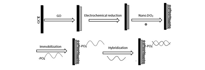
Figure 1.
Illustration for fabricating the modified electrode of nanoZrO2/ERGO/GCE


The genetically modified organisms(GMOs) are the organisms(i.e. plants, animals or microorganisms) whose genome is modified by introducing an exogenous gene to confer resistance to pathogens and herbicides and for better nutrient profiles[1]. However, the genetically modified technology can also lead to unpredicted harmful changes in the nutritional status of foods, potential gene flows to other organisms, agricultural diversity destruction, and gastrointestinal problems[2-3]. Therefore, in the past few years, the use of genetic engineering in the production of food has attracted more public attention. Despite the arguments on the GMOs′ potential health risks have still existed, especial measures have been engaged to implement labelling regulations for bioengineered foods in China, Japan, Australia, New Zealand, and EU(European Union) countries[3]. According to European legislation, labeling of GM crops and their products are mandatory when the content of any authorized GMO ingredient exceeds 0.9% of the food or feed products[4]. The U.S. legislation requests the food companies to label the GMOs voluntarily, and get the approval of FDA(Food and Drug Administration) before these foods are launched into market[5]. Therefore, it has become crucial to developing reliable and rapid methods of detection, identification, tracing and quantification of GMOs for proper food labeling.
The traditional methods, such as polymerase chain reaction(PCR)[6], ELISA(enzyme-linked immunosorbent assay)[7], and real-time PCR[8] can be used for monitoring and verifying the presence and the content of GMOs in various food products. However, these methods have too many drawbacks to conduct on-site or in-field determination. Instead of traditional methods, the electrochemical DNA biosensor, as one inexpensive and convenient tool, has been used for GMOs determination[9]. For DNA biosensor fabrication, the immobilization of ssDNA probes is a crucial step for achieving good performance[10]. Various nanomaterials and methods have been used for immobilizing ssDNA probes[11-12].
Graphene(GR) is two-dimensional nanomaterial, and has been extensively studied and used in many regions since its founding in 2004. Graphene oxide(GO) is a precursor for graphene preparation. It can be easily electrochemically reduced to GO(ERGO) by cyclic voltammetry or potentiostatic method on an electrode surface[13]. ERGO has shown a better performance than GO[14-18] because the reduction degree of ERGO can be controlled by electrochemical technologies. ERGO has been used as a platform for developing biosensors and sensors[19]. Using ERGO modified carbon ionic liquid electrode(CILE) as working electrode, Sun et al[20] fabricated an electrochemical DNA biosensor for detection of transgenic maize MON810. Using ERGO modified electrode, Gao et al[21] have set up a new method for sensitive detection of rutin.
Nano zirconia(nanoZrO2) as an inorganic oxide has many good physicochemical properties including thermal stability, chemical inertness, resistance to poison and strong affinity for oxygen-containing groups(such as phosphoric group), therefore, nanoZrO2 and its hybridized materials are often used for immobilizing the biomolecules that contained oxygenal groups to fabricate biosensors and sensors[22-23]. By electrodepositing nanoZrO2 on the graphene, Sun et al[24] fabricated an electrochemical DNA biosensor for detection of Staphylococcus aureus nuc gene sequence.
In this research, the GO suspension solution was cast on a glassy carbon electrode(GCE) and then the GO was reduced directly to ERGO by cyclic voltammetry. After that, the nanoZrO2 was casted on the surface of ERGO. The ERGO and nanoZrO2 composites were used as a platform for immobilizing DNA probe, and accordingly an electrochemical DNA biosensor for detection of the specific genetic sequence in transgenic maize was fabricated(Fig. 1). The good conductivity of ERGO and the affinity of nanoZrO2 for immobilizing DNA probes play synergistic effect for sensing DNA hybridization, therefore, the proposed sensor shows many good performances, such as fine selectivity, high sensitivity, good stability, and simplicity.
All electrochemical experiments were performed on a CHI 660 D electrochemical workstation(Shanghai CH Instruments Co., China), which is connected with a glassy carbon electrode(GCE, Ф=3.0 mm) or modified GCE working electrode, a platinum wire auxiliary electrode and a saturated calomel reference electrode(SCE).
Phosphinothricin acetyltransferase(PAT) gene fragments were purchased from Beijing SBS Genetec Co., Ltd(Beijing, China). It contains DNA probes(ssDNA:5′-GCC ACA AAC ACC ACA AGA GT-3′), complementary DNA(csDNA:5′-ACT CTT GTG GTG TTT GTG GC-3′, target DNA, namely a 20-base fragment of PAT gene sequence, which was selected according to transgenic maize), and non-complementary DNA(ncDNA:5′-CAT GGT TGA TCC GTT CGC TG-3′). GO was purchased from Nanjing Jicang Nano Co., Ltd(Nanjing, China); Tris-EDTA buffer solution was prepared with 1.0 mol/L Tris-HCl buffer solution and 1.0 mL pH=8.0 EDTA solution, which was used in preparing DNA solution for DNA hybridization. The phosphate buffer solution(PBS) was prepared by mixing 0.1 mol/L NaH2PO4 and 0.1 mol/L Na2HPO4 and the pH was adjusted with 0.1 mol/L H3PO4 or 0.1 mol/L NaOH. Na2HPO4, NaH2PO4, H3PO4 and NaOH were all obtained from Tianjin Chemical Co., Ltd.(Tianjin, China). All chemicals are of analytical grade and used without further purification. Triply redistilled water was used in all experiments.
Nano zirconia(nanoZrO2) was synthesized as the following procedures: First, 0.932 g ZrCl4 was dissolved in 10 mL water, and then 6.0 mol/L NH3·H2O was added. After the solution pH was adjusted to pH=10.0 and stayed for 3 h, the solution was centrifuged and dried. Last, the solid material was put into a muffle furnace and heated at 500 ℃ for 4 h. The reaction equations are as follows [25]:
|
$ \begin{array}{l} {\rm{ZrC}}{{\rm{l}}_4} + {{\rm{H}}_2}{\rm{O}} \to {\rm{ZrOC}}{{\rm{l}}_2} + 2{\rm{HCl}}\\ {\rm{ZrOC}}{{\rm{l}}_2} + 2{\rm{N}}{{\rm{H}}_3} \cdot {{\rm{H}}_2}{\rm{O}} \to {\rm{Zr}}{\left( {{\rm{OH}}} \right)_4} + 2{\rm{N}}{{\rm{H}}_4}{\rm{Cl}}\\ {\rm{Zr}}{\left( {{\rm{OH}}} \right)_4} \to {\rm{Zr}}{{\rm{O}}_2} + 2{{\rm{H}}_2}{\rm{O}} \end{array} $ |
Prior to modification, the GCE was polished with 0.3 μm alumina slurry and washed thoroughly with water. The freshly polished electrodes were then cleaned with isopropanol and followed by sonication in water. The GCE modified with GO was fabricated by drop casting. In brief, dropping 6.0 μL GO(1.0 g/L) on the GCE surface, and dried at room temperature in the air. The electrode was taken out and rinsed with ultrapure water, then treated with cyclic scanning in the potential range of -1.7~0.2 V at 50 mV/s for 10 cycles using 0.2 mol/L KCl as electrolyte solution. The modified electrode was denominated as the ERGO/GCE. And then 6.0 μL nanoZrO2(1.0 g/L) was dropped on the ERGO/GCE surface, and dried at room temperature in the air. The final electrode is recorded as nanoZrO2/ERGO/GCE. The modified electrode could be used repeatedly after rinsed with triply distilled water to remove the physically adsorbed and unreacted species from the electrode surface and dried in the air at ambient temperature. The morphology of the nanoZrO2 and the ERGO on the modified GCE were not characterized by scanning electron microscopy because the preparation of them is according to references.
The ssDNA probes can be immobilized on the nanoZrO2/ERGO/GCE by immersing the electrode in 1.0 μmol/L ssDNA solution under the constant potential of +0.5 V for 800 s. After that, the electrode was rinsed by 0.5% sodium dodecyl sulfate(SDS) solution and triply redistilled water, and it was dried at room temperature. For hybridization, the target DNA was dropped onto the electrode surface and kept for 500 s at room temperature. The resulting electrode was washed thoroughly with 2.0 g/L SDS solution and triply redistilled water and used for differential pulse voltammetry(DPV) measurement.
The DPV was used for quantitative determination of PAT gene using the mixture of 1.0 mmol/L [Fe(CN)6]3-/4- and 0.1 mol/L KCl as indicator. The DPV was recorded in the potential range from -0.3 to 0.7 V with pulse amplitude of 50 mV, pulse width of 50 ms and pulse period of 200 ms.
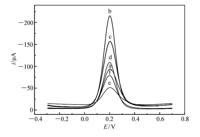
The fabrication course of the modified electrode and the hybridization of DNA were monitored by DPV technique using [Fe(CN)6]3-/4- as indicators. As shown in Fig. 2a, on the bare GCE, [Fe(CN)6]3-/4- had a sensitive peak current at about 0.2 V. After the ERGO was formed on the bare GCE surface(Fig. 2b), the reduction peak current increased significantly compared with that on the bare electrode, which can be attributed to the excellent conductivity and the large effective area of ERGO. After a layer of nanoZrO2 was deposited on the ERGO/GCE, the reduction current of [Fe(CN)6]3-/4- decreased apparently(Fig. 2c), which was caused by the poor conductivity of nanoZrO2. When the ssDNA probes were immobilized on the nanoZrO2, the peak current decreased more(Fig. 2d) relative to that on the nanoZrO2/ERGO/GCE because of the electrostatic repulsion between the ssDNA probes and the [Fe(CN)6]3-/4-, indicating that the ssDNA probes were successfully immobilized. After the csDNA hybridized with the ssDNA probes, the smallest peak current was obtained(Fig. 2e). The hybridization constitutes the DNA phosphate skeleton double helix structure, which makes the membrane more compact and stronger electronegativity, therefore, the [Fe(CN)6]3-/4- can not reach to the surface of electrode and transfer electrons easily, so the curve peak current declined further. However, after the ncDNA hybridized with the ssDNA probes(Fig. 2f), the electrochemical response was lower than that of Fig. 2a, because gene sequences can not complement each other. These results showed that this method was suitable for detection of PAT gene.
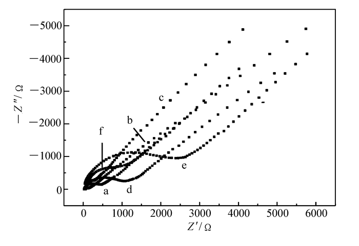
Using [Fe(CN)6]3-/4- as electrochemical probes, different electrodes were characterized carefully by EIS and the Nyquist plots were shown in the Fig. 3. First, let us see the electron transfer resistances(Ret)(the semicircle section on Nyquist plots) of the bare GCE(Fig. 3a), ERGO/GCE (Fig. 3b) and nanoZrO2/ERGO/GCE(Fig. 3c). The smallest and the biggest Ret were obtained on the bare GCE(Fig. 3a) and ERGO/GCE(Fig. 3b), respectively, among of these three electrodes, which could be ascribed to the good properties of ERGO and relatively large area surface of nanoZrO2. After the ssDNA was immobilized on the nanoZrO2/ERGO/GCE, the Ret increased significantly(Fig. 3d), and after the ssDNA hybridized with the csDNA, the Ret reached the biggest value(Fig. 3f) comparing with that of the others electrodes. However, after the ssDNA hybridized with the nsDNA, interestingly, the Ret of the ssDNA/nanoZrO2/ERGO/GCE is almost as the same as that of the nsDNA/nanoZrO2/ERGO/GCE(Fig. 3f), which showed that only a little of nsDNA can be hybridized with ssDNA and immobilized on the electrode.
The amount of ERGO and nanoZrO2 had deeply influence on the immobilization of DNA probes and electron transfer. After optimization, 6.0 μL GO(1.0 g/L) and 6.0 μL nanoZrO2(1.0 g/L) were selected as the optimal amount, and the GO was reduced to ERGO by cyclic voltammetry in the -1.7~0.2 V for 20 cycles.
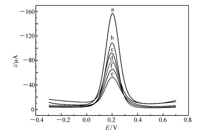
The adsorption time of the DNA probes was also optimized and the results were showed in the Fig. 4. It was found that the DPV current of the [Fe(CN)6]3-/4- decreased with the adsorption time, and the smallest current was obtained at 800 s, then the current increased with the adsorption time. Therefore, 800 s was selected as the optimal adsorption time.
The time for hybridization was also investigated in the range of 100~800 s. The results showed that, at the same concentration of target DNA, the smallest DPV current value was obtained when the time for hybridization was 500 s. If hybridizing for a longer time, the DPV peak current did not decrease any more, indicating that the ssDNA probes on the modified electrode had been hybridized completely. So the hybridization was carried out for 500 s at room temperature.
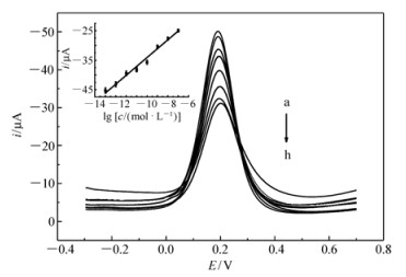
The hybridization of ssDNA/nanoZrO2/ERGO/GCE with different concentrations of the PAT gene sequence was investigated by DPV and the results were shown in the Fig. 5. The results showed that the DPV peak current decreased with the concentration of the PAT gene sequence and the further study showed that the values of the DPV peak current at ssDNA/nanoZrO2/ERGO/GCE after the ssDNA probes hybridized with the PAT gene sequence, were linearly dependent on the logarithm of the concentrations of the PAT gene sequence in the range of 1.0×10⁻⁶~1.0×10⁻¹³ mol/L. The regression equation of the calibration line was I(μA)=2.997×10⁻⁶lg [c/(mol·L-1)]-7.137×10-5(n=8) with correlation coefficient R=0.9933, and the detection limit was 2.0×10⁻¹⁵ mol/L obtained according to a signal-to-noise ratio of 3. The plot of the DPV peak current values versus the logarithm of the concentrations of the PAT gene sequence was showed in Fig. 5, Inset.
The comparison of the proposed biosensor with the other zirconia-based DNA electrochemical biosensors was listed in the Table 1. The results showed that the proposed sensor had lower detection limit and wider detection range than the other sensors based on zirconia. The good performance of the proposed biosensor was ascribed to the synergistic effects of the ERGO and the mesoporous nanoZrO2 for immobilizing ssDNA and electron transfer.

 下载:
导出CSV
下载:
导出CSV
| Composing of the electrodes | Detection techniques | Linear range/(mol·L-1) | Detection limit/(mol·L-1) | References |
| ZrO2/SWNTs/PDC/GCE | EIS | 1.0×10⁻¹¹~1.0×10⁻⁶ | 1.38×10⁻¹² | [26] |
| ZrO2/nanoAuNPs/GCE | DPV | 1.0×10⁻¹⁰~1.0×10⁻⁶ | 3.1×10⁻¹¹ | [23] |
| PAN-nanoZrO2/PTyr/GCE | EIS | 1.0×10⁻¹³~1.0×10⁻⁶ | 2.68×10⁻¹⁴ | [27] |
| ZrO2-Carbon paste electrode | DPV | 2.25×10⁻¹⁰~2.25×10⁻⁷ | ≤2×10⁻¹⁰ | [28] |
| nanoZrO2/ERGO/GCE | DPV | 1.0×10⁻¹³~1.0×10⁻⁶ | 2.0 × 10⁻¹⁵ | This work |
According to previous study[28], this kind of biosensor can be used for detecting the PCR products of transgenic maize samples with satisfactory results. Because in this study, the csDNA, i.e. the 20-base fragment of phosphinothricin acetyltransferase(PAT) gene sequence, was selected from transgenic maize, we do not conduct related experiments.
The repeatability of this sensor was verified by parallel detecting the DPV changes before and after the hybridization of the ssDNA probes and csDNA for five times. The RSD was 3.48%(n=5), which showed this sensor had good repeatability.
The electrochemically reduced graphene oxide(ERGO) and nanoZrO2 are very suitable for immobilization of ssDNA probes and sensing the phosphinothricin acetyltransferase(PAT) gene segments. They play synergistic effects on improving the performance of this sensor. Due to its broad dynamic linear range and low detection limit, the sensor can be used for determination of the PAT gene segments in transgenic maize with good selectivity, fast response and high sensitivity.
Tam P D. Genetically Modified Organism(GMO) Detection by Biosensor Based on SWCNT Material[J]. Curr Appl Phys, 2015, 15(3): 397-401. doi: 10.1016/j.cap.2015.01.017
Deisingh A K, Badrie N. Detection Approaches for Genetically Modified Organisms in Foods[J]. Food Res Int, 2005, 38: 639-649. doi: 10.1016/j.foodres.2005.01.003
Arugula M A, Zhang Y, Simonian A L. Biosensors as 21st Century Technology for Detecting Genetically Modified Organisms in Food and Feed[J]. Anal Chem, 2014, 86(1): 119-129.
European Commission. Regulation(EC) No.1830/2003 Concerning the Traceability and Labelling of Genetically Modified Organisms and the Traceability of Food and Feed Products Produced from Genetically Modified Organisms and Amending Directive 2001/18/EC[J]. Off J Eur Union, 2003, 268: L24-L28.
Ahmed F E. Detection of Genetically Modified Organism in Food[J]. Trends Biotechnol, 2002, 20(5): 215-223. doi: 10.1016/S0167-7799(01)01920-5
Vollenhofer S, Burg K, Schmidt J. Genetically Modified Organisms in Food Screening and Specific Detection by Polymerase Chain Reaction[J]. J Agric Food Chem, 1999, 47(12): 5038-5043. doi: 10.1021/jf990353l
Mafra I, Ferreira I, Oliveira M. Food Authentication by PCR-Based Methods[J]. Eur Food Res Technol, 2008, 227(3): 649-665. doi: 10.1007/s00217-007-0782-x
Gašparič M B, Tengs T, La Paz J L. Comparison of Nine Different Real-Time PCR Chemistries for Qualitative and Quantitative Applications in GMO Detection[J]. Anal Bioanal Chem, 2010, 396(6): 2023-2029. doi: 10.1007/s00216-009-3418-0
Manzanares-Palenzuela C L, Mafra I, Costa J. Electrochemical Magneto-Assay Coupled to PCR as a Quantitative Approach to Detect the Soybean Transgenic Event GTS40-3-2 in Foods[J]. Sens Actuators B, 2016, 222: 1050-1057. doi: 10.1016/j.snb.2015.09.013
Ma Y, Jiao K, Yang T. Sensitive PAT Gene Sequence Detection by Nano-SiO2/p-Aminothiophenol Self-assembled Films DNA Electrochemical Biosensor Based on Impedance Measurement[J]. Sens Actuators B, 2008, 131(2): 565-571. doi: 10.1016/j.snb.2007.12.046
Yang T, Zhang W, Du M. A PDDA/Poly(2, 6-Pyridinedicarboxylic Acid)-CNTs Composite Film DNA Electrochemical Sensor and Its Application for the Detection of Specific Sequences Related to PAT Gene and NOS Gene[J]. Talanta, 2008, 75(4): 987-994. doi: 10.1016/j.talanta.2007.12.049
Jiang C, Yang T, Jiao K. A DNA Electrochemical Sensor with Poly-L-Lysine/Single-Walled Carbon Nanotubes Films and Its Application for the Highly Sensitive EIS Detection of PAT Dene Fragment and PCR Amplification of NOS Gene[J]. Electrochim Act, 2008, 53(6): 2917-2924. doi: 10.1016/j.electacta.2007.11.015
Zhang D D, Li L Z, Ma W N. Electrodeposited Reduced Graphene Oxide Incorporating Polymerization of L-Lysine on Electrode Surface and Its Application in Simultaneous Electrochemical Determination of Ascorbic Acid, Dopamine and Uric Acid[J]. Mater Sci Eng C, 2017, 70: 241-249. doi: 10.1016/j.msec.2016.08.078
Zhou M, Wang Y L, Zhai Y M. Controlled Synthesis of Large-Area and Patterned Electrochemically Reduced Graphene Oxide Films[J]. Chem Eur J, 2009, 15: 6116-6120. doi: 10.1002/chem.v15:25
Guo H L, Wan X F, Qian Q Y. A Green Approach to the Synthesis of Graphene Nanosheets[J]. ACS Nano, 2009, 3(9): 2653-2659. doi: 10.1021/nn900227d
Wang J F, Yang S L, Guo D Y. Comparative Studies on Electrochemical Activity of Graphene Nanosheets and Carbon Nanotubes[J]. Electrochem Comm, 2009, 11(10): 1892-1895. doi: 10.1016/j.elecom.2009.08.019
Shao Y Y, Wang J, Engelhard M. Facile and Controllable Electrochemical Reduction of Graphene Oxide and Its Applications[J]. J Mater Chem, 2010, 20: 743-748. doi: 10.1039/B917975E
Yang T, Li X, Li Q H. Electrochemically Reduced Graphene Oxide-Enhanced Electropolymerization of Poly-Xanthurenic Acid for Direct[J]. Polym Chem, 2013, 4: 1228-1234. doi: 10.1039/C2PY20655B
Xu Y, Gao M, Zhang G. Electrochemically Reduced Graphene Oxide with Enhanced Electrocatalytic Activity Toward Tetracycline Detection[J]. Chinese J Catal, 2015, 36(11): 1936-1942. doi: 10.1016/S1872-2067(15)60956-1
Sun W, Lu Y, Wu Y. Electrochemical Sensor for Transgenic Maize MON810 Sequence with Electrostatic Adsorption DNA on Electrochemical Reduced Graphene Modified Electrode[J]. Sens Actuators B, 2014, 202(31): 160-166.
Gao F, Qi X, Cai X. Electrochemically Reduced Graphene Modified Carbon Ionic Liquid Electrode for the Sensitive Sensing of Rutin[J]. Thin Solid Films, 2012, 520(15): 5064-5069. doi: 10.1016/j.tsf.2012.03.002
Fang M M, David M K, Anthony C S. A "Mix and Match" Ionic-Covalent Strategy for Self-Assembly of Inorganic Multilayer Films[J]. J Am Chem Soc, 1997, 119(50): 12184-12191. doi: 10.1021/ja972569e
Zhang W, Yang T, Jiang C. DNA Hybridization and Phosphinothricin Acetyltransferase Gene Sequence Detection Based on Zirconia/Nanogold Film Modified Electrode[J]. Appl Surf Sci, 2008, 254(15): 4750-4756. doi: 10.1016/j.apsusc.2008.01.102
Sun W, Wang X, Wang W. Electrochemical DNA Sensor for Staphylococcus aureus Nuc Gene Sequence with Zirconia and Graphene Modified Electrode[J]. J Solid State Electrochem, 2015, 19(8): 2431-2438. doi: 10.1007/s10008-015-2893-9
Renuka L, Anantharaju K S, Sharma S C. A Comparative Study on the Structural, Optical, Electrochemical and Photocatalytic Properties of ZrO2 Nanooxide Synthesized by Different Routes[J]. J Alloys Comp, 2017, 695: 382-395. doi: 10.1016/j.jallcom.2016.10.126
Yang J, Jiao K, Yang T. A DNA Electrochemical Sensor Prepared by Electrodepositing Zirconia on Composite Films of Single-Walled Carbon Nanotubes and Poly(2, 6-Pyridinedicarboxylic Acid), and Its Application to Detection of the PAT Gene Fragment[J]. Anal Bioanal Chem, 2007, 389(3): 913-921.
Yang J, Wang X L, Shi H Q. An Electrochemical DNA Biosensor for Highly Sensitive Detection of Phosphinothricin Acetyltransferase Gene Sequence Based on Polyaniline-(Mesoporous Nanozirconia)/Poly-Tyrosine Film[J]. Sens Actutators B, 2012, 162(1): 178-183. doi: 10.1016/j.snb.2011.12.064
Zou Y J, Sun L, Xu X F. Biosensor Based on Polyaniline-Prussian Blue/Multi-walled Carbon Nanotubes Hybrid Composites[J]. Biosens Bioelectron, 2007, 22(11): 2669-2674. doi: 10.1016/j.bios.2006.10.035

Figure 2 Differential pulse voltammograms of [Fe(CN)6]3-/4- on different electrodes GCE(a), ERGO/GCE(b), nanoZrO2/ERGO/GCE(c), ssDNA/nanoZrO2/ERGO/GCE(d), csDNA/nanoZrO2/ERGO/GCE(e), nsDNA/nanoZrO2/ERGO/GCE(f)

Figure 3 Nyquist curves of [Fe(CN)6]3-/4- on different electrodes GCE(a), ERGO/GCE(b), nanoZrO2/ERGO/GCE(c), ssDNA/nanoZrO2/ERGO/GCE(d), nsDNA/nanoZrO2/ERGO/GCE(e), csDNA/nanoZrO2/ERGO/GCE(f)

Figure 4 Differential pulse voltammograms of nanoZrO2/ERGO/GCE in [Fe(CN)6]3-/4- after the nanoZrO2/ERGO/GCE was immersed in 1.0 μmol/L ssDNA solution for different time:0(a), 200(b), 400(c), 600(d), 800(e), and 1000 s(f)

Figure 5 Differential pulse voltammograms of ssDNA/nanoZrO2/ERGO/GCE in [Fe(CN)6]3-/4- after it hybridized with different concentrations of target DNA sequences. Inset:the linear relationship between the logarithm of the concentration and the peak current. 1.0×10⁻¹³(a), 1.0×10⁻¹²(b), 1.0×10⁻¹¹(c), 1.0×10⁻¹⁰(d), 1.0×10⁻⁹(e), 1.0×10⁻⁸(f), 1.0×10⁻⁷(g) and 1.0×10⁻⁶(h) mol/L
Table 1. Comparison of the performance of the proposed biosensor with that of other zirconia-based DNA biosensors
| Composing of the electrodes | Detection techniques | Linear range/(mol·L-1) | Detection limit/(mol·L-1) | References |
| ZrO2/SWNTs/PDC/GCE | EIS | 1.0×10⁻¹¹~1.0×10⁻⁶ | 1.38×10⁻¹² | [26] |
| ZrO2/nanoAuNPs/GCE | DPV | 1.0×10⁻¹⁰~1.0×10⁻⁶ | 3.1×10⁻¹¹ | [23] |
| PAN-nanoZrO2/PTyr/GCE | EIS | 1.0×10⁻¹³~1.0×10⁻⁶ | 2.68×10⁻¹⁴ | [27] |
| ZrO2-Carbon paste electrode | DPV | 2.25×10⁻¹⁰~2.25×10⁻⁷ | ≤2×10⁻¹⁰ | [28] |
| nanoZrO2/ERGO/GCE | DPV | 1.0×10⁻¹³~1.0×10⁻⁶ | 2.0 × 10⁻¹⁵ | This work |
 下载: 导出CSV

 扫一扫看文章


扫一扫关注我们

 下载:
下载: