Figure 1.
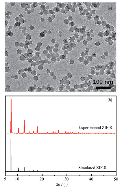
(a) TEM image of ZIF-8 nanoparticles prepared with nZn/nHmim=1/4; (b) Experimental XRD pattern of as-synthesized ZIF-8 powder and XRD pattern simulated from ZIF-8 crystal structure data
Controlling Distribution of Gold Nanoparticles in Au@ZIF-8 Core-Shell Structures for Sensing Fluorescent Molecules with Photoluminescence
Wei ZHANG , Yu-Yi ZHANG , Ya-Jie BIAN , Meng-Di CHEN , Xiao-Lei ZHANG , Qing-Yuan JIN , Bing-Wen HU

Core-shell nanoparticles have been widely investigated for multifunctional applications[1]. In general, the properties of core-shell nanoparticles can be rationally designed by playing with either the core or the shell materials, i. e., the compositions, the size of the core, and the thickness of the shell. Moreover, the core distribution in the core-shell nanoparticle makes a difference, for example, in the catalytic properties[2-4].
Metal@MOF is a core-shell type configuration which encapsulates metal nanoparticles into metal organic framework. Due to the combination of optical and catalytic properties with tunable porosity, metal @MOF has been considered as outstanding platforms for drug delivery[5] and sensing[6-7]. However, controlling the distribution of metal nanoparticles in metal@MOF core-shell nanoparticles is not easy to realize. In the case of Au nanoparticles (Au NPs) encapsulated in zeolitic imidazolate frameworks (Au@ZIF-8), pioneer work from Huo's group[8] has shown that the polyvinylpyrrolidone (PVP) assisted process can tune the spatial distribution of Au within ZIF-8 crystals by adjusting the addition sequence. In addition, it has been found that the dominant growth mode at early stage of ZIF-8 determines the formation of single or multi-core-shell Au@ZIF-8 structures[9]. Therefore, the strategies which allow finely controlled formation of ZIF-8 would be feasible approaches to tune the distribution of Au cores.
Coordination modulation method has been demonstrated to be effective in controlling the crystal growth of MOF nanocrystals[10]. Based on the strategy of simultaneous addition of two capping ligands, significant progress has been made to put the synthesis of ZIF-8 nanocrystals on a more rational basis[11-13]. In this work, we attempted to apply this dual-ligand strategy to the synthesis of Au@ZIF-8 core-shell nanoparticles, aiming to manipulate the distribution of Au NPs in ZIF-8 crystals. Furthermore, we envisioned that Au@ZIF-8 core-shell structure with photoluminescent information will have promising applications in sensing. We investigated two differently sized fluorescent molecules which match the aperture size of ZIF-8 to explore if the distribution of Au NPs in ZIF-8 would affect the luminescence intensity.
Zn(NO3)2·6H2O was obtained from Alfa Aesar. PVP (Mw=55 000) was bought from Sigma. Trisodium citrate, 2-methylimidazole(Hmim), 1-methylimidazole (1-MI), MeOH and HAuCl4 were purchased from Aladdin. All the reagents were used without further purification. Scanning electron microscopy (SEM) measurements were performed using a Zeiss sigma300 field emission microscope at an accelerating voltage of 3 kV. Transmission electron microscope (TEM) measurements were performed using a G2F20. The accelerating voltage of TEM was 200 kV. Dynamic light scattering (DLS) were measured using a Malvern Zetasizer Nano ZS laser diffraction analyzer. Powder X-ray diffraction (XRD) measurements were performed using a Rigaku Smartlab SE with a high-density Cu Kα radiation (λ= 0.154 178 nm) at 35 kV and 25 mA, in the 2θ range of 5°-50° with a step size of 0.01° and a scan rate of 10 (°) ·min-1. A liquid nitrogen cooled charge coupled device (CCD) spectrometer (Princeton Instruments) was used to detect the PL spectra while a microchannel plate photomultiplier tube (Hamamatsu) combined with time-correlated single photon counting technique (Edinburgh Instruments) was used for photon counting and lifetimes measurements. A pulse picosecond diode laser at 375 nm (Advance Laser System) was used to excite the samples.
Au NPs were prepared by a sodium citrate reduction method of HAuCl4. In a typical procedure for the synthesis of 13 nm Au NPs, an aqueous solution of HAuCl4 (0.01%, 150 mL) was brought to a vigorous boil with rapid stirring in a round bottom flask (250 mL) fitted with a reflux condenser. When the solution started to boil, an aqueous solution of trisodium citrate (1%, 4.5 mL) was added. The mixture was refluxed with stirring for another 20 min. The resulting deep red suspension was then removed from the heat solution. After the Au NP sol was cooled to room temperature, a solution of PVP (0.5 g) in water (20 mL) was added dropwise to the Au NP sol with stirring, and the mixture was further stirred at room temperature for 24 h. The PVP-stabilized Au NPs were collected by centrifugation at 14 000 r·min-1 for 30 min, washed by methanol for three times, and finally dispersed in 20 mL methanol.
To synthesis the ZIF-8 crystal with different molar ratios (nZn/nHmim), 0.037 g Zn(NO3)2·6H2O were dissolved in 5 mL MeOH. To this solution, then another solution of Hmim (0.04, 0.16, 0.28 g) was added and mixed vigorously for 10 s, and the resulting solution was incubated at room temperature for 24 h. The formed particles were collected by centrifugation and washed with MeOH several times before they were redispersed in MeOH.
The particles of ZIF-8 with 1-MI as capping ligand were synthesized as follows. 0.037 g Zn(NO3)2· 6H2O were dissolved in 5 mL MeOH. To this solution, another solution of 0.04 g Hmim and 1-MI (50 or 100 or 150 or 200 mmol·L-1) in MeOH (5 mL) was added and mixed vigorously for 10 s, and the resulting solution was incubated at room temperature for 24 h. The formed particles were collected by centrifugation and washed with MeOH several times before they were redispersed in MeOH. According to the amount of 1-MI added, the samples were named ZIF-8-50, ZIF-8-100, ZIF-8-150 and ZIF-8-200.
The nanoparticles were synthesized using the established methods and their surfaces were functionalized with PVP either during or after synthesis. The encapsulation procedure was demonstrated initially for 13 nm Au NPs. In a typical experiment, methanolic solutions of Hmim (100 mmol·L-1, 5 mL) and 1-MI (50 or 100 or 150 or 200 mmol·L-1) were added into methanolic solutions of Zn(NO3)2 (25 mmol·L-1, 5 mL) and then Au NPs (1 mL) were mixed briefly and then kept at room temperature for 24 h without stirring. The formed particles were collected by centrifugation and washed with MeOH several times, then they were redispersed in MeOH. According to the amount of 1-MI added, the samples were named Au@ZIF-8-50, Au@ZIF-8100, Au@ZIF-8-150 and Au@ZIF-8-200.
Instead of directly investigating the dual-ligand synthesis of Au@ZIF-8 core-shell nanoparticles, we firstly tried to find out the optimum ratio of Zn(NO3)2· 6H2O (zinc salt) to Hmim for the ZIF-8 precursor without introducing the second ligand. To obtain nanoparticles with uniform size, the ideal situation is that all nuclei form at the same time with the same size. In addition, all the nuclei will experience the same subsequent growth. Therefore, it is highly desirable to have nucleation to occur within a very short period of time. It has been found that adding Hmim in excess to the zinc source could yield a high nucleation rate and deliver stable colloidal dispersion[14]. Fig. 1a displays the TEM image of the product prepared with nZn/nHmim= 1/4. The polydispersity index (PDI) was measured to be 0.18 by DLS. The XRD pattern shown in Fig. 1b confirms that the as-synthesized product is single-phase ZIF-8 material. When the n Zn/nHmim was further increased to 1/7, the PDI could be optimized to 0.12. But the size of the obtained nanoparticles decreased significantly (Fig.S1, Supporting information). A compromise has to be made between the polydispersity and the size of obtained particles, i.e., the nucleation rate, as preliminary work[8] reported that if the ratio is higher than 1.6, the encapsulation of Au in ZIF-8 would partially or completely fail.
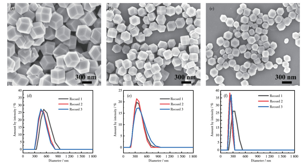
Thus, synthesis strategies which could slow down the nucleation rate a bit while keeping a low level of polydispersity are highly desirable. 1-MI with pKa < 10.3 competes with Hmim in the reaction of zinc ions and Hmim, resulting in a lower nucleation rate[11]. On the basis of n Zn/nHmim=1/4, different amounts of the modulating ligand 1-MI were introduced to the ZIF-8 precursor. When a small amount of 1-MI (50 mmol·L-1) was added to the ZIF-8 precursor, the size of obtained ZIF-8 particles (Fig. 2a and 2d) increased a lot, compared to the particles shown in Fig. 1a. At the same time, the PDI also increased from 0.18 to 0.29. As the concentration of 1-MI further increased to 100 mmol· L-1, the ZIF-8 particles became smaller while delivered a much smaller PDI of 0.15 (Fig. 2b and 2e). Too concentrated 1-MI (c≥150 mmol·L-1) can cause the size distribution to be bimodal, which is the case shown in Fig. 2c and 2f and also in other reported work[13].
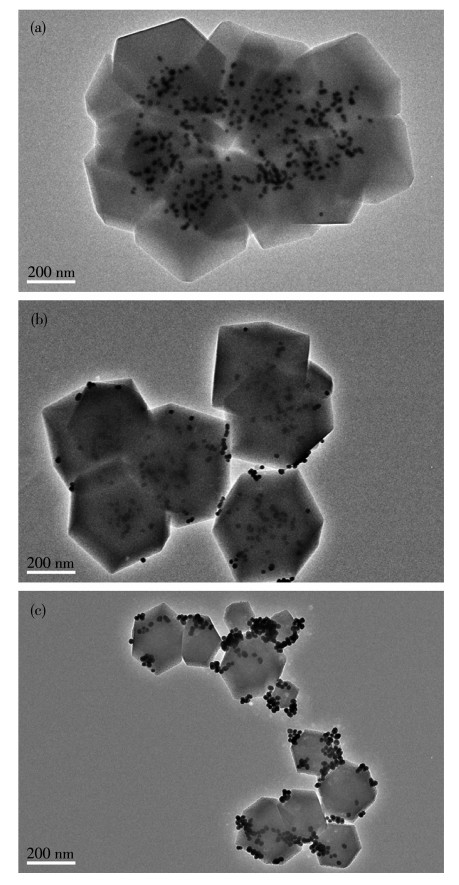
The dual-ligand ZIF-8 precursor containing both Hmim and 1 -MI was adopted to encapsulate the PVP functionalized Au NPs. Presumably due to the low concentrations and/or small sizes of Au NPs, only ZIF-8 produces its pattern in the XRD characterization of gold -loaded materials (Fig.S2). In Fig. 3, one can find that both the size and polydispersity of the obtained ZIF-8 crystals grown in presence of Au NPs are consistent with the ones obtained from pure precursor solution (Fig. 2). However, the distribution of Au NPs in the ZIF-8 crystals differed a lot. With the increasing amount of 1-MI, the Au NPs tended to spread and disperse more in space to the boarder of ZIF-8 crystals. When 200 mmol·L-1 1 -MI was used in the ZIF-8 precursor, the Au NPs started to agglomerate and only occupied the edges of ZIF-8 crystals, as displayed in Fig. 3c. The agglomeration of Au NPs can also be identified by the colour of the obtained sample (Fig. S3). It has also been found that 1-MI alone can destabilize the PVP functionalized gold nanoparticles (Fig.S4). Therefore, finely tuning the amount of the second ligand 1-MI could manipulate the distribution of Au NPs in ZIF-8 crystals. More TEM images can be found in Fig. S5. Over concentrated 1-MI would on one hand excessively slow down the nucleation rate of ZIF-8, and on the other hand cause the agglomeration of Au NPs.
For the further exploration of Au@ZIF-8 in optical sensing molecules, the photoluminescence (PL) detection of Au@ZIF-8 to two different sized molecules was studied. The aperture of ZIF-8 shell would act as an analyte filter to allow the smaller molecules to pass through the pores and reach the Au NPs inside, while the larger ones would be blocked.
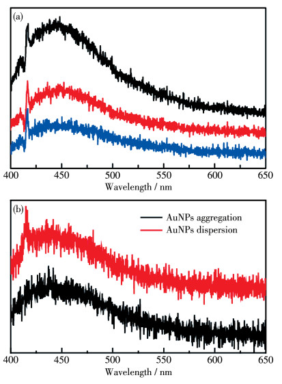
As depicted in Fig. 4a, ZIF-8-150 exhibited a weak emission band at 450 nm under 375 nm laser excitation. We also obtained the fluorescent spectra of ZIF-8-50 and ZIF-8-100, with the emission peak getting stronger. In Liu's work[15], the fluorescence emission peak of ZIF-8 may come from the free ligand Hmim, which means the emission peak will not be affected by 1-MI. Furthermore, we examined PL emission spectra of Au@ZIF-8 (Fig. 4b). The aggregation and dispersion of Au NPs in ZIF-8 are shown in the TEM images (Fig. 3a and 3b). The similar spectral features with the peak at 450 nm demonstrate that the determinant of the spectrum is the ligand Hmim other than the distribution of Au NPs.
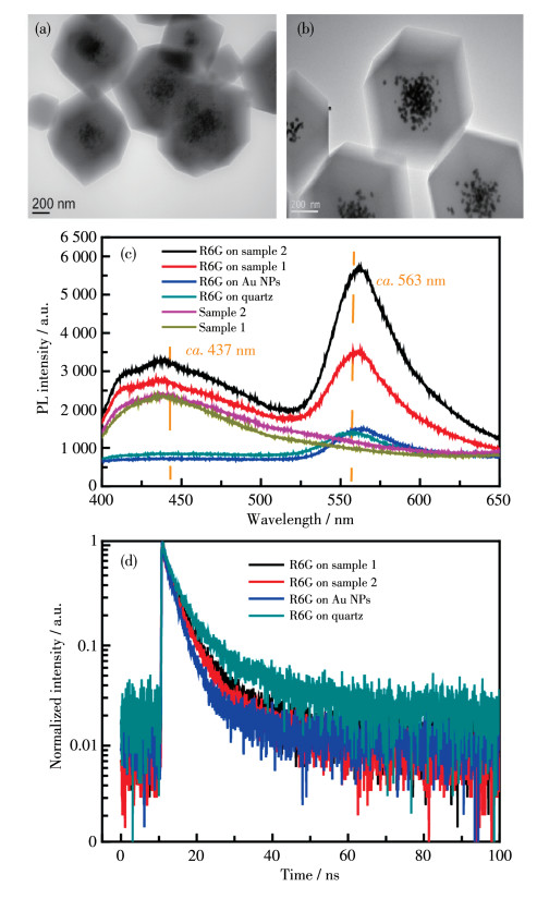
Rhodamine 6G (R6G) molecules with hydrodynamic diameter being 1 nm[18, 26] were used. It is larger than the size of ZIF-8 aperture[16] (about 0.34 nm). First of all, Au@ZIF-8 particles were aggregated into a tiny spot (about 0.5 cm2). As 10 μL of their ethanol solution was completely evaporated, the size of the aggregate was slightly dependent on the nature of the solvent (Fig. S6). Then R6G submonolayers were thermally sublimated to Au@ZIF-8 substrate in vacuum (p=10-4 Pa) at room temperature. For comparison, we also prepared the R6G samples evaporated on quartz and Au NPs substrates. The TEM images of Au@ZIF-8 are given in Fig. 5a and 5b. Fewer Au NPs were present when diluted (1∶1, V/V) Au NPs dispersion was applied in the synthesis of Au@ZIF-8. The shell thickness of Au@ZIF-8 with fewer Au NPs (Fewer-Au@ZIF-8) was around 300 nm (Fig. 5a) and Au@ZIF-8 with more Au NPs (MoreAu@ZIF-8) was around 250 nm (Fig. 5b). Fig.S5 shows the TEM image of Au NPs. The PL spectra of Au@ZIF-8 samples and R6G submonolayers on different substrates are shown in Fig. 5c, exhibiting that the luminescence intensity of R6G on Au@ZIF-8 was enhanced compared with that of R6G on quartz or Au NPs with a stable peak position at 563 nm. By synthetically taking account for the R6G emission peak (ca. 563 nm), Au@ZIF-8 emission band (430-450 nm), and the different extent of the luminescence enhancement for the different substrates, the enhancement mechanism may be attributed to the strong adsorption of ZIF-8 to organic dyes, leading to the molecules enrichment. Also, we compared the R6G on Fewer-Au@ZIF-8 (red line) with R6G on More-Au@ZIF-8 (black line) where ZIF-8 packaged different amounts of Au NPs. The results demonstrate that increasing Au NPs aggregation in ZIF8 will enhance the molecular fluorescence due to the plasmonic resonant excitation enhancement. If R6G molecules are absorbed directly on Au NPs, charge and energy transfer between dyes and metals usually lead to decreasing luminescence (blue line). The fluorescence decay curves as shown in Fig. 5d exhibit faster decay processes (blue line) for R6G on Au NPs than that for R6G on quartz (green line). Detailed lifetimes at 560 nm peak are listed in Table 1. A two-exponential function was fitted to the decay process of excited R6G molecules on quartz, with a shorter component τ1 of 3.28 ns and a longer component τ2 of 13.05 ns. For the R6G on Au NPs sample, significant changes in decay processes occurred and a very fast nonradiative decay channel began to emerge representing a shorter component τ1 of 2.56 ns and a longer component τ2 of 7.93 ns. Considering R6G attaching to Au NPs, nonradiative decay processes of the molecular excited state should be achieved by several charge and energy transfer channels, such as π-π stacking of neighboring molecules[17, 19], surface interactions between molecules and the polyelectrolyte layer[20], and molecules-metal coupling[19]. The lifetimes of R6G on Au@ZIF-8 were between those of R6G on quartz and those of R6G on Au NPs, which similar to those of R6G on quartz sample. τ1 and τ2 components can correspond to the decay of monomers and excimers or aggregates affected by intermolecular interaction via Förster resonance energy transfer mechanism respectively[18-19].
 下载:
导出CSV
下载:
导出CSV
| Sample | τ1/ns | A1 | τ2/ns | A2 |
| R6G on quartz | 3.28 | 52 | 13.05 | 48 |
| R6G on Au NPs | 2.56 | 79 | 7.93 | 21 |
| R6G on Fewer-Au@ZIF-8 | 2.53 | 48 | 8.71 | 52 |
| R6G on More-Au@ZIF-8 | 2.67 | 52 | 8.25 | 48 |
| HPBI | 1.87 | 18 | 4.48 | 82 |
| HPBI/Au@ZIF-8-50 | 1.65 | — | — | — |
| HPBI/Au@ZIF-8-100 | 1.95 | — | — | — |
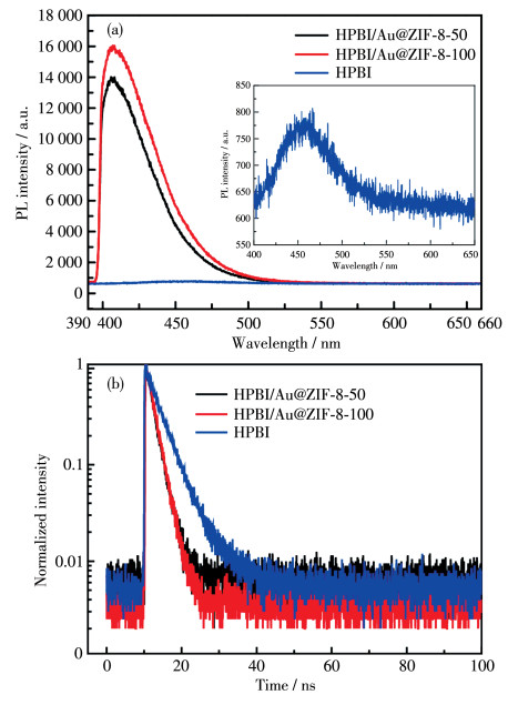
For further exploration of the selective and sensitive detection of ZIF-8, we also chose another molecule, 2-(2-hydroxyphenyl)-1H-benzimidazole (HPBI), whose size is close to that of ZIF-8 aperture. The PL spectra in Fig. 6a demonstrate that the distribution of Au NPs in ZIF-8 will affect the luminescence intensity of HPBI. Compared with pure HPBI solution, the intensity (Fig. 6a) was increased approximately 18 times of HPBI with Au NPs aggregated in ZIF-8 (Au@ZIF-8-50) and 21 times of HPBI with Au NPs dispersed in ZIF-8 (Au@ZIF-8-100), which was much larger than the enhancement factor (less than 4) of R6G on Au@ZIF-8 (Fig. 5c). Except for the HPBI molecule adsorbed around the ZIF-8 aperture which can enhance the emission intensity, the size of HPBI molecules (around 0.3 nm×0.7 nm) may play an important role. Although the diameter of HPBI is slightly larger than the aperture size of ZIF-8 (about 0.34 nm), the molecules can still pass through the windows of ZIF-8 because of the flexible structure of ZIF-8[16, 21-23]. The HPBI molecules can get closer to the Au NPs encapsulated in ZIF-8, which increases its excitation rate by the enhanced local near field[17]. As shown in Fig. 6b, the fluorescence decay curves of HPBI/Au@ZIF-8 mixture exhibited faster decay processes than pure HPBI.
Inset: enlarged PL spectrum of HPBI solution
Table 1 lists the lifetimes of HPBI and HPBI/ Au@ZIF-8 mixture. Two-exponential function fitted to the decay process of the excited HPBI solution, with a shorter component τ1 of 1.87 ns and a longer component τ2 of 4.48 ns. However, after mixing Au@ZIF-8 with HPBI, the lifetime can be well fitted by a singleexponential function, indicating the quenching effect of the metallic NPs[17]. The luminescence enhancement and the fast one-component exponential decay behavior illustrate the competition of the amplification and inhibition effects of the near field of metallic NPs in ZIF-8 on the molecular fluorescence.
We described a dual-ligand strategy which is effective in controlling the distribution of Au NPs in ZIF-8 crystals. The presence of proper amount of the second ligand 1-MI could slow down the nucleation rate without sacrificing the polydispersity of ZIF-8 crystals. By finely adjusting the amount of 1-MI, the distribution of Au NPs in ZIF-8 crystals can be tuned. Au NPs scattered from the centre to the edge of the ZIF-8 crystals with the increasing concentration of 1-MI. Over concentrated 1-MI could cause the agglomeration of Au NPs. Furthermore, the spectral feature and decay lifetime of R6G and HPBI molecules separately with Au@ZIF-8 were investigated. The observations show that the porous ZIF-8 shells selectively allow suitablesized molecules to pass through the apertures and Au NPs in Au@ZIF-8 nanostructures will sensitively affect the molecular luminescent enhancement and fluorescent quenching in the near field of metallic NPs. Our findings suggest that controlling the distribution of Au NPs in Au@ZIF-8 core-shell nanostructures can effectively sense fluorescent molecules with photoluminescence.
El-Toni A M, Habila M A, Labis J P, ALOthman Z A, Alhoshan M, Elzatahry A A, Zhang F. Design, Synthesis and Applications of Core-Shell, Hollow Core, and Nanorattle Multifunctional Nanostructures[J]. Nanoscale, 2016, 8: 2510-2531. doi: 10.1039/C5NR07004J
An W, Liu P. Rationalization of Au Concentration and Distribution in AuNi@Pt Core-Shell Nanoparticles for Oxygen Reduction Reaction[J]. ACS Catal., 2015, 5: 6328-6336. doi: 10.1021/acscatal.5b01656
Wang X K, Liu J, Zhang L, Dong L Z, Li S L, Kan Y H, Li D S, Lan Y Q. Monometallic Catalytic Models Hosted in Stable Metal-Organic Frameworks for Tunable CO2 Photoreduction[J]. ACS Catal., 2019, 9: 1726-1732. doi: 10.1021/acscatal.8b04887
Shiga T, Kumamaru R, Newton G N, Oshio H. Heteroleptic Iron (Ⅱ) Complexes with Naphthoquinone-Type Ligands[J]. Dalton Trans., 2020, 49: 1485-1491. doi: 10.1039/C9DT03946E
Chen X R, Tong R L, Shi Z Q, Yang B, Liu H, Ding S P, Wang X, Lei Q F, Wu J, Fang W J. MOF Nanoparticles with Encapsulated Autophagy Inhibitor in Controlled Drug Delivery System for Antitumor[J]. ACS Appl. Mater. Interfaces, 2018, 10: 2328-2337. doi: 10.1021/acsami.7b16522
Qiao X Z, Su B S, Liu C, Song Q, Luo D, Mo G, Wang T. Selective Surface Enhanced Raman Scattering for Quantitative Detection of Lung Cancer Biomarkers in Superparticle@MOF Structure[J]. Adv. Mater., 2018, 30: 1702275. doi: 10.1002/adma.201702275
He L C, Liu Y, Liu J Z, Xiong Y S, Zheng J Z, Liu Y L, Tang Z Y. Core-Shell Noble-Metal@Metal-Organic-Framework Nanoparticles with Highly Selective Sensing Property[J]. Angew. Chem. Int. Ed., 2013, 125: 3829-3833. doi: 10.1002/ange.201209903
Lu G, Li S Z, Guo Z, Farha O K, Hauser B G, Qi X Y, Wang Y, Wang X, Han S Y, Liu X G. Imparting Functionality to a Metal-Organic Framework Material by Controlled Nanoparticle Encapsulation[J]. Nat. Chem., 2012, 4: 310-316. doi: 10.1038/nchem.1272
Chen L Y, Peng Y, Wang H, Gu Z Z, Duan C Y. Synthesis of Au@ZIF-8 Single-or Multi-core-Shell Structures for Photocatalysis[J]. Chem. Commun., 2014, 50: 8651-8654. doi: 10.1039/C4CC02818J
Tsuruoka T, Furukawa S, Takashima Y, Yoshida K, Isoda S, Kitagawa S. Nanoporous Nanorods Fabricated by Coordination Modulation and Oriented Attachment Growth[J]. Angew. Chem. Int. Ed., 2009, 121: 4833-4837. doi: 10.1002/ange.200901177
Cravillon J, Nayuk R, Springer S, Feldhoff A, Huber K, Wiebcke M. Controlling Zeolitic Imidazolate Framework Nano- and Microcrystal Formation: Insight into Crystal Growth by Time-Resolved In Situ Static Light Scattering[J]. Chem. Mater., 2011, 23: 2130-2141. doi: 10.1021/cm103571y
Yanai N, Granick S. Directional Self-Assembly of a Colloidal Metal-Organic Framework[J]. Angew. Chem. Int. Ed., 2012, 124: 5736-5739. doi: 10.1002/ange.201109132
Yanai N, Sindoro M, Yan J, Granick S. Electric Field-Induced Assembly of Monodisperse Polyhedral Metal-Organic Framework Crystals[J]. J. Am. Chem. Soc., 2013, 135: 34-37. doi: 10.1021/ja309361d
Cravillon J, Münzer S, Lohmeier S J, Feldhoff A, Huber K, Wiebcke M. Rapid Room-Temperature Synthesis and Characterization of Nanocrystals of a Prototypical Zeolitic Imidazolate Framework[J]. Chem. Mater., 2009, 21: 1410-1412. doi: 10.1021/cm900166h
Liu S, Xiang Z H, Hu Z, Zheng X P, Cao D P. Zeolitic Imidazolate Framework-8 as a Luminescent Material for the Sensing of Metal ions and Small Molecules[J]. J. Mater. Chem., 2011, 21: 6649-6653. doi: 10.1039/c1jm10166h
Ding Q Q, Wang J, Chen X Y, Liu H, Li Q J, Wang Y L, Yang S K. Quantitative and Sensitive SERS Platform with Analyte Enrichment and Filtration Function[J]. Nano Lett., 2020, 20: 7304-7312. doi: 10.1021/acs.nanolett.0c02683
Niu J X, Pan C D, Liu Y T, Lou S T, Wu E, Wu B T, Zhang X L, Jin Q Y. Plasmon-Enhanced Fluorescence of Submonolayer Porphyrins by Silver-Polymer Core-Shell Nanoparticles[J]. Opt. Express, 2018, 26: 3489-3496. doi: 10.1364/OE.26.003489
Popov P, Steinkerchner L, Mann E K. Molecular Dynamics Study of Rhodamine 6G Diffusion at n-Decane-Water Interfaces[J]. Phys. Rev. E, 2015, 91: 053308. doi: 10.1103/PhysRevE.91.053308
Zhang X L, Chen L G, Lv P, Gao H Y, Wei S J, Dong Z C, Hou J G. Fluorescence Decay of Quasimonolayered Porphyrins near a Metal Surface Separated by Short-Chain Alkanethiols[J]. Appl. Phys. Lett., 2008, 92: 223118. doi: 10.1063/1.2938861
Reineck P, Gómez D, Ng S H, Karg M, Bell T, Mulvaney P, Bach U. Distance and Wavelength Dependent Quenching of Molecular Fluorescence by Au@SiO2 Core-Shell Nanoparticles[J]. ACS Nano, 2013, 7: 6636-6648. doi: 10.1021/nn401775e
Zheng G C, de Marchi S, López-Puente V, Sentosun K, Polavarapu L, Pérez-Juste I, Hill E H, Bals S, Liz-Marzán L M, Pastoriza-Santos I. Encapsulation of Single Plasmonic Nanoparticles within ZIF-8 and SERS Analysis of the MOF Flexibility[J]. Small, 2016, 12: 3935-3943. doi: 10.1002/smll.201600947
Carrillo-Carrión C, Martínez R, Navarro Poupard, M F, Pelaz B, Polo E, Arenas-Vivo A, Olgiati A, Taboada P, Soliman M G, Catalán Ú. Aqueous Stable Gold Nanostar/ZIF-8 Nanocomposites for Light-Triggered Release of Active Cargo Inside Living Cells[J]. Angew. Chem. Int. Ed., 2019, 58: 7078-7082. doi: 10.1002/anie.201902817
Morabito J V, Chou L Y, Li Z H, Manna C M, Petroff C A, Kyada R J, Palomba J M, Byers J A, Tsung C K. Molecular Encapsulation beyond the Aperture Size Limit Through Dissociative Linker Exchange in Metal-Organic Framework Crystals[J]. J. Am. Chem. Soc., 2014, 136: 12540-12543. doi: 10.1021/ja5054779
Dulkeith E, Morteani A, Niedereichholz T, Klar T, Feldmann J, Levi S, Van Veggel F, Reinhoudt D, Möller M, Gittins D. Fluorescence Quenching of Dye Molecules Near Gold Nanoparticles: Radiative and Nonradiative Effects[J]. Phys. Rev. Lett., 2002, 89: 203002. doi: 10.1103/PhysRevLett.89.203002
Anger P, Bharadwaj P, Novotny L. Enhancement and Quenching of Single-Molecule Fluorescence[J]. Phys. Rev. Lett., 2006, 96: 113002. doi: 10.1103/PhysRevLett.96.113002
Wang D, Pevzner L, Li C, Peneva K, Li C Y, Chan D Y, Müllen K, Mezger M, Koynov K, Butt H J. Layer with Reduced Viscosity at Water-Oil Interfaces Probed by Fluorescence Correlation Spectroscopy[J]. Phys. Rev. E, 2013, 87: 012403. doi: 10.1103/PhysRevE.87.012403

Figure 1 (a) TEM image of ZIF-8 nanoparticles prepared with nZn/nHmim=1/4; (b) Experimental XRD pattern of as-synthesized ZIF-8 powder and XRD pattern simulated from ZIF-8 crystal structure data

Figure 2 (a-c) SEM images and (d-f) DLS measurements of (a, d) ZIF-8-50, (b, e) ZIF-8-100 and (c, f) ZIF-8-150

Figure 3 TEM images of Au@ZIF-8 prepared using dual-ligand strategy: (a) Au@ZIF-8-50, (b) Au@ZIF-8-100 and (c) Au@ZIF-8-200

Figure 4 (a) PL spectra of ZIF-8-50 (black), ZIF-8-100 (blue) and ZIF-8-150 (red); (b) PL spectra of Au@ZIF-8 with Au NPs aggregation (Au@ZIF-8-50) and dispersion (Au@ZIF-8-100)

Figure 5 TEM images of Au@ZIF-8: (a) Fewer-Au@ZIF-8 (sample 1) and (b) More-Au@ZIF-8 (sample 2); (c) PL spectra of Au@ZIF-8, R6G on quartz, R6G on Au NPs, and R6G on Au@ZIF-8; (d) Dynamics of PL decay of R6G on quartz, R6G on Au NPs, and R6G on Au@ZIF-8

Figure 6 (a) PL spectra of Au@ZIF-8 mixing with HPBI; (b) Dynamics of PL decay of HPBI and Au@ZIF-8 mixing with HPBI
Inset: enlarged PL spectrum of HPBI solution
Table 1. Lifetimes of R6G on Au@ZIF-8 and Au@ZIF-8+HPBI
| Sample | τ1/ns | A1 | τ2/ns | A2 |
| R6G on quartz | 3.28 | 52 | 13.05 | 48 |
| R6G on Au NPs | 2.56 | 79 | 7.93 | 21 |
| R6G on Fewer-Au@ZIF-8 | 2.53 | 48 | 8.71 | 52 |
| R6G on More-Au@ZIF-8 | 2.67 | 52 | 8.25 | 48 |
| HPBI | 1.87 | 18 | 4.48 | 82 |
| HPBI/Au@ZIF-8-50 | 1.65 | — | — | — |
| HPBI/Au@ZIF-8-100 | 1.95 | — | — | — |
 下载: 导出CSV

 扫一扫看文章

扫一扫关注我们
 下载:
下载: