
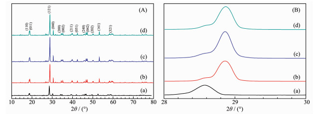
Figure 1.
XRD patterns (A) and the magnified XRD patterns at 2θ=28°~30° (B) of as-fabricated samples
(a) BiVO4, (b) F-BiVO4-0.5, (c) F-BiVO4-1, (d) F-BiVO4-1.5


In the past decades, nanoscaled semiconductor photocatalysts have been widely investigated for the applications in the fields such as solar energy conversion and the degradation of environmental poll-utants[1-2]. To date, TiO2 is the most popular photocata-lyst for its high photocatalytic activity, good chemical stability, non-toxicity, and low cost. Unfortunately, TiO2 has a large band gap of 3.2 eV and responds only to ultraviolet light, which greatly restricts its practical application for the low utilization of solar energy. Therefore, it is highly desirable to develop novel photocatalysts with visible-light-responding photocatalytic ability.
Among various novel visible-light-responding photocatalysts, monoclinic bismuth vanadate, with a relatively narrow band gap (~2.4 eV), is considered as an important visible-light-driven semiconductor photo-catalyst due to its exceptional optical and electronic properties, chemical stability and non-toxicity properties[3]. It has been used in organic pollutants degradation[4-5] and water splitting under visible light irradiation[6-7]. A number of studies have been focused on the controlled preparation of the effective monoclinic BiVO4 photocatalyst with special morphology, high surface area, or exposed high-energy facets[4-8]. The photocatalytic performance of the individual BiVO4, however, has not been ideal for practical application owing to the poor transportation and separation of photogenerated holes and electrons. Many methods have been used to enhance the photocatalytic perfor-mance of a photocatalyst, such as the fabrication of the upconversion nanoparticles based hetero-structures[9-11], hollow nanostructures[12], and pyroelectric materials[13]. According to some reports[14-16], the photocatalytic performance of BiVO4 would be greatly increased by doping BiVO4 with nonmetal atoms for the effective reduction of the recombination rate of photo-induced electron-hole pairs. For example, Wang et al.[14] synth-esized N-doped BiVO4 by using the complexing sol-gel method and observed that N-doped BiVO4 exhibits the enhanced photocatalytic performance in the degradation of methyl orange under visible light irradiation. Guo et al.[15] found that in the degradation of methylene blue under visible light illumination, the photocatalytic activity of S-doped BiVO4 photocatalyst is much higher than that of BiVO4 photocatalyst because an appropriate amount of S2- ions effectively improve the separation efficiency of photogenerated electron-hole pairs. Yin et al.[16] fabricated C-doped BiVO4 photocatalyst with fine hierarchical structures using a novel sol-gel method, showing extremely high photocatalytic performance in O2 production from water splitting under visible light irradiation.
Recently, our group[17] and Li et al.[18] have synthesized F-BiVO4 using the two-step hydrothermal strategy, however, the method is complicated. Herein, in this study, we have prepared fluorine doped BiVO4 material by using a simple one-step alcohol-hydrothermal method. The F-BiVO4 samples show better photocatalytic activity toward the degradation of phenol under visible-light irradiation than the as-prepared BiVO4 samples.
F-doped BiVO4 photocatalysts were fabricated using the alcoho-hydrothermal method with Bi(NO3)3·5H2O and NH4VO3 as inorganic source, dodecylamine (DA) as surfactant, ethanol and ethylene glycol (EG) as solvent, and NH4F as fluoride source. In a typical synthesis process, 5 mL of concentrated nitric acid (67%(w/w)) and 30 mmol of DA, were dissolved in a mixed solvent of 25 mL of ethanol and 25 mL of EG under stirring. Bi(NO3)3·5H2O (10 mmol) and NH4VO3 (10 mmol) were added to the above mixed solution. Then the desired amount of NH4F was added under stirring (nominal nF/nBi=0.5, 1.0, and 1.5). When NH4F was dissolved completely, a certain amount of NaOH solution (2 mol·L-1) containing absolute ethanol and EG (VEtOH:VEG=1) was used to adjust the pH value to 1.5. The final mixture was transferred into a 100 mL Teflon-lined stainless steel autoclave and maintained at 100 ℃ for 12 h. The precipitate was collected, washed three times with deionized water and absolute ethanol, and then dried at 60 ℃ overnight. Finally, the powder was calcined in air at 450 ℃ for 4 h. The as-obtained material were named as BiVO4, F-BiVO4-0.5, F-BiVO4-1, and F-BiVO4-1.5 according to the nominal nF/nBi, respectively.
All these chemicals (AR) were purchased from Beijing Chemicals Company and were used without further purification.
The as-fabricated F-doped BiVO4 catalysts were characterized by X-ray diffraction (XRD) using an X-ray diffractometer (Bruker/AXS D8 Advance) operated at 40 kV and 35 mA with a Cu Kα X-ray radiation source and a nickel filter (λ=0.154 06 nm). Scanning electron microscopy (SEM) was performed on a Gemini Zeiss Supra 55 apparatus (operated at 10 kV). X-ray photoelectron spectroscopic (XPS) analysis was conducted on a Thermo Scientic K-Alpha, with Mg Kα (hν=1 253.6 eV) as the excitation source. Ultraviolet-visible diffuse reflectance spectra (UV-Vis DRS) were measured with a Shimadzu UV-2450 spectrophotometer, using BaSO4 as the reflectance standard. PL spectra were measured on an F-7000 fluorescence spectrometer at room temperature (wavelength of excitation light: 420 nm).
Photocatalytic activities of the as-prepared catalysts for the removal of phenol were evaluated in a quartz reactor (QO250, Beijing Changtuo Sci. & Technol. Co. Ltd.) under visible-light irradiation with a 300 W Xe lamp and a 400 nm cut-off filter. The photocatalytic process was conducted at RT as follows: 0.2 g of the as-prepared F-doped BiVO4 sample and 0.6 mL of H2O2 solution (30%(w/w)) were added to an aqueous solution of phenol (200 mL, initial phenol concentration C0=0.2 mmol·L-1). Before illumination, the mixed solution was ultrasonicated for 0.5 h and then stirred for 3 h in the dark to establish the adsorption-desorption equilibrium of phenol on the surface of the samples. Then the reaction system was magnetically stirred and exposed to the visible-light irradiation. 5 mL of the suspension was collected at 15 min intervals and separated from the photocatalyst particles for analysis. The concentration (Ct) of phenol after reaction for some time was determined by monitoring the absorbance of phenol in the solution at ca. 280 nm during on the aforementioned UV-Vis equipment. The ratio (Ct/C0) of phenol was used to evaluate the photocatalytic performance. All recycle photocatalytic tests were carried out under the same experimental conditions. The sample after every trial was collected by centrifugation, washed with a mixture of water and ethanol (1:1, V/V), and dried.
Fig. 1 shows the XRD patterns of BiVO4, F-BiVO4 -0.5, F-BiVO4-1, and F-BiVO4-1.5 samples. It is showed that all the peaks could be indexed as (110), (011), (121), (040), (200), (002), (211), (051), (240), (042), (202), (161), and (321) planes, which is in good agreement with that of pure monoclinic BiVO4 (PDF No.14-0688) without impurities. It is seen that the doping of fluorine did not change the crystal type of the BiVO4 sample. A similar result was also reported by Li and his coworkers[18]. The diffraction peaks of F-BiVO4-0.5, F-BiVO4-1, and F-BiVO4-1.5 samples were sharp and intense, indicating the highly crystalline character of these samples. As a result, the average crystalline size of the samples calculated according to the line width of the (121) diffraction peak based on the Scherrer formula were 29, 33, 36 and 35 nm, corresponding to BiVO4, F-BiVO4-0.5, F-BiVO4-1, and F-BiVO4-1.5, respectively. The crystalline sizes of the F-doped BiVO4 samples were slightly bigger than that of BiVO4 sample, attributed to a slight lattice distortion in the F-doped BiVO4 samples[14]. In addition, from Fig. 1(B), it can be seen that the (121) diffraction peak in the XRD patterns of F-doped BiVO4 samples showed an obvious shift towards the higher diffraction angle, indicating the presence of compressive strain in the F-doped BiVO4 samples[19].
(a) BiVO4, (b) F-BiVO4-0.5, (c) F-BiVO4-1, (d) F-BiVO4-1.5
Fig. 2 shows the SEM images of pure BiVO4 sample and F-doped BiVO4 samples. From Fig. 2, an olive-like structure with a porous structure is observed for the BiVO4, F-BiVO4-0.5 and F-BiVO4-1 samples. There were numerous mesopores and macropores on the surface of the olive-like particles. It can be demonstrated that the doping of the small amount of F had little effect on the morphology of the BiVO4 samples. Similar phenomenon was also observed in the citric acid assisted preparation of the B-doped samples[20]. However, the F-BiVO4-1.5 sample was composed of nano-sized particles and the olive-like micro-particles, indicating that the excess doping of fluorine could change the particle morphology of the BiVO4 sample.
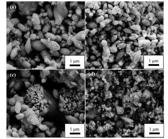
(a) BiVO4, (b) F-BiVO4-0.5, (c) F-BiVO4-1, (d) F-BiVO4-1.5
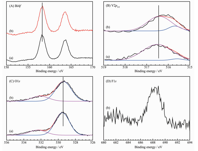
In order to certify the doping of fluorine, XPS was performed to study the surface composition of BiVO4 sample and F-BiVO4-1 sample, as shown in Fig. 3. From Fig. 3(A), it can be seen that the Bi4f spectra of the as-obtained samples were consisted of two symmetrical peaks at binding energy(BE)=158.5 and 163.9 eV, corresponding to Bi4f7/2 and Bi4f5/2 signals respectively, which were characteristic of the Bi3+ species[21]. It can be concluded that the doping of fluorine had no effect on the chemical state of Bi. In Fig. 3(B), the asymmetric peaks of V2p3/2 were decom-posed into two peaks with Gaussian distributions for the BiVO4 sample and the F-BiVO4-1 sample at BE=515.5 and 516.4 eV, attributable to the surface of V4+ (in minority) and V5+ (in majority) species of the two samples[22]. The molar ratio of V4+ to V5+ (0.31) of F-BiVO4-1 sample was higher than that (0.12) of BiVO4 sample. According to the electro-neutrality principle, the as-prepared samples were oxygen-deficient and the amount of nonstoichiometric oxygen on the surface was dependent on the surface molar ratios (nV4+/nV5+). From Fig. 3(C), it can be seen that the asymmetric O1s were deconvoluted into two components at BE=529.1 eV (in majority) and 532.1 eV (in minority), which could be assigned to surface lattice oxygen (Olatt) and adsorbed oxygen (Oads) species, respectively[23-24]. The molar ratios of nOads/nOlatt in BiVO4 sample and F-BiVO4-1 sample were 0.27 and 0.50, respectively. Therefore, the F-doped BiVO4 sample contained more surface oxygen vacancies than the un-doped BiVO4 sample, which could be helpful for the enhancement of the photocatalytic activity of BiVO4 samples, as confirmed by the activity data shown later. From Fig. 3(D), the strong symmetric peak at BE=688.0 eV could be assigned to the fluorine ions in the lattice[25], indicating that the fluorine ions could be doped in the lattice of BiVO4 crystal by the simple one-step alcohol-hydrothermal method.
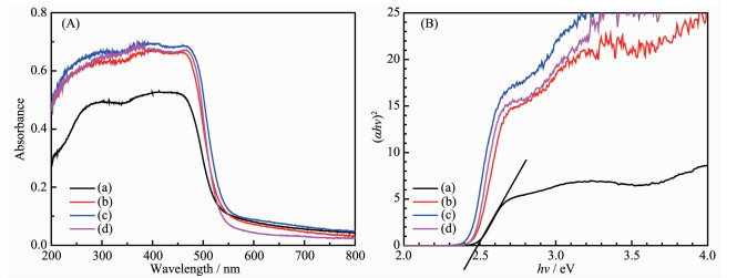
The optical properties of the as-obtained samples were characterized by UV-Vis DRS, as shown in Fig. 4. According to Fig. 4, all of the samples displayed strong absorption in the UV and visible light regions. It is clear that the absorption intensity of the F-doped BiVO4 samples was stronger than the un-doped BiVO4 sample, indicating that the F-doped BiVO4 samples could better respond to visible light. A similar phenomenon was also observed by Shan and his coworkers[26]. The band gap could be determined by the Kubelka-Munk equation: αhν=A(hν-Eg)n/2, where α, A, hν, and Eg are the absorption coefficient, a constant, the discrete photon energy, and the band gap. The value of n depends on the characteristics of the transition in the semiconductor (n=1 for direct transition and n=4 for indirect transition). For BiVO4, the value of n is 1. Fig. 4(B) presents the plots of (αhν)2 versus hν of the as-prepared samples, the band gaps(Eg) of BiVO4 sample, F-BiVO4-0.5 sample, F-BiVO4-1 sample and F-BiVO4-1.5 sample were estimated to be 2.48, 2.46, 2.43 and 2.44 eV, respectively. Compared to the un-doped BiVO4 sample, the band gaps of fluorine doped BiVO4 samples decreased slightly. In accordance with XRD and XPS analysis, the result might be due to the increased oxygen vacancies in the F-doped BiVO4 samples[27].
Effective separation of photogenerated charge carriers is an important factor for excellent photo-catalytic activity of the photocatalyst. PL spectra are helpful to determine the separation efficiency of the photogenerated charge carriers. As shown in Fig. 5, a broad PL peak centered at 530 nm could be observed for all the samples, however, the PL intensities of F-doped BiVO4 samples were lower than that of BiVO4 sample, indicating that the doping of fluorine could inhibit the recombination of photogenerated charge carriers and hence enhance the photocatalytic perfor-mance of BiVO4 photocatalyst[28]. The result would be confirmed by the following photocatalytic activity tests.
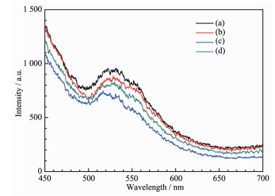
(a) BiVO4, (b) BiVO4-F-0.5, (c) BiVO4-F-1, (d) BiVO4-F-1.5
To demonstrate the photocatlytic performance of fluorine doped BiVO4 materials, the photocatalytic degradation of phenol in the presence of a small amount of H2O2 under visible light irradiation were investigated, as shown in Fig. 6. It should be noticed that after visible light illumination for 90 min, the concentration of phenol in the presence of H2O2 was not changed and the conversion of phenol was only 8% over the F-BiVO4-1 catalyst without H2O2. The F-doped BiVO4 sample, however, showed high photo-catalytic performance in the presence of H2O2 under visible light irradiation. It is indicated that there is a synergistic effect between H2O2 and the photocatalyst. H2O2, as an efficient electron scavenger, could trap the photoinduced electrons and inhibit the recombination of photoinduced electrons and photoinduced holes[29]. After irradiation for 90 min, nearly 95% of phenol was degraded by the F-BiVO4-1 sample, while the other samples, including the pure BiVO4 sample, F-BiVO4-0.5 sample and F-BiVO4-1.5 sample, exhibited lower degradation rates of ca. 77%, 90% and 92%, respectively. Compared with the un-doped BiVO4 sample, all of the F-doped BiVO4 samples showed an obvious enhancement on the photodegradation of phenol, due to higher molar ratios of nV4+/nV5+, more oxygen vacancy densities, and higher separation effici-ency of photogenerated charge carriers of the F-doped BiVO4 samples than those of the un-doped BiVO4 sample, as confirmed by XPS and PL analysis. Furthermore, the F-doped BiVO4 samples had stronger light absorption in the visible light region and lower ban-gap energies than the un-doped BiVO4 sample, which might also contribute to the enhanced photo-catalytic performance of fluorine-doped BiVO4 samples.
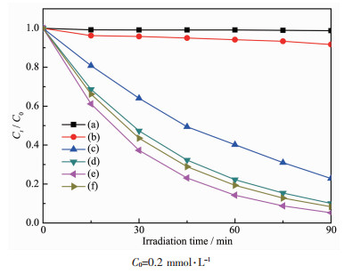
C0=0.2 mmol·L-1
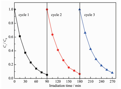
To investigate the photostability of the fluorine-doped BiVO4 photocatalyst in the photocatalytic reaction under visible light irradiation, the recycle experiments were performed. Fig. 7 displays the results of three successive runs for the photodegradation of phenol over F-BiVO4-1 photocatalyst under the identical experimental conditions. As can be seen in Fig. 7, the photocatalytic performance of F-BiVO4-1 photocatalyst did not exhibited a significant loss after three successive runs, indicating the excellent photostability of F-doped BiVO4 photocatalyst under visible light illumination.
The photocatalytic experiments were performed with assistance of H2O2 to achieve efficient degradation of phenol. The hydroxyl radicals (·OH), deriving from the decomposition of H2O2, are efficient active species to oxidize phenol[30]. Under visible-light irradiation, F-BiVO4 photocatalyst is inspired to generate photo-generated carries. The electrons from the valence band (VB) are transferred to the conduction band (CB), leaving lots of holes in VB. The photo-generated holes react with surface hydroxyl to form ·OH radicals[31]. The photo-generated electrons react with absorbed O2 on the surface of photocatalyst or dissolved O2 in water to generate ·O2- radicals. The photo-generated electrons might also reacted with H2O2 to produce ·OH radicals[30]. ·OH and ·O2- radicals with strong oxidation ability are responsible for the degradation of phenol. The main reaction steps for the photodegra-dation of phenol in addition of H2O2 under visible-light irradiation might be proposed as Eq.(1)~(6).
|
$ {\rm{F}} - {\rm{BiV}}{{\rm{O}}_{\rm{4}}}{\rm{ + }}h\nu \to {\rm{F}} - {\rm{BiV}}{{\rm{O}}_{\rm{4}}}{\rm{}}\left( {{{\rm{e}}_{{\rm{CB}}}}^ - } \right){\rm{ + F}} - {\rm{BiV}}{{\rm{O}}_{\rm{4}}}{\rm{}}\left( {{{\rm{h}}_{{\rm{VB}}}}^{\rm{ + }}} \right) $ |
(1) |
|
$ {\rm{F}} - {\rm{BiV}}{{\rm{O}}_{\rm{4}}}\left( {{{\rm{h}}_{{\rm{VB}}}}^ + } \right) + {\rm{O}}{{\rm{H}}^ - } \to \cdot{\rm{OH + F}} - {\rm{BiV}}{{\rm{O}}_{\rm{4}}} $ |
(2) |
|
$ {\rm{F}} - {\rm{BiV}}{{\rm{O}}_{\rm{4}}}\left( {{{\rm{e}}_{{\rm{CB}}}}^ - } \right) + {{\rm{H}}_{\rm{2}}}{{\rm{O}}_{\rm{2}}} \to 2\cdot{\rm{OH + F}} - {\rm{BiV}}{{\rm{O}}_{\rm{4}}} $ |
(3) |
|
$ {\rm{F}} - {\rm{BiV}}{{\rm{O}}_{\rm{4}}}\left( {{{\rm{e}}_{{\rm{CB}}}}^ - } \right) + {{\rm{O}}_{\rm{2}}} \to \cdot {\rm{O}}_2^ - {\rm{ + F}} - {\rm{BiV}}{{\rm{O}}_{\rm{4}}} $ |
(4) |
|
$ \cdot {\rm{OH}} + {\rm{phenol}} \to {\rm{Degraded}}\;{\rm{products}} $ |
(5) |
|
$ \cdot {\rm{O}}_2^ - + {\rm{phenol}} \to {\rm{Degraded}}\;{\rm{products}} $ |
(6) |
In summary, fluorine-doped BiVO4 photocatalysts were prepared by adopting a simple one-step alcohol-hydrothermal strategy with NH4F as fluorine source. It was found that the doping of fluorine do not change the crystal type of BiVO4. Compared to the un-doped BiVO4, the fluorine-doped BiVO4 samples had higher crystallinity and separation efficiency of photogenerated charge carriers, more surface oxygen vacancy, stronger optical absorbance performance, and lower bandgap energy. The fluorine doped BiVO4 sample with a nominal nF/nBi of 1.0 and a bandgap energy of 2.43 eV exhibited excellent photocatalytic activity for the degradation of phenol in the presence of a small amount of H2O2 under visible-light illumination. The excellent photocatalytic activity of fluorine-doped BiVO4 can be attributed to higher surface oxygen vacancy density and separation efficiency of photo-generated charge carriers, stronger optical absorbance performance, and lower bandgap energy.
Xin X, Lang J Y, Wang T T, et al. Appl. Catal. B, 2016, 181:197-209 doi: 10.1016/j.apcatb.2015.07.052
Zhao D, Wu Q, Yang C F, et al. Appl. Surf. Sci., 2015, 356:308-316 doi: 10.1016/j.apsusc.2015.08.008
Li Y, Sun Z H, Zhu S M, et al. Carbon, 2015, 94:599-606 doi: 10.1016/j.carbon.2015.07.042
Zhang X F, Du L L, Wang H, et al. Microporous Mesoporous Mater., 2013, 173:175-180 doi: 10.1016/j.micromeso.2013.02.029
Ji K M, Dai H X, Deng J G, et al. Appl. Catal. B, 2015, 168-169:274-282 doi: 10.1016/j.apcatb.2014.12.045
Hernández S, Thalluri S M, Sacco A, et al. Appl. Catal. A, 2015, 504:266-271 doi: 10.1016/j.apcata.2015.01.019
Thalluri S M, Suarez C M, Hernández S, et al. Chem. Eng. J., 2014, 245:124-132 doi: 10.1016/j.cej.2014.02.017
Thalluri S M, Hussain M, Saracco G, et al. Ind. Eng. Chem. Res., 2014, 53(7):2640-2646 doi: 10.1021/ie403999g
Zhang F, Zhang C L, Wang W N, et al. ChemSusChem, 2016, 9(12):1449-1454 doi: 10.1002/cssc.v9.12
Wang W N, Huang C X, Zhang C Y, et al. Appl. Catal. B, 2018, 224:854-862 doi: 10.1016/j.apcatb.2017.11.037
李耀武, 黄辰曦, 陶伟, 等.无机化学学报, 2017, 33(3):361-376 http://www.wjhxxb.cn/wjhxxbcn/ch/reader/view_abstract.aspx?file_no=20170301
Zhang C Y, Liu H H, Wang W N, et al. Appl. Catal. B, 2018, 239:309-316 doi: 10.1016/j.apcatb.2018.08.027
Xu X L, Xiao L B, Jia Y M, et al. Energy Environ. Sci., 2018, 11:2198-2207 doi: 10.1039/C8EE01016A
Wang M, Liu Q, Che Y S, et al. J. Alloys Compd., 2013, 548:70-76 doi: 10.1016/j.jallcom.2012.08.140
Guo M N, Wang Y, He Q L, et al. RSC Adv., 2015, 5:58633-58639 doi: 10.1039/C5RA07603J
Yin C, Zhu S M, Chen Z X, et al. J. Mater. Chem. A, 2013, 1:8367-8378 doi: 10.1039/c3ta11833a
Jiang H Y, Dai H X, Deng J G, et al. Solid State Sci., 2013, 17:21-27 doi: 10.1016/j.solidstatesciences.2012.12.009
Li J Q, Guo Z Y, Liu H, et al. J. Alloys Compd., 2013, 581:40-45 doi: 10.1016/j.jallcom.2013.06.141
Jagadale T C, Takale S P, Sonawane R S, et al. J. Phys. Chem. C, 2008, 112(37):14595-14602 doi: 10.1021/jp803567f
Wang M, Zheng H Y, Liu Q, et al. Spectrochim. Acta Part A, 2013, 114:74-79 doi: 10.1016/j.saa.2013.05.032
Poulston S, Price N J, Weeks C, et al. J. Catal., 1998, 178(2):658-667 doi: 10.1006/jcat.1998.2200
Liu W, Lai S Y, Dai H X, et al. Catal. Lett., 2007, 113(3/4):147-154 doi: 10.1007/s10562-007-9023-y
Colón G, Hidalgo M C, Munuera G, et al. Appl. Catal. B, 2006, 63:45-59 doi: 10.1016/j.apcatb.2005.09.008
Kulkarni G U, Rao C N R, Roberts M W. J. Phys. Chem., 1995, 99(10):3310-3316 doi: 10.1021/j100010a048
Yu J C, Yu J G, Ho W K, et al. Chem. Mater., 2002, 14(9):3808-3816 doi: 10.1021/cm020027c
Shan L W, Wang G L, Suriyaprakash J, et al. J. Alloys Compd., 2015, 636:131-137 doi: 10.1016/j.jallcom.2015.02.113
Zhang A P, Zhang J Z. Spectrochim. Acta Part A, 2009, 73(2):336-341 doi: 10.1016/j.saa.2009.02.028
Tang J T, Liu Y H, Li H Z, et al. Chem. Commun., 2013, 49:5498-5500 doi: 10.1039/c3cc41090k
Shang M, Wang W Z, Sun S M, et al. J. Phys. Chem. C, 2009, 113(47):20228-20233 doi: 10.1021/jp9067729
Wang Q L, Li H Y, Yang J H, et al. Appl. Catal. B, 2016, 192:182-192 doi: 10.1016/j.apcatb.2016.03.047
Zhu C S, Zheng J T, Fang L Y, et al. J. Mol. Catal. A:Chem., 2016, 424:135-144 doi: 10.1016/j.molcata.2016.08.028

Figure 1 XRD patterns (A) and the magnified XRD patterns at 2θ=28°~30° (B) of as-fabricated samples
(a) BiVO4, (b) F-BiVO4-0.5, (c) F-BiVO4-1, (d) F-BiVO4-1.5

Figure 2 SEM images of as-fabricated samples
(a) BiVO4, (b) F-BiVO4-0.5, (c) F-BiVO4-1, (d) F-BiVO4-1.5

Figure 3 XPS spectra of Bi4f (A), V2p3/2 (B), O1s (C) and F1s (D) of BiVO4 sample (a) and F-BiVO4-1 sample (b)

Figure 4 UV-Vis diffuse reflectance spectra (A) and plots of (αhν)2 versus hν (B) of BiVO4 (a), F-BiVO4-0.5 (b), F-BiVO4-1 (c) and F-BiVO4-1.5 (d)

Figure 5 Room-temperature PL spectra of as-fabricated samples
(a) BiVO4, (b) BiVO4-F-0.5, (c) BiVO4-F-1, (d) BiVO4-F-1.5

Figure 6 Phenol concentration versus visible-light irradiation time for degradation of phenol aqueous solution under visible-light (≥400 nm) irradiation: (a) direct photolysis in the presence of H2O2; (b) F-BiVO4-1 in the absence of H2O2; (c) BiVO4, (d) BiVO4-F-0.5, (e) BiVO4-F-1 and (f) BiVO4-F-1.5 in the presence of H2O2
C0=0.2 mmol·L-1

 扫一扫看文章


扫一扫关注我们
 下载:
下载:
 下载:
下载:

 下载:
下载: