表 1
Crystal data and structure refinement for the complex
Table 1.
Crystal data and structure refinement for the complex
Crystal Structure and Bioactivities of Mn(Ⅱ) Coordination Polymer with Biological Ligand of 2-Quinolinecarboxylic-carboxylate
Ren-Shu WANG , Jing FENG , Yi-Zhu LEI , Dang YANG , Ming-Chang ZHU , Jia-Ling XING , Ming-Lei LIAN

Owing to their wide and potential applications in the fields of adsorption, catalysis, magnetism, lumine-scent, anti-cancer activity, and so on, great attention has been drawn to metal-organic coordination poly-mers[1-5]. These extended coordination polymers have molecular topological structures, which are mainly based on N, N-bidentate bridging ligands such as 1, 2-bis(2-pyridyl)ethylene and 4, 4′-trimethylenedipyrid- ine[6]. Heteroaromatic N, N-bidentateligands is a kind of molecule with rigid structure[7], which means the effects of π…π and C-H…π are involved in the synthesis of the coordination polymers[8]. More recently, attractive forces of π…π and C-H…π effect have been proved to play significant roles in a variety of biological phenomena[9-10]. On the other hand, the flexible structure that connected with the nitrogen-containing heterocyclic of the aliphatic chain owns various lengths, which means, the carbon chain of the N, N-bidentate ligands exhibit various efficiencies in DNA-binding rate for DNA of biological cell and the cytotoxic activity[11].
N-O chelate ligands are widely-used and strong chelating agents coordinated to transition metal cations. 2-Quinolinecarboxylic acid, has three potential coordination sites, including two oxygens of carboxy-late groups and one pyridinic nitrogen[12-13]. Meanwhile, as the quinaldic acid is a bioactive molecule in regard for the metabolism of tryptophan, it should be conducive to applications in pharmaceutical[14].
Conventional small molecule of metal-based anti-cancer drugs applied to clinical trials, such as species Ru(Ⅲ) complexes are restricted for demerits of general toxicity and drug resistance[15-17]. Due to their small sizes, small molecule complexes cannot accumulate efficient enough at tumor sites. Similarly, the complexes may get lost rapidly from the blood stream due to their poor biocompatible[18].
As an ingredient of glycosyltransferase and phosphoenolpyruvate carboxylase, manganese is nece-ssary for human beings[19-21]. Additionally, some special Mn(Ⅱ) complexes could imitate as catalase, to decrease the transporting channels with ATP-related Ca2+ under certain conditions. Thus special Mn(Ⅱ) complexes can induce the apoptosis of tumour cells[22-24].
Consequently, the researchers have focused on developing low-toxic anti tumor agents, thus the metal complexes cultivated by constituents of Mn(Ⅱ), 2-quinolinecarboxylic acid and 4, 4′-trimethylenedipyri-dine are selected. In this paper, the synthesis and characterization of the coordinator polymer {[Mn(Qina)2(Bpp)]·2/3H2O}n will be presented specifically, as well as the evaluation for ct-DNA-binding abilities by using fluorescence spectroscopy. Its cleavage behavior toward pBR322-DNA and cytotoxicity of HeLa and MCF-7 in vitro will also be investigated.
All reagents are purchased through commercial channels and directly used without any treatment. As for analysis, a model Finnigan EA 1112 is employed for Elemental analyses (including C, H and N); and a Thermo Nicolet 380 FT-IR spectrophotometer for IR analysis by KBr disks method. And simultaneous thermogravimetry-differential scanning calorimetry (TG-DSC) was measured on a NETZSCH STA 449 F5 instrument under a dynamic argon atmosphere, and the sample was heated from room temperature to 600 ℃ at a heating rate of 10 ℃·min-1. As well as a Perkin-Elmer LS55 fluorescence spectrofluorimeter for emission spectrum and a JS-380A gel electrophoresis spectrometer for gel electrophoresis.
For all reagents, the concentration was prepared as 15 mmol·L-1. Firstly, an ethanol-water mixtures (1:1, V/V, 10 mL) containing 1.5 mmol of 2-quinoline-carboxylic acid was added into 10 mL of aqueous solution containing 1.5 mmol of MnCl2 dropwise under stirring. After reacting for 6 h at room temperature, another 10 mL of mixture prepared by 1.5 mmol of 4, 4′-trimethylenedipyridineand ethanol-water mixtures (1:1, V/V) was added and continued the reaction for another 10 h. Subsequently, the pH value of the mixture was adjusted to 8.37 using KOH aqueous solution to generate yellowish-brown sediment after filtration, and then the sediment was dissolved with acetonitrile-water mixtures (1:1, V/V) and stored in the open air. A few days later, the desired yellow transparent crystals were generated and then sequen-tially filtered and rinsed with water. Anal. Calcd. for C33H25N4O4Mn·2/3H2O(%):C 65.13, H 5.48, N 9.21; Found(%): C 63.61, H 4.79, N 9.76. IR (KBr, cm-1): 3 237(m), 3 080(w), 2 925(w), 1 708(s), 1 601(m), 1 571(m), 1 405(m), 1 372(m), 1 213(m), 805(m), 520(w).
The single-crystal X-ray diffraction analysis for complex was conducted by a Bruker SMART APEX2 CCD diffractometer with Mo Kα irradiation source (λ=0.071 073 nm) at 296 K, and intensity data were collected within 2θ range of 3.786°~50.842° by a ω scan technique. Data indexing, integration and absorp-tion correction were done with APEX 2 software package. Structure solution and refinement were done by using SHELXL-2014 software[25-26]. There is an alert level B because the unordered object water molecules are unable to be added the hydrogen atoms. To facilitate the analysis of crystal structure, the highly disordered solvent molecules in the structure were squeezed and refined by used the PLATON and the SHELXL. All non-hydrogen atoms of complex were determined by successive difference Fourier syntheses and refined by full-matrix least squares on F2. It is found that the locations of all hydrogen atoms are theoretically correct. Selected crystal data of the complex is listed in Table 1.
 下载:
导出CSV
下载:
导出CSV
| Formula weight | 608.51 | F(000) | 1 256 | |
| Crystal system | Monoclinic | Crystal size/mm | 0.22×0.2×0.18 | |
| Space group | P21n | Index ranges | -15 ≤h ≤ 15, —16 ≤ k≤ 16, —19≤ l ≤0 | |
| a / nm | 1.338 030(10) | Reflection collected | 10 586 | |
| b / nm | 1.413 840(10) | Independent reflection | 5 461(Rint=0.046 2) | |
| c / nm | 1.619 370(10) | Data, restraint, parameter | 5 461, 4, 379 | |
| β/(°) | 95.159 0(10) | Goodness-of-fit on F2 | 0.908 | |
| Volume / nm3 | 3.051 05(4) | Final R indexes [I≥2σ(I)] | R1=0.076 8, wR2=0.215 2 | |
| Z | 1 | Final R indexes (all data) | R1=0.125 7, wR2=0.244 3 | |
| Dc /(g·cm-3) | 1.325 | Largest diff. peak and hole / (e · nm-3) | 0.925 and -425 | |
| μ/ mm-1 | 0.478 |
CCDC: 1550803.
The analytical cif file of the complex molecule was carried out by the CrystalExplorer software[27]. And all the Hirshfeld surfaces were output at the highest resolution. Moreover, the translated command whose coordinate range is from 0.08 to 0.3 nm was selected. And it was applied to all the fingerprint plots.
The molecule of ethidium bromide (EtBr) for fluorophore labeling, has a conjugate planar, the fluorescence intensity of which is extremely weak. But fluorescence intensity is significantly increased if EtBr is specifically intercalated among the base pairs of double strands. The effects of complex difference in concentration are competitive binding with DNA-ethidium bromide (cDNA=5.0 μmol·L-1, cEtBr=1 μmol·L-1, ccomplex/cDNA=1.0, 2.0, 3.0, 4.0). The buffer of 50 mmol·L-1 Tris-HCl, pH 7.4, which contains 10 mmol·L-1 NaCl, was used in the binding studies. The sample was firstly incubated at temperature of 20 ℃ for 4 h before spectral analysis[28]. The excitation wavelength for fluorescence measurement was 615 nm, while the emission range was set in scope of 550 and 750 nm.
As regards to the gel electrophoresis experiments, the pBR322 DNA (0.33 mg·mL-1) was mixed with the complex and then the mixture was incubated at room temperature for 1.5 h. The samples were electro-phoresed at 90 V for 1.5 h on 0.8% agarose gel in Tris-acetate buffer (50 mmol·L-1 Tris-acetate, 18 mmol·L-1 NaCl buffer, pH=7.4). Finally, the gels were stained with 1.0 mg·mL-1 EtBr and then photographed under UV light[29].
The MTT assay was used to evaluate the growth inhibitory effect of metal complex on the HeLa and MCF-7 cells. Briefly, cells cultured in RPMI-1640 containing 10% fetal bovine serum, was placed in 37 ℃ and 5% CO2 incubator. The 96-well culture plate was accurately inoculated with 3×10⁴ cells suspension in 100 μL and then incubated for 24 h. RPMI-1640 was used to dilute the solution of complex and 150 μL solution was joined to each well. Besides, the highest concentration of DMSO concentration was not higher than 0.1% and each concentration was under three replication settings. After 72 h, the old solution was cleaned by using 200 μL of saline for each well. MTT solution was diluted for 10 times with RPIM1640. In addition, 100 μL of MTT solution (0.5 mg·mL-1) was added into each well and then was cultivated for 3 h. Subsequently, the media containing MTT was removed, and the formazan crystals were dissolved with 150 μL of DMSO under ambient temperature for 0.5 h. The absorbance value of each hole was measured respectively at 492 and 630 nm[30]. However, the effects of the cisplatin on cell growth were studied in another experiment which was treated as a control team.
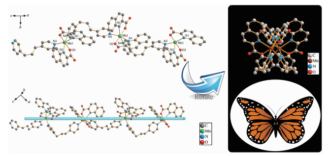
Results of single-crystal X-ray analyses indicated that complex was monoclinic system with P21/n space group. The single crystal X-ray diffraction analysis showed that each manganese(Ⅱ) ion is six-coordinated by two nitrogen atoms originated from two molecules of 4, 4′-trimethylenedipyridine, as well as two oxygen atoms and two nitrogen atoms that separately from two respective quinoline-2 carboxylic acid ligands (N1-O2, N2-O3). The base structure of polymer complex is shown in Fig. 1, and the chosen bond lengths and bond angles are tabulated as Table 2. Besides, along c axis, the fragments of [Mn(Qina)2] formed from centric Mn(Ⅱ) ions were in turn bridged by Bpp nitrogen atoms, resulting in a 1D chain, as shown in Fig. 2.
下载:
导出CSV
| Mn1-N1 | 0.237 2(3) | Mn1-N4ⅰ | 0.227 0(2) | N4-Mn1ⅱ | 0.227 0(2) |
| Mn1-N2 | 0.237 0(3) | Mn1-02 | 0.211 4(2) | ||
| Mn1-N3 | 0.226 1(3) | Mn1-03 | 0.211 3(2) | ||
| N2-Mn1-N1 | 94.32(9) | 03-Mn1-N1 | 101.97(10) | C16-N2-Mn1 | 130.9(2) |
| N3-Mn1-N1 | 163.32(10) | 03-Mn1-N2 | 73.23(10) | C21-N3-Mn1 | 121.1(2) |
| N3-Mn1-N2 | 91.79(9) | 03-Mn1-N3 | 94.65(9) | C25-N3-C21 | 116.6(3) |
| N3-Mn1-N4ⅰ | 92.40(9) | 03-Mn1-N4ⅰ | 86.05(9) | C25-N3-Mn1 | 122.2(2) |
| N41-Mn1-N1 | 87.39(8) | 03-Mn1-02 | 174.17(9) | C31-N4-C32 | 115.4(3) |
| N41-Mn1-N2 | 159.12(10) | C2-N1-C6 | 118.4(3) | C31-N4-Mn1ⅱ | 122.6(2) |
| 02-Mn1-N1 | 73.41(9) | C2-N1-Mn1 | 110.3(2) | C32-N4-Mn1ⅱ | 120.8(2) |
| 02-Mn1-N2 | 103.27(10) | C6-N1-Mn1 | 131.1(2) | C1-02-Mn1 | 122.4(2) |
| 02-Mn1-N3 | 90.09(9) | C12-N2-C16 | 118.4(3) | C11-03-Mn1 | 121.9(3) |
| 02-Mn1-N4ⅰ | 97.17(9) | C12-N2-Mn1 | 110.6(3) | ||
| Symmetry codes: ⅰ -1/2+x, 1/2-y, 1/2+z; ⅱ 1/2+x, 1/2-y, -1/2+z | |||||
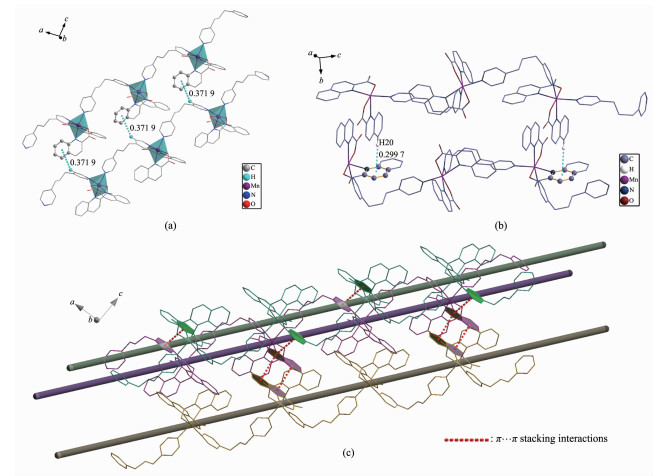
Additionally, stacking interactions of C-H…π and π…π between the two one-dimension chains was found in the complex. The H30 from pyridine ring of Bpp molecule, will produce the stacking interactions of C-H…π by an aromatic ring from Qina in the different chain. The distances between H30 from the Bpp molecule and the benzene ring of the Qina molecule are 0.371 9 nm, which is shorter than the general one of 0.38 nm that was observed in inter-molecular C-H…π interactions[31], as shown in Fig. 3a. Meanwhile, the H20 from benzene ring of Qina molecule, will also produce the stacking interactions of C-H…π by a pyridine ring from Qina in the different chain, as shown in Fig. 3b. The centroid-to-centroid distance between a pair of Qina ligands and Bpp ligands possessing π…π stacking interactions is 0.363 0 and 0.379 6 nm, respectively, which is also within normal ranges[32], as shown in Fig. 3c. Finally, the 3D supramolecular architecture is directed by 1D-interchains via weak stacking interactions of C-H…π and π…π (Fig. 4).
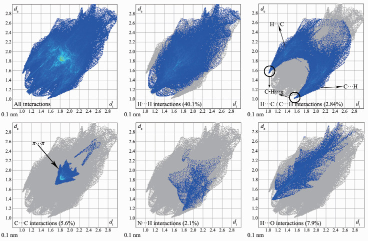
Hirshfeld surface is a way to analyze the intermolecular interactions in a crystal[33]. As shown in Fig. 5, there is Hirshfeld dnorm surfaces, shape index and curvedness of complex. The red spots or regions on the 3D Hirshfeld surfaces indicate the close-contact interactions, the blue regions represent longer contacts, and the white and green regions represent the distance of contacts. In order to illustrate the weak forces in the crystal better, there are two different observation directions in each type of analysis diagram.
The red parts of the dnorm surfaces diagram represents strong intermolecular interactions which represent the C-H…π and π…π interactions. The red concave with the corresponding positions in shape index and the blue convex of the surrounding receptors complement each other, which has confirmed the existence of such effects[34]. In addition, the white part of the dnorm surfaces diagram represents the weak intermolecular interaction which represents the H…H interactions[35]. The curvedness diagram also shows the same information expressed in the previous two graphs.
Furthermore, the quantitative relationship between the intermolecular interactions of the crystals has also been measured by the 2D fingerprint plots of the CrystalExplorer program. As shown in Fig. 6, H…H interactions account for the largest proportion of the 2D fingerprint plots. Additionally, the C…H interac-tions are shown on the finger print to represent the symmetrical wing shape, which illustrate that the main intermolecular force between the complex is the C-H…π interactions. Moreover, the C…C interactions are the evidences that there are weak π…π effects in the intermolecular of the crystal[36-37]. Besides, it can be obtained from the fingerprint plots that the N…H interaction comprises 2.1%, and H…O comprises 7.9% of total Hirshfeld surfaces. All Hirshfeld analysis was consistent with single crystal structure analysis.
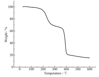
As shown in the thermogravimetric analysis curve (Fig. 7), the complex exhibits three steps of weight losses. The first weight loss corresponding to the removal of lattice solvent is 4.54% (Calcd. 3.94%) from 190 to 290℃, the second weight loss correspon-ding to the removal of Bpp ligand is 27.07% (Calcd. 32.58%) from 190 to 300 ℃, and the third weight loss corresponding to the removal of Qina ligand is 52.14% (Calcd. 56.92%) from 300 to 550 ℃.The final formation of the complex is metal oxide MnO (Calcd. 11.66%).
Since the complex molecule binds to DNA more strongly than EB, its addition would quench the DNA-induced EB emission[38]. The fluorescence spectra of EB and DNA-bound EB in the absence and presence of the complex is provided as Fig. 8.

In the classical Stern-Volmer equation: I0/I=1+Ksqr, I0 and I separately represent the fluorescence intensity with and without the complex; r stands for the total concentration ratio of the complex to that of DNA (ccomplex/cDNA); Ksq is the linear Stern-Volmer quen-ching constant depending on the ratio of the bound concentration of EB to the concentration of DNA, which is generated as the slope of linear curve of I/I0 against r[39]. The fluorescence quenching curves of DNA-bound EtBr by the complex is also provided in Fig. 8. The Ksq value for the complex is 0.046 6, which indicates that the complex displaced DNA-bound EB and was bound to DNA with a weak bound at the intercalative mode, due to certain partial weak effect intercalation of the aromatic ring. And the spectrophotometric studies show that the main mode of complex interacting with DNA was electrostatic interaction[40-41].
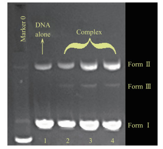
The behavior of complex in DNA cleaving was evaluated by monitoring the conversion of the tertiary structure of pBR322 DNA. When circular plasmid DNA is subjected to electrophoresis, the intact supercoiled form (Form Ⅰ) shows a relatively fast migration. While, when scission occurs on one strand of supercoil, it will generate an open circular form (Form Ⅱ)which exhibited a slower-moving migration, and when both strands were cleaved and migrated between Form Ⅰ and Form Ⅱ, it will form a linear form (Form Ⅲ)[42]. The complex can significantly induce the cleavage of the pBR322 DNA at the concentration of 3.23, 6.46 and 12.92 μmol·L-1. As shown in Fig. 9, with the increase of concentration of complex, the pBR322 supercoiled DNA (Form Ⅰ) decreases gradually, and the open circular (Form Ⅱ) comes into existence. However, the characteristic of linear (Form Ⅲ) was not obviously changed. The complex has the moderate intensity cleavage of pBR322 DNA, which is in agreement with the trend found in fluorescence quenching experiment.
The prepared complex was measured in term of the in vitro cytotoxic activity by MTT assay. IC50 values estimated for two human tumor cell lines of HeLa and MCF-7 are summarized in Table 3. The complex showed a certain level of anti-tumor activity and cytotoxicity[43].
下载:
导出CSV
| Complex | IC50 / (μmol·L-1) | |
| HeLa | MCF-7 | |
| {[Mn(Qina)2(Bpp)]·2/3H2O}n | 35.17±0.24 | 30.32±0.25 |
| Cisplatin | 9.90±0.23 | 9.95±0.24 |
A coordination polymer of {[Mn(Qina)2(Bpp)]· 2/3H2O}n was synthesized and characterized in aspects of FT-IR spectroscopy, elemental analysis as well as single-crystal X-ray diffraction. The properties of the complex in DNA binding were tested via fluorescence spectra, and results indicated that the complex behaves varied binding affinities in DNA interaction. The cleavage capability of the complex with respect to pBR322 DNA was reviewed by agarose gel electro-phoresis, and the results showed that the complex is efficient in DNA cleaving. Through in vitro cytotoxi-city experiment, it was testified that the complex possesses a level of toxicity to inhibited Hela and MCF-7 cell lines.
Noro S I, Fukuhara K, Kubo K, et al. Cryst. Growth Des., 2011, 11:2379-2385 doi: 10.1021/cg2001392
Zhou S B, Wang X F, Du C C, et al. CrystEngComm, 2017, 19:3124-3137 doi: 10.1039/C7CE00394C
邓青峰, 于良民, 李霞.无机化学学报, 2017, 33(9):1561-1567 doi: 10.11862/CJIC.2017.210DENG Qing-Feng, YU Liang-Min, LI Xia. Chinese J. Inorg. Chem., 2017, 33(9):1561-1567 doi: 10.11862/CJIC.2017.210
Zhang X, Wang N, Liu P F, et al. Polyhedron, 2017, 129:149-156 doi: 10.1016/j.poly.2017.03.042
Etaiw S E H, Amer S A, Bendary M M E. J. Inorg. Organomet. Polym. Mater., 2011, 21:662-672 doi: 10.1007/s10904-011-9532-4
Taleghani S, Mirzaei M, Eshtiagh H H, et al. Coord. Chem. Rev., 2016, 309:84-106 doi: 10.1016/j.ccr.2015.10.004
Haque A, Ilmi R, Busaidi I J A, et al. Coord. Chem. Rev., 2017, 350:320-339 doi: 10.1016/j.ccr.2017.07.008
Guo Y, Xu L, Liu H, et al. Adv. Mater., 2015, 27:985-1013 doi: 10.1002/adma.201403846
Gao E J, Lin L, Zhang Y, et al. Eur. J. Med. Chem., 2011, 46:2546-2554 doi: 10.1016/j.ejmech.2011.03.044
Singh M, Butcher R J, Singh N K. Polyhedron, 2009, 28:95-100 doi: 10.1016/j.poly.2008.10.008
Gao E J, Zhu M C, Liu L, et al. Inorg. Chem., 2010, 49:3261-3270 doi: 10.1021/ic902176e
Ye J W, Wang Q Q, Gao H Z, et al. Inorg. Chim. Acta, 2012, 384:1-7 doi: 10.1016/j.ica.2011.11.028
Gao E J, Zhu M C, Zhang W Z, et al. Russ. J. Coord. Chem., 2009, 35:621-627 doi: 10.1134/S1070328409080107
Zhou P, O'Hagan D, Mocek U, et al. J. Am. Chem. Soc., 1989, 111:7274-7276 doi: 10.1021/ja00200a065
Casolaro M, Cini R, Del B B, et al. Biomacromolecules, 2009, 10:944-949 doi: 10.1021/bm8014939
Sullivan S T, Ciccarese A, Fanizzi F P, et al. Inorg. Chem., 2009, 39:836-842 https://www.unisalento.it/scheda-utente/-/people/antonella.ciccarese/ricerca
Caruso F, Rossi M, Benson A, et al. J. Med. Chem., 2012, 55:1072-1081 doi: 10.1021/jm200912j
Sun W, Li S, Haupler B, et al. Adv. Mater., 2017, 29:1603702 doi: 10.1002/adma.201603702
Yang P, Klimistavantzis D J. Biol. Trace Elem. Res., 1998, 64:275-288 doi: 10.1007/BF02783343
Miller R S, Mildvan A S, Chang H C, et al. J. Biol. Chem., 1968, 243:6030-6040 http://www.ncbi.nlm.nih.gov/pubmed/4673667
Rico H, Gómez Raso N, Revilla M, et al. Eur. J. Obstet. Gynecol. Reprod. Biol., 2000, 90:97-101 doi: 10.1016/S0301-2115(99)00223-7
Miguel L L. Cancer Lett., 2007, 252:1-8 doi: 10.1016/j.canlet.2006.10.029
Zhou D F, Chen Q Y, Qi Y, et al. Inorg. Chem., 2011, 50:6929-6937 doi: 10.1021/ic200004y
Chen Q Y, Zhou D F, Huang J, et al. J. Inorg. Biochem., 2010, 104:1141-1147 doi: 10.1016/j.jinorgbio.2010.06.012
Sheldrick G M. Acta Crystallogr. Sect. A:Found. Crystallogr., 2015, A71:3-8 http://europepmc.org/articles/PMC4294323
Sheldrick G M. Acta Crystallogr. Sect. C:Cryst. Struct. Commun., 2015, C71:3-8 https://pbn.nauka.gov.pl/sedno-webapp/works/858229
Liu Q L, Yang L J, Luo Y H, et al. Res. Chem. Intermed., 2016, 42:6947-6957 doi: 10.1007/s11164-016-2506-y
Gao E J, Zhang Y, Lin L, et al. J. Coord. Chem., 2011, 64:3992-4005 doi: 10.1080/00958972.2011.634910
Rossiter C S, Mathews R A, Morrow J R. J. Inorg. Biochem., 2009, 101:925-934 http://www.hort.purdue.edu/rhodcv/hort640c/referen/ms.htm
Hsu C P, Kao T K, Chang W L, et al. Eur. J. Surg. Oncol., 2011, 37:140-147 doi: 10.1016/j.ejso.2010.12.003
Tsuzuki S. Annu. Rep. Prog. Chem. Sect. C:Phys. Chem., 2012, 108:69-95 doi: 10.1039/c2pc90003c
Oszajca M, Nitek W, Rafalska L A, et al. Cryst. Res. Technol., 2015, 50:781-790 doi: 10.1002/crat.201500084
冯超, 张舵, 周士艳, 等.无机化学学报, 2016, 32(7):1215-1222 doi: 10.11862/CJIC.2016.147FENG Chao, ZHANG Duo, ZHOU Shi-Yan, et al. Chinese J. Inorg. Chem., 2016, 32(7):1215-1222 doi: 10.11862/CJIC.2016.147
Seth S K, Sarkar D, Kar T. CrystEngComm, 2011, 13:4528-4535 doi: 10.1039/c1ce05037k
Seth S K. J. Mol. Struct., 2014, 1064:70-75 doi: 10.1016/j.molstruc.2014.01.068
Spackman M M, McKinnon J J. CrystEngComm, 2002, 4:378-392 doi: 10.1039/B203191B
McKinnon J J, Jayatilaka D, Spackman M A. Chem. Commun., 2007:3814-3816 http://pubs.rsc.org/en/Content/ArticleLanding/CC/2007/B704980C
Zhu M C, He W X, Gao E J, et al. Life. Sci., 2012, 90:519-524 doi: 10.1016/j.lfs.2012.01.006
王慧, 甘国庆, 瞿阳, 等.无机化学学报, 2012, 28(6):1217-1221 http://www.wjhxxb.cn/wjhxxbcn/ch/reader/view_abstract.aspx?file_no=20120623WANG Hui, GAN Guo-Qing, QU Yang, et al. Chinese J. Inorg. Chem., 2012, 28(6):1217-1221 http://www.wjhxxb.cn/wjhxxbcn/ch/reader/view_abstract.aspx?file_no=20120623
Satyanarayana S, Dabrowiak J C, Chaires J B. Biochemistry, 1993, 32:2573-2548 doi: 10.1021/bi00061a015
杨浩.高等学校化学学报, 2007, 28(5):872-876 http://pub.chinasciencejournal.com/article/getArticleRedirect.action?doiCode=10.3724/SP.J.1095.2012.00328YANG Hao. Chem. J. Chinese Universities, 2007, 28(5):872-876 http://pub.chinasciencejournal.com/article/getArticleRedirect.action?doiCode=10.3724/SP.J.1095.2012.00328
Rajendiran V, Karthik R, Palaniandavar M, et al. Inorg. Chem., 2007, 46:8208-8221 doi: 10.1021/ic700755p
史蕾, 杨文聪, 曾淑莹, 等.高等学校化学学报, 2016, 37(6):1059-1068 http://kns.cnki.net/KCMS/detail/detail.aspx?filename=gdxh201606006&dbname=CJFD&dbcode=CJFQSHI Lei, YANG Wen-Cong, ZENG Shu-Ying, et al. Chem. J. Chinese Universities, 2016, 37(6):1059-1068 http://kns.cnki.net/KCMS/detail/detail.aspx?filename=gdxh201606006&dbname=CJFD&dbcode=CJFQ

Figure 1 Structure of the complex
H atoms were omitted for clarity; displacement ellipsoids are drawn at the 30% probability level; Symmetry codes: ⅰ-1/2+x, 1/2-y, 1/2+z; ⅱ 1/2+x, 1/2-y, -1/2+z

Figure 2 One dimensional polymeric chain of coordination polymer
H atoms were omitted for clarity

Figure 3 C-H…π and π…π stacking interactions in the complex: (a) C-H…π stacking interactions between Bpp molecule and Qina molecule; (b) C-H…π stacking interactions between two Qina molecules; (c) π…π stacking interactions between multiple 1D molecular chains

Figure 4 Three dimensional structure of coordination polymer: (a) 3D structure is composed by multiple 1D chains; (b) weak effect superimposed in the supramolecular structure

Figure 8 (a) Fluorescence spectra of DNA-EB in the absence and presence of the complex; (b) Stern-Volmer plot for the complex
1~5: r=ccomplex/cDNA=0.0, 1.0, 2.0, 3.0, 4.0, respectively

Figure 9 DNA strand break in HeLa cells treated with the complex
Line 0: Marker; Line 1: DNA alone; Line 2~4: in the different concentrations of the complex: 3.23, 6.46, 12.92 μmol·L-1, respectively
Table 1. Crystal data and structure refinement for the complex
| Formula weight | 608.51 | F(000) | 1 256 | |
| Crystal system | Monoclinic | Crystal size/mm | 0.22×0.2×0.18 | |
| Space group | P21n | Index ranges | -15 ≤h ≤ 15, —16 ≤ k≤ 16, —19≤ l ≤0 | |
| a / nm | 1.338 030(10) | Reflection collected | 10 586 | |
| b / nm | 1.413 840(10) | Independent reflection | 5 461(Rint=0.046 2) | |
| c / nm | 1.619 370(10) | Data, restraint, parameter | 5 461, 4, 379 | |
| β/(°) | 95.159 0(10) | Goodness-of-fit on F2 | 0.908 | |
| Volume / nm3 | 3.051 05(4) | Final R indexes [I≥2σ(I)] | R1=0.076 8, wR2=0.215 2 | |
| Z | 1 | Final R indexes (all data) | R1=0.125 7, wR2=0.244 3 | |
| Dc /(g·cm-3) | 1.325 | Largest diff. peak and hole / (e · nm-3) | 0.925 and -425 | |
| μ/ mm-1 | 0.478 |
 下载: 导出CSV
Table 2. Selected bond lengths (nm) and angels (°)
| Mn1-N1 | 0.237 2(3) | Mn1-N4ⅰ | 0.227 0(2) | N4-Mn1ⅱ | 0.227 0(2) |
| Mn1-N2 | 0.237 0(3) | Mn1-02 | 0.211 4(2) | ||
| Mn1-N3 | 0.226 1(3) | Mn1-03 | 0.211 3(2) | ||
| N2-Mn1-N1 | 94.32(9) | 03-Mn1-N1 | 101.97(10) | C16-N2-Mn1 | 130.9(2) |
| N3-Mn1-N1 | 163.32(10) | 03-Mn1-N2 | 73.23(10) | C21-N3-Mn1 | 121.1(2) |
| N3-Mn1-N2 | 91.79(9) | 03-Mn1-N3 | 94.65(9) | C25-N3-C21 | 116.6(3) |
| N3-Mn1-N4ⅰ | 92.40(9) | 03-Mn1-N4ⅰ | 86.05(9) | C25-N3-Mn1 | 122.2(2) |
| N41-Mn1-N1 | 87.39(8) | 03-Mn1-02 | 174.17(9) | C31-N4-C32 | 115.4(3) |
| N41-Mn1-N2 | 159.12(10) | C2-N1-C6 | 118.4(3) | C31-N4-Mn1ⅱ | 122.6(2) |
| 02-Mn1-N1 | 73.41(9) | C2-N1-Mn1 | 110.3(2) | C32-N4-Mn1ⅱ | 120.8(2) |
| 02-Mn1-N2 | 103.27(10) | C6-N1-Mn1 | 131.1(2) | C1-02-Mn1 | 122.4(2) |
| 02-Mn1-N3 | 90.09(9) | C12-N2-C16 | 118.4(3) | C11-03-Mn1 | 121.9(3) |
| 02-Mn1-N4ⅰ | 97.17(9) | C12-N2-Mn1 | 110.6(3) | ||
| Symmetry codes: ⅰ -1/2+x, 1/2-y, 1/2+z; ⅱ 1/2+x, 1/2-y, -1/2+z | |||||
下载: 导出CSV
Table 3. Cytotoxicity of the complex against selected human tumor cells after 72 h of incubation
| Complex | IC50 / (μmol·L-1) | |
| HeLa | MCF-7 | |
| {[Mn(Qina)2(Bpp)]·2/3H2O}n | 35.17±0.24 | 30.32±0.25 |
| Cisplatin | 9.90±0.23 | 9.95±0.24 |
下载: 导出CSV

 扫一扫看文章

扫一扫关注我们


 下载:
下载: