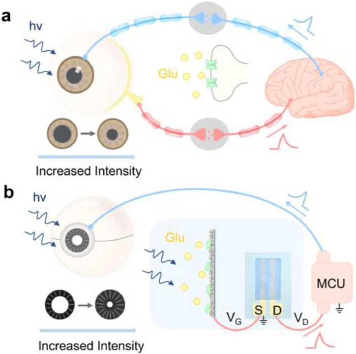
Figure 1.
(a) Schematic illustration of human VNS to control the pupil. (b) Schematic illustration of artificial VNS using GluDh/WO3-photogated photoelectrochemical transistor to control artificial pupil.

An organic photoelectrochemical transistor biosensor mimicking visual nociceptive system with self-adaptability
Xing-Shi Liu , Cheng Yuan , Feng-Zao Chen , Yuan-Cheng Zhu , Jin Hu , Yazhou Wang , Hui-Min Jia , Wei-Wei Zhao


Until recently, it has been challenging to approach artificial biosensory systems that demonstrate specific responsiveness to biochemical stimuli in aqueous environments [1–12]. Among various human biosensory systems, natural selection has produced exquisite visual nociceptive systems (VNS) to protect our eyes so as to increase our survival likelihood in challenging environments [13]. By this inspiration, artificial VNS have drawn increasing interest with booming development of humanoids [14]. However, the operation of existing VNS generally relies on physical principles of solid-state optoelectronics with high-voltages, overlooking the pivotal role of chemically synaptic transmission that is critical for biological vision [15,16]. More essentially, the VNS functions through the neurotransmitter glutamate (Glu)-mediated transmission in aqueous media [17]. Such features signify that high-level mimicry of VNS requires not only a closed-loop of light perception, integration and actuation, but also the Glu biosensing, which, nevertheless, remains unachieved according to recent comprehensive reviews [18–20].
Featured by versatile light-biochemical perception, we have proposed the concept of organic photoelectrochemical transistor (OPECT), which has been shown promising towards next-generation biosensing, optoelectronics and neuromorphic exploration [21–36]. Recently, Santoro et al. reported an OPECT synapse based on the photo-isomerization of azobenzene moieties [23]. Inal et al. explored new semiconducting polymers for OPECT optoelectronics [24]. Hu et al. reported a hydrogel-based synapse for color perception based on specific chromogenic reactions [25]. Yuan et al. reported an aquatic autonomous nervous system with both excitatory and inhibitory response [26]. Huang et al. explored OPECT for visual-gustatory multisensory integration [27]. Recently, Wang et al. reported the fabrication of a larger n-type OPECT array for motion recognition and neural network [28]. Very recently, Huang et al. reported the fabrication of OPECT array by wafer-scale photolithography [29]. Despite the progress, no effort has been dedicated to emulating the large family of human nociceptive systems. In this work, we report on the first OPECT biosensor capable of mimicking VNS with self-adaptability, i.e., Glu-dependent automatic actuation of artificial pupil to emulate the autonomous protection of biological eyes toward harmful light.
Fig. 1a illustrated the mechanism of VNS that could detect and respond to harmful ambient lights. Once the noxious light exceeds a certain threshold, the pain-perceptual ability of VNS would be activated, transmitting pain signals to cerebral cortex to guide the adaptable closure of pupils to avoid the retinal photochemical damage, one of the major harms to the human eye in ophthalmology [37]. In this process, Glu was the predominant neurotransmitter playing a critical role in pain modulation. Fig. 1b gave a general view of the structure of our OPECT device, which innovated in combining Glu biosensing, processing and self-adaptability in a single system, i.e., the light-Glu perception in a Glu dehydrogenase (GluDh)/WO3-photogated organic transistor with the assistance of a micro controller unit (MCU) [38,39] to control the artificial pupil responding to the incident light passing through it. Once noxious light exceeded the threshold, the device would process and transmit the "pain" signals to the MCU thus helping to avoid the potential damage. Significantly, highly akin to its role in biology, Glu can amplify the "pain" signals, making it easier to reach the threshold and trigger the autonomous protection.
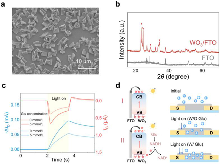
Fig. 2a showed the scanning electron microscopy (SEM) image of the WO3 with a uniform plate-like morphology, with dimensions of 3–4 μm and thickness of 150–200 nm. The X-ray diffraction (XRD) spectra in Fig. 2b, the energy-dispersive spectroscopy mapping images in Fig. S1 (Supporting information) and UV–vis absorption spectrum in Fig. S2 (Supporting information) indicated the successful fabrication of WO3 [40,41]. The as-fabricated GluDh/WO3-photogated OPECT showed a highly reproducible and sensitive gate voltage (VG) control and good operational stability (Fig. S3 in Supporting information) [42]. The yield rates were also examined across different batches, with results showing a minimal variation (Fig. S4 in Supporting information). The Glu response was studied by investigating the channel current (ID) and gate current (IG) variation upon photonic pulse in the absence and presence of Glu, respectively. As shown, in the presence of 5 mmol/L Glu, both the ∆IG (red curves) and ∆ID (blue curves) were substantially improved (Fig. 2c). The underlying mechanism of such phenomena could be illustrated from charge transfer and ion migration [43–45]. Under light illumination, the photoinduced electrons would transmit from the conduction band (CB) of WO3 to FTO, leading to the injection of cations into the channel and thus decreased channel conductance (Fig. 2d(Ⅰ)). By contrast, in the presence of Glu, the catalytically generated electron donor NADH could consume the photo-induced holes, thus boosting the electron transfer and enhancing the cation injection (Fig. 2d(Ⅱ)). The corresponding enzymatic catalysis was also illustrated (Figs. S5-S7 in Supporting information).
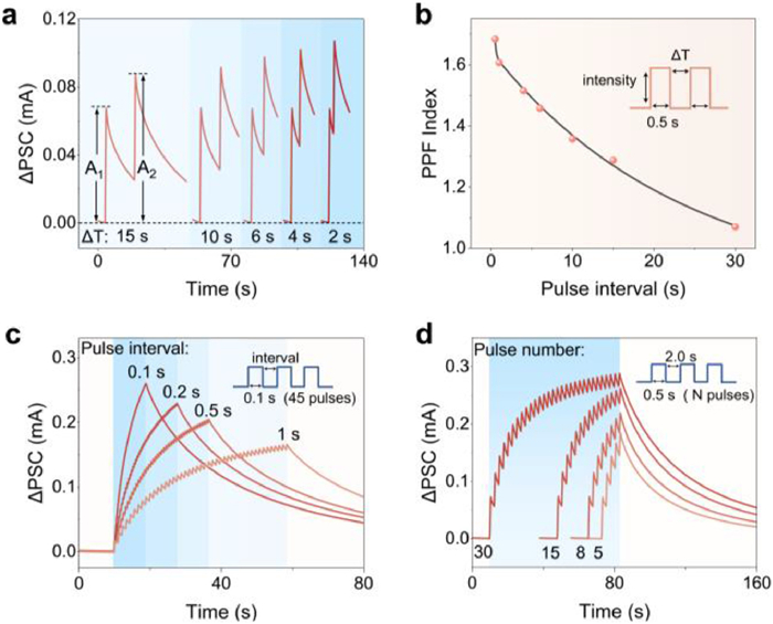
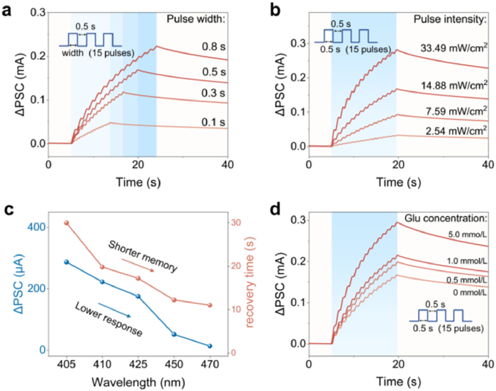
Glu-mediated synaptic plasticity serves as the cornerstone of the biological VNS [46]. As a typical short-term plasticity, paired pulse facilitation (PPF) is critical for processing temporal information [47,48]. Here, paired photonic pulses with different intervals (∆t) were applied to emulate such behavior. As shown, the postsynaptic current (∆PSC) showed typical PPF behavior in which the second spike value (A2) was higher than the first one (A1) (Fig. 3a). With increased ∆t, the PPF gradually weakened as indicated by the decreased PPF index (A2/A1) which could be described by a double exponential function (Fig. 3b). Spike-dependent plasticity is another fundamental plasticity in VNS, which revealed the relationship between synaptic weight and the characteristics of spikes (e.g., the rate, width, intensity and number) [49]. Such functions could also be simulated by regulating corresponding conditions. As shown, with decreased pulse interval and increased pulse number, ∆PSC gradually enhanced and exhibited a progressively slower retention to the initial level, emulating the spike-rate dependent plasticity (SRDP) (Fig. 3c) and spike-number dependent plasticity (SNDP) (Fig. 3d), respectively. On the other hand, with increased pulse width and intensity, ∆PSC exhibited increased value and recovery time, emulating the spike-width dependent plasticity (SWDP) (Fig. 4a) and spike-intensity dependent plasticity (SIDP) (Fig. 4b). Moreover, biological retinal damage induced by light stimulation is highly dependent on the wavelength of light, and the intensity threshold for causing damage decreases as the wavelength decreases [17]. Analogous to such phenomenon, when wavelength increased at a constant light intensity, VNS exhibited a lower ∆PSC and shorter recovery time due to the reduction in photon energy and ultraviolet-visible absorption (Fig. 4c). Significantly, with increased Glu concentrations from 0 mmol/L to 5 mmol/L, the ∆PSC remarkably enhanced and exhibited prolonged recovery time (Fig. 4d), which was highly consistent with the Glu-mediated plasticity in biological VNS [50]. Such results indicated the capability of the as-developed device to perform the biomimicry functions of human VNS.
Furthermore, human VNS has evolved unique Glu-dependent threshold to evaluate the harm degree of external stimulus, the sensitivity of which was determined by Glu level [51]. The stronger light stimuli, the stronger responses over the threshold and the prolonged recovery time. To emulate such intricate characteristics, variable photonic pulses and Glu concentrations were applied to mimic the harmful lights and internal pain sensitivities, respectively. As shown, in the case of 0 mmol/L Glu (representing pain relief status), with increased light intensity from 32.4 mW/cm2 to 124.2 mW/cm2 (Fig. 5a1), both the ∆PSC and recovery time gradually increased, whereas all the stimulus cannot induce ∆PSC exceeding the pain threshold (30 µA) (Fig. 5a2). However, in the case of 0.5 mmol/L Glu (representing normal pain status), it generated higher ∆PSC and improved recovery time with the last 2 spikes exceeding the threshold (Fig. 5a3). In the case of 5.0 mmol/L Glu (representing pain sensitization status), the ∆PSC and the recovery time further increased with 4 spikes exceeding the threshold (Fig. 5a4). A linear function was used to model the relationship between ΔPSC and light intensity at various glutamate concentrations (Fig. S8 in Supporting information).
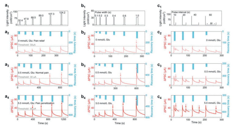
The duration of photonic stimuli was also found to influence the response. With increased pulse width from 0.1 s to 1 s (Fig. 5b1), it also generated increased ∆PSC with enhanced recovery time. In the case of 0, 0.5 and 5.0 mmol/L Glu, there were 1, 2 and 3 ∆PSC spikes that could exceed the set threshold, respectively (Figs. 5b2-5b4). Such results were highly analogous to the Glu-dependent threshold behavior in human VNS. The effect of photonic stimuli width could be fitted by an exponential equation based on ion migration (Fig. S9 in Supporting information). This indicated that Glu enhanced pain sensitivity, enabling the device to respond to harmful stimuli with a shorter duration and avoid further damage.
Another critical property of VNS is the Glu-dependent sensitization, which can amplify the response to subsequent weak stimulus after an intense stimulus, thus avoiding repeated injury [52]. Normally, such increased severity of the injury would gradually reduce over time, a process known as desensitization [53]. Two vital factors in such a process, i.e., the amplified magnitude and the recovery time, was related to the Glu concentrations. To emulate such a feature, paired photonic pulses with different intervals were applied and the intensity of the first one was substantially higher than the second one (Fig. 5c1). In the case of 0 mmol/L Glu, with increased intervals, both the ∆PSC and its recovery time gradually decreased, none of which could exceed the threshold (Fig. 5c2). In the case of 0.5 mmol/L Glu, the ∆PSC and its recovery time also decreased with increased intervals, whereas their values remarkably enhanced, leading to 1 spike exceeding the threshold (Fig. 5c3). In the case of 5.0 mmol/L Glu, both the ∆PSC and its recovery time further increased, leading to 4 spikes exceeding the threshold (Fig. 5c4). To further study the effect of stimulus interval on the sensitization of artificial VNS, an exponential equation was proposed to fit the sensitization and desensitization behaviors in our artificial visual nociceptive system (Fig. S10 in Supporting information). Such behaviors well mimicked biological sensitization and desensitization processes.
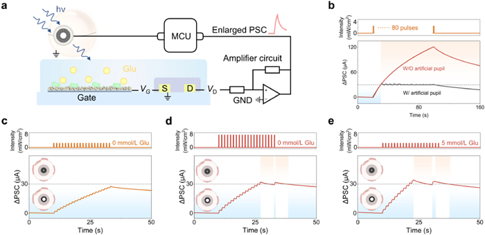
Based on the Glu-dependent threshold and sensitizing behaviors, the device was then applied for managing an artificial pupil with self-adaptability (Fig. 6a). As soon as the "pain" signal exceeded the threshold, the artificial pupil would be closed of a self-driven way (Figs. S11 and S12 in Supporting information). Moreover, the artificial pupil was then exactly placed in the path between the light source and the device, once the "pain" signal exceeded the threshold, the self-driven closure of the artificial pupil would block the light illumination on the device (Fig. S13 in Supporting information). Compared with the case without artificial pupil-enabled protection, the ∆PSC could hardly exceed the threshold under the protection of artificial pupil, emulating the protective function of the human VNS (Fig. 6b). Upon such a configuration, the effects of light intensities and Glu concentrations were further studied. When 20 light pulses with relatively lower intensity (2.54 mW/cm2) were applied, the ∆PSC gradually increased but could not reach threshold (Fig. 6c). However, when the pulse intensity increased to 7.59 mW/cm2, the ∆PSC substantially increased and the 15th spike could exceed the threshold, resulting in the closure of the artificial pupil, which simulated the self-protection behavior of human eyes under repeated strong light stimuli (Fig. 6d). On the other hand, with Glu concentration increasing from 0 mmol/L to 5 mmol/L, the 11th spike could exceed the threshold and lead to the closure of pupil (Fig. 6e). Such behaviors well mimicked the reaction with higher internal pain sensitivity [54]. Incidentally, its performance metrics against some state-of-the-art reports were also compared (Table S1 in Supporting information) [15,16,55,56].
In conclusion, this is a first report of real neurotransmitter-mediated artificial nociceptive system, as exemplified by an OPECT Glu biosensor with chemically synaptic transmission as well as self-adaptability. The correlation between the specific enzyme-catalyzed Glu reaction and the sensitive response of OPECT made possible the Glu-dependent synaptic behaviors. Significantly, the dependence of pain on Glu, highly analogous to its actual role in biology, was successfully emulated by the glutamatergic modulation of spike-dependent plasticity. By converting the biosensory perception into digital data, the self-adaptive pupil protection of eyes against harmful light was realized. By bridging real neurotransmitter perception with nociceptive systems, this work takes a key step in bringing artificial nociceptors closer to biological systems for seamless neuro-electronics.
Xing-Shi Liu: Writing – original draft, Visualization, Formal analysis, Data curation. Cheng Yuan: Writing – original draft, Visualization, Methodology, Conceptualization. Feng-Zao Chen: Investigation, Formal analysis. Yuan-Cheng Zhu: Formal analysis. Jin Hu: Resources, Data curation. Yazhou Wang: Investigation. Hui-Min Jia: Investigation. Wei-Wei Zhao: Writing – review & editing, Supervision, Project administration, Methodology, Conceptualization.
The authors declare that they have no known competing financial interests or personal relationships that could have appeared to influence the work reported in this paper.
We thank National Natural Science Foundation of China (Nos. 22374066, 22174063), and Excellent Research Program of Nanjing University (No. ZYJH004).
Supplementary material associated with this article can be found, in the online version, at doi:
Y. Gao, J. Zhang, D. Liu, et al., Chin. Chem. Lett. 35 (2024) 108582. doi: 10.1016/j.cclet.2023.108582
J. Xu, C. Liang, Z. Gao, Y.Y. Song, Chin. Chem. Lett. 34 (2023) 107863. doi: 10.1016/j.cclet.2022.107863
H. Zhou, Y. Zhang, S. Wang, et al., Chin. Chem. Lett. (2025), doi: 10.1016/j.cclet.2025.111643.
G. Gong, Y. Zhou, Q. Li, et al., Adv. Mater. (2025), doi: 10.1002/adma.202505420.
X. Zhao, H. Zou, M. Wang, et al., Adv. Mater. 36 (2024) 2403444. doi: 10.1002/adma.202403444
S.T. Keene, C. Lubrano, S. Kazemzadeh, et al., Nat. Mater. 19 (2020) 969–973. doi: 10.1038/s41563-020-0703-y
T. Sarkar, K. Lieberth, A. Pavlou, et al., Nat. Electron. 5 (2022) 774–783. doi: 10.1038/s41928-022-00859-y
S. Wang, X. Chen, C. Zhao, et al., Nat. Electron. 6 (2023) 281–291. doi: 10.1038/s41928-023-00950-y
Y.T. Xu, S.Y. Yu, Z. Li, et al., Proc. Natl. Acad. Sci. U. S. A. 121 (2024) e2403143121. doi: 10.1073/pnas.2403143121
S.Y. Yu, J. Hu, Z. Li, et al., J. Am. Chem. Soc. 146 (2024) 27022–27029. doi: 10.1021/jacs.4c08833
Z.M. Duan, Y.T. Xu, Z. Li, et al., Angew. Chem. Int. Ed. 64 (2025) e202420602. doi: 10.1002/anie.202420602
X. Zhang, J.X. Pang, Y.H. Kang, et al., Anal. Chem. 97 (2025) 22548–22554. doi: 10.1021/acs.analchem.5c03531
C.J. Woolf, Q. Ma, Neuron 55 (2007) 353–364. doi: 10.1016/j.neuron.2007.07.016
G. Feng, J. Jiang, Y. Li, et al., Adv. Funct. Mater. 31 (2021) 2104327. doi: 10.1002/adfm.202104327
Y. Lin, W. Wang, R. Li, et al., Nano Energy 121 (2024) 109267. doi: 10.1016/j.nanoen.2024.109267
Z. Li, G. Zou, Y. Xiao, et al., Nano Energy 127 (2024) 109733. doi: 10.1016/j.nanoen.2024.109733
D.T. Organisciak, D.K. Vaughan, Prog. Retinal Eye Res. 29 (2010) 113–134. doi: 10.1016/j.preteyeres.2009.11.004
G. Ding, H. Li, J. Zhao, et al., Chem. Rev. 124 (2024) 12738–12843. doi: 10.1021/acs.chemrev.4c00369
K. He, C. Wang, Y. He, J. Su, X. Chen, Chem. Rev. 123 (2023) 13796–13865. doi: 10.1021/acs.chemrev.3c00527
P. Gkoupidenis, Y. Zhang, H. Kleemann, et al., Nat. Rev. Mater. 9 (2024) 134–149.
C.S. Wang, B.Y. Zhou, Y.F. Wang, et al., Chin. Chem. Lett. 36 (2025) 110080. doi: 10.1016/j.cclet.2024.110080
J.H. Zhu, X. Yang, Y. Zheng, et al., ACS Nano 19 (2025) 20247–20256. doi: 10.1021/acsnano.5c06982
F. Corrado, U. Bruno, M. Prato, et al., Nat. Commun. 14 (2023) 6760. doi: 10.1038/s41467-023-41083-2
V. Druet, D. Ohayon, C.E. Petoukhoff, et al., Nat. Commun. 14 (2023) 5481. doi: 10.1038/s41467-023-41313-7
J. Hu, M.J. Jing, Y.T. Huang, et al., Adv. Mater. 36 (2024) 2405887. doi: 10.1002/adma.202405887
C. Yuan, K.X. Xu, Y.T. Huang, J.J. Xu, W.W. Zhao, Adv. Mater. 36 (2024) 2407654. doi: 10.1002/adma.202407654
Y.T. Huang, Z. Li, C. Yuan, et al., Adv. Mater. 37 (2025) 2503030. doi: 10.1002/adma.202503030
Y. Wang, W. Shan, H. Li, et al., Nat. Commun. 16 (2025) 1615. doi: 10.1038/s41467-025-56814-w
X. Liu, S. Dai, Y. Jin, et al., Nat. Commun. 17 (2025) 197. doi: 10.2528/pierc24111401
Z. Li, Q.Q. Wu, M.H. Chen, et al., eScience 5 (2025) 100374. doi: 10.1016/j.esci.2025.100374
Q.Q. Wu, Z. Li, M.H. Chen, et al., Adv. Mater. 37 (2025) 2504338. doi: 10.1002/adma.202504338
Y. Zheng, J.H. Zhu, S. Wang, et al., Adv. Funct. Mater. 35 (2025) 2418158. doi: 10.1002/adfm.202418158
F.Z. Chen, L. Hou, Y. Gao, et al., Adv. Funct. Mater. 34 (2024) 2408186. doi: 10.1002/adfm.202408186
C. Yuan, Q. Wu, K.X. Xu, et al., Biosens. Bioelectron. 257 (2024) 116346. doi: 10.1016/j.bios.2024.116346
D. Leng, T. Wu, X. Ren, et al., Adv. Funct. Mater. 36 (2026) e16018. doi: 10.1002/adfm.202516018
C.S. Wang, L. Hou, S.J. Liu, et al., Adv. Funct. Mater. 36 (2026) e27361. doi: 10.1002/adfm.202527361
D. Ghezzi, M.R. Antognazza, R. Maccarone, et al., Nat. Photonics 7 (2013) 400–406. doi: 10.1038/nphoton.2013.34
Z. Li, M.H. Chen, Q.Q. Wu, et al., Natl. Sci. Rev. 12 (2025) nwaf213. doi: 10.1093/nsr/nwaf213
Z. Li, F.Q. Liu, Q.Q. Wu, et al., ACS. Nano 19 (2025) 20029–20036. doi: 10.1021/acsnano.5c03749
Y.T. Huang, K.X. Xu, X.S. Liu, et al., Anal. Chem. 95 (2023) 17912–17919. doi: 10.1021/acs.analchem.3c04263
Z. Li, Q.Q. Wu, Y. Wang, et al., Adv. Mater. 38 (2026) e07054. doi: 10.1002/adma.202507054
F.J. Yu, Z. Yang, K.X. Xu, et al., Anal. Chem. 97 (2025) 21459–21465. doi: 10.1021/acs.analchem.5c03368
J. Hu, Z. Li, Y.T. Huang, et al., Adv. Funct. Mater. 35 (2025) 2412928. doi: 10.1002/adfm.202412928
C. -S. Wang, Y. Jiang, Z. Li, et al., Adv. Funct. Mater. 35 (2025) 2500235. doi: 10.1002/adfm.202500235
K.X. Xu, C. Yuan, H. Lou, et al., Chin. J. Chem. 42 (2024) 1999–2004. doi: 10.1002/cjoc.202400206
A.E. Dubin, A. Patapoutian, J. Clin. Invest. 120 (2010) 3760–3772. doi: 10.1172/JCI42843
D.M. Blitz, K.A. Foster, W.G. Regehr, Nat. Rev. Neurosci. 5 (2004) 630–640. doi: 10.1038/nrn1475
Z. Zhang, L. Shi, B. Wang, et al., Chin. Chem. Lett. 36 (2025) 109687. doi: 10.1016/j.cclet.2024.109687
K. Zhu, S. Pazos, F. Aguirre, et al., Nature 618 (2023) 57–62. doi: 10.1038/s41586-023-05973-1
A.I. Basbaum, D.M. Bautista, G. Scherrer, D. Julius, Cell 139 (2009) 267–284. doi: 10.1016/j.cell.2009.09.028
T. Hucho, J.D. Levine, Neuron 55 (2007) 365–376. doi: 10.1016/j.neuron.2007.07.008
M.S. Gold, G.F. Gebhart, Nat. Med. 16 (2010) 1248–1257. doi: 10.1038/nm.2235
J.H. Yoon, Z. Wang, K.M. Kim, et al., Nat. Commun. 9 (2018) 417. doi: 10.1038/s41467-017-02572-3
S. Qu, L. Sun, S. Zhang, et al., Nat. Commun. 14 (2023) 7181. doi: 10.1038/s41467-023-42240-3
L. Zheng, J. Ren, Y. Liu, et al., Chem. Eng. J. 512 (2025) 162436. doi: 10.1016/j.cej.2025.162436
J. Li, Y. Zhou, Y. Li, et al., ACS Photonics 11 (2024) 4578–4587. doi: 10.1021/acsphotonics.4c00877

Figure 1 (a) Schematic illustration of human VNS to control the pupil. (b) Schematic illustration of artificial VNS using GluDh/WO3-photogated photoelectrochemical transistor to control artificial pupil.

Figure 2 (a) The SEM image of WO3. (b) The XRD spectra of fluorine-doped tin oxide coated glass (FTO) and WO3/FTO electrode. (c) ID and IG responses of the (GluDh)/WO3-photogated photoelectrochemical transistor with different concentrations of Glu. (d) Schematic of the corresponding mechanism of electron transfer and ion migration.

Figure 3 (a) PPF and (b) the corresponding PPF indexes upon light pulses with variable pulse intervals. The emulation of (c) SRDP and (d) SNDP.

Figure 4 (a) SWDP and (b) SIDP of the as-prepared device. (c) The ∆PSC and recovery time responses to different wavelengths. (d) The ∆PSC responses in the absence and presence of Glu with different concentrations from 0 mmol/L to 5.0 mmol/L.

Figure 5 (a1) The as-applied 6 light pulses with different intensity. The corresponding ∆PSC (red curves) and recovery time (blue histogram) in the case of (a2) 0 mmol/L Glu, (a3) 0.5 mmol/L Glu and (a4) 5 mmol/L Glu. (b1) The as-applied 6 light pulses with different duration. The corresponding ∆PSC (red curves) and recovery time (blue histogram) in the case of (b2) 0 mmol/L Glu, (b3) 0.5 mmol/L Glu and (b4) 5 mmol/L Glu. (c1) The as-applied 4 paired light pulses with different intervals (the intensity of the first one is 124.2 mW/cm2 and that of the second one is 32.4 mW/cm2). The corresponding ∆PSC (red curve) and the recovery time (blue histogram) in the case of (c2) 0 mmol/L Glu, (c3) 0.5 mmol/L Glu and (c4) 5 mmol/L Glu.

Figure 6 (a) Schematic illustration of the VNS controlling the artificial pupil. (b) The ∆PSC responses upon light stimuli with and without the protection of the artificial pupil. The ∆PSC responses under 20 light pulses (c) with lower intensity and in the absence of Glu, (d) with higher intensity and in the absence of Glu. (e) with lower intensity and in the presence of 5 mmol/L Glu.

 扫一扫看文章


扫一扫关注我们
 DownLoad:
 下载:
 下载: