
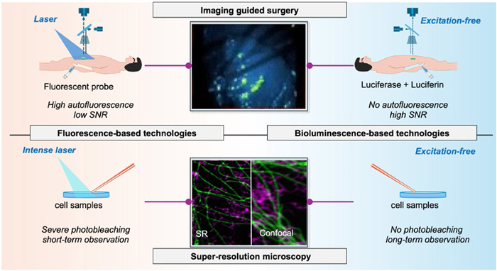
Figure 1.
Schematic illustration of fluorescence (FL) and BL based imaging guided surgery and super resolution microscopy.


Optical imaging is an indispensable tool in modern biomedicine and life sciences, characterized by its unparalleled spatiotemporal resolution, non-invasive nature, and real-time readout capabilities [1–9]. Over the past three decades, fluorescent probe-based technologies have undergone explosive development, with two transformative applications at their forefront: Fluorescence-guided surgery (FGS) and fluorescence super-resolution microscopy (FL-SRM) (Fig. 1) [10–15].
FGS has redefined the clinical standard of care for solid tumor resection by leveraging targeted fluorescent probes to enable real-time visualization of tumor margins and micrometastatic lesions, maximizing resection completeness while sparing adjacent healthy tissue [16–18]. Its clinical performance is fundamentally dictated by three core physicochemical properties of fluorescent probes: Targeting specificity, luminescent brightness, and photostability [19,20]. Multiple targeted probes, including the Food and Drug Administration (FDA)-approved near-infrared dye indocyanine green (ICG), have entered clinical use or trials, significantly improving R0 resection rates for multiple solid tumor types [21–24]. Recent advances have further extended FGS capabilities into the second near-infrared window (NIR-Ⅱ, 1000–1700 nm) [25], where reduced tissue scattering and autofluorescence enable deeper imaging and higher contrast. For instance, the recent development of ultrabright NIR-Ⅱ quantum dots [26], highlights the ongoing optimization of fluorescent probes for clinical FGS applications [27–29].
As the second transformative technology of fluorescent probe-based optical imaging, FL-SRM has broken the Abbe diffraction limit of conventional optical microscopy to achieve nanoscale spatial resolution (<200 nm), enabling direct visualization of subcellular organelles, protein-protein interactions, and genomic organization at the molecular level [30–34]. Its three mainstream modalities, stimulated emission depletion (STED) microscopy, structured illumination microscopy (SIM), and single-molecule localization microscopy (SMLM), each impose distinct, stringent requirements on the photophysical and photochemical properties of fluorescent probes: STED demands fluorophores with ultra-high photostability to withstand high-power depletion laser irradiation; SIM requires high-brightness, photostable dyes for long-term dynamic in vivo imaging; and SMLM, including its widely used derivatives STORM and PALM, relies on fluorophores with well-controlled blinking behavior and high photon output per on-state switching cycle [31,35]. To meet these strict performance criteria, rhodamine derivatives have emerged as a particularly versatile and widely adopted fluorophore scaffold for super-resolution imaging: Their intrinsic equilibrium between a fluorescent ring-opened zwitterionic form and a non-fluorescent spirocyclic closed form can be precisely tuned via rational structural modification, enabling targeted optimization of the photophysical properties required for different super-resolution modalities, most notably the controllable single-molecule blinking that is indispensable for SMLM [36,37].
Despite their revolutionary impact, both technologies are confined by a shared, mechanism-derived bottleneck: Absolute dependence on external excitation light. For FGS, excitation light scattering and endogenous autofluorescence limit tissue penetration and imaging signal-to-noise ratio (SNR), preventing reliable detection of microscopic metastases; prolonged excitation also causes fluorophore photobleaching and off-target phototoxicity, compromising imaging quality and clinical safety. For FL-SRM, high-power excitation exacerbates these issues, precluding long-term in vivo dynamic imaging and degrading resolution accuracy.
In this perspective, we propose that bioluminescence (BL), which generates light via enzyme-catalyzed chemical reactions without the requirement for external excitation, represents a fundamentally distinct paradigm capable of overcoming these shared limitations. We first summarize the current state of FGS and FL-SRM, systematically dissecting their notable achievements and persistent challenges. We then introduce BL as a promising alternative light source, elaborating on its unique reaction mechanism, inherent advantages, and the specific performance requirements for its adaptation to surgical guidance and super-resolution imaging applications. Finally, we identify the key chemical and biological bottlenecks that must be addressed, particularly in the areas of substrate engineering and targeted enzyme delivery and highlight promising recent advances that delineate a future trajectory in which BL assumes a central role in both the operating room and the biomedical imaging suite.
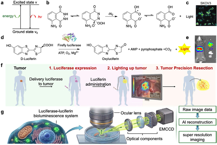
To address this fundamental limitation, excitation-free optical imaging modalities, represented by chemiluminescence (CL) and BL, have garnered widespread attention in the chemistry and materials science communities [38–40]. CL generates photon emission via non-enzymatic chemical reactions, a process that typically yields ultra-high peak brightness, a key property that has enabled its successful implementation in single-molecule super-resolution microscopy (Figs. 2a–c) [41–44].
However, CL systems suffer from inherent drawbacks including rapid irreversible signal decay, limited tunability of emission kinetics, and insufficient biocompatibility for long-term in vivo and clinical applications. In contrast, BL produces photons through luciferase-catalyzed oxidation of its cognate luciferin substrate without external excitation, with four core inherent advantages that directly overcome the mechanism-derived bottlenecks of fluorescence imaging: (1) Zero autofluorescence background and high SNR from excitation-free emission; (2) superior deep-tissue penetration by eliminating dual optical losses of excitation light; (3) no photobleaching and fully tunable and sustainable photon emission via enzymatic substrate turnover; and (4) negligible phototoxicity and excellent biocompatibility validated in preclinical and early clinical studies. These four unique, mechanism-endowed advantages position BL as a powerful, transformative alternative to conventional fluorescent probes, underpinning its disruptive potential to redefine next-generation FGS and FL-SRM.
Despite its transformative promise, the widespread application of BL-based guided surgery (BGS) and super-resolution microscopy (BL-SRM) faces two fundamental, interconnected challenges: (1) Rational engineering of luciferin substrates to meet the distinct performance requirements of different imaging modalities; and (2) safe, targeted, and controllable delivery of luciferase to achieve site-specific expression in target tissues or cells (Figs. 2d and e) [45,46]. Subsequent sections systematically dissect these two core bottlenecks in sequence: Section 3 focuses on rational substrate engineering tailored to BGS and BL-SRM, while Section 4 addresses the targeted delivery of luciferase, the other critical barrier to clinical translation (Figs. 2f and g).
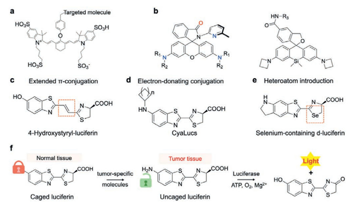
The design of specialized fluorescent probes is pivotal for the advancement of FGS and FL-SRM. As a gold-standard optical probe, ICG can be modified with various targeting ligands to achieve precision-guided surgery across diverse tissue types [12]. Furthermore, extending the π-conjugation system or implementing rational structural modifications to induce a bathochromic shift can afford deeper tissue penetration, allowing for effective guidance in deep-seated lesions (Fig. 3a) [10]. In super-resolution imaging, rhodamine and its derivatives deliver outstanding performance, primarily due to their spirocyclization equilibrium (Fig. 3b) [33,47]. This mechanism supports robust reversible fluorescence switching and self-blinking, which are essential for SMLM to overcome the optical diffraction limit. Moreover, replacing the bridging oxygen atom in the rhodamine scaffold with Group 14 heteroatoms (e.g., Si, Ge, Sn) effectively lowers the lowest unoccupied molecular orbital (LUMO) energy level and induces a significant bathochromic shift into the NIR region, thereby reducing autofluorescence background and enhancing imaging depth. In contrast, cyanine- and boron-dipyrromethene (BODIPY)-based probes enable super-resolution imaging through distinct photophysical and photochemical switching pathways [30,32,48]. Unlike fluorescence imaging, which relies solely on the structural design of fluorescent probes, the performance of BL imaging depends on the synergistic interplay between two key components: The luciferin substrate and the luciferase enzyme [49,50]. Because luciferase protein engineering is methodologically more complex, labor-intensive, and time-consuming compared to small-molecule synthesis, a substantial portion of current research has prioritized rational substrate engineering to meet these distinct imaging requirements.
For the clinical translation of BGS, luciferin substrates must meet four core performance criteria that directly address the limitations of conventional FGS: (1) Photon emission from the luciferase-luciferin catalytic reaction within the red/NIR-Ⅰ/NIR-Ⅱ optical window to maximize deep-tissue penetration and reduce autofluorescence interference; (2) high BL quantum yield and brightness to enable high-sensitivity detection of tumor lesions; (3) in vivo metabolic stability to sustain emission throughout the full surgical window; and (4) excellent biocompatibility and low systemic toxicity to meet clinical safety standards.
Considerable advances have been achieved in rational substrate engineering to address these requirements over the past decade. For the targeted red-shifting of emission wavelength, three primary strategies have been widely exploited: Extension of the π-conjugation system [51], conjugation with electron-donating moieties [39,53], and heteroatom incorporation (Figs. 3c–e) [52]. Importantly, these strategies follow fundamental design principles parallel to those of fluorescent probe engineering, centered on π-conjugation extension to red-shift emission [54,55]. Notably, structural modifications to the substrate tend to compromise its binding affinity with wild-type luciferase, which in such cases necessitates the remodeling of the enzyme's active site via rational design. Landmark advances include the development of AkaLumine, a D-luciferin analog paired with the mutant luciferase AkaLuc, which has an emission peak at 675 nm and enabled deep-tissue BL imaging in non-human primates [56]. More recently, engineered luciferin analogs paired with mutant railroad worm luciferases have achieved NIR-Ⅱ emission peaks beyond 700 nm, with >3-fold higher brightness than the AkaLumine-AkaLuc system [57].
For brightness and stability optimization, structural modifications to the phenolic hydroxyl and carboxyl groups of luciferins have significantly enhanced substrate-luciferase binding affinity, resulting in 10- to 100-fold increases in BL brightness [58]. Furthermore, tumor microenvironment (TME)-activatable caged luciferin substrates have been developed by masking the luciferin with moieties responsive to specific TME biomarkers (e.g., matrix metalloproteinases, acidic pH, or reactive oxygen species). These substrates exclusively release active luciferin within tumor tissues, drastically improving the tumor-to-normal tissue ratio and imaging specificity. Ultimately, this precise activation effectively distinguishes tumor margins from inflammatory backgrounds, serving as a key determinant for the clinical utility of BGS (Fig. 3f) [50,59]. These advances have laid a solid material foundation for the preclinical validation and clinical translation of BGS, directly addressing the core limitations of conventional fluorescence-based FGS.
For BL-SRM, the performance requirements for luciferin substrates are far more stringent and modality-specific, with each requirement tailored to overcome a core limitation of conventional fluorescence super-resolution: (1) For STED and SIM: Substrates must deliver ultra-high photon output, exceptional brightness, and long-term enzymatic stability to maintain high spatial resolution and contrast during continuous imaging, eliminating the photobleaching issue that plagues long-term fluorescence STED/SIM imaging. (2) For SMLM: Substrates must exhibit controllable, reversible blinking behavior (switchable on/off emission) with a low duty cycle and high photon output per on-state cycle, which is the prerequisite for single-molecule localization and super-resolution reconstruction, without relying on external light activation that causes phototoxicity.
While significant advancements have been made in enhancing BL brightness and stability for super-resolution imaging, the field remains in its infancy, with a critical unmet gap in the development of blinking-controllable BL substrates. For brightness optimization, the NanoLuc/furimazine system, which exhibits 150-fold higher brightness than conventional firefly luciferase systems, has demonstrated great potential for BL-SRM (Fig. 4a) [60].
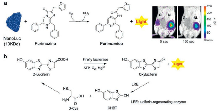
For blinking control, a transformative, untapped design strategy has recently emerged: Redox-switchable luciferin substrates. In this system, the oxidized luminescent product of the luciferase reaction can be reduced back to an inactive, non-luminescent state by endogenous or exogenous reductases, enabling reversible, enzyme-controlled on/off switching of BL emission (Fig. 4b) [61]. Unlike photoactivatable fluorescent probes for SMLM, this light-free switching mechanism inherently eliminates photobleaching and phototoxicity, directly addressing the long-standing limitation of external light-dependent blinking in conventional SMLM. While extensive work has optimized BL substrates for emission wavelength, brightness, and stability, the development of switchable, blinking-controllable substrates remains largely unexplored, representing the highest-priority research direction to unlock the full potential of BL-SRM.
Alongside rational substrate engineering, efficient, site-specific, and safe delivery of luciferase to target cells or tissues is the fundamental prerequisite for both BGS and BL-SRM. For clinical translation, delivery strategies must be tailored to the distinct requirements of each imaging modality: BGS requires transient, tumor-restricted expression aligned with the perioperative window, while BL-SRM demands tunable expression for long-term dynamic imaging. Below, we systematically review the state-of-the-art delivery strategies, their advantages, limitations, and remaining challenges for clinical BL imaging (Fig. 5).
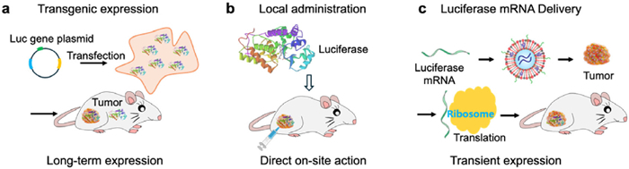
Adeno-associated virus and lentiviral vectors have been widely used for targeted delivery of luciferase-encoding plasmids in preclinical animal models, with high transduction efficiency and long-term ectopic expression [62,63]. However, the risk of insertional mutagenesis, immunogenicity, and lack of temporal control over expression limit their clinical utility for intraoperative imaging. Such long-term ectopic expression is fundamentally incompatible with the transient, perioperative timeframe required for BGS.
Messenger RNA (mRNA) encoding luciferase drives rapid, direct expression of functional luciferase protein in the cytoplasm of target cells post-delivery, thereby eliminating the risk of genomic insertional mutagenesis and conferring a well-validated, excellent biosafety profile for clinical translation. Meanwhile, the transient and controllable expression profile of mRNA aligns perfectly with the short-term perioperative imaging requirements of BGS, avoiding the risks associated with long-term exogenous protein expression. However, naked mRNA is easily degraded by nucleases in the bloodstream, necessitating the use of delivery vectors for effective in vivo delivery [46].
Lipid nanoparticle (LNP) formulations are the only clinically approved mRNA delivery platform with an established safety record in human populations. Nevertheless, two core limitations remain to be addressed for its widespread clinical translation: First, the inherent hepatic organotropism of conventional LNP systems, which results in predominant off-target accumulation in the liver rather than tumor tissues; second, the lack of precise, quantitative control over the kinetics and magnitude of luciferase expression [64,65]. Fortunately, recent preclinical studies have validated the feasibility of employing ligand engineering, formulation optimization, nanostructure design, and biological barrier modulation to enhance tumor-specific mRNA delivery. By overcoming the core bottleneck of LNP hepatic organotropism, these emerging strategies hold substantial promise for breaking through current translational barriers in BGS applications.
For localized solid tumors (e.g., gliomas, breast cancer, hepatocellular carcinoma), local intratumoral, intraperitoneal, or intracavitary injection of luciferase or luciferase-loaded nanoparticle formulations represents a simple, safe, and directly actionable strategy for BGS. However, direct intracellular delivery of luciferase remains a substantial challenge: As a hydrophilic biomacromolecule, luciferase suffers from inefficient cell internalization and poor cytosolic access [66]. Moreover, interactions with nanoparticle carriers often lead to protein denaturation and inactivation.
To overcome these limitations, multiple strategies have been developed to improve intracellular delivery efficiency and preserve enzymatic activity, including cell-penetrating peptide conjugation, mild-condition soft nanocarriers such as LNPs and exosomes, and physical delivery approaches such as electroporation [66]. Together, these approaches enable efficient intracellular delivery and stabilization of luciferase, greatly boosting the sensitivity and reliability of protein-based BGS.
While BL exhibits broad application prospects in surgical guidance and super-resolution microscopy, its clinical translation and practical application still face several translational challenges. For BGS, the precise delivery of luciferase protein or its mRNA to tumor tissues with high spatiotemporal controllability remains a critical technical hurdle that urgently needs to be overcome. Although certain progress has been made, the tumor-targeting capabilities and responsive release mechanisms of enzyme- or mRNA-loaded delivery systems, as well as the structure-activity relationships between carrier architectures and delivery efficiencies, require further in-depth investigation. Furthermore, the long-term biosafety of these delivery vectors and the potential immunogenicity induced by repeated administration are critical issues that must be systematically evaluated and optimized prior to clinical translation. Meanwhile, in the field of BL-SRM, the most severe challenge lies in its relatively low photon flux. Although novel BL systems with enhanced brightness have been developed in recent years, a significant gap in absolute photon output remains when compared to traditional fluorescent probes, which directly limits the localization precision of super-resolution imaging. Moreover, how to achieve controllable blinking of BL molecules via chemical modulation (e.g., introducing redox systems) and accurately characterizing their blinking frequencies and photophysical kinetics remains to be fully elucidated by future profound mechanistic studies.
Fluorescence-based optical imaging has profoundly transformed clinical oncologic surgery and basic life science research, yet its intrinsic reliance on external excitation light imposes fundamental, insurmountable limitations. BL, endowed with an excitation-free emission mechanism and exhibiting inherently high SNR, excellent biocompatibility and sustainable photon emission, has the unique potential to overcome these bottlenecks. While significant progress has been made in BL substrate engineering and luciferase delivery, critical challenges remain, particularly in the optimization of photon flux, the development of blinking-controllable substrates for BL-SRM, and targeted mRNA/luciferase delivery systems for BGS. By addressing these challenges, BL technology will not only redefine the standard of care for image-guided oncologic surgery, but also open new frontiers for nanoscale dynamic imaging in life science research, providing transformative tools for both precision medicine and foundational biological discovery.
Zhaoming Chen: Writing – original draft, Conceptualization. Qinglong Qiao: Supervision, Funding acquisition. Yalin Huang: Data curation. Zhaochao Xu: Writing – review & editing, Supervision, Funding acquisition.
The authors declare that they have no known competing financial interests or personal relationships that could have appeared to influence the work reported in this paper.
This work is supported by the National Natural Science Foundation of China (Nos. 22225806, 22522816, 22578443, 22378385, 22278394, 22507121, 22508383, 22541705, U25A20620); The Chinese Academy of Sciences Project for Young Scientists in Basic Research (No. YSBR-104); The Energy Revolution S&T Program of Yulin Innovation Institute of Clean Energy (No. E412050705); Liaoning Binhai Laboratory (No. LBLD-2024-07); Dalian Institute of Chemical Physics (Nos. DICP I202436, DICP I202522, DICP I202512, DICP&SIA UN202502, DMU-1&DICP UN202301, DMU-1&DICP UN202302, DMU-2&DICP UN202502); Natural Science Foundation of Liaoning (Nos. 2025-MS-061, 2025-BS-0147); Dalian Science and Technology Innovation Fund Program (No. 2022JJ11CG007).
S. Jiao, Exp. Biol. Med. 245 (2020) 269–272. doi: 10.1177/1535370220909543
R.R. Huang, Q.L. Qiao, D. Seah, et al., J. Am. Chem. Soc. 147 (2025) 5258–5268. doi: 10.1021/jacs.4c16087
W. Zhou, X. Fang, Q. Qiao, et al., Chin. Chem. Lett. 32 (2021) 943–946. doi: 10.1016/j.cclet.2021.02.003
J. Chen, W. Liu, X. Fang, et al., Chin. Chem. Lett. 33 (2022) 5042–5046. doi: 10.1016/j.cclet.2022.03.120
W. Liu, L. Miao, X.L. Li, et al., Coord. Chem. Rev. 429 (2021) 213646. doi: 10.1016/j.ccr.2020.213646
S. Yokomizo, M. Henary, E.R. Buabeng, et al., Adv. Sci. 9 (2022) 2201416. doi: 10.1002/advs.202201416
W. Liu, J. Chen, Q. Qiao, et al., Chin. Chem. Lett. 33 (2022) 4943–4947. doi: 10.1016/j.cclet.2022.03.121
G. Wang, Q. Qiao, W. Jia, et al., Chin. Chem. Lett. 36 (2025) 110130. doi: 10.1016/j.cclet.2024.110130
C. Yan, Q. Qiao, W. Zhou, et al., Chin. Chem. Lett. 36 (2025) 110258. doi: 10.1016/j.cclet.2024.110258
P.S. Low, S. Singhal, M. Srinivasarao, Curr. Opin. Chem. Biol. 45 (2018) 64–72. doi: 10.1016/j.cbpa.2018.03.002
H.A. Galema, R.P.J. Meijer, L.J. Lauwerends, et al., Eur. J. Surg. Oncol. 48 (2022) 810–821. doi: 10.1016/j.ejso.2021.10.005
S.R. Thammineedi, A.R. Saksena, S. Nusrath, et al., J. Surg. Oncol. 123 (2021) 1679–1698. doi: 10.1002/jso.26469
M. Zhao, H. Li, H. Li, et al., RSC Adv. 5 (2015) 86355–86358. doi: 10.1039/C5RA15384K
W. Jiang, J. Chen, Y. Ruan, et al., ACS Sens. 10 (2025) 5266–5275. doi: 10.1021/acssensors.5c01623
Y. Shao, W. Jia, G. Qin, et al., Coord. Chem. Rev. 543 (2025) 216944. doi: 10.1016/j.ccr.2025.216944
X. Yang, C. Fan, J. Gao, et al., Spectrochim. Acta Part A 330 (2025) 125666. doi: 10.1016/j.saa.2024.125666
G.M. van Dam, G. Themelis, L.M.A. Crane, et al., Nat. Med. 17 (2011) 1315–1319. doi: 10.1038/nm.2472
S. Van Keulen, M. Hom, H. White, et al., Mol. Imaging Biol. 25 (2023) 36–45. doi: 10.1007/s11307-022-01772-8
Z. Li, Q. Qiao, N. Xu, et al., Chin. Chem. Lett. 35 (2024) 108824. doi: 10.1016/j.cclet.2023.108824
K. An, Q. Qiao, Lovelesh, et al., Chin. Chem. Lett. 36 (2025) 109786. doi: 10.1016/j.cclet.2024.109786
Q.R. Tummers, F.P. Verbeek, B.E. Schaafsma, et al., Eur. J. Surg. Oncol. 40 (2014) 850–858. doi: 10.1016/j.ejso.2014.02.225
Q.R. Tummers, C.E. Hoogstins, A.A. Peters, et al., PLoS One 10 (2015) e0129766. doi: 10.1371/journal.pone.0129766
M. Janicko, S. Drazilova, P. Jarcuska, J. Clin. Transl. Hepatol. 11 (2023) 757–760.
J. Li, Q. Qiao, N. Xu, et al., Chin. Chem. Lett. 35 (2024) 108348. doi: 10.1016/j.cclet.2023.108348
Y. Chen, L. Xue, Q. Zhu, et al., Front. Chem. 9 (2021) 750404. doi: 10.3389/fchem.2021.750404
Z. Li, L. Du, B. Du, et al., Theranostics 15 (2025) 5616–5665. doi: 10.7150/thno.112204
J. Mieog, F. Achterberg, A. Zlitni, et al., Nat. Rev. Clin. Oncol. 19 (2022) 9–22. doi: 10.1038/s41571-021-00548-3
L.J. Lauwerends, P. van Driel, R.J.B. de Jong, et al., Lancet Oncol. 22 (2021) E186–E195. doi: 10.1016/S1470-2045(20)30600-8
X. Zhou, L. Miao, W. Zhou, et al., Chin. Chem. Lett. 36 (2025) 110984. doi: 10.1016/j.cclet.2025.110984
X. Fang, Q. Qiao, Z. Li, et al., J. Am. Chem. Soc. 147 (2025) 22253–22267. doi: 10.1021/jacs.5c08833
X. Zhou, L. Miao, W. Zhou, et al., Chem. Sci. 16 (2025) 10476–10486. doi: 10.1039/d5sc02442k
Q. Qiao, W. Yin, X. Wu, et al., Angew. Chem. Int. Ed. 64 (2025) e202503916. doi: 10.1002/anie.202503916
Q. Qiao, A. Song, K. An, et al., Angew. Chem. Int. Ed. 64 (2025) e202417469. doi: 10.1002/anie.202417469
A. Song, Q. Qiao, N. Xu, et al., Chin. Chem. Lett. 36 (2025) 110643. doi: 10.1016/j.cclet.2024.110643
I. Kubalová, A. Němečková, K. Weisshart, et al., Int. J. Mol. Sci. 22 (2021) 1903. doi: 10.3390/ijms22041903
L.D. Lavis, R.T. Raines, ACS Chem. Biol. 9 (2014) 855–866. doi: 10.1021/cb500078u
J.B. Grimm, A.N. Tkachuk, R. Patel, et al., J. Am. Chem. Soc. 145 (2023) 23000–23013. doi: 10.1021/jacs.3c05273
A.J. Syed, J.C. Anderson, Chem. Soc. Rev. 50 (2021) 5668–5705. doi: 10.1039/d0cs01492c
W. Wu, J. Su, C. Tang, et al., Anal. Chem. 89 (2017) 4808–4816. doi: 10.1021/acs.analchem.6b03510
X. Tian, Z. Li, C. Lau, et al., Anal. Chem. 87 (2015) 11325–11331. doi: 10.1021/acs.analchem.5b03712
N. Hananya, D. Shabat, ACS Cent. Sci. 5 (2019) 949–959. doi: 10.1021/acscentsci.9b00372
Z. Zhang, J. Dong, Y. Yang, et al., Nat. Commun. 14 (2023) 7993. doi: 10.1038/s41467-023-43640-1
C. Dodeigne, L. Thunus, R. Lejeune, Talanta 51 (2000) 415–439. doi: 10.1016/S0039-9140(99)00294-5
J. Huang, Y. Jiang, J. Li, et al., Angew. Chem. Int. Ed. 60 (2021) 3999–4003. doi: 10.1002/anie.202013531
I.M. Mostafa, A. Abdussalam, Y.T. Zholudov, et al., Chem. Biomed. Imaging 1 (2023) 297–314. doi: 10.1021/cbmi.2c00002
Z. Chen, Y. Tian, J. Yang, et al., J. Am. Chem. Soc. 145 (2023) 24302–24314. doi: 10.1021/jacs.3c09143
Q. Qiao, A. Song, G. Jiang, et al., Angew. Chem. Int. Ed. 64 (2025) e202503177. doi: 10.1002/anie.202503177
Q. Qiao, W. Liu, J. Chen, et al., Angew. Chem. Int. Ed. 61 (2022) e202202961. doi: 10.1002/anie.202202961
M. Evans, J. Chaurette, S. Adams, et al., Nat. Methods. 11 (2014) 393–395. doi: 10.1038/nmeth.2839
X. Yang, X. Qin, H. Ji, et al., Org. Biomol. Chem. 20 (2022) 1360–1372. doi: 10.1039/D1OB01940F
S. Iwano, R. Obata, C. Miura, et al., Tetrahedron 69 (2013) 3847–3856. doi: 10.1016/j.tet.2013.03.050
N.R. Conley, A. Dragulescu-Andrasi, J. Rao, et al., Angew. Chem. Int. Ed. 51 (2012) 3350–3353. doi: 10.1002/anie.201105653
Y. Ikeda, T. Saitoh, K. Niwa, et al., Chem. Commun. 54 (2018) 1774–1777. doi: 10.1039/c7cc09720d
Z. Chen, M. Li, Q. Gu, et al., Adv. Sci. 10 (2023) 2207003. doi: 10.1002/advs.202207003
X. Fang, Q. Qiao, F. Deng, et al., Chin. Chem. Lett. 36 (2025) 110980. doi: 10.1016/j.cclet.2025.110980
T. Kuchimaru, S. Iwano, M. Kiyama, et al., Nat. Commun. 7 (2016) 11856. doi: 10.1038/ncomms11856
V.R. Viviani, V.R. Bevilaqua, D.R. de Souza, et al., Int. J. Mol. Sci. 22 (2021) 303.
M. Endo, T. Ozawa, Int. J. Mol. Sci. 21 (2020) 6538. doi: 10.3390/ijms21186538
Z. Chen, Y. Yang, Y. Tian, et al., Anal. Chem. 96 (2024) 9236–9243. doi: 10.1021/acs.analchem.4c01462
C.G. England, E.B. Ehlerding, W. Cai, Bioconjug. Chem. 27 (2016) 1175–1187. doi: 10.1021/acs.bioconjchem.6b00112
Z. Zheng, T. Xia, T. Wang, et al., Biosens. Bioelectron. 220 (2023) 114860. doi: 10.1016/j.bios.2022.114860
L. Zwi-Dantsis, S. Mohamed, G. Massaro, et al., Viruses 17 (2025) 239. doi: 10.3390/v17020239
L.R. Lee, L. Peacock, L. Lisowski, et al., Mol. Ther. Methods Clin. Dev. 15 (2019) 101–111. doi: 10.1016/j.omtm.2019.08.010
Z. Chen, Y. Yang, X. Qiu, et al., J. Am. Chem. Soc. 146 (2024) 34209–34220. doi: 10.1021/jacs.4c14500
C. Forenzo, N. Arnold, J. Larsen, Nanomedicine 20 (2025) 2357–2374. doi: 10.1080/17435889.2025.2542110
A. Chan, A. Tsourkas, BME Front. 5 (2024) 0035. doi: 10.34133/bmef.0035

Figure 1 Schematic illustration of fluorescence (FL) and BL based imaging guided surgery and super resolution microscopy.

Figure 2 (a) Schematic illustration of the CL mechanism. (b) Simplified mechanism of the classic luminol CL reaction: Oxidation of the luminol heterocycle generates the key α-hydroxyperoxide intermediate, whose decomposition produces the aminophthalate anion with concomitant photon emission. Reproduced with permission [43]. Copyright 2000, Elsevier B.V. (c) SKOV3 cells visualized by CL. Copied with permission [44]. Copyright 2021, Wiley Publishing Group. (d) Schematic mechanism of BL. Reproduced with permission [45]. Copyright 2023, American Chemical Society. (e) Selective hepatic imaging in mice achieved via BL. Copied with permission [46]. Copyright 2023, American Chemical Society. Schematic of BL-guided surgery (f) and BL-super resolution imaging (g).

Figure 3 Classic structures of representative probes for FGS (a) and FL-SRM (b). Three primary strategies for the red-shifting of BL emission: (c) Extension of the π-conjugation system (copied with permission [51]. Copyright 2013, Elsevier B.V.), (d) conjugation with electron-donating moieties (copied with permission [39]. Copyright 2017, American Chemical Society), and (e) heteroatom incorporation (copied with permission [52]. Copyright 2012, Wiley Publishing Group). (f) Design principle and working mechanism of TME-activatable caged luciferin substrates.

Figure 4 (a) Principle of the NanoLuc/furimazine BL system and its application for in vivo BL imaging in mice. Copied with permission [60]. Copyright 2016, American Chemical Society. (b) Recycling mechanism of luciferin catalyzed by the LRE reductase in the firefly luciferase/luciferin BL system. Reproduced with permission [61]. Copyright 2017, Elsevier B.V.

 扫一扫看文章


扫一扫关注我们
 DownLoad:
 下载:

 下载: