
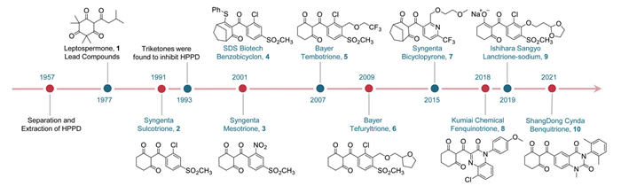
Figure 1.
History of the discovery of commercialized triketone HPPD.


Triketones are structural motifs containing three carbonyl groups that can tautomerize to their enol forms. Owing to their unique structure, triketones can act as both hydrogen bond donors and acceptors upon binding to biological targets, and they can also function as metal-chelating moieties, thereby exhibiting diverse biological activities and demonstrating broad application potential in both agricultural and pharmaceutical fields. Initially attracting attention for their potent herbicidal properties as 4-hydroxyphenylpyruvate dioxygenase (HPPD) inhibitors, these compounds play a crucial role in herbicide research by disrupting the biosynthesis of plastoquinone and tocopherol in photosynthesis [1,2]. Furthermore, through structural optimization and functional expansion, triketones have emerged as promising multifunctional agrochemical candidates, exhibiting inhibitory effects against parasites, insects, and plant viruses, thereby offering innovative solutions for agricultural pest management [3–5].
Notably, triketone compounds also show significant antimicrobial [6], antiviral [7], anticancer [8], anti-inflammatory [9], and antioxidant activities [10] in the pharmaceutical field, providing a valuable starting point for developing novel anti-infectives, anticancer therapeutics, and antioxidants. Remarkably, some triketone compounds are being explored in materials science, where their structural diversity and tunable chemical reactivity enable potential applications in acidichromic colorants and high-performance polymer materials [11,12]. For instance, their excellent chemical stability makes them suitable as monomers for advanced polymeric materials.
There are few prior reviews on triketones. Jhala et al. reviewed the history and U.S. application of HPPD-inhibitor herbicides and discussed emerging resistance issues [13]. Lin et al. analyzed the binding modes of various HPPD inhibitors to enhance mechanistic understanding [2]. Ma et al. focused on the molecular design and structural characteristics guiding novel HPPD inhibitor development [14]. However, these studies did not cover the antifungal and insecticidal roles of triketones in agriculture, nor their growing significance in medicine and materials science. In this review, we emphasize the multidisciplinary potential of triketone scaffolds and provides a valuable reference for future research. To better align with the term "triketone compounds", all compound structures in this article are presented in their triketone forms.
As a versatile class of chemical scaffolds, triketones exert herbicidal activity primarily by inhibiting 4-hydroxyphenylpyruvate dioxygenase (HPPD), thereby disrupting tyrosine metabolism and blocking the biosynthesis of plastoquinone and tocopherol essential for photosynthesis. The resulting impairment of carotenoid-mediated photoprotection leads to leaf bleaching and ultimately weed death [2,15–17]. Accordingly, structural optimization of triketone-based herbicides has progressed beyond aromatic ring modification to systematic tuning of the triketone core itself. Approaches including ring-size expansion, heteroatom incorporation, and fused-ring construction have been widely explored to improve physicochemical properties and biological performance. Notably, triketone derivatives have also exhibited insecticidal and antiviral activities, highlighting their potential for the multifunctional development of agrochemicals.
In recent decades, HPPD inhibitors have been shown to effectively control weeds by inhibiting the conversion of 4-hydroxyphenylpyruvic acid (HPPA) to homogentisic acid (HGA) in tyrosine metabolism pathways. This blockage halts the biosynthesis of essential substances, such as plastoquinone and tocopherol, necessary for photosynthesis [18]. The triketone structure and its analogs, as one of the pharmacophores of HPPD inhibitors, are crucial for herbicidal activity because they form chelation interactions with the catalytically active site metal ions of HPPD [19,20]. This importance places triketone compounds in a significant position among HPPD inhibitors. To date, nine triketone-based HPPD inhibitors have been registered and launched commercially, including seven triketone compounds, one triketone prodrug, and one sodium salt (Fig. 1). In the following sections, these inhibitors will be described in detail based on the historical development of triketone HPPD inhibitors and their structural characteristics.
The discovery of HPPD inhibitor herbicides began in 1977 when researchers at the Western Research Center in California, led by Reed Gray, observed that the density of weeds around Callistemon spp. was significantly lower than around other plants. Gray and his team extracted a potentially active substance from these plants and found that it caused seedling bleaching in germination tests. Structural identification confirmed the active substance as the natural product leptospermone (1, Fig. 1), a volatile phytotoxin found in Myrtaceae plants. In greenhouse herbicidal activity tests, leptospermone demonstrated moderate control efficacy against grass weeds at an application rate of 1000 g ai/ha [21].
Researchers have identified a series of leptospermone derivatives bearing distinct hydrophobic chains at the exo-carbonyl position (compounds 11–13, Fig. 2A). Despite their natural abundance, these compounds exhibit inferior pharmacokinetic parameters compared to commercially available triketones synthesized through systematic optimization, thereby limiting their practical application [22]. However, recent discoveries in natural product-based bioherbicides have revitalized interest in essential oils (rich in triketone compounds) for sustainable weed management in organic farming systems. Frabboni et al. demonstrated that undiluted essential oils from Origanum vulgare and Rosmarinus officinalis significantly inhibit the weed growth in Amaranthus retroflexus, Lamium maculatum, and Lactuca virosa compared to 50% diluted formulations [23]. Kanatas et al. developed a bioherbicide containing 68% (w/v) pelargonic acid and 6.55% (w/v) Carum carvi essential oil, which reduced the fresh weight of Echinochloa crus-galli and Sorghum halepense by 30% and 50%, respectively, versus untreated controls [24]. Greenhouse trials by Barickman et al. revealed that triketone-enriched manuka oil extract exhibited herbicidal efficacy comparable to 2% glyphosate against Amaranthus palmeri and Digitaria sanguinalis. Notably, the triketone extract achieved 70% growth inhibition in Cyperus esculentus within 9 days post-application [25].
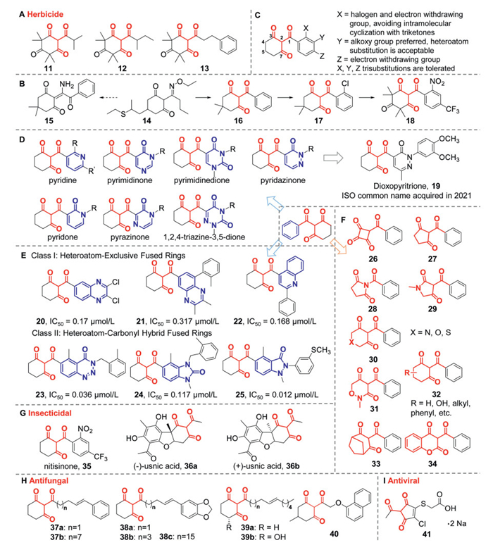
The actual development of HPPD inhibitor herbicides stemmed from an unexpected discovery during the development of another mechanism-based herbicide (Fig. 2B). In 1982, researchers at the Western Research Center were modifying the structure of the commercial herbicide sethoxydim (14) to develop new ACCase inhibitor herbicides. They unexpectedly obtained a novel benzoyl cyclohexane-1,3-dione structure (16) instead of the expected enamine cyclohexane-1,3-dione structure (15) when reacting diketones with various substituted benzoyl cyanides. Although the new structure did not exhibit significant herbicidal activity, subsequent optimization of the substituents on this compound type led to improved herbicidal activity. Further structural optimization revealed that compound 17, effectively controlled broadleaf weeds at an application rate of 2000 g ai/ha. Interestingly, the symptoms observed in treated plants were highly similar to the bleaching effects caused by leptospermone, rather than resembling those of sethoxydim analogs. Therefore, the researchers replaced the cyclohexanedione fragment in compound 17 with the cyclohexatrione fragment from leptospermone, thereby optimizing the structure to yield compound 18. In greenhouse herbicidal activity tests, compound 18 demonstrated good herbicidal efficacy at 62.5 g ai/ha and provided effective pre-emergence control of both grass and broadleaf weeds [26]. These two serendipitous discoveries spurred major agrochemical companies to develop this class of structures.
Although HPPD was partially purified and its enzymatic properties preliminarily characterized as early as 1956 [27], the definitive identification of HPPD as the molecular target of triketone inhibitors dates only to 1993, when Schulz and colleagues provided compelling experimental evidence: Mesotrione (3) was shown to disrupt the biosynthesis of plastoquinone and tocopherol, significantly inhibiting the incorporation of radiolabeled tyrosine into lipid-soluble quinones in wheat; the growth inhibition of Lemna seedlings caused by mesotrione could be fully reversed by supplementing the culture medium with HGA, whereas addition of its substrate, HPPA, had no effect, demonstrating that the inhibition occurs specifically at the step of HPPA conversion to HGA; in vitro assays revealed that mesotrione exhibited extremely potent inhibitory activity against HPPD partially purified from etiolated maize seedlings, with an IC50 as low as 4.5 × 10⁻⁸ mol/L; furthermore, HPPD from rat liver was also sensitive to mesotrione, indicating high structural and functional conservation of this enzyme across plants and animals [28,29]. In 1997, Lee further elucidated the structure–activity relationship of HPPD inhibitors, identifying 2-benzoylvinyl-1-ol as the minimal pharmacophore required for effective inhibition [30], a finding that laid the chemical foundation for the rational design of novel HPPD inhibitors.
In 1991, Syngenta successfully developed sulcotrione (2), which effectively controls broadleaf weeds in corn fields when applied post-emergence at 300–450 g ai/ha [31]. Ten years later, the company developed the second triketone herbicide, mesotrione (3), through structural modification of sulcotrione. Mesotrione has a more pronounced herbicidal efficacy, with a post-emergence application dose of 70–150 g ai/ha, an order of magnitude higher activity than sulcotrione. It has been found that mesotrione achieves high selectivity between corn and weeds because corn can rapidly metabolize and inactivate the active substance [32].
The introduction of sulcotrione and mesotrione sparked the development of triketone HPPD inhibitors by major agrochemical companies. The exploration of monocyclic HPPD inhibitors has led to the discovery of thousands of additional triketones. Substituents were introduced at the ortho, meta, and para positions relative to the carbonyl group, and structure-activity relationship studies revealed that introducing electron-withdrawing substituents at the ortho and para positions enhances herbicidal activity. However, the substituent bulk should not be too large. The meta position can accommodate a wider variety of substituents, making it a key focus for modifications, such as the alkoxy or cycloalkyl groups (Fig. 2C). On this basis, Bayer introduced tembotrione (5) in 2007 and tefuryltrione (6) in 2009, while Kumiai Chemical developed lancotrione-sodium (9) in 2019 [33–35]. Tembotrione is mainly used post-emergence for weed control in corn fields, and its efficacy is further enhanced with the addition of the safener isoxadifen-ethyl, significantly improving crop safety for corn. Tefuryltrione, the second triketone HPPD inhibitor developed for rice fields, requires a lower application rate compared to the previously developed benzobicyclon. At 300 g ai/ha, tefuryltrione effectively controls common broadleaf weeds and sedges, and shows significant efficacy against sulfonylurea-resistant weeds. The development of tefuryltrione has been beneficial for controlling noxious and resistant weeds. Lancotrione-sodium, a triketone HPPD inhibitor for paddy fields, was registered and marketed in sodium salt form. It provides adequate control of broadleaf weeds, grass weeds, and sedges in paddy fields and is highly safe for rice when applied pre-emergence or post-emergence. Other substituents at position Y, such as nitrogen-containing, sulfur-containing, and cyclic substituents, have not yet led to successful commercial products, but most compounds exhibit excellent weed control efficacy [36,37]. These results also demonstrate that substituent optimization is crucial in enhancing herbicidal activity and crop safety.
Following aromatic ring substitutions, structures such as nitrogen atoms and carbonyl groups have been introduced to the aromatic rings, leading to the discovery of diverse aromatic heterocyclic compounds. For example, In 1992, SDS Biotech synthesized a pyridine-containing triketone HPPD inhibitor (benzobicyclon, 4), which was commercialized in 2001. Benzobicyclon is a triketone prodrug; its hydrolytic metabolite is the active triketone HPPD inhibitor. It exhibits broad-spectrum activity against annual grasses, sedges, and broadleaf weeds while showing excellent crop safety in rice. Subsequently, Syngenta developed and commercialized a third triketone HPPD-inhibiting herbicide featuring a similar pyridine structure, bicyclopyrone (7). It is extensively used in the United States, Canada, and other regions for pre-plant and pre-emergence control of grass weeds and broadleaf weeds (application rate: 50–300 g ai/ha), and demonstrates high crop safety for maize, cereals, and sugarcane. Bicyclopyrone is a low-toxicity herbicide, exhibiting minimal toxicity to bees, birds, fish, and trematode species [38]. In 2012, Syngenta incorporated a pyridazinone fragment to generate dioxopyritrione (19, Fig. 2D). At application rates of 125–250 g ai/ha, dioxopyritrione showed 100% control against Abutilon theophrasti, Amaranthus retroflexus, and Echinochloa crus-galli, with no phytotoxicity observed in barley [39,40]. Other aromatic heterocycles, including, pyrimidione, pyrimidinedione, pyridone, pyrazinone, and 1,2,4-triazine-3,5-dione were also acceptable for herbicidal activity [41]. However, these compounds regrettably failed to progress to further development.
The aromatic ring constitutes a critical component of HPPD inhibitors. Researchers have synthesized fused-ring HPPD inhibitors by extending monocyclic frameworks. Some early fused-ring structures (e.g., saccharin derivatives) were synthesized, and their activity showed limited improvement over monocyclic analogues while incurring substantial cost increases [42]. Following the elucidation of HPPD-inhibitor cocrystal structures in 2004, researchers demonstrated that extending conjugation systems on monocyclic frameworks could significantly enhance binding affinity [18,43]. Concurrently, fused-ring architectures provided opportunities to circumvent patent restrictions and discover novel active molecules, thereby accelerating the development of fused-ring HPPD inhibitors.
In 2018, Kumiai Chemical launched fenquinotrione (8), a paddy-field herbicide containing a quinoxalin-2-one moiety. As the first commercialized HPPD inhibitor featuring a benzoheterocyclic system, it established a valuable paradigm for subsequent HPPD inhibitor design. Fenquinotrione exhibits exceptional crop safety in rice at application rates of 125–250 g ai/ha. It shows broad-spectrum efficacy against dicotyledonous weeds, gramineous weeds, and sedges, by pre- and early post-emergence applications. Notably, it effectively controls some acetolactate synthase (ALS) inhibitor-resistant weeds [44]. In 2021, benquitrione (10), a sorghum-field herbicide containing a quinazoline-2,4-dione scaffold, was successfully commercialized. At application rates of 75–150 g ai/ha, this sorghum-selective herbicide shows superior crop safety in sorghum while maintaining acceptable safety profiles in wheat, maize, and sugarcane. It effectively controls annual weeds in sorghum fields, particularly showing exceptional efficacy against pernicious weeds, such as Setaria viridis and Echinochloa crus-galli. The registration of benquitrione addresses long-standing challenges in sorghum weed management by providing full growth cycle control while reducing overall herbicide input [45].
Recent exploration of fused-ring HPPD inhibitors remains a focal point in herbicide development. These fused-ring systems are further categorized into two classes (Fig. 2E). One is heteroatom-exclusive fused rings. In 2021, Hu et al. employed structure-based drug design to create quinoxaline-incorporated HPPD inhibitors, with selected compounds exhibiting enzymatic inhibition (20, IC50 = 0.17 μmol/L) comparable to mesotrione (IC50 = 0.23 μmol/L). However, most analogues showed reduced potency. A lead compound demonstrated > 90% efficacy against Echinochloa crus-galli and Setaria faberi at 150 g ai/ha [46]. Similarly, Zheng et al. developed novel triketone-quinoxaline hybrids (21), with some compounds showing superior in vitro inhibition of Arabidopsis thaliana HPPD (AtHPPD) compared to mesotrione. However, herbicidal activity data were absent in this study [47]. Further progress emerged in 2023 when Zhao et al. designed quinoline-based inhibitors (22) with enzymatic activity matching mesotrione. Complete weed suppression (Abutilon juncea, Echinochloa crus-galli, Setaria faberii) was achieved at 150 g ai/ha [48].
The other type is heteroatom-carbonyl hybrid fused rings, which generally exhibit better herbicidal activity than the first type for HPPD and weed control. Yan et al. discovered benzotriazine-4-one (23) scaffolds through pharmacophore-linked fragment virtual screening, achieving nanomolar AtHPPD inhibition and > 80% weed control across five species [49]. In the same year, benquitrione-derived benzimidazol-2-one inhibitors (24) were designed via ring contraction. These compounds outperformed mesotrione in enzymatic assays and exhibited exceptional field efficacy at 150 g ai/ha, coupled with complete peanut safety. These compounds maintained effective weed control at application rates of 30–120 g ai/ha [50]. Chen et al. introduced the indazolone fragment via active substructure splicing, resulting in compound 25 with an IC50 of 12 nmol/L against AtHPPD. This compound exhibited broad-spectrum herbicidal activity at 30–120 g ai/ha while demonstrating high safety in peanut [51].
Researchers have systematically modified the triketone scaffold through four primary approaches: (Ⅰ) Ring size reduction from six-membered to five-/four-membered systems; (Ⅱ) heteroatom (N/O/S) incorporation; (Ⅲ) cyclohexanedione ring substituent modification; (Ⅳ) cyclization strategies (Fig. 2F). Early efforts in the 1990s to shrink the cyclohexanedione ring or introduce heteroatoms produced derivatives with unsatisfactory herbicidal activity, such as compound 26–31 [26,40,52]. Notably, nitrogen substitution at the triketone linker generated N-aroyl analogues (28) showing moderate efficacy against Echinochloa crus-galli and Abutilon theophrasti, though lacking broad-spectrum utility [53]. Structure-activity relationship (SAR) studies revealed that introducing substituents (32) at the 5-position of cyclohexanedione significantly reduced potency, with the following activity hierarchy: Unsubstituted (IC50 = 0.354 μmol/L) > 5-phenyl (IC50 = 0.356 μmol/L) > 5-CH3 (IC50 = 1.568 μmol/L) > 5-diCH3 (IC50 = 2.789 μmol/L) [48]. Introduction of substituents, such as methyl groups, to the cyclohexanedione ring, generally enhances the herbicidal activity, particularly against grasses. However, this modification also increases toxicity to crops, such as maize, presumably due to blocking potential metabolic sites. Resistance in weeds is associated with the metabolism of triketones into 5-hydroxy derivatives, resulting in a nearly complete loss of herbicidal activity [54]. Similarly, hydroxylated metabolic products at the 4- or 6-position also exhibit significantly reduced efficacy, and 4- or 6-hydroxylation markedly diminishs efficacy [55,56]. However, forming a bridging ring structure (33), such as bicyclopyrone (7), is acceptable for activity. Hybrid architectures merging HPPD pharmacophores with coumarin fragments (34) exhibited selective root growth inhibition in rape, yet comprehensive greenhouse validation remains pending [57].
The essential oils of many Myrtaceae plants, particularly those containing mānuka oil, leptospermone (1), flavesone (11), and isoleptospermone (12), are primarily found due to their herbicidal activity. The efficacy of these compounds is mainly attributed to their triketone moieties, which therapeutically block tyrosine catabolism by inhibiting HPPD. However, studies have also indicated that these triketones exhibit efficacy against arthropods and other pests, a mechanism associated with HPPD inhibition.
In 2023, Bošković et al. found that triketones from essential oils exhibited contact toxicity against Drosophila suzukii, suggesting their potential as alternatives to conventional insecticides [58]. Subsequent research by McComic identified HPPD-targeting herbicides as toxic to hematophagous species, including Aedes aegypti and Amblyomma americanum, with nitisinone (NTBC, 35) showing the highest toxicity (Fig. 2G) [3]. These findings aligned with the toxicity data for Aedes aegypti reported by Vergaray Ramirez and Sterkel et al., as well as congruent mosquito studies [59,60]. In 2021, Sterkel found that NTBC can kill blood-fed tsetse flies through oral and topical disruption of tyrosine metabolic pathways, indicating the potential for mitigating African trypanosomiasis transmission [61]. Parallel studies by Alvarez Costa et al. evaluated triketone-rich essential oils from another plant species, Eucalyptus nitens, showing both repellent effects and larvicidal activity against Aedes aegypti and Aedes albopictus [62]. In 2008, Huseyin et al. found that the lichen secondary metabolites (-)-usnic acid (36a) and (+)-usnic acid (36b) exhibited strong larvicidal activity against Culex pipiens L. larvae, causing 100% mortality within 24 h at doses of 5 and 10 ppm against third- to fourth-instar larvae, with LC50 values of 0.8 and 0.9 ppm, respectively [63]. In 2021, Koc et al. also found that a reservoir solution of (-)-usnic acid (36a) dissolved in acetone was toxic to Aedes aegypti larvae, with a median lethal concentration (LC50) of 3.8 ppm and inducing 61.3% mortality at 10 ppm [64]. This insecticidal activity against blood-sucking insects may be attributed to the accumulation of tyrosine in the blood due to the use of triketone compounds.
In 2025, Nammunige et al. first identified the acaricidal efficacy of triketone compounds against all developmental stages of scabies mites (Sarcoptes scabiei). Exposure to 150 mmol/L concentrations of any tested triketone for 4 h resulted in complete mortality in motile stages, whereas ova required extended exposure beyond this threshold for complete eradication [4]. This builds upon earlier findings by Jeong et al., who documented the miticidal activity of triketones against diverse mite species, including Dermatophagoides farinae, Dermatophagoides pteronyssinus, and Tyrophagus putrescentiae [22].
The antimicrobial properties of plant essential oils have been extensively studied, with most research focusing on their effects against non-plant pathogenic fungi. However, the potential antifungal activity of key constituents, especially triketones, against plant pathogens remains an area that should not be overlooked.
In 2014, Ferreira investigated the antifungal properties of dichloromethane extracts from the aerial parts of Peperomia alata and Peperomia trineura against Cladosporioides and Cladosporium sphaerospermum. Cytotoxicity assays conducted on K562 and Nalm-6 leukemia cell lines revealed significant efficacy in both antifungal and cytotoxic evaluations. The authors identified the active components in the extracts, which shared a common 2-acylcyclohexane-1,3-dione scaffold (compounds 37–39, Fig. 2H). The antifungal activity and cytotoxicity of these compounds were influenced by side-chain length, as well as the type and position of substituents. Among them, compounds 37a and 38a exhibited superior antifungal activity [65]. In a subsequent study in 2018, Gundoju further explored the fungicidal activity of similar compounds [66].
Yu et al. confirmed the potent antifungal activity of (+)-usnic acid (36b) against key agricultural pathogens (Sclerotinia sclerotiorum, Fusarium graminearum, and Rhizoctonia solani). They also elucidated the crystal structure of the Zymoseptoria tritici HPPD (ZtHPPD)-(+)-usnic acid complex, revealing that HPPD is a crucial target for the antifungal activity of usnic acid. A molecularly optimized compound 40, showed enhanced inhibitory activity in bioassays against Sclerotinia sclerotiorum and Rhizoctonia solani, outperforming classic HPPD inhibitors, such as mesotrione and (+)-usnic acid. Based on prior mechanistic studies, the antibacterial activity of usnic acid is attributed to the inhibition of DNA and RNA synthesis, as well as the disruption of bacterial cell membranes. The authors propose that its antifungal activity likely arises from a combination of HPPD inhibition and the disruption of normal cell structures [67].
In 2012, Lapshina reported the discovery that the disodium salt of cyclopentene triketone (41, Fig. 2I) enhances the activity of hydrolytic enzymes, such as protease and ribonuclease, resulting in abnormalities such as swelling, thinning, and fracturing of tobacco mosaic virus particles in aqueous tobacco leaf extracts. These effects may be attributed to a cellular defense mechanism that enhances plant resistance to viral infections [5].
Triketones in plant extracts have garnered significant attention in recent years due to their considerable potential in various biological activities. These compounds exhibit notable antibacterial, antifungal, and antiviral properties, as well as anticancer, anti-inflammatory, antidiabetic, and free radical scavenging effects. Numerous studies have identified plant-derived natural products with a triketone structure, and it has been showed that their biological activity can be further enhanced through structural modifications and synthetic chemical approaches. These advancements have expanded their potential for use in treating various diseases.
The antimicrobial activity of triketone compounds has been extensively studied since the early discovery of bactericidal constituents in plant essential oils. Through structural characterization and bioactivity evaluations of natural products, researchers have confirmed the antimicrobial potential of triketones (Fig. 3A). Killee et al. found that triketone compounds exhibit antibacterial activity against Gram-positive bacteria, including methicillin-resistant Staphylococcus aureus (MRSA). Among the tested derivatives leptospermone (1), flavesone (11), isoleptospermone (12), grandiflorone (13), and trimethylated acylphloroglucinol derivatives (nortriketones 42–45), grandiflorone (13) exhibited the highest potency, with an IC50 value of 750 nmol/L against HPPD. In contrast, nortriketones 42, 43, and 45 showed no inhibitory activity toward HPPD at 100 μmol/L, most likely due to reduced binding affinity caused by enol tautomerization [68].
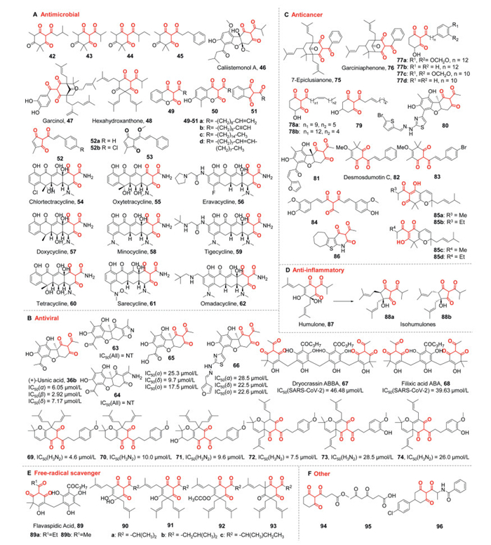
Further studies on plant triketone extracts have explored the antimicrobial effects of leptospermone (1) against foodborne pathogens, underscoring its potential as a natural food preservative [69]. Callistemonols A (46) also exhibited potent bactericidal activity against MRSA, while garcinol (47) showed significant inhibitory effects against Gram-positive bacteria, including Staphylococcus aureus, Escherichia coli, and Streptococcus mutans [70,71]. Hexahydroxanthone (48) demonstrated antibacterial efficacy against Clostridium perfringens [72]. Additionally, usnic acid (36a and 36b) has been shown to exert differential antimicrobial activities against Staphylococcus aureus, Bacillus subtilis, Enterococcus faecalis, and Mycobacterium tuberculosis, potentially through mechanisms involving membrane disruption and inhibition of nucleic acid synthesis [73].
Researchers have synthesized various triketone derivatives and systematically investigated their SAR of antibacterial activity, and potential inhibitory mechanisms (Fig. 3A). Boccalini et al. synthesized a series of 4-hydroxycoumarin-based long-chain 3-acyl derivatives (49–51), with compounds 49a, 49b, and 50a exhibiting selective inhibition against S. aureus, S. epidermidis, and Propionibacterium acnes. The SAR studies revealed that both the triketone moiety and the long-chain fatty acyl group are critical for antibacterial activity. In contrast, modifications of the coumarin core have a minimal effect on potency [74].
Babu et al. systematically modified three structural elements of coruscanone B (52a): The cyclopentanedione ring, enol methoxy group, and styrene side chain, and identified compound 52b with triketone modifications as a moderately active antifungal agent [75]. Shestak et al. further showed that free cyclopentene-containing triketones exhibit superior antifungal activity compared to methyl enol ether derivatives (53), which were more effective against yeast and fungi [76]. Khan et al. revealed that the mechanism of action of these compounds involves the inhibition of fungal lanosterol 14α-demethylase (CYP51A1), a key enzyme in ergosterol biosynthesis [77].
Tetracycline antibiotics are broad-spectrum bacteriostatic agents integral to anti-infective therapy since their introduction in the 1940s. The tetracycline class originated with the isolation of chlortetracycline (54) from Streptomyces aureofaciens in 1948, followed by the discovery of successive generations: Oxytetracycline (55), eravacycline (56), doxycycline (57), minocycline (58), tigecycline (59), tetracycline (60), sarecycline (61), and omadacycline (62), whose structures are shown in Fig. 3A. Structurally, these antibiotics share a tetracyclic naphthacene carboxamide nucleus as the core, with 2-carboxamidocyclohexane-1,3-diones and a C4 dimethylamino group as essential active moieties. Their mechanism of action involves binding to the 16S rRNA of the 30S ribosomal subunit, blocking the A-site to prevent the attachment of aminoacyl-tRNA, thereby inhibiting peptide chain elongation and protein synthesis. This mechanism provides efficacy against various pathogens, including Gram-positive bacteria, some Gram-negative bacteria, and certain anaerobes [78,79].
Early studies discovered that leptospermone (1) and flavesone (11) exhibit anti-herpes zoster activity [80]. The antiviral mechanism of these triketones may involve interference with the viral envelope structure or the masking of compounds necessary for viral adsorption or entry into host cells. Additionally, researchers identified low antiviral activity of usnic acid against Herpes simplex type 1 and Polio type 1 viruses [81]. Subsequent investigations focused on the activity of triketone compounds against influenza viruses. In 2012, the anti-H1N1 influenza virus (pdm09) activity of usnic acid was first explored, revealing the critical role of stereochemistry in antiviral activity. (-)-Usnic acid (36a, ED50 = 14.5 μmol/L) exhibited superior activity, while structural modifications reversed this trend, with (+)-usnic acid analogs showing enhanced potency (Fig. 3B). Researchers hypothesized that usnic acid might indirectly reduce viral transmission by inhibiting host pro-viral pathways [82].
In 2022, usnic acid demonstrated in vitro inhibitory activity against SARS-CoV-2 variants α, β, and δ, with efficacy comparable to remdesivir (a COVID-19 emergency therapeutic), although its activity varied across strains. Mechanistic studies suggested that (+)-usnic acid (36b) likely targets the SARS-CoV-2 Mpro [83]. Another study confirmed the antiviral activity of usnic acid and its derivatives against SARS-CoV-2 strains (α, δ, and ο). Structural modifications, such as cyclization (63) or amination (64) at the triketone moiety, resulted in the loss of antiviral activity, while alterations at other positions (65) caused fluctuating potency [7]. In 2023, thiazole-hydrazone substitutions (66) on usnic acid significantly enhanced its anti-SARS-CoV-2 activity. These studies collectively revealed that usnic acid derivatives do not primarily inhibit viral proteases but effectively block viral entry, potentially by binding to the N-terminal domain of the spike glycoprotein S, overlapping with hemoglobin binding regionsm [84]. However, the exact mechanism remains unknown.
In 2022, Jin et al. found that dryocrassin ABBA (67) and filixic acid ABA (68) exert their effects in inhibiting SARS-CoV-2 infection by targeting the Mpro enzyme, showing broad-spectrum antiviral activity against coronaviruses. Among these, dryocrassin ABBA emerged as a promising therapeutic candidate for the development of COVID-19 drugs [85]. Lee et al. further explored chromenone derivatives derived from plant extracts for the inhibition of influenza virus neuraminidase (NA), particularly effective against the H3N2 subtype. These chromenone derivatives (69–74), characterized by a 2-acetyl-1,3-cyclohexanedione core structure, exhibited significant variations in inhibitory activity and binding patterns due to distinct functional and conformational modifications. Notably, competitive inhibitors, such as chromenones 69 and 73, interacted with the enzyme at the catalytic site, while noncompetitive inhibitors, including chromenones 70, 71, 72, and 74, bound to a metastable site on the enzyme [86].
Many triketone natural products extracted from plants exhibit anticancer activity. For example, in 1998, Stephan evaluated the ability of hexahydrocolupulone (48) to inhibit tumor cell growth in vitro (Fig. 3A). Hexahydrocolupulone inhibited the proliferation of various human tumor cell lines, including those resistant to currently used chemotherapeutic drugs. Its mechanism may involve action on macromolecular pathways, thereby inhibiting tumor cell proliferation in vitro [87]. In 2004, Tanaka et al. isolated isomers of 7-epiclusianone (75, Fig. 3C) from Hypericum perforatum, which demonstrated moderate antitumor activity against human lung cancer cells (A549) and breast cancer cells (MCF-7) [88]. Subsequent studies have shown that 7-epiclusianone also exhibits antineoplastic activity in a glioblastoma model, a tumor with intrinsic chemoresistance, affecting cell growth, cell cycle dynamics, apoptosis, and colony-forming ability [89]. Similar compounds, such as garciniaphenone (76), were later isolated from Garcinia leaves, exhibiting cytotoxic activity against various cancer cell lines. Additionally, garciniaphenone (76) shows antioxidant and anti-inflammatory properties that help protect cells from oxidative stress-induced damage [90]. In 2007, Li et al. extracted various triketone compounds (77 and 78) from Peperomia dindygulensis Miq., some of which inhibited human umbilical vein endothelial cell (HUVEC) proliferation and suppressed HUVEC tube formation [91]. In 2008, Denny et al. isolated a new triketone compound (79) from the leaves of Virola sebifera, which exhibited strong toxicity and selectivity against the ovarian cancer cell line OVCAR03 and the multidrug-resistant breast cancer cell line NCI-ADR [92].
Usnic acid, a widely studied natural compound, displays multi-target antitumor activity. Its mechanism may involve regulating the expression of apoptosis-related proteins in gastric cancer cells, inducing cell cycle arrest and autophagy, thus exerting antiproliferative and pro-apoptotic effects. Usnic acid induces G1-phase arrest in human gastric cancer BGC823 cells, G2-phase arrest in SGC7901 cells, and S-phase arrest in colon cancer HCT-116 cells [93,94]. It also inhibits the proliferation of human osteosarcoma MG-63 cells and melanoma A375 cells in a time- and dose-dependent manner [95]. Additionally, usnic acid suppresses the growth and angiogenesis of mouse H22 tumors by inhibiting the secretion of vascular endothelial growth factor (VEGF) and basic fibroblast growth factor (bFGF), and inhibits breast tumor angiogenesis and growth by blocking VEGFR2-mediated AKT and ERK1/2 signaling pathways [96,97]. Recent studies have further elucidated the anticancer mechanisms of usnic acid. In 2021, Sun et al. found that usnic acid reduces PD-L1 protein synthesis by synergistically suppressing STAT3 and RAS pathways, thereby decreasing PD-L1 expression in HeLa cells, enhancing the cytotoxicity of co-cultured T cells against tumor cells, and inhibiting cervical cancer cell proliferation [98]. In 2024, Varlı et al. discovered that usnic acid targets 14–3–3 proteins to inhibit cancer progression. Usnic acid binds to and degrades 14–3–3 proteins, disrupting critical processes such as cancer cell invasion, cell cycle progression, aerobic glycolysis, and mitochondrial biogenesis. Furthermore, it modulates Akt/mTOR, JNK, STAT3, and NF-κB signaling pathways to suppress the metabolism and survival of cancer cells [99]. Mariraj et al. observed morphological changes in G361 human melanoma cells via DAPI staining, which intensified with increasing usnic acid concentration and exposure time, confirming that usnic acid induces apoptosis. Computational simulations suggest that usnic acid may specifically bind to cancer-associated proteins, such as CYP3A4 and B-Raf kinase [100]. Azhamuthu et al. demonstrated that usnic acid inhibits the growth and proliferation of human oral cancer cells through ROS-dependent mitochondrial-mediated apoptosis. Usnic acid downregulates the expression of the anti-apoptotic protein Bcl-2 while upregulating pro-apoptotic proteins p53, Bax, cytochrome c, and Caspase-3/9, significantly activating mitochondrial apoptotic pathways [101]. In another study, the combination of usnic acid and ABT-199 synergistically promoted the expression of integrated stress response-related genes ATF4, CHOP, and NOXA via heme-regulated inhibitory kinase, as well as the degradation of the anti-apoptotic protein MCL-1, which may increase the sensitivity of ABT-199 to chemotherapy in acute myeloid leukemia [102].
Researchers have also investigated the anticancer properties of usnic acid analogues. In 2023, Zakharenko et al. introduced hydrazonothiazole fragments (80) into usnic acid derivatives, which inhibited tyrosyl-DNA phosphodiesterase (TDP1) at submicromolar concentrations and enhanced the in vivo antitumor and antimetastatic effects of topotecan (an anticancer drug) [103]. In 2025, Roney et al. synthesized compound 81 by incorporating α, β-unsaturated groups, which exhibited significant cytotoxicity against breast cancer cell lines MCF7 and T47D. Molecular docking studies revealed that compound 81 binds to the active site of the AKT2 (PKB-BETA) protein [104].
Researchers have also modified the structures of other natural products to obtain triketone compounds with anticancer activity. In 2005, Nakagawa synthesized a series of desmosdumotin C (82) analogs and found that altering the substituents on the terminal benzene ring could increase antitumor activity, with the 4-bromophenyl derivative (83) showing the strongest activity, 4–6 times higher than that of desmosdumotin C [105]. In 2006, Lin et al. synthesized over 40 curcumin derivatives containing unsaturated conjugated linking fragments. Compounds with a triketone structure (84) inhibited androgen receptor signaling, blocking the proliferation and survival of prostate cancer cells [106]. Further research in 2015 showed that curcumin derivative 84 effectively reduced EGFR phosphorylation and expression in lung adenocarcinoma cells and mitigated gefitinib-induced gastrointestinal damage [107]. In 2008, Tanaka et al. proposed a biosynthetic pathway for petiolins (85a-d), which exhibited moderate cytotoxic activity against the L1210 and KB cancer cell lines. Notably, while the keto form maintains a uniform structural framework, the corresponding enol forms display configurational variations [108]. In 2017, Shaaban synthesized a series of cyclopenta[b]thiophene derivatives, with compound 86 containing a triketone fragment showing reasonable antitumor activity against three human cancer cell lines (HepG2, MCF-7, and HT-29) and significant antifungal activity against Aspergillus species [8].
Isohumulones (88) are a class of lipophilic compounds extracted from hops, derived through the isomerization of humulones (87), and are known for their diverse biological activities (Fig. 3D). Studies have shown that isohumulones can reduce metabolic inflammation and insulin resistance while positively influencing dyslipidemia and obesity. These effects have been validated in various cell lines, animal models, and human intervention trials. Isohumulones (88a and b) exert their actions through pleiotropic mechanisms, including regulating gene expression, epigenetic modulation, enzyme inhibition, and cell signaling. Specific mechanistic studies indicate that isohumulones have anti-inflammatory and antidiabetic effects by downregulating pro-inflammatory gene expression and enhancing insulin signaling. Additionally, isohumulones have been shown to improve metabolic syndrome and obesity-related metabolic disorders by increasing HDL cholesterol levels, decreasing triglycerides, and reducing hepatic fat storage. In summary, isohumulones, as a key bioactive agent, possess significant therapeutic potential in medical nutrition, particularly for managing diseases associated with chronic inflammation and insulin resistance [109].
Similarly, 7-epiclusianone (75) has been found to exhibit comparable effects. 7-Epiclusianone reduces inflammation and obesity in obese rats by modulating the expression of various genes and proteins. Specifically, it decreases the expression of fatty acid synthase and lipoprotein lipase, increases the expression of the anti-inflammatory cytokine interleukin-10, and reduces levels of the pro-inflammatory cytokine tumor necrosis factor-α (TNF-α). Furthermore, 7-epiclusianone enhances antioxidant capacity by upregulating the expression of peroxisome proliferator-activated receptor-γ. These combined mechanisms work synergistically to reduce weight gain and fat accumulation in obese rats while alleviating inflammation [9].
In 2003, Lee and collaborators discovered that methanolic extracts from Korean plants exhibited strong antioxidant activity, particularly in inhibiting lipid peroxidation. However, their scavenging capacities for DPPH free radicals and superoxide anions were weak. The authors suggested that the potent lipid peroxidation inhibition could be attributed to the stabilising effects of flavaspidic acids (89a and b, triketone analogues in the extract, Fig. 3E), which may chelate with iron, thereby enhancing their activity. This insight highlighted the potential of triketones as antioxidants [110]. Supporting this, Tagashira et al. chemically modified humulone (90–93) to investigate its DPPH free radical scavenging and lipid peroxidation inhibitory activities. They found that derivatives lacking the 5-hydroxyl group exhibited higher lipid peroxidation inhibition, suggesting the importance of the triketone structure for this activity [111].
In 2018, Liu extended this understanding by conducting oxygen radical absorbance capacity measurements on hexahydro-β-acids, noting that triketones exhibited superior antioxidant capacity compared to their hydrogenated derivatives. This enhanced activity was attributed to the enol double bond in the triketone structure of hexahydroxanthone (48), which is chemically reactive and unstable, leading to stronger antioxidant effects [112]. Furthermore, in 2023, Peng and Xiao developed solid dispersions of hexahydroxanthone to improve its water solubility and stability, enhancing its antioxidant capacity. This formulation was shown to delay the browning of apple juice, preserving its nutritional quality [10].
In addition, other natural compounds exhibited strong antioxidant properties. For instance, Arwa et al. demonstrated that 7-epiclusianone (75) regulates oxidative stress by inhibiting NADPH oxidase and stimulating reactive oxygen species (ROS) production in human neutrophils. Its analogs also exhibited potent inhibition of AAPH-induced hemolysis and lipid peroxidation in human erythrocytes [113]. Moreover, 7-epiclusianone's ability to absorb UVB radiation further prevents oxidative stress and lipid peroxidation, suggesting potential as a sunscreen agent [114]. Similarly, Fernández-Moriano et al. showed that usnic acid (36) protects against H2O2-induced cytotoxicity and oxidative stress by upregulating phase Ⅱ antioxidant enzymes via the Nrf2 pathway, while Erfani et al. provided evidence that usnic acid exerts neuroprotective effects, alleviating memory impairment after ischemia-reperfusion injury by reducing cell death, inflammation, and oxidative stress [115,116]. Additionally, the photodegradation derivatives of usnic acid exhibited significant UV protection activity and lower toxicity to human liver and skin cells compared to the parent compound [117].
Nitisinone (35, Fig. 2G) was initially developed as a herbicide, but in 1991, studies found that it could prevent the accumulation of toxic metabolites by inhibiting HPPD activity. This discovery led to its use as a life-saving drug for infants with type Ⅰ tyrosinemia [118]. In 2016, Annalisa Santucci found that nitisinone also showed efficacy in treating alkaptonuria, a condition with no other available treatments [119]. Neither of these diseases involves mutations in the HPPD gene. The other HPPD mutation associated with type Ⅰ tyrosinemia, known as hawkinsinuria, may not significantly alter the binding mechanism, allowing nitisinone to be well tolerated by infants and enabling its use during their first few years of life in hawkinsinuria [120]. However, nitisinone can cause side effects, including visual disturbances, liver failure, convulsions, and cognitive difficulties, due to the accumulation of tyrosine in the blood [121]. Additionally, there is currently no treatment for oculocutaneous albinism caused by mutations in the tyrosinase gene. While oral administration of nitisinone does not result in iris melanin content, it has been shown to increase ocular and dermal pigmentation in a mouse model of albinism [122].
Kim synthesized novel succinylacetone analogs (94, Fig. 3F). Initial evaluations revealed that compound 94 exhibited an IC50 value for the mouse allogeneic mixed lymphocyte reaction similar to that of succinylacetone (95), indicating that alkylacyloxy enol esters are a promising class of novel lead compounds for use as immunosuppressive agents [123].
In 2017, Scala introduced the potential of triketone compounds as inhibitors of the cytosolic protease CPB2.8ΔCTE from Leishmania mexicana. The triketone structure binds to the thiol group of the active site cysteine, forming an unstable hemithioacetal. These compounds exhibit reversible covalent inhibition, showing a certain degree of selectivity and activity. Compound 96 was predicted to be non-mutagenic, non-carcinogenic, and non-neurotoxic, making it a promising candidate for the development of antileishmanial drugs [124].
In 2005, Yang was the first to utilize a triketone moiety as an acid- and base-sensitive group to design and synthesize acidichromic colorants (97 and 98). The molecular structure of these compounds consists of an acid-sensitive triketone moiety and a base-sensitive aniline molecule within an extended conjugated system. This structure enables the compounds to undergo two different reversible color changes under strongly acidic and basic conditions (Fig. 4A) [11,125]. Building on this work, the group modified the molecule in 2007 by synthesizing a coumarin ring and introducing various substituents to modulate the strength of hydrogen bonds within the molecules. This modulation affects the deprotonation ability of the alcohol-based hydrogen, although it does not influence the color itself. Among the compounds, the 7-bromo-substituted derivatives (99) were more readily deprotonated by ammonia than the 7-methoxy-substituted compounds. These colourants can be dissolved in organic solvents or incorporated into polycarbonate films, serving as acid-base sensors or reversible colour-change devices [126].
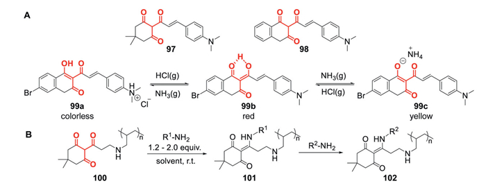
A significant advancement in polymer chemistry was reported by Helms et al., who pioneered the spontaneous condensation of triketones (100) with amines under ambient conditions, establishing a catalyst-free dynamic covalent click chemistry approach (Fig. 4B) [12,127]. This innovative strategy enabled the synthesis of closed-loop recyclable polydiketoenamine (PDK, 101) plastics via spontaneous click polycondensation. The inherent hydrolytic lability of diketoenamine linkages in strongly acidic environments facilitated the efficient chemical recycling of triketone monomers and corresponding amines. Remarkably, the resulting PDK elastomers exhibited exceptional resistance to creep deformation at elevated temperatures [128]. Building on Helms' foundational work, Trachsel et al. systematically explored the integration of triketones into vinyl polymer architectures for post-polymerization modification. Their studies revealed that diketoenamine bonds retain thermodynamic stability under ambient conditions and exhibit dynamic transamination behavior (101 and 102) at elevated temperatures, without the need for catalytic mediation. These findings significantly broaden the application scope of these dynamic covalent systems [129].
Since its discovery, HPPD has become crucial due to its involvement in human genetic diseases and its role in the biosynthesis of compounds related to photosynthesis in plants. In living organisms, tyrosine is first catalyzed by tyrosine aminotransferase (TAT) to form HPPA. In the presence of oxygen, HPPD catalyzes the conversion of HPPA to HGA, releasing CO2. In different organisms, HGA can be further metabolized into growth-promoting substances (Fig. 5A). In humans and other mammals, HGA is catalyzed by homogentisate dioxygenase (HGD) to form maleylacetoacetate (MAA), which can be further metabolized and excreted [130]. In microorganisms, HGA can be oxidized to form pigments and metabolized to MAA, which serves as an energy source for the microorganisms [131]. In plants, HGA can be catalyzed by various enzymes to produce plastoquinone and tocopherol, which play essential roles in photosynthesis. Inhibition of human HPPD results in the accumulation of toxic intermediates, causing diseases such as tyrosinemia. In plants, HPPD inhibition leads to carotenoid deficiency, which results in chlorophyll degradation, impaired photosynthesis, and ultimately results in plant bleaching and death [15].
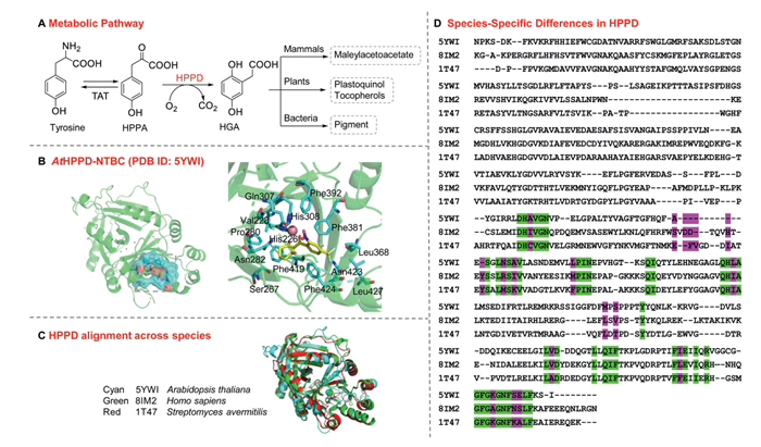
Understanding the structure of HPPD is critical for designing targeted enzyme inhibitors. For example, in the NTBC-AtHPPD complex structure (PDB ID: 5YWI, Fig. 5B), at the active site, the two carbonyl groups of NTBC chelate with the metal ion (Co2+), while His206, His308, Glu394, and a water molecule also participate in the metal coordination, forming a six-coordinate structure for the metal ion. Additionally, the cyclohexanedione structure of NTBC is surrounded by Val228, Ser267, Pro280, Asn282, Gln307, and Phe419. The phenyl group forms notable π-π stacking interactions with Phe381 and Phe424, the nitro group is adjacent to Gln307, His308, and Phe392, while the trifluoromethyl group interacts with a hydrophobic region composed of Leu368, Phe381, Asn423, Phe424, and Leu427. The binding modes of other HPPD inhibitors are highly consistent with that of NTBC: The triketone moiety forms a bidentate chelation with the metal ion in the active site, the aromatic ring engages in π-π stacking interactions with a phenylalanine residue, and the substituents establish hydrophobic interactions with nonpolar amino acids lining the interior wall of the cavity pocket.
The structural and biological comparison of HPPD from different species is crucial for developing highly selective inhibitors targeting HPPD. By comparing the amino acid sequence differences of HPPD structures from representative sources such as AtHPPD (PDB ID: 5YWI), Homo sapiens HPPD (hHPPD, PDB ID: 8IM2), and Streptomyces avermitilis HPPD (SaHPPD, PDB ID: 1T47), and superimposing these structures with 5YWI, the RMSD values between 8IM2 and 1T47 were found to be 0.935 and 0.910 Å, respectively (Fig. 5C). The amino acids within 8 Å (Fig. 5D) of the NTBC ligand in the complexes were defined as the target for study, totaling 54 amino acids. Identical amino acid residues in the HPPD active sites across species are marked in green. In contrast, sites where amino acid residues differ but the position remains the same are marked in magenta, with 18 such sites identified. Further analysis revealed that within a 6 Å range around the NTBC ligand, there were 3–5 amino acid differences, while within a 4 Å range, the amino acids were identical (Fig. S2 in Supporting information). This indicates a certain degree of conservation of amino acids in the HPPD active site across species, with the inhibitor binding site being relatively conserved. This explains why HPPD inhibitors, such as NTBC, can exhibit herbicidal, insecticidal, and fungicidal activities, in addition to their therapeutic use in treating human tyrosine metabolism-related diseases. Furthermore, the differences in the inhibitory activities of HPPD inhibitors across species are also influenced by their pharmacokinetic properties.
Early studies on the toxicity of triketone compounds indicated that while nitisinone lacks acute toxicity, it induces ocular toxicity upon repeated exposure. This effect is linked to tyrosine accumulation caused by the inhibition of HPPD, with species-specific variations observed. For instance, male rats treated with HPPD inhibitors exhibited plasma tyrosine levels as high as ~3 mmol/L, whereas females showed approximately half the concentration. Notably, intraocular tyrosine levels in rats exceeded those in plasma. Subchronic administration of nitisinone at doses as low as 1 mg kg-1 d-1 for 6 weeks resulted in reversible corneal lesions in both rats and beagles. In contrast, mice treated with HPPD inhibitors showed peak plasma tyrosine concentrations of ~0.8 mmol/L, and no corneal damage was observed in mice, rabbits, or rhesus macaques even after 90 days of dosing at 10 mg kg-1 d-1. These corneal pathologies resembled those observed in rats fed diets enriched in tyrosine. Adverse effects were absent when plasma tyrosine levels remained below 0.5 mmol/L, suggesting that ocular lesions induced by triketones are not due to intrinsic compound toxicity but rather the result of excessive systemic tyrosine [132]. Potential mechanisms for this include intracellular tyrosine crystal formation or redox imbalance due to tyrosyl radical generation, although alternative pathways cannot be excluded. Clinical reports have also documented reduced cognitive function and lower intelligence quotient scores in children undergoing nitisinone therapy, alongside learning and behavioural deficits. This phenomenon may arise from tyrosine crossing the blood-brain barrier via concentration gradients, leading to cerebral tyrosine accumulation rather than direct drug effects. Tyrosine aminotransferase (TAT) activity is crucial in regulating plasma tyrosine levels. Triketones inhibit HPPD, causing the accumulation of HPPA, which is a noncompetitive inhibitor of TAT. This suppression of TAT activity exacerbates tyrosine accumulation. Species-specific differences in baseline TAT activity, such as higher TAT activity in mice, likely explain their resistance to ocular lesions [130].
Pharmacokinetic disparities further explain interspecies differences. In healthy males, mesotrione (4 mg/kg) exhibited a half-life of approximately 1 h and was rapidly excreted unchanged in urine. Under controlled dietary conditions, plasma tyrosine levels increased from 0.036 mmol/L to 0.105 mmol/L, peaking at 0.3 mmol/L, and returning to baseline within 48 h. In contrast, nitisinone induced rapid HPPD inhibition with slow dissociation kinetics (half-life: ~53 h), elevating plasma tyrosine from 0.076 mmol/L (on an ad libitumdiet) to 1.1 mmol/L over 120 h, with normalization requiring up to two months [133,134]. Both TAT activity and drug elimination half-life are crucial factors influencing tyrosine-associated toxicities in humans.
The differential toxicity profiles, dynamics of tyrosine accumulation, and pharmacokinetic behaviors of triketones directly inform their applications. Nitisinone's prolonged HPPD inhibition and significant tyrosine elevation support its therapeutic use in hereditary tyrosinemia type Ⅰ (T1T) and alkaptonuria [134,135]. In contrast, mesotrione's rapid metabolism, minimal tyrosine accumulation, and low mammalian toxicity make it suitable for widespread agricultural use as a herbicide.
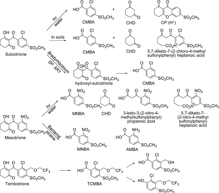
Triketone herbicides have been widely adopted due to their high herbicidal activity; however, they exhibit significant differences in toxicity, metabolism, and environmental behavior. Among them, sulcotrione, mesotrione, and tembotrione are currently the most thoroughly studied, particularly with respect to their degradation and transformation under soil conditions, aquatic environments, and photolytic processes. In neutral water, sulcotrione primarily undergoes photolysis to form two main products: 2-Chloro-4-mesylbenzoic acid (CMBA) and 1,3-cyclohexanedione (CHD). In acidic water, it mainly degrades to 1,9-dione-3,4-dihydro-6-methylsulfonyl-xanthene (CP). Microbial degradation in soil predominantly yields CMBA and CHD, with the additional presence of 5,7-diketo-7-(2-chloro-4-methylsulfonylphenyl)heptanoic acid in acidic soils (Fig. 6) [136]. Similar degradation pathways have been observed in Bradyrhizobium sp. SR1, which produces CMBA without generating CHD [137]. Mesotrione exhibits a photolysis half-life (DT50) of 89–94 days in water and 23.9 days in soil, which is significantly longer than its rapid degradation on crop surfaces (DT50 = 2 h). Photolysis primarily yields four products, indicating that photolysis is a rapid dissipation process on crops. Microbial degradation is the primary pathway in soil, with strains such as Bacillus species and Bradyrhizobium sp. SR1, E.coli, and Pantoea species can degrade mesotrione. These bacteria convert mesotrione through two metabolic routes: (Ⅰ) Nitroreduction to form AMBA; (Ⅱ) oxidation leading to diketone cleavage, producing 4-methylsulfonyl-2-nitrobenzoic acid (MNBA) [138]. Subsequent studies have elucidated the molecular mechanisms of mesotrione metabolism by Bacillus sp., identifying NfrA1 and NfrA2/YcnD as key enzymes in the degradation process [139]. Tembotrione's hydrolysis and photolysis rates increase with rising pH, and its degradation rate in soil positively correlates with soil organic matter content. However, no strains capable of degrading tembotrione have been identified. Under aerobic conditions in soil, it primarily metabolizes to TCMBA, along with two further metabolites [140]. Most degradation products of these herbicides exhibit greater stability in soil and sediments than their parent compounds, and sunlight can accelerate the degradation process of triketones and their metabolites.
Regarding toxicity, the triketone herbicide family has not been definitively linked to significant environmental or human health risks. Most of the nine commercialized HPPD inhibitors show low or minimal toxicity based on terrestrial ecotoxicology, aquatic ecotoxicology, and health-related information reported during pesticide registration (Table S1 in Supporting information, PPDB database). However, certain compounds exhibit toxicity to specific organisms [1]. Mesotrione generally displays favorable toxicological and environmental characteristics, and under proper usage conditions, poses low risk to humans and non-target organisms. Nevertheless, some studies report moderate toxicity of mesotrione to earthworms (LC50 > 437 mg/kg), and low concentrations can induce DNA damage in fish [137,141]. Notably, mesotrione metabolites such as MNBA and AMBA are more toxic than the parent compound. Similarly, sulcotrione itself is generally safe, but among its photodegradation products, CP exhibits particularly high toxicity, causing 7–9 times greater harm to organisms than the parent compound or its major metabolites [142]. Benzobicyclon shows high acute toxicity to aquatic organisms, with a 96 h LC50 of 0.489 mg/L for rainbow trout and a 48 h LC50 of 0.368 mg/L for Daphnia magna. Existing ecotoxicological evidence indicates that the observed acute toxicity of benzobicyclon to aquatic organisms is primarily due to the parent compound rather than its transformation products. Tembotrione is moderately to highly toxic to algae and Daphnia in aquatic ecosystems and also exhibits moderate toxicity to earthworms. Due to its high solubility and persistent residues, tembotrione may contribute to water contamination through runoff or leaching, posing potential food safety concerns [140,143]. Fenquinotrione also shows moderate toxicity to algae. Developmental toxicity studies in rabbits with bicyclopyrone demonstrated structural abnormalities (e.g., extra ribs, craniofacial and cardiac defects) at doses as low as 10 mg kg-1 d-1, indicating teratogenic effects in this species. In contrast, no teratogenic effects were observed in rat developmental studies. Benquitrione exhibits an excellent toxicological profile, showing very low toxicity to non-target organisms. It causes no significant eye or skin irritation, does not induce skin sensitization, and long-term studies have provided no evidence of carcinogenicity, teratogenicity, or reproductive toxicity [144]. Overall, these triketone HPPD inhibitors generally pose low acute risk to mammals and non-target aquatic animals. However, their effects on algae and the potential toxicity of their metabolites should be carefully considered in environmental risk assessments, as the sensitivity of algae may be related to the inhibition of HPPD and the consequent disruption of photosynthesis.
Triketone HPPD inhibitors are primarily metabolized by cytochrome P450 enzymes in plants. For instance, resistant weeds such as Amaranthus palmeri, Conyza canadensis, and Setaria viridis accelerate the hydroxylation of mesotrione by upregulating P450 enzymes, including the CYP71A and CYP72A families, which produce less active or inactive metabolites, such as 4-OH-mesotrione and 5-OH-mesotrione [54–56,145]. Moreover, in Amaranthus palmeri-resistant populations, the increased expression of the HPPD gene suggests that resistance arises from a combination of metabolic detoxification and overexpression of the target enzyme. Similarly, crops like maize rapidly hydroxylate mesotrione into inactive products via the cytochrome P450 enzyme CYP71C3v2. Other metabolic enzymes, such as the glycosyltransferase MdUGT91AJ2 in apples, also participate in detoxification by glycosylating sulcotrione [146]. Additionally, in rice, the triketone dioxygenase (TDO) enzyme can metabolize sulcotrione into less active or inactive forms such as 5-OH-mesotrione and oxy-mesotrione [147]. Overall, weed resistance mainly results from enhanced metabolic processes rather than mutations in the target gene. In contrast, the metabolic differences between crops and susceptible weeds contribute to the selective herbicidal activity.
This review introduces an up-and-coming class of chemical scaffolds and triketone compounds with broad applications spanning agriculture, medicine, and materials science. Among these, the study of HPPD inhibitors, particularly those derived from plants, is the most prominent. These inhibitors are utilized as bleaching herbicides in agrochemicals, with commercial products such as sulcotrione and mesotrione demonstrating high herbicidal efficacy and strong environmental adaptability. However, the increasing prevalence of herbicide resistance due to cytochrome P450-mediated metabolic detoxification in weeds underscores the need to develop next-generation triketone herbicides that can overcome enzymatic degradation pathways. Additionally, the metabolism of triketone compounds in different environments should be considered to prevent environmental pollution, including bacterial metabolism in soil, photodegradation in aquatic environments, and decomposition on plant surfaces.
The binding sites of HPPD from different species exhibit a high degree of similarity in their protein sequences and binding modes with the ligand, which allows triketone compounds to effectively inhibit HPPD in other species. This makes them potential therapeutic agents for treating conditions such as those caused by blood-feeding insects, various bacterial infections, and human diseases like tyrosinemia type Ⅰ, alkaptonuria, and possibly hawkinsinuria. Notably, the structural flexibility of triketones allows for chemical modifications that enhance their potency and selectivity. However, due to certain toxicity issues, particularly ocular toxicity caused by tyrosine accumulation when treating human diseases, more in-depth pharmacokinetic studies and optimized drug designs are necessary.
Triketone compounds that do not function as HPPD inhibitors also exhibit significant bioactivity, including antibacterial, anticancer, antiviral, and anti-inflammatory properties, paving the way for new avenues in drug discovery. However, the mechanisms of action have only been elucidated for a subset of these compounds, leaving many pathways yet to be explored. In materials science, integrating triketones into polymer chemistry has led to the development of new strategies for dynamic covalent networks and the development of sustainable materials. These compounds enable the formation of recyclable and processable polymers, aligning with global trends promoting green chemistry and circular economy principles. Future research into triketone-based polymer systems is expected to drive advancements in high-performance recyclable materials.
Further exploration of structure-activity relationships, mechanisms of action, metabolic pathways, and biocompatibility will be essential for maximizing the potential of triketones. Interdisciplinary collaborations involving computational modeling, synthetic chemistry, and chemical biology are expected to accelerate the development of multifunctional triketones. As research continues to uncover the potential of these compounds, they are poised to make sustainable contributions and enhance global capabilities in addressing agricultural and health-related challenges.
The authors have declared no conflict of interest
Lijun Chen: Writing – review & editing, Writing – original draft, Visualization. Biao Li: Visualization, Investigation. Dawei Wang: Writing – review & editing, Funding acquisition, Conceptualization. Guangfu Yang: Conceptualization.
This work was supported by the National Key Research and Development Program of China (No. 2024YFE0214300), self-determined research funds of CCNU from the College Basic Research and Operation of MOE (Nos. CCNU24JCPT023, CCNU24JCPT029).
Supplementary material associated with this article can be found, in the online version, at doi:
F. Ndikuryayo, B. Moosavi, W.C. Yang, G.F. Yang, J. Agric. Food Chem. 65 (2017) 8523–8537. doi: 10.1021/acs.jafc.7b03851
T. Ma, S. Gao, L.X. Zhao, F. Ye, Y. Fu, J. Agric. Food Chem. 72 (2024) 17125–17137. doi: 10.1021/acs.jafc.4c01171
S.E. McComic, S.O. Duke, E.R. Burgess, D.R. Swale, Pestic. Biochem. Physiol. 194 (2023) 105532. doi: 10.1016/j.pestbp.2023.105532
N.A. Nammunige, K.A. Agnew-Francis, D.D. Fernando, et al., Phytomedicine 136 (2025) 156321. doi: 10.1016/j.phymed.2024.156321
L.A. Lapshina, A.V. Reunov, V.P. Nagorskaya, et al., Appl. Biochem. Microbiol. 49 (2013) 59–63. doi: 10.1134/S0003683813010110
S.A. Obeng-Darko, J. Sloan, R.M. Binks, et al., J. Agric. Food Chem. 71 (2023) 7703–7709. doi: 10.1021/acs.jafc.3c00673
O.I. Yarovaya, A.S. Filimonov, D.S. Baev, et al., Viruses 16 (2024) 215. doi: 10.3390/v16020215
S.A. Abdel-Rahman, N.S. El-Gohary, E.R. El-Bendary, et al., Eur. J. Med. Chem. 140 (2017) 200–211. doi: 10.1016/j.ejmech.2017.08.066
M.E.C. Moreira, D.I.G. Natal, R.C.L. Toledo, et al., J. Funct. Foods 29 (2017) 143–153. doi: 10.1016/j.jff.2016.11.001
Y. Zhang, F. Liu, Y. Cao, et al., Food Chem. 424 (2023) 136367. doi: 10.1016/j.foodchem.2023.136367
C.T. Lin, J.H. Shih, C.L. Chen, D.Y. Yang, Tetrahedron Lett. 46 (2005) 5033–5037. doi: 10.1016/j.tetlet.2005.05.070
P.R. Christensen, A.M. Scheuermann, K.E. Loeffler, B.A. Helms, Nat. Chem. 11 (2019) 442–448. doi: 10.1038/s41557-019-0249-2
A.J. Jhala, V. Kumar, R. Yadav, et al., Weed Technol. 37 (2023) 1–14. doi: 10.1017/wet.2022.79
H.Y. Lin, J. Dong, J.Q. Dong, W.C. Yang, G.F. Yang, Trends. Biochem. Sci. 48 (2023) 568–584. doi: 10.1016/j.tibs.2023.02.006
G.R. Moran, Arch. Biochem. Biophys. 433 (2005) 117–128. doi: 10.1016/j.abb.2004.08.015
L.J. Chen, G.Y. Huang, J. Dong, et al., Adv. Agrochem. 2 (2023) 163–172. doi: 10.1016/j.aac.2023.05.001
E. Rocaboy-Faquet, L. Barthelmebs, C.C. Blanchard, T. Noguer, Talanta 146 (2016) 510–516. doi: 10.1016/j.talanta.2015.09.030
H.Y. Lin, J.F. Yang, D.W. Wang, et al., FEBS. J. 286 (2019) 975–990. doi: 10.1111/febs.14747
J. Dong, J.Q. Dong, X.H. Yu, et al., Adv. Agrochem. 1 (2022) 174–181. doi: 10.1016/j.aac.2022.10.002
T.L. Yang, J. Dong, X.L. Wang, et al., Adv. Agrochem. 3 (2024) 174–181.
F.E. Dayan, J. Howell, J.P. Marais, D. Ferreira, M. Koivunen, Weed. Sci. 59 (2011) 464–469. doi: 10.1614/WS-D-11-00043.1
E.Y. Jeong, M.G. Kim, H.S. Lee, Pest. Manage Sci. 65 (2008) 327–331.
L. Frabboni, A. Tarantino, F. Petruzzi, G. Disciglio, Agronomy 9 (2019) 475. doi: 10.3390/agronomy9090475
P. Kanatas, S. Zavra, A. Tataridas, et al., Agronomy 12 (2022) 1755. doi: 10.3390/agronomy12081755
T.C. Barickman, C.L. Cantrell, A. Reichley, ACS. Agric. Sci. Technol. 4 (2024) 907–915. doi: 10.1021/acsagscitech.4c00225
R. Beaudegnies, A.J.F. Edmunds, T.E.M. Fraser, et al., Bioorg. Med. Chem. 17 (2009) 4134–4152. doi: 10.1016/j.bmc.2009.03.015
S.E. Hager, R.I. Gregerman, W.E. Knox, J. Biol. Chem. 225 (1957) 935–947. doi: 10.1016/S0021-9258(18)64891-4
A. Schulz, O. Ort, P. Beyer, FEBS Lett. 318 (1993) 162–163. doi: 10.1016/0014-5793(93)80013-K
I.C. Barta, P. Böger, Pestic. Sci. 45 (1995) 286–287. doi: 10.1002/ps.2780450315
D.L. Lee, M.P. Prisbylla, T.H. Cromartie, et al., Weed. Sci. 45 (1997) 601–606. doi: 10.3348/jkrs.1997.36.4.601
A.T. Halle, J. Wiszniowski, A. Hitmi, et al., Pest. Manage Sci. 65 (2009) 14–18. doi: 10.1002/ps.1637
G. Mitchell, D.W. Bartlett, T.E.M. Fraser, et al., Pest. Manage Sci. 57 (2001) 120–128. doi: 10.1002/1526-4998(200102)57:2<120::AID-PS254>3.0.CO;2-E
A.V. Almsick, J. Benet-Buchholz, B. Olenik, L. Willms, Bayer CropSci. J. 62 (2009) 5–16.
H. Erwin, R.C. Hugh, U. Chieko, B.P. Georg, Z. Frank, Pantent, WO2010136165A2, 2010.
N. Umetsu, Y. Shirai, J. Pestic. Sci. 45 (2020) 54–74. doi: 10.1584/jpestics.d20-201
A.A. Van, L. Willms, T. Auler, H. Bieringer, et al., Patent, WO0218352, 2022.
T. Tamai, K. Tsukuda, S. Yamada, S. Kajita, Y. Miyashita, Patent, WO2009116290, 2009.
A.J.F. Edmunds, Chimia 76 (2022) 641–646. doi: 10.2533/chimia.2022.641
J. Dallimore, Patent, WO2012136703, 2012.
C. Chen, W. Geng, K. Li, et al., Chin. Chem. Lett. 36 (2025) 110902. doi: 10.1016/j.cclet.2025.110902
D.M. Barber, J. Agric. Food Chem. 70 (2022) 11075–11090. doi: 10.1021/acs.jafc.1c07910
H. Walter, P. Plath, M. Witschel, et al., Patent, US6083880, 2000.
I.M. Fritze, L. Linden, J. Freigang, et al., Plant Physiol. 134 (2004) 1388–1400. doi: 10.1104/pp.103.034082
A. Nagamatsu, K. Ueda, R. Tamai, S. Tani, S. Yamamoto, J. Pestic. Sci. 47 (2022) 139–145. doi: 10.1584/jpestics.j22-02
D.W. Wang, H.Y. Lin, R.J. Cao, et al., J. Agric. Food Chem. 62 (2014) 11786–11796. doi: 10.1021/jf5048089
W. Hu, S. Gao, L. Zhao, et al., Pest. Manage Sci. 78 (2022) 938–946. doi: 10.1002/ps.6703
B.F. Zheng, Y.C. Yan, C. Fu, et al., Front. Agr. Sci. Eng. 9 (2022) 133–145. doi: 10.15302/j-fase-2021401
L.X. Zhao, W. Hu, Z.B. Jiang, et al., J. Agric. Food Chem. 71 (2023) 17678–17688. doi: 10.1021/acs.jafc.3c04651
Y.C. Yan, W. Wu, G.Y. Huang, et al., J. Agric. Food Chem. 70 (2022) 6644–6657. doi: 10.1021/acs.jafc.2c01507
G.F. Yang, Y.C. Yan, R.Y. Qu, et al., Patent, CN115960049, 2023.
L.J. Chen, R.N. Ying, X.Q. Wang, et al., J. Agric. Food Chem. 73 (2025) 1112–1121. doi: 10.1021/acs.jafc.4c08544
Y.Q. Zhu, X.M. Zou, F.Z. Hu, et al., J. Agric. Food Chem. 53 (2005) 9566–9570. doi: 10.1021/jf051510l
Y. Fu, D. Zhang, S.Q. Zhang, et al., J. Agric. Food Chem. 67 (2019) 11839–11847. doi: 10.1021/acs.jafc.9b01412
H. Maeda, K. Murata, N. Sakuma, et al., Science 1979 365 (2019) 393–396. doi: 10.1126/science.aax0379
J.C.T. Concepcion, S.S. Kaundun, J.A. Morris, et al., J. Agric. Food Chem. 72 (2024) 5595–5608. doi: 10.1021/acs.jafc.3c06903
Y. Lan, Y. Cao, Y. Sun, R. Wang, Z. Huang, Agronomy 14 (2024) 2399. doi: 10.3390/agronomy14102399
X.H. Xu, B. Liu, L.G. Xie, Patent, CN102146068, 2011.
D. Bošković, S. Vuković, S. Lazić, Plants 12 (2023) 3727. doi: 10.3390/plants12213727
M.A.V. Ramirez, M. Sterkel, A.J. Martins, J. Bp Lima, P.L. Oliveira, Pest Manage. Sci. 78 (2021) 692–702.
M. Sterkel, H.D. Perdomo, M.G. Guizzo, et al., Curr. Biol. 26 (2016) 2188–2193. doi: 10.1016/j.cub.2016.06.025
M. Sterkel, L.R. Haines, A. Casas-Sánchez, PLoS. Biol. 19 (2021) e3000796. doi: 10.1371/journal.pbio.3000796
A.A. Costa, C.V. Naspi, A. Lucia, H.M. Masuh, J. Med. Entomol. 54 (2017) 670–676. doi: 10.1093/jme/tjw222
C. Huseyin, T.C. Ozge, O.T. Ayse, et al., Parasitol. Res. 102 (2008) 1277–1279. doi: 10.1007/s00436-008-0905-8
S. Koc, O. Tufan-Cetin, M. Candan, A. Turk, H. Cetin, Fresenius Environ. Bull. 11 (2021) 11938–11941.
E.A. Ferreira, J.B. Reigada, M.V. Correia, et al., J. Nat. Prod. 77 (2014) 1377–1382. doi: 10.1021/np500130x
N.R. Gundoju, R. Bokam, N.R. Yalavarthi, et al., J. Asian Nat. Prod. Res. 21 (2019) 262–269. doi: 10.1080/10286020.2018.1460362
X.H. Yu, J. Dong, C.P. Fan, et al., J. Agric. Food Chem. 71 (2023) 19396–19407. doi: 10.1021/acs.jafc.3c05260
D.P. Killeen, L. Larsen, F.E. Dayan, et al., J. Nat. Prod. 79 (2016) 564–569. doi: 10.1021/acs.jnatprod.5b00968
E.Y. Jeong, M.J. Lee, H.S. Lee, Food Sci. Biotechnol. 27 (2018) 1541–1547. doi: 10.1007/s10068-018-0391-4
J.W. Wu, B.L. Li, C. Tang, et al., J. Nat. Prod. 82 (2019) 1917–1922. doi: 10.1021/acs.jnatprod.9b00064
L. Lozano, C. Ramírez, J.M. Lozano, et al., Bol. Latinoam. Caribe Plant. Med. Aromat. 21 (2022) 309–322.
W. Zhang, Y. Lu, M. Ma, et al., Front. Vet. Sci. 11 (2024) 1362292. doi: 10.3389/fvets.2024.1362292
W.Z. Nie, Q.K. Shen, Z.S. Quan, H.Y. Guo, Y.M. Li, Mini-rev. Med. Chem. 24 (2024) 1368–1384. doi: 10.2174/0113895575277085231123165546
G. Cravotto, S. Tagliapietra, R. Cappello, et al., Arch. Pharm. 339 (2006) 129–132. doi: 10.1002/ardp.200500127
K.S. Babu, X.C. Li, M.R. Jacob, et al., J. Med. Chem. 49 (2006) 7877–7886. doi: 10.1021/jm061123i
O.P. Shestak, V.L. Novikov, E.A. Martyyas, M.M. Anisimov, Pharm. Chem. J. 43 (2009) 16–19. doi: 10.1007/s11094-009-0237-8
H. Khan, D.A. Sabbah, M. Zafar, M.S. Mubarak, Life Sci. 209 (2018) 332–340. doi: 10.1016/j.lfs.2018.07.059
X. Wen, M.S. Nobakht, Y. Yang, et al., Ann. Clin. Microbiol. Antimicrob. 22 (2023) 83. doi: 10.1186/s12941-023-00628-5
W. Qiao, L. Wang, Y. Luo, T. Yang, Eur. J. Med. Chem. 285 (2025) 117267. doi: 10.1016/j.ejmech.2025.117267
J. Reichling, C. Koch, E. Stahl-Biskup, C. Sojka, P. Schnitzler, Planta Med. 71 (2005) 1123–1127. doi: 10.1055/s-2005-873175
V.P. Zambare, L.P. Christopher, Pharm. Biol. 50 (2012) 778–798. doi: 10.3109/13880209.2011.633089
D.N. Sokolov, V.V. Zarubaev, A.A. Shtro, et al., Bioorg. Med. Chem. Lett. 22 (2012) 7060–7064. doi: 10.1016/j.bmcl.2012.09.084
A. Gupta, N. Sahu, A.P. Singh, et al., Appl. Biochem. Biotechnol. 194 (2022) 6386–6406. doi: 10.1007/s12010-022-04103-3
O.I. Yarovaya, A.S. Filimonov, D.S. Baev, New. J. Chem. 47 (2023) 19865–19879. doi: 10.1039/d3nj03598k
Y.H. Jin, S. Jeon, J. Lee, et al., Pharmaceutics 14 (2022) 376. doi: 10.3390/pharmaceutics14020376
Y.H. Lee, Y. Wang, J.Y. Kim, et al., J. Mol. Struct. 1293 (2023) 136243. doi: 10.1016/j.molstruc.2023.136243
E. Tyrrell, R. Archer, M. Tucknott, et al., Phytochem. Lett. 5 (2012) 144–149. doi: 10.1016/j.phytol.2011.11.011
N. Tanaka, Y. Takaishi, Y. Shikishima, et al., J. Nat. Prod. 67 (2004) 1870–1875. doi: 10.1021/np040024+
L. Sales, J.A. Pezuk, K.S. Borges, et al., BMC Complem. Altern. Med. 15 (2015) 393. doi: 10.1186/s12906-015-0911-1
J.P. Dzoyem, A.M. Lannang, H. Fouotsa, et al., Phytochem. Lett. 14 (2015) 153–158. doi: 10.1016/j.phytol.2015.10.003
N. Li, J.L. Wu, T. Hasegawa, et al., J. Nat. Prod. 70 (2007) 998–1001. doi: 10.1021/np070089n
C. Denny, M.E. Zacharias, A.L. Ruiz, et al., PhytOther Res. 22 (2008) 127–130. doi: 10.1002/ptr.2251
M. Backorova, M. Backor, J. Mikes, R. Jendzelovsky, P. Fedorocko, Toxicol. Vitr. 25 (2011) 37–44. doi: 10.1016/j.tiv.2010.09.004
X. Geng, X. Zhang, B. Zhou, et al., Med. Sci. Monit. 24 (2018) 556–566. doi: 10.12659/MSM.908568
M. Grudzińska, P. Paśko, D. Wróbel-Biedrawa, I. Podolak, A. Galanty, Chem. Biodivers. 19 (2022) e202200408. doi: 10.1002/cbdv.202200408
Y. Song, F. Dai, D. Zhai, et al., Angiogenesis 15 (2012) 421–432. doi: 10.1007/s10456-012-9270-4
K. Petrová, M. Bačkorová, Z. Demčišáková, et al., Life 12 (2022) 1444. doi: 10.3390/life12091444
T.X. Sun, M.Y. Li, Z.H. Zhang, et al., PhytOther Res. 35 (2021) 3916–3935. doi: 10.1002/ptr.7103
M. Varlı, S.R. Bhosle, E. Kim, et al., JACS. Au 4 (2024) 1521–1537. doi: 10.1021/jacsau.3c00774
M. Mariraj, M. Gundappa, S. Velayuthaprabhu, et al., N-S Arch. Pharmacol. 398 (2025) 5101–5117. doi: 10.1007/s00210-024-03584-9
T. Azhamuthu, S. Kathiresan, I. Senkuttuvan, et al., Cell Biochem. Funct. 42 (2024) e4074. doi: 10.1002/cbf.4074
D.J. Wu, M. Li, Y.N. Hong, et al., J. Adv. Res. 74 (2025) 621–635. doi: 10.1016/j.jare.2024.10.003
A.L. Zakharenko, N.S. Dyrkheeva, O.A. Luzina, et al., Genes 14 (2023) 1931. doi: 10.3390/genes14101931
M. Roney, Comput. Biol. Chem. 115 (2025) 108303. doi: 10.1016/j.compbiolchem.2024.108303
K. Nakagawa-Goto, J.H. Wu, K.F. Bastow, C.C. Wu, K.H. Lee, Bioorg. Med. Chem. 13 (2005) 2325–2330. doi: 10.1016/j.bmc.2004.12.040
L. Lin, Q. Shi, A.K. Nyarko, et al., J. Med. Chem. 49 (2006) 3963–3972. doi: 10.1021/jm051043z
K. Wada, J.Y. Lee, H.Y. Hung, et al., Bioorg. Med. Chem. 23 (2015) 1507–1514. doi: 10.1016/j.bmc.2015.02.003
N. Tanaka, T. Kubota, H. Ishiyama, et al., Bioorg. Med. Chem. 16 (2008) 5619–5623. doi: 10.1016/j.bmc.2008.03.076
J.S. Bland, D. Minich, R. Lerman, et al., PharmaNutrition 3 (2015) 46–52. doi: 10.1016/j.phanu.2015.03.001
S.M. Lee, M.K. Na, R.B. An, H.K. L B.S. Min, Biol. Pharm. Bull. 26 (2003) 1354–1356. doi: 10.1248/bpb.26.1354
M. Tagashira, M. Watanabe, N. Uemitsu, Biosci. Biotechnol. Biochem. 59 (1995) 740–742. doi: 10.1271/bbb.59.740
Y.M. Liu, N. Lu, J. Tang, J. Mol. Struct. 1175 (2019) 721–727. doi: 10.1016/j.molstruc.2018.08.026
P.S. Arwa, M.L. Zeraik, V.F. Ximenes, et al., J. Ethnopharmacol 174 (2015) 410–418. doi: 10.1016/j.jep.2015.08.041
S.A. Figueiredo, F.M. Vilela, C.A. da Silva, et al., J. Photochem. Photobiol. B 131 (2014) 65–73. doi: 10.1016/j.jphotobiol.2014.01.004
C. Fernández-Moriano, P.K. Divakar, A. Crespo, M.P. Gómez-Serranillos, Food Chem. Toxicol. 105 (2017) 262–277. doi: 10.1016/j.fct.2017.04.030
S. Erfani, T. Valadbeigi, N. Aboutaleb, et al., Iran. J. Basic Med. Sci. 23 (2020) 1225–1231.
S.P. Kwong, H.X. Wang, L. Shi, et al., J. Photochem. Photobiol. B 205 (2020) 111814. doi: 10.1016/j.jphotobiol.2020.111814
A. Schlune, E. Thimm, D. Herebian, U. Spiekerkoetter, J. Inherit. Metab. Dis. 35 (2012) 831-336. doi: 10.1007/s10545-012-9450-9
M. Laschi, G. Bernardini, E. Dreassi, et al., ChemMedChem. 11 (2016) 674–678. doi: 10.1002/cmdc.201500578
J.M. Brownlee, B. Heinz, J. Bates, G.R. Moran, Biochemistry 49 (2010) 7218–7226. doi: 10.1021/bi1008112
L.R. Ranganath, E.E. Psarelli, J.B. Arnoux, et al., Lancet Diabetes. Endocrinol. 8 (2020) 762–772. doi: 10.1016/S2213-8587(20)30228-X
D.R. Adams, S. Menezes, R. Jauregui, et al., JCI. Insight. 4 (2019) 124387. doi: 10.1172/jci.insight.124387
T.H. Kim, D.R. Oh, H.S. Na, H.C. Lee, Arch. Pharm. Res. 26 (2003) 192–196. doi: 10.1007/BF02976828
A. Scala, A. Rescifina, N. Micale, et al., Chem. Biol. Drug Des. 91 (2017) 597–604.
Y.S. Chen, P.Y. Kuo, T.L. Shie, D.Y. Yang, Tetrahedron 62 (2006) 9410–9416. doi: 10.1016/j.tet.2006.07.053
S.L. Lin, P.Y. Kuo, D.Y. Yang, Molecules 12 (2007) 1316–1324. doi: 10.3390/12071316
E.A. Dailing, P. Khanal, A.R. Epstein, et al., ACS. Cent. Sci. 10 (2024) 54–64. doi: 10.1021/acscentsci.3c01096
A.R. Epstein, J. Demarteau, B.A. Helms, K.A. Persson, J. Am. Chem. Soc. 145 (2023) 8082–8089. doi: 10.1021/jacs.3c00772
L. Trachsel, K.A. Stewart, D. Konar, et al., J. Am. Chem. Soc. 146 (2024) 16257–16267. doi: 10.1021/jacs.4c04664
A. Santucci, G. Bernardini, D. Braconi, E. Petricci, F. Manetti, J. Med. Chem. 60 (2017) 4101–4125. doi: 10.1021/acs.jmedchem.6b01395
E. Rocaboy-Faquet, T. Noguer, S. Romdhane, C. Bertrand, et al., Appl. Microbiol. Biotechnol. 98 (2014) 7243–7252. doi: 10.1007/s00253-014-5793-5
R.W. Lewis, J.W. Botham, Crit. Rev. Toxicol. 43 (2013) 185–199. doi: 10.3109/10408444.2013.764279
M.G. Hall, M.F. Wilks, W.M. Provan, S. Eksborg, B. Lumholtz, Brit. J. Clinical. Pharma. 52 (2001) 169–177. doi: 10.1046/j.0306-5251.2001.01421.x
E.A. Lock, Metabolites 12 (2022) 902. doi: 10.3390/metabo12100902
A.S. Davison, A.M. Milan, J.A. Gallagher, L.R. Ranganath, J. Inherited Metab. Dis. 39 (2016) 203–210. doi: 10.1007/s10545-015-9902-0
J. Wiszniowskia, A.T. Hallea, C. Richarda, A. Hitmic, G. Ledoigt, Chemosphere 74 (2009) 1224–1230. doi: 10.1016/j.chemosphere.2008.11.017
H. Barchanska, A. Kluza, K. Krajczewska, J. Maj, J. Soils. Sediments 16 (2016) 125–133. doi: 10.1007/s11368-015-1188-1
L. Carles, M. Joly, P. Joly, Clean-Soil Air Water 45 (2017) 1700011. doi: 10.1002/clen.201700011
L. Carles, P. Besse-Hoggan, M. Joly, et al., Biochem. J. 473 (2016) 1443–1453. doi: 10.1042/BJ20151366
X. Dong, Z. Chen, Y. Chu, et al., Environ. Sci. Pollut. Res. 30 (2023) 72389–72397. doi: 10.1007/s11356-023-27501-4
L.D.S. Piancini, I.C. Guiloski, H.C. Silva de Assis, M.M. Cestari, Toxicol. Rep. 2 (2015) 1157–1163. doi: 10.1016/j.toxrep.2015.08.007
E. Dumas, M. Giraudo, E. Goujon, et al., J. Hazard. Mater. 325 (2017) 136–156. doi: 10.1016/j.jhazmat.2016.11.059
M. Velki, S. Ecimovic, Environ. Toxicol. Pharmacol. 40 (2015) 774–784. doi: 10.1016/j.etap.2015.09.009
H.Y. Lin, X. Chen, J.N. Chen, et al., Research 2019 (2019) 2602414.
J. Wu, X. Wen, X.L. Wang, Adv. Agrochem. 5 (2026) 41–48. doi: 10.1016/j.aac.2025.05.005
A. Zhao, X. Teng, Y. Ma, et al., Plants 13 (2024) 1796. doi: 10.3390/plants13131796
S.M.G. Duff, M. Zhang, F. Zinnel, et al., BBA-Gen. Subjects 1868 (2024) 130504. doi: 10.1016/j.bbagen.2023.130504

Figure 3 Applications of triketones in the field of medicine. Some compounds are represented in their enol forms in the referenced literature; however, for consistency with the notation used in this manuscript, they are depicted in their triketone forms in the figure.

Figure 4 Acidichromic switching of triketone colorants and their application in catalyst-free post-polymerization modification.

Figure 5 Molecular basis of catalytic function and species-specific differences in HPPD. (A) Catalytic reaction pathways of HPPD in different organisms. (B) Schematic representation of the binding mode of AtHPPD-NTBC (PDB ID: 5YWI) at the active site; chelation is represented by yellow dashed lines and π–π stacking interactions by gray dashed lines. (C) Stacked plots of Arabidopsis thaliana HPPD (cyan, 5YWI), Homo sapiens HPPD (green, 8IM2), and Streptomyces avermitilis HPPD (red, 1T47) in complex with NTBC. (D) Comparison of catalytic site residues within an 8 Å radius among the three species; conserved residues are shown in green, and divergent residues in magenta.

 扫一扫看文章


扫一扫关注我们
 DownLoad:

 下载:
下载:


 下载:
下载: