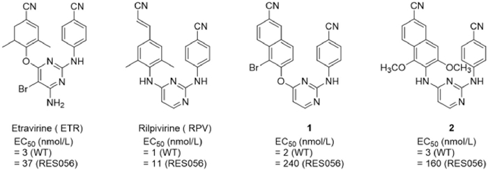
Figure 1.
The chemical structure of ETR, RPV and other DAYP compounds.

Discovery of novel and potent non-nucleoside reverse transcriptase inhibitors through structure-based drug design for HIV treatment
Yin-Xiang Zhang , Christophe Pannecouque , Erik De Clercq , Enzo Tramontano , Angela Corona , Laura Dettori , Phuong-Thao Tran , Xu-Dong Li , Shuo Su , Shuai Wang , Fen-Er Chen


Non-nucleoside reverse transcriptase inhibitors (NNRTIs) constitute an essential component of antiretroviral therapy (ART) for human immunodeficiency virus (HIV) treatment [1]. Etravirine (ETR) and rilpivirine (RPV), representative of diarylpyrimidine (DAPY) structures, are the second-generation NNRTIs approved by Food and Drug Administration (FDA) due to their favorable pharmacological profiles and excellent potency against diverse clinically relevant viral mutants (Fig. 1) [2–4]. The intrinsic structural flexibility of DAPY scaffolds enables compounds such as ETR and RPV to conformationally adapt to diverse allosteric binding pockets of both wild-type (WT) and mutant reverse transcriptase (RT) [5–7]. However, despite these advantages, both ETR and RPV display poor aqueous solubility and suboptimal pharmacokinetic (PK) properties inherent to their chemical structures. For example, the solubility of RPV is only about 20 ng/mL, while that of ETR is well below 1 µg/mL, rendering its oral bioavailability difficult to evaluate [8,9]. Furthermore, serious side effects like hepatotoxicity and hypersensitivity are continuously observed in patients receiving ETR and RPV regime, and the rapid emergency of drug resistance have greatly hampered their clinically applications [10,11].
In our previous work, we sought to overcome these deficiencies by modifying the cyanovinyl moiety of RPV (a known Michael acceptor) into a substituted cyano-naphthalene group (Fig. 1). The resulting compounds 1 and 2 exhibited potent antiviral activity against WT HIV-1, which was attributed to enhanced aromatic–aromatic interactions [12,13]. However, their efficacy against the RES056 (K103N + Y181C) strain was significantly diminished [14]. Furthermore, a simple replacement with a naphthalene ring was unlikely to improve solubility in theory. Recently, compound 3 with a bromo-substituted quinoline ring on the left side was reported to exhibit significantly increased solubility of 26.02 µg/mL at pH 7.4 [15]. Although 3 exhibited potent activity against WT HIV-1 (median effect concentration (EC50) = 1.8 nmol/L), it was largely inactive against a panel of mutant strains (Figs. 2A and D). By comparing the structure of 1, 2, 3, ETR and RPV, these findings suggest that the key structural determinants governing the resistance profiles of ETR and RPV are likely located in the terminal nitrile (cyanovinyl) moiety and/or the dimethyl substituent, features that influence the dihedral angle between the central pyrimidine ring and the left-hand aromatic ring [16,17].
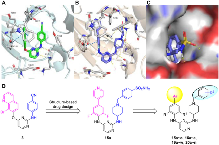
In this work, the dimethyl group on the left ring of the newly designed compounds was either retained or replaced with an isosteric difluoro moiety. Furthermore, inspired by the “necessary nitrogen atom” effect on solubility observed in compound 3, a pyridine ring, a bioisostere of the cyano or cyanovinyl moiety, was introduced at the terminal position of the difluoro/dimethylaniline group (Fig. 2D) [18]. The electron-deficient pyridine ring promotes the formation of favorable face-to-face π–π stacking interactions, primarily driven by electrostatic complementarity, thereby enhancing binding affinity [19]. Fig. 2A illustrates this interaction between 3 and RT. In order to improve the resistance profile, the para-cyanophenyl ring on the right wing was replaced by privileged 4-aminopiperidine-linked benzyl scaffold, which was predicted to extend into the protein–solvent interface (Figs. 2B–D) [20,21]. Concurrently, the solubility was anticipated to be improved, as it disrupted the molecular symmetry characteristic of DAPY compounds and introduced several solubilizing motifs, such as the pyridine ring, an ionizable organic basic ring and the neutral hydrophilic group [22–26].
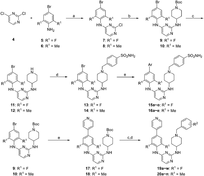
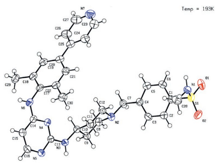
The synthetic routes of target compounds 15a–o, 16a–e, 19a–w and 20a–n were illustrated in Scheme 1. Using excess hydrochloric acid, starting material 4 was treated with 5 to harvest 7, followed by a second nucleophilic substitution with excess 4-boc-aminopiperidine to afford 9. Then 11 was obtained by the deprotection of 9 and treated with 4-sulfonamide benzyl bromide to afford the common intermediate 13, which was coupled with aromatic boronic acids or esters to afford target compounds 15a–o [27,28]. The similar Suzuki–Miyaura coupling reaction was carried out on 9 to obtain 17, followed by deprotection and continuous nucleophilic substitution to afford target compounds 19a–w. A similar procedure was performed to give dimethyl derivatives 16a–e and 20a–n. To verify the regioselectivity of 2,4-dichlorinepyrimidine, the X-ray single crystal structure of 20b was conducted and shown in Fig. 3.
All of the target compounds 15a–o, 16a–e, 19a–w and 20a–n were evaluated for their biological activity against WT HIV-1 (ⅢB) and RES056 strains in MT-4 cell utilizing the MTT method [29]. Selected compounds were further screened for the antiviral activity against clinically frequently observed single/double mutant strains. Nevirapine (NPV), efavirenz (EFV) and ETR were selected as reference drugs. The values of EC50 (antiviral efficacy), concentration of cytotoxicity 50% (CC50) (cytotoxicity), selectivity index (SI, the ratio of CC50/EC50) and half maximal inhibitory concentration (IC50) (enzymatic inhibitory potency) were organized in Tables 1–3.

 DownLoad:
CSV
DownLoad:
CSV
 |
|||||||||||
| Compd. | R1 | Ar | EC50 (WT) (μmol/L) | CC50 (μmol/L) | SI | Compd. | R1 | Ar | EC50 (WT) (μmol/L) | CC50 (μmol/L) | SI |
| 15a | F |  |
0.007 ± 0.003 | 3.3 ± 0.92 | 669 | 15k | F |  |
0.073 ± 0.029 | 4.1 ± 0.78 | 56 |
| 15b | F |  |
0.011 ± 0.004 | 4.1 ± 0.52 | 360 | 15l | F |  |
0.021 ± 0.004 | 23.5 ± 2.82 | 832 |
| 15c | F |  |
0.010 ± 0.008 | 8.2 ± 0.6 | 855 | 15m | F |  |
0.007 ± 0.005 | 6.1 ± 1.43 | 1111 |
| 15d | F |  |
0.010 ± 0.003 | 6.9 ± 0.91 | 668 | 15n | F |  |
0.036 ± 0.013 | 22.8 ± 2.31 | 255 |
| 15e | F |  |
0.012 ± 0.004 | 10.2 ± 3.03 | 350 | 15o | F |  |
0.007 ± 0.001 | 2 ± 0.57 | 288 |
| 15f | F |  |
0.026 ± 0.008 | 5.5 ± 0.98 | 311 | 16a | Me |  |
0.008 ± 0.003 | 1.2 ± 0.42 | 0.24 |
| 15g | F |  |
0.015 ± 0.004 | 4.1 ± 0.47 | 214 | 16b | Me |  |
0.006 ± 0.001 | 4.2 ± 0.63 | 0.34 |
| 15h | F |  |
0.044 ± 0.012 | 2.9 ± 0.72 | 183 | 16c | Me |  |
0.007 ± 0.001 | 2.9 ± 0.92 | 3.94 |
| 15i | F |  |
0.016 ± 0.007 | 5.7 ± 1.08 | 270 | 16d | Me |  |
0.007 ± 0.002 | 4.2 ± 0.51 | 0.51 |
| 15j | F |  |
0.050 ± 0.011 | 4.8 ± 0.9 | 98 | 16e | Me |  |
0.007 ± 0.001 | 3.9 ± 0.46 | 0.26 |
| NVP | 0.188 ± 0.066 | >15 | >80 | ETR | 0.003 ± 0.001 | >4.59 | >1340 | ||||
| EFV | 0.004 ± 0.002 | >6.34 | >1425 | ||||||||
DownLoad:
CSV
 |
|||||||||||
| Compd. | R1 | R2 | EC50 (WT) (μmol/L) | CC50 (μmol/L) | SI | Compd. | R1 | R2 | EC50 (WT) (μmol/L) | CC50 (μmol/L) | SI |
| 15a | F | 4-SO2NH2 | 0.007 ± 0.003 | 3.3 ± 0.92 | 669 | 19s | F |  |
0.003 ± 0.001 | 2.5 ± 0.99 | 724 |
| 19a | F | 4-SO2NHCH3 | 0.006 ± 0.001 | 3.7 ± 0.96 | 604 | 19t | F |  |
0.005 ± 0.001 | 3.8 ± 0.79 | 786 |
| 19b | F | 4-SO2CH3 | 0.003 ± 0 | 4.7 ± 1.18 | 1584 | 19u | F |  |
0.007 ± 0.001 | 3.8 ± 0.71 | 535 |
| 19c | F | 4-CONH2 | 0.004 ± 0.001 | 2.1 ± 0.28 | 544 | 19v | F |  |
0.004 ± 0.001 | 3.7 ± 0.69 | 1081 |
| 19d | F | 4-CONHCH3 | 0.004 ± 0.001 | 8.7 ± 0.95 | 2156 | 19w | F |  |
0.013 ± 0.005 | 22.2 ± 2.67 | 1695 |
| 19e | F | 4-COCH3 | 0.007 ± 0.003 | 4.6 ± 1.14 | 655 | 20a | Me | 4-SO2NH2 | 0.005 ± 0.001 | 3.4 ± 0.75 | 760 |
| 19f | F | 4-COOMe | 0.051 ± 0.013 | 14.8 ± 9.28 | 288 | 20b | Me | 4-SO2NHCH3 | 0.005 ± 0.001 | 1.3 ± 0.25 | 258 |
| 19g | F | 4-F | 0.012 ± 0.004 | 5 ± 0.64 | 418 | 20c | Me | 4-CONH2 | 0.006 ± 0.003 | 2.3 ± 1.12 | 408 |
| 19h | F | 3-F | 0.007 ± 0.002 | 5.2 ± 0.6 | 719 | 20d | Me | 3-CONH2 | 0.006 ± 0.002 | 3.2 ± 0.77 | 511 |
| 19i | F | 2-F | 0.012 ± 0.001 | 19.3 ± 8.34 | 1597 | 20e | Me | 4-CONHCH3 | 0.002 ± 0.001 | 3.1 ± 0.81 | 1962 |
| 19j | F | 4-CN | 0.012 ± 0.004 | 3.7 ± 0.92 | 320 | 20f | Me |  |
0.006 ± 0.002 | 5.3 ± 12.42 | 923 |
| 19k | F | 3-CN | 0.009 ± 0.006 | 4.3 ± 0.91 | 488 | 20g | Me |  |
0.005 ± 0.002 | 0.9 ± 0.27 | 180 |
| 19l | F | 2-CN | 0.007 ± 0.002 | 1.7 ± 0.23 | 488 | 20h | Me |  |
0.005 ± 0.001 | 3.7 ± 0.57 | 763 |
| 19m | F | 4-Cl | 0.017 ± 0.005 | 4.9 ± 0.92 | 288 | 20i | Me |  |
0.004 ± 0.001 | 3.1 ± 0.69 | 710 |
| 19n | F | 4-CF3 | 0.023 ± 0.008 | 4.1 ± 1.05 | 178 | 20j | Me |  |
0.006 ± 0.001 | 1.1 ± 0.04 | 189 |
| 19o | F | 4-Me | 0.006 ± 0.002 | 3.3 ± 1.08 | 521 | 20k | Me |  |
0.007 ± 0.001 | 1.5 ± 0.57 | 214 |
| 19p | F |  |
0.008 ± 0.002 | 0.8 ± 0.15 | 98 | 20l | Me |  |
0.006 ± 0.002 | 5.5 ± 0.38 | 971 |
| 19q | F |  |
0.004 ± 0.001 | 0.9 ± 0.14 | 226 | 20m | Me |  |
0.003 ± 0.001 | 3.3 ± 0.56 | 1215 |
| 19r | F |  |
0.014 ± 0.005 | 4.3 ± 0.98 | 310 | 20n | Me |  |
0.006 ± 0.001 | 2.6 ± 0.88 | 453 |
| NVP | 0.188 ± 0.066 | >15 | >80 | ETR | 0.003 ± 0.001 | >4.59 | >1340 | ||||
| EFV | 0.004 ± 0.002 | >6.34 | >1425 | ||||||||
DownLoad:
CSV
| Compd. | EC50 (µmol/L) | IC50 (µmol/L) | ||||||
| L100I | K103N | Y181C | Y188L | E138K | F227L + V106A | RES056 | ||
| 15a | 0.156 ± 0.041 | 0.041 ± 0.016 | 0.127 ± 0.057 | ≥0.943 | 0.048 ± 0.018 | ≥5.892 | ≥5.892 | 0.100 ± 0.018 |
| 15b | 0.350 ± 0.053 | 0.063 ± 0.013 | 0.072 ± 0.012 | >4.12 | 0.098 ± 0.067 | >4.12 | >4.12 | 0.091 ± 0.029 |
| 16a | 0.054 ± 0.007 | 0.008 ± 0 | 0.042 ± 0.003 | 0.923 ± 0.032 | 0.055 ± 0.012 | >0.264 ± 0 | 0.248 ± 0.064 | 0.182 ± 0.009 |
| 16b | 0.061 ± 0.013 | 0.008 ± 0.002 | 0.036 ± 0.003 | 0.245 ± 0.045 | 0.053 ± 0.006 | 0.355 ± 0.097 | 0.247 ± 0.05 | 0.148 ± 0.001 |
| 16d | 0.065 ± 0.011 | 0.006 ± 0.002 | 0.034 ± 0.012 | 0.286 ± 0.07 | 0.034 ± 0.013 | 0.234 ± 0.124 | 0.209 ± 0.097 | 0.199 ± 0.019 |
| 19b | 0.094 ± 0.008 | 0.008 ± 0.001 | 0.059 ± 0.02 | >1.344 | 0.017 ± 0.005 | >0.926 | >4.675 | 0.091 ± 0.031 |
| 19c | 0.004 ± 0.001 | 0.004 ± 0.001 | 0.004 ± 0.001 | >2.139 | 0.024 ± 0.005 | >2.139 | >2.139 | 0.074 ± 0.008 |
| 19d | 0.208 ± 0.158 | 0.021 ± 0.012 | 0.057 ± 0.019 | 1.185 ± 0.118 | 0.018 ± 0.002 | >8.653 | >8.653 | 0.135 ± 0.025 |
| 19s | 0.194 ± 0.25 | 0.006 ± 0.002 | 0.025 ± 0.008 | 0.722 ± 0.208 | 0.015 ± 0.002 | >3.836 | >3.836 | 0.109 ± 0.050 |
| 19v | 0.36 ± 0.136 | 0.056 ± 0.01 | 0.108 ± 0.034 | >3.74 | 0.042 ± 0.003 | 7.295 ± 1.574 | 3.106 ± 0.361 | 0.178 ± 0.052 |
| 20a | 0.024 ± 0.014 | 0.004 ± 0.001 | 0.012 ± 0.005 | 0.205 ± 0.064 | 0.017 ± 0.007 | 0.383 ± 0.07 | 0.12 ± 0.016 | 0.087 ± 0.043 |
| 20b | 0.017 ± 0.006 | 0.004 ± 0.001 | 0.016 ± 0.008 | 0.213 ± 0.022 | 0.029 ± 0.008 | 0.690 ± 0.476 | 0.093 ± 0.011 | 0.132 ± 0.009 |
| 20e | 0.026 ± 0.008 | 0.003 ± 0.001 | 0.010 ± 0.001 | 0.365 ± 0.055 | 0.015 ± 0.001 | 0.550 ± 0.343 | 0.176 ± 0.046 | 0.164 ± 0.061 |
| 20m | 0.011 ± 0.004 | 0.001 ± 0.001 | 0.006 ± 0.001 | 0.108 ± 0.047 | 0.008 ± 0.003 | 0.196 ± 0.084 | 0.032 ± 0.011 | 0.209 ± 0.002 |
| NVP | 1.79 ± 1.426 | 5.872 ± 2.777 | 9.019 ± 2.619 | >15 | 0.141 ± 0.039 | >15 | >15 | 0.976 ± 0.338 |
| EFV | 0.030 ± 0.015 | 0.084 ± 0.033 | 0.008 ± 0.003 | 0.303 ± 0.127 | 0.008 ± 0.003 | 0.267 ± 0.368 | 0.327 ± 0.194 | 0.011 ± 0.025 |
| ETR | 0.007 ± 0.005 | 0.003 ± 0.001 | 0.016 ± 0.005 | 0.02 ± 0.008 | 0.009 ± 0.004 | 0.015 ± 0.013 | 0.041 ± 0.026 | 0.030 ± 0.005 |
| 3 | 0.340 ± 0.072 | 0.069 ± 0.014 | 0.220 ± 0.096 | 7.19 ± 4.64 | 0.045 ± 0.022 | 2.53 ± 1.06 | 11.57 ± 9.062 | 0.011 ± 0.001 |
First of all, we explored the effect of aromatic-difluoro/dimethylaniline fragment on their antiviral activity. Pleasingly, the newly synthesized compounds 15a–o and 16a–e showed moderate to potent inhibitory potency against WT HIV-1 (ⅢB) (EC50 = 0.006–0.073 µmol/L) as depicted in Table 1. Based on the structural features and biological results, preliminary structure–activity relationship (SAR) was summarized. The antiviral activity was largely independent of the position of the pyridine nitrogen atom but was markedly governed by the substituents on the pyridine ring. For instance, the comparable potency of 15a (EC50 = 0.007 µmol/L) and 15d (EC50 = 0.010 µmol/L) resulted in a marginal delta pEC50 (ΔpEC50) value of −0.15 ± 0.23 (Table S1 in Supporting information), indicating no significant difference in their activity. Typically, fluoro- and methoxy-substituents on the pyridine ring exhibited minimal positive effect on antiviral activity, whereas methyl, dimethylamino and trifluoromethyl groups led to significant reduction in potency. The methyl-substituted derivatives 15f (EC50 = 0.026 µmol/L) and 15i (EC50 = 0.050 µmol/L) showed 3.7- and 7.1-fold decrease in activity compared to 15a, respectively. Interestingly, the pyrimidine analog 15L also exhibited significantly reduced antiviral activity (EC50 = 0.020 µmol/L). Subsequent introduction of a methyl group at the 2-positition of the pyrimidine ring further diminished the potency (15n, EC50 = 0.036 µmol/L) albeit with lower cytotoxicity (CC50 = 22.8 µmol/L), conversely, methoxy substitution (15m) restored the potency (EC50 = 0.008 µmol/L). Besides, the comparable activity observed with the 4-cyanophenyl substitution (15o) to that of 15a supported the feasibility of the pyridine ring replacement. Furthermore, the dimethyl analogs (16a–e) also exhibited potent activity against WT HIV-1 (EC50 = 0.006–0.007 µmol/L), which was substantially superior to NVP (EC50 = 0.188 µmol/L) and comparable to EFV (EC50 = 0.004 µmol/L) and ETR (EC50 = 0.003 µmol/L), as indicated by their ΔpEC50 values of 0.25 ± 0.29 and 0.37 ± 0.24 relative to 15a. Based on these findings, the pyridine ring was identified as the preferred moiety attached to the left-hand difluoro/dimethylaniline ring.
In the second round of optimization, structurally diverse hydrogen donators/acceptors were installed on the right-hand phenyl scaffold to interrogate the protein/solvent interface and enable more favorable interactions. As depicted in Table 2, 19a–w and 20a–n maintained low nanomolar inhibitory activity against WT HIV-1 (ⅢB) with EC50 values ranging from 0.002 µmol/L to 0.051 µmol/L. More specifically, the methyl-masking analog (19a) exhibited nearly identical antiviral activity to 15a, while the slightly more lipophilic methylsulfonyl analog (19b) showed an approximately 2.3-fold increase in potency (EC50 = 0.003 µmol/L), with a ΔpEC50 value of 0.37 ± 0.19 supporting this enhancement (Table S2 in Supporting information). In this way, amide derivatives 19c–d and ketone 19e showed comparable antiviral potency relative to 15a. Subsequently, more hydrophobic substituents (e.g., ester, fluoro, chloro, cyano, trifluoromethyl and methyl) were incorporated into the scaffold. Most of these modifications led to equivalent or marginally reduced activity, whereas ester (19f) and highly lipophilic trifluoromethyl groups (19n) caused the pronounced loss in potency against WT strain, reducing it by 7.3- and 3.3-fold compared with 15a, respectively. To further investigate the effects of spatial occupancy and lipophilicity on biological activity, cyclic and bulkier substituents were also explored (19p–q, 19t–v), which showed no significantly statistical differences in anti-WT activity relative to 15a. More intriguingly, compound 19s, bearing a methoxylethylsulfonamide terminus, exhibited superior anti-WT activity (EC50 = 0.003 µmol/L) compared to the hydroxyethylsulfonamide-terminated analog 19r (EC50 = 0.014 µmol/L) and hydroxyethylamide-containing derivative 19w (EC50 = 0.013 µmol/L), which featured a terminus of the same two-carbon chain length. The identical anti-WT activity trend was also observed in dimethyl series (20a–n). The order of antiviral potency for the short-chain hydrophilic groups was 4-CONHCH3 (20e, EC50 = 0.002 µmol/L) >4-SO2NH2 ≈ 4-SO2NHCH3 ≈ 4-CONH2 ≈ 3-CONH2, with the activities of the latter four groups clustered in the range of 0.005–0.006 µmol/L. Besides, the analogs with cyclic and sterically demanding substituents (20f–k) exhibited excellent anti-HIV-1 (ⅢB) activity, with no significant difference in potency observed among them. Regarding to two-carbon chain end, 20m (EC50 = 0.003 µmol/L) with methoxylethylamide appendage was more potent than analogs 20L and 20n (EC50 = 0.006 µmol/L).
Subsequently, compounds 15a–b, 16a–b, 16d, 19b–c, 19s, 19v, 20a–b, 20e and 20m with high potency against WT HIV-1 (ⅢB) were further selected to assess their efficacy against clinically prevalent single mutant strains (L100I, K103N, Y181C, Y188L and E138K) and double mutant strains (F227L + V106A and RES056) as well as WT HIV-1 RT. As depicted in Table 3, it was noticeable that all tested compounds exhibited superior potency against the L100I, K103N, Y181C, and E138K mutant strains. However, against the Y188L strain and the two double mutants (F227L + V106A and RES056), a clear divergence was observed between the difluoro and dimethyl series (15a–b vs. 16a–b, 16d; 19b–c, 19s, 19v vs. 20a–b, 20e, 20m) that the difluoro analogs were more sensitive and even substantially lost their activities, whereas the dimethyl analogs largely maintained their potency. Among them, 20m demonstrated to be the most potent inhibitors, with EC50 values of 0.011 µmol/L (L100I), 0.001 µmol/L (K103N), 0.006 µmol/L (Y181C), 0.108 µmol/L (Y188L), 0.008 µmol/L (E138K), 0.196 µmol/L (F227L + V106A), 0.032 µmol/L (RES056), outperforming 3 and EFV, slightly inferior to ETR. Concurrently, to confirm the binding target of these compounds, their inhibitory activity against the recombinant WT HIV-1 RT were also evaluated with IC50 values of 0.074–0.209 µmol/L, being superior to NVP (IC50 = 0.976 µmol/L). Among them, 15b (IC50 = 0.091 µmol/L), 19b (IC50 = 0.091 µmol/L), 19c (IC50 = 0.074 µmol/L) and 20a (IC50 = 0.087 µmol/L) were identified as the four most effective inhibitors with robust RT inhibitory potency. Notably, these compounds exhibited marked disparity in potency, with cellular-level inhibition substantially exceeding that at the enzyme level in vitro. This observation may be attributed to multiply factors, such as cellular accumulation yielding elevated local concentration at the target site, intracellular metabolic activation, prolonged target residence time and potential multi-target synergistic effects. Collectively, the HIV-1 RT inhibition assay demonstrated potent binding of these novel derivatives to RT, indicating that they could be regarded as HIV-1 NNRTIs.
In order to explain the discrepancy of antiviral activity, molecular docking was employed to predict the binding modes of 20m or difluoro analogs with WT HIV-1 RT and mutants. As depicted in Fig. 4A, 20m binds to the active binding pocket of WT HIV-1 RT in a “U-shaped” conformation, similar to classic NNRTIs. The left-hand pyridine-dimethylaniline fragment was capable of fitting well into the hydrophobic tunnel constituted by residues Y181, Y183, Y188, F227 and highly conserved W229. Especially for Y188 residue, the electron-deficient pyridine ring is conducive to forming preferable face-to-face π-π stacking, an interaction more driven by electrostatic complementarity in nature than aromatic hydrophobic effects, thereby contributing to improvement in binding affinity. Besides, an important hydrogen bond between 20m and K101 is retained, consistent with binding of other DAPYs. Additionally, water-mediated hydrogen bonds are also observed at the position of N atom of central pyrimidine or the N atom of piperidine ring. At the protein/solvent interface, the terminal oxygen atom of 20m engages in another hydrogen bond with residue V106. Collectively, Given the multitude of critical interactions observed, it is rational that 20m exhibits high potency against WT HIV-1 RT.
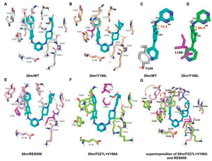
However, with respect to Y188L mutant, the robust π-π stacking is disrupted and a weaker aromatic hydrophobic interaction between Y181 and dimethyl-phenyl ring enforces to strengthen as a compensation, which partially compromised the antiviral activity of 20m against the Y188L strain (Fig. 4B). More interestingly, the dihedral angle between dimethylaniline ring and central pyrimidine ring, that is C(14)-N(6)-C(17)-C(22) of 20m, varies greatly when 20m adapts itself to WT or Y188L RT (Figs. 4C and D). when bound to WT HIV-1 RT, 20m adopts a dihedral angle of 74.5°, whereas the angle shifts to 86.4° in the Y188L RT complex. The dihedral angle of 20m in the WT complex is closer to that of observed in the X-ray structure of 20b (−70.8°, Fig. 3), which means that the conformation of 20m in the WT complex likely resembles its dominant solution conformation and requires minimal torsional or rotational adjustment to adapt to the binding pocket. In contrast, there will be more entropy loss of 20m in the Y188L RT complex. As for RES056 mutant, 20m exhibited robust inhibitory potency which can be evidenced by Fig. 4E. Despite Y181C and K103N double mutations, the pyridine-dimethylaniline fragment enhances interactions with residue Y188 and the conserved W229. Furthermore, an additional hydrogen bond between the terminal amide NH and the backbone oxygen atom of K104 is observed, further contributing to the binding affinity. In the case of the F227L + V106A mutant, however, the scenario differs significantly. The π-π stacking is markedly weakened due to a substantial shift of the entire binding pocket (Fig. 4F). Structural superposition of the 20m/F227L + V106A and RES056 complexes reveals notable displacements: the centroid of residue Y183 is shifted by 6.3 Å, and that of Y188 by 2.8 Å (Fig. 4G). Furthermore, residue L227 is positioned further away compared to F227. Although the hydrogen bond interacted with A106 is retained, the critical aromatic interactions are substantially lost, which mainly accounts for the weakened activity against this mutant. Moreover, the predicted dihedral angle of 20m counts again, that is 54.7° in F227L + V106A RT complex, shift 19.8° away from that in WT complex. Besides, other docking dihedral angles are 78.9° (L100I), 64.6° (K103N), 81.1° (Y181C), 68.1° (E138K) and 68.4° (RES056).
Another phenomenon is that difluoro derivatives are mainly sensitive to the mutation of HIV-1 RT, which can be attributed to their dihedral configurations. The preferred conformations of 20b (Fig. 3) or 20m (Fig. 4D) support that the dimethylaniline moiety is closely perpendicular to the central pyrimidine ring (−70.8° and 74.5°) and the lone pairs on the N4H and N6 atoms favor a trans-figuration. Thus, a steric clash is likely to happen when the methyl substitute approach to the N4 atom due to lone pair repulsion. In contrast, the favorable interaction of a N → F (nN → σ*F) will be formed that induces the fluorine atom more closely to pyrimidine ring resulting to smaller dihedral angle. As expected, the docking dihedral angle of 15a in WT complex is 62° convinces this hypothesis. Consequently, the relatively rigid structures of difluoro analogs reflect reduced flexibility when adapted to diverse binding pockets of mutants, necessitating the overcoming of greater rotational energy barriers and leading to inferior anti-resistant activity.
To further elucidate the potency, 20m complexed with the RES056 are subjected to molecular dynamics (MD) simulation. From Fig. S1 (Supporting information), it can be seen that their root-mean-square deviation (RMSD) values were kept below 3.5 Å, indicating the strong binding affinity and low probability of dissociation. Four primary types of interactions exist: ionic interactions, hydrogen bonds, hydrophobic contacts, and water-bridged bonds. The analysis demonstrates that residue K101 form a very significant and stable hydrogen bond with 20m for 97% of the simulation time during the trajectory process. Besides, the three critical water-bridge/hydrogen bonds formed by residues K101, V106, and Q222. The binding is further stabilized by significant π-π stacking interactions between the left-wing heterobiaryl fragment of 20m and residues Y188, F227, and W229. The mean dihedral angle for 20m is 68.44° ± 9.63°, which is consistent with the value measured in the molecular docking analysis. Collectively, the protein-ligand contacts effectively account for the potent potency of 20m against RES056. Eventually, the aqueous solubility of 20m was also assessed in different phosphate buffer solutions (pH 7.4, 4.5 and 2.0) in Table 4. Gratifyingly, the aqueous solubility of 20m was markedly enhanced, particularly in acidic buffers, reaching >3954.5 µg/mL at pH 4.5 and >7380.6 µg/mL at pH 2.0, far surpassing the solubility of both compound 3 and ETR.
DownLoad:
CSV
| Compd. | Solubility (µg/mL) | ||
| pH 2.0 | pH 4.5 | pH 7.4 | |
| 20m | >7380.6 | >3954.5 | 0.32 |
| 3 | 28.49 | N/A | 26.02 |
| ETR | 127 | N/A | ≪1 |
In conclusion, we have synthesized a series of novel compounds incorporating a substituted pyridine–difluoro/dimethylaniline fragment and a 4-aminopiperidine moiety by employing structure-based drug design strategy. The electron-deficient pyridine ring enhanced π–π stacking interactions, thereby improving binding affinity. At the protein–solvent interface, hydrophilic substitutions proved more favorable than lipophilic groups. SAR analysis further revealed that dimethyl analogs displayed stronger antiviral activity than their difluoro counterparts, particularly against the Y188L mutant and the double mutations (F227L + V106A, RES056). This difference in activity was likely associated with variations in the dihedral angle between the pyridine–difluoro/dimethylaniline fragment and the adjacent moiety. The dimethyl analog adopted a larger dihedral angle, whereas the difluoro analog were predicted to have a smaller one and conferred a more rigid conformation, reducing flexibility to compromise its ability to adapt to diverse mutant binding pockets and thus leading to a significant decrease in potency. The dimethyl derivative 20m demonstrated the efficacy of this strategy, exhibiting not only excellent antiviral potency against WT HIV-1 and seven mutant strains (EC50 = 0.003–0.196 µmol/L) but also significantly improve solubility profile (S > 3954.5 µg/mL at pH 4.5; S > 7380.6 µg/mL at pH 2.0), dramatically higher than that of 3 and ETR.
The authors declare that they have no known competing financial interests or personal relationships that could have appeared to influence the work reported in this paper.
Yin-Xiang Zhang: Writing – original draft, Formal analysis, Data curation. Christophe Pannecouque: Writing – original draft, Resources, Formal analysis, Data curation. Erik De Clercq: Writing – review & editing, Conceptualization. Enzo Tramontano: Writing – review & editing, Resources. Angela Corona: Writing – review & editing, Resources. Laura Dettori: Writing – original draft, Resources. Phuong-Thao Tran: Writing – review & editing, Project administration. Xu-Dong Li: Writing – review & editing, Writing – original draft, Formal analysis, Data curation. Shuo Su: Writing – review & editing, Formal analysis, Data curation. Shuai Wang: Writing – review & editing, Supervision, Project administration, Funding acquisition, Data curation, Conceptualization. Fen-Er Chen: Writing – review & editing, Supervision, Resources, Project administration, Conceptualization.
This work was financially supported by the National Natural Science Foundation of China (Nos. 82574236 and 82304297), Shanghai Pujiang Programme (No. 23PJD005), the State Key Laboratory of Natural and Biomimetic Drugs (No. K202408), and Shanghai Pilot Program for Basic Research-Fudan University 21TQ1400100 (No. 25TQ011).
Supplementary material associated with this article can be found, in the online version, at doi:
Q. Hao, X. Ling, C. Pannecouque, et al., Chin. Chem. Lett. 34 (2023) 107663. doi: 10.1016/j.cclet.2022.07.006
K. Zhang, Y.J. Zhang, M. Li, et al., Med. Res. Rev. 45 (2024) 426–483.
L.M. Zhao, C. Pannecouque, E. De Clercq, et al., Chin. Chem. Lett. 34 (2023) 108261. doi: 10.1016/j.cclet.2023.108261
S. Du, X. Liu, X. Hu, et al., Chin. Chem. Lett. 36 (2025) 110378. doi: 10.1016/j.cclet.2024.110378
V. Poongavanam, V. Namasivayam, M. Vanangamudi, et al., WIREs Comput. Mol. Sci. 8 (2017) e1328.
E.B. Lansdon, K.M. Brendza, M. Hung, et al., J. Med. Chem. 53 (2010) 4295–4299. doi: 10.1021/jm1002233
K. Das, P.J. Lewi, S.H. Hughes, et al., Prog. Biophys. Mol. Biol. 88 (2005) 209–231. doi: 10.1016/j.pbiomolbio.2004.07.001
Y. Sang, S. Han, C. Pannecouque, et al., J. Med. Chem. 62 (2019) 11430–11436. doi: 10.1021/acs.jmedchem.9b01446
X.M. Chen, Q.Q. Hao, C. Pannecouque, et al., Acta Pharm. Sin. B 15 (2025) 4115–4136. doi: 10.1016/j.apsb.2025.06.016
Z. Wang, W.A. Zalloum, W. Wang, et al., J. Med. Chem. 64 (2021) 13658–13675. doi: 10.1021/acs.jmedchem.1c01015
Z. Wang, P.P. Sharma, B. Rathi, et al., J. Med. Chem. 66 (2023) 8643–8665. doi: 10.1021/acs.jmedchem.3c00275
Y.H. Liang, X.Q. Feng, Z.S. Zeng, et al., ChemMedChem 4 (2009) 1537–1545. doi: 10.1002/cmdc.200900212
X.Q. Feng, Y.H. Liang, Z.S. Zeng, et al., ChemMedChem 4 (2009) 219–224. doi: 10.1002/cmdc.200800334
Y.H. Liang, Q.Q. He, Z.S. Zeng, et al., Biorg. Med. Chem. 18 (2010) 4601–4605. doi: 10.1016/j.bmc.2010.05.036
R.L. Zhou, C. Yu, C. Pannecouque, et al., Bioorg. Chem. 140 (2023) 106821. doi: 10.1016/j.bioorg.2023.106821
J. Che, X. Dai, J. Gao, et al., J. Med. Chem. 64 (2021) 12163–12180. doi: 10.1021/acs.jmedchem.1c00815
J. Chandrasekhar, R. Dick, J. Van Veldhuizen, et al., J. Med. Chem. 61 (2018) 6858–6868. doi: 10.1021/acs.jmedchem.8b00797
L.D. Pennington, D.T. Moustakas, J. Med. Chem. 60 (2017) 3552–3579. doi: 10.1021/acs.jmedchem.6b01807
M.A.M. Subbaiah, N.A. Meanwell, J. Med. Chem. 64 (2021) 14046–14128. doi: 10.1021/acs.jmedchem.1c01215
K. Zhang, L. Zhao, T. Xing, et al., Chin. Chem. Lett. 37 (2026) 111283. doi: 10.1016/j.cclet.2025.111283
B. Meng, Z. Zhuo, H. Yu, et al., Chin. Chem. Lett. 35 (2024) 100827.
M. Ishikawa, Y. Hashimoto, J. Med. Chem. 54 (2011) 1539–1554. doi: 10.1021/jm101356p
F. Lovering, J. Bikker, C. Humblet, J. Med. Chem. 52 (2009) 6752–6756. doi: 10.1021/jm901241e
F. Lovering, MedChemComm 4 (2013) 515–519. doi: 10.1039/c2md20347b
X. Lin, H. Shi, W. Zhang, et al., J. Med. Chem. 62 (2019) 10352–10361. doi: 10.1021/acs.jmedchem.9b01421
Y. Wu, L. Zhang, Z. Sun, et al., J. Med. Chem. 66 (2023) 8545–8563. doi: 10.1021/acs.jmedchem.3c00231
M. Yang, Y. Liu, X. Qi, et al., Green Syn. Catal. 5 (2024) 211–269.
B. Qiu, Y.F. Shi, X.D. An, et al., Green Syn. Catal. 6 (2025) 110–113.
C. Pannecouque, D. Daelemans, E. De Clercq, Nat. Protoc. 3 (2008) 427–434. doi: 10.1038/nprot.2007.517

Figure 2 (A) The docking mode of 3 in WT HIV-1 RT (PDB ID: 2ZD1). (B, C) the docking mode and surface pattern of 15a in WT HIV-1 RT (PDB ID: 6C0N), respectively. (D) Rational design of target compounds.

Scheme 1 Reagents and conditions: (a) IPA/H2O, HCl (conj, excess), r.t., 12–36 h; (b) 4-amino-1-Boc-piperidine (5.0 equiv.), neat, 120 ℃, 12 h; (c) TFA, DCM, r.t., 4 h; (d) substituted benzyl bromide or chloride, DMF, K2CO3, r.t., 12 h; (e) aromatic boronic acid or esters, Pd(dppf)Cl2, dioxane/H2O, Cs2CO3, 110 ℃, N2, 12 h.

Figure 4 The docking pattern of 20m with WT HIV-1 RT and mutants. (A) WT (PDB code: 6C0N, in gray). (B) Y188L (PDB code: 6C0N, in wheat). (C, D) Different dihedral configurations of C(14)-N(6)-C(17)-C(22) of 20m (in cyan or green) in WT and Y188L, respectively. (E) RES056 (PDB code: 6C0R, in light pink). (F) F227L + V106A (PDB code: 6DUF, in limon). (G) Superimposition of 20m with F227L + V106A and RES056. Mutated residues are depicted as magentas sticks (or slate sticks). Hydrogen bonds are showed as yellow dashed lines and π-π stacking contacts are illustrated as marine dashed lines.
Table 1. Antiviral activity of 15a–o and 16a–e against HIV-1 (ⅢB) strains in MT-4 cells.
|
|||||||||||
| Compd. | R1 | Ar | EC50 (WT) (μmol/L) | CC50 (μmol/L) | SI | Compd. | R1 | Ar | EC50 (WT) (μmol/L) | CC50 (μmol/L) | SI |
| 15a | F | |
0.007 ± 0.003 | 3.3 ± 0.92 | 669 | 15k | F | |
0.073 ± 0.029 | 4.1 ± 0.78 | 56 |
| 15b | F | |
0.011 ± 0.004 | 4.1 ± 0.52 | 360 | 15l | F | |
0.021 ± 0.004 | 23.5 ± 2.82 | 832 |
| 15c | F | |
0.010 ± 0.008 | 8.2 ± 0.6 | 855 | 15m | F | |
0.007 ± 0.005 | 6.1 ± 1.43 | 1111 |
| 15d | F | |
0.010 ± 0.003 | 6.9 ± 0.91 | 668 | 15n | F | |
0.036 ± 0.013 | 22.8 ± 2.31 | 255 |
| 15e | F | |
0.012 ± 0.004 | 10.2 ± 3.03 | 350 | 15o | F | |
0.007 ± 0.001 | 2 ± 0.57 | 288 |
| 15f | F | |
0.026 ± 0.008 | 5.5 ± 0.98 | 311 | 16a | Me | |
0.008 ± 0.003 | 1.2 ± 0.42 | 0.24 |
| 15g | F | |
0.015 ± 0.004 | 4.1 ± 0.47 | 214 | 16b | Me | |
0.006 ± 0.001 | 4.2 ± 0.63 | 0.34 |
| 15h | F | |
0.044 ± 0.012 | 2.9 ± 0.72 | 183 | 16c | Me | |
0.007 ± 0.001 | 2.9 ± 0.92 | 3.94 |
| 15i | F | |
0.016 ± 0.007 | 5.7 ± 1.08 | 270 | 16d | Me | |
0.007 ± 0.002 | 4.2 ± 0.51 | 0.51 |
| 15j | F | |
0.050 ± 0.011 | 4.8 ± 0.9 | 98 | 16e | Me | |
0.007 ± 0.001 | 3.9 ± 0.46 | 0.26 |
| NVP | 0.188 ± 0.066 | >15 | >80 | ETR | 0.003 ± 0.001 | >4.59 | >1340 | ||||
| EFV | 0.004 ± 0.002 | >6.34 | >1425 | ||||||||
 下载: 导出CSV
Table 2. Antiviral activity of 19a–w and 20a–n against HIV-1 (ⅢB) strains in MT-4 cells.
|
|||||||||||
| Compd. | R1 | R2 | EC50 (WT) (μmol/L) | CC50 (μmol/L) | SI | Compd. | R1 | R2 | EC50 (WT) (μmol/L) | CC50 (μmol/L) | SI |
| 15a | F | 4-SO2NH2 | 0.007 ± 0.003 | 3.3 ± 0.92 | 669 | 19s | F | |
0.003 ± 0.001 | 2.5 ± 0.99 | 724 |
| 19a | F | 4-SO2NHCH3 | 0.006 ± 0.001 | 3.7 ± 0.96 | 604 | 19t | F | |
0.005 ± 0.001 | 3.8 ± 0.79 | 786 |
| 19b | F | 4-SO2CH3 | 0.003 ± 0 | 4.7 ± 1.18 | 1584 | 19u | F | |
0.007 ± 0.001 | 3.8 ± 0.71 | 535 |
| 19c | F | 4-CONH2 | 0.004 ± 0.001 | 2.1 ± 0.28 | 544 | 19v | F | |
0.004 ± 0.001 | 3.7 ± 0.69 | 1081 |
| 19d | F | 4-CONHCH3 | 0.004 ± 0.001 | 8.7 ± 0.95 | 2156 | 19w | F | |
0.013 ± 0.005 | 22.2 ± 2.67 | 1695 |
| 19e | F | 4-COCH3 | 0.007 ± 0.003 | 4.6 ± 1.14 | 655 | 20a | Me | 4-SO2NH2 | 0.005 ± 0.001 | 3.4 ± 0.75 | 760 |
| 19f | F | 4-COOMe | 0.051 ± 0.013 | 14.8 ± 9.28 | 288 | 20b | Me | 4-SO2NHCH3 | 0.005 ± 0.001 | 1.3 ± 0.25 | 258 |
| 19g | F | 4-F | 0.012 ± 0.004 | 5 ± 0.64 | 418 | 20c | Me | 4-CONH2 | 0.006 ± 0.003 | 2.3 ± 1.12 | 408 |
| 19h | F | 3-F | 0.007 ± 0.002 | 5.2 ± 0.6 | 719 | 20d | Me | 3-CONH2 | 0.006 ± 0.002 | 3.2 ± 0.77 | 511 |
| 19i | F | 2-F | 0.012 ± 0.001 | 19.3 ± 8.34 | 1597 | 20e | Me | 4-CONHCH3 | 0.002 ± 0.001 | 3.1 ± 0.81 | 1962 |
| 19j | F | 4-CN | 0.012 ± 0.004 | 3.7 ± 0.92 | 320 | 20f | Me | |
0.006 ± 0.002 | 5.3 ± 12.42 | 923 |
| 19k | F | 3-CN | 0.009 ± 0.006 | 4.3 ± 0.91 | 488 | 20g | Me | |
0.005 ± 0.002 | 0.9 ± 0.27 | 180 |
| 19l | F | 2-CN | 0.007 ± 0.002 | 1.7 ± 0.23 | 488 | 20h | Me | |
0.005 ± 0.001 | 3.7 ± 0.57 | 763 |
| 19m | F | 4-Cl | 0.017 ± 0.005 | 4.9 ± 0.92 | 288 | 20i | Me | |
0.004 ± 0.001 | 3.1 ± 0.69 | 710 |
| 19n | F | 4-CF3 | 0.023 ± 0.008 | 4.1 ± 1.05 | 178 | 20j | Me | |
0.006 ± 0.001 | 1.1 ± 0.04 | 189 |
| 19o | F | 4-Me | 0.006 ± 0.002 | 3.3 ± 1.08 | 521 | 20k | Me | |
0.007 ± 0.001 | 1.5 ± 0.57 | 214 |
| 19p | F | |
0.008 ± 0.002 | 0.8 ± 0.15 | 98 | 20l | Me | |
0.006 ± 0.002 | 5.5 ± 0.38 | 971 |
| 19q | F | |
0.004 ± 0.001 | 0.9 ± 0.14 | 226 | 20m | Me | |
0.003 ± 0.001 | 3.3 ± 0.56 | 1215 |
| 19r | F | |
0.014 ± 0.005 | 4.3 ± 0.98 | 310 | 20n | Me | |
0.006 ± 0.001 | 2.6 ± 0.88 | 453 |
| NVP | 0.188 ± 0.066 | >15 | >80 | ETR | 0.003 ± 0.001 | >4.59 | >1340 | ||||
| EFV | 0.004 ± 0.002 | >6.34 | >1425 | ||||||||
下载: 导出CSV
Table 3. Inhibitory activity of representative compounds toward seven mutant strains and HIV-1 RT.
| Compd. | EC50 (µmol/L) | IC50 (µmol/L) | ||||||
| L100I | K103N | Y181C | Y188L | E138K | F227L + V106A | RES056 | ||
| 15a | 0.156 ± 0.041 | 0.041 ± 0.016 | 0.127 ± 0.057 | ≥0.943 | 0.048 ± 0.018 | ≥5.892 | ≥5.892 | 0.100 ± 0.018 |
| 15b | 0.350 ± 0.053 | 0.063 ± 0.013 | 0.072 ± 0.012 | >4.12 | 0.098 ± 0.067 | >4.12 | >4.12 | 0.091 ± 0.029 |
| 16a | 0.054 ± 0.007 | 0.008 ± 0 | 0.042 ± 0.003 | 0.923 ± 0.032 | 0.055 ± 0.012 | >0.264 ± 0 | 0.248 ± 0.064 | 0.182 ± 0.009 |
| 16b | 0.061 ± 0.013 | 0.008 ± 0.002 | 0.036 ± 0.003 | 0.245 ± 0.045 | 0.053 ± 0.006 | 0.355 ± 0.097 | 0.247 ± 0.05 | 0.148 ± 0.001 |
| 16d | 0.065 ± 0.011 | 0.006 ± 0.002 | 0.034 ± 0.012 | 0.286 ± 0.07 | 0.034 ± 0.013 | 0.234 ± 0.124 | 0.209 ± 0.097 | 0.199 ± 0.019 |
| 19b | 0.094 ± 0.008 | 0.008 ± 0.001 | 0.059 ± 0.02 | >1.344 | 0.017 ± 0.005 | >0.926 | >4.675 | 0.091 ± 0.031 |
| 19c | 0.004 ± 0.001 | 0.004 ± 0.001 | 0.004 ± 0.001 | >2.139 | 0.024 ± 0.005 | >2.139 | >2.139 | 0.074 ± 0.008 |
| 19d | 0.208 ± 0.158 | 0.021 ± 0.012 | 0.057 ± 0.019 | 1.185 ± 0.118 | 0.018 ± 0.002 | >8.653 | >8.653 | 0.135 ± 0.025 |
| 19s | 0.194 ± 0.25 | 0.006 ± 0.002 | 0.025 ± 0.008 | 0.722 ± 0.208 | 0.015 ± 0.002 | >3.836 | >3.836 | 0.109 ± 0.050 |
| 19v | 0.36 ± 0.136 | 0.056 ± 0.01 | 0.108 ± 0.034 | >3.74 | 0.042 ± 0.003 | 7.295 ± 1.574 | 3.106 ± 0.361 | 0.178 ± 0.052 |
| 20a | 0.024 ± 0.014 | 0.004 ± 0.001 | 0.012 ± 0.005 | 0.205 ± 0.064 | 0.017 ± 0.007 | 0.383 ± 0.07 | 0.12 ± 0.016 | 0.087 ± 0.043 |
| 20b | 0.017 ± 0.006 | 0.004 ± 0.001 | 0.016 ± 0.008 | 0.213 ± 0.022 | 0.029 ± 0.008 | 0.690 ± 0.476 | 0.093 ± 0.011 | 0.132 ± 0.009 |
| 20e | 0.026 ± 0.008 | 0.003 ± 0.001 | 0.010 ± 0.001 | 0.365 ± 0.055 | 0.015 ± 0.001 | 0.550 ± 0.343 | 0.176 ± 0.046 | 0.164 ± 0.061 |
| 20m | 0.011 ± 0.004 | 0.001 ± 0.001 | 0.006 ± 0.001 | 0.108 ± 0.047 | 0.008 ± 0.003 | 0.196 ± 0.084 | 0.032 ± 0.011 | 0.209 ± 0.002 |
| NVP | 1.79 ± 1.426 | 5.872 ± 2.777 | 9.019 ± 2.619 | >15 | 0.141 ± 0.039 | >15 | >15 | 0.976 ± 0.338 |
| EFV | 0.030 ± 0.015 | 0.084 ± 0.033 | 0.008 ± 0.003 | 0.303 ± 0.127 | 0.008 ± 0.003 | 0.267 ± 0.368 | 0.327 ± 0.194 | 0.011 ± 0.025 |
| ETR | 0.007 ± 0.005 | 0.003 ± 0.001 | 0.016 ± 0.005 | 0.02 ± 0.008 | 0.009 ± 0.004 | 0.015 ± 0.013 | 0.041 ± 0.026 | 0.030 ± 0.005 |
| 3 | 0.340 ± 0.072 | 0.069 ± 0.014 | 0.220 ± 0.096 | 7.19 ± 4.64 | 0.045 ± 0.022 | 2.53 ± 1.06 | 11.57 ± 9.062 | 0.011 ± 0.001 |
下载: 导出CSV
Table 4. The aqueous solubility of 20m.
| Compd. | Solubility (µg/mL) | ||
| pH 2.0 | pH 4.5 | pH 7.4 | |
| 20m | >7380.6 | >3954.5 | 0.32 |
| 3 | 28.49 | N/A | 26.02 |
| ETR | 127 | N/A | ≪1 |
下载: 导出CSV

 扫一扫看文章


扫一扫关注我们


 下载:
下载: