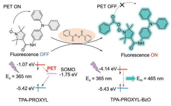
Figure 1.
Schematic illustration of the TPA-PROXYL fluorescence probe for the detection of BPO and the PET molecular orbital transition mechanism of TPA-PROXYL.

Rapid OFF–ON photoinduced electron transfer probe based on triphenylamine and PROXYL radical specifically activated by benzoyl peroxide
Xinfang Cui , Zehong Wang , Yingjiang Li , Yufeng Xiong , Tong Zhou , Xiangdong Peng , Guanghui Zhao , Lijie Dong


Stable radicals are usually defined as a class of organic radical substances that have long-lasting stability and longer lifetimes [1]. In contrast to the diamagnetic character [2] of conventional closed-shell organic molecules, stable radicals exhibit intrinsic paramagnetic behavior [3] due to their unique open-shell electronic configurations, which are considered to be effective in quenching fluorophores. This arises from interactions between the unpaired spin of the radical and the conjugated system of the fluorophore, altering the multiplicity of electronic states [4]. Therefore, the integration of stable radicals to fluorophores enables the rational design of sensitive fluorescence probes [5,6]. Nitrogen-oxygen radical fluorescent probe has been widely used in detection of reactive oxygen species in biological systems, redox substances and imaging polymer degradation [7,8]. When nitrogen-oxygen radicals react with redox substances or reactive oxygen species, fluorescence emission turns on, thereby enabling detection of these reactive substances [9]. Tetramethyl-1-piperidinyloxyl (TEMPO) radicals are typically designed as fluorescent probes for detecting redox substances or reactive oxygen species [10]. Ishii et al. [11] covalently attached phthalocyanine silicon (SiPc) to TEMPO radicals as fluorescent probes for straightforwardly detecting ascorbic acid in biological systems. Maki et al. [12] employed perylenebisimide-linked nitroxide (PBILN) as a fluorescent reagent for the simple and sensitive detection of ascorbic acid, with a detection limit of 0.28 μmol/L. Despite the extensive design of piperidine-based TEMPO radicals as practical and sensitive fluorescence probes, their utility is frequently compromised by limited selectivity. In contrast, the pyrroline scaffold of PROXYL radicals confers enhanced robustness in redox-competent milieus [13]. Kinoshita et al. [14] characterized the reaction kinetics of diverse nitrogen-oxygen radicals with ascorbic acid, revealing that carbamoyl-PROXYL retained over 80%, markedly more stable to hydroxy–TEMPO and oxo-TEMPO, which underwent near-complete depletion within 10 min. Therefore, the strategic development of PROXYL radical-fluorophore conjugates present substantial promise for advanced sensing platforms, leveraging the synergistic interplay of paramagnetic quenching and stability in complex biological matrices.
Triphenylamine (TPA) is a typical non-planar propeller molecule with unique optical properties. It can be connected to a TPA donor via a receptor group to form a donor-acceptor (D-A) aggregation-induced emission (AIE) fluorophore [15,16], exhibiting intramolecular charge transfer (ICT) or twisted intramolecular charge transfer (TICT) states, enabling fluorescence state switching for fluorescence detection purposes. TPA possesses strong electron-donating capabilities, conferring TPA-based AIEgens with higher sensitivity and better responsiveness to external stimuli [17]. Therefore, the photophysical properties of TPA-based AIEgens can be altered by functionalizing triphenylamine with different receptor sidechain units, thereby changing the fluorescence state and enabling fluorescence detection [18]. Yang et al. [19] reported an AIE-type probe using triphenylamine as the fluorescent moiety and cyano or vinyl as the acceptor, where the coordination interaction between ferric ion (Fe3+) and the cyano group, along with the nucleophilic addition of cyanide ion (CN-) and sulfite (SO32-) to the vinyl group, causes fluorescence quenching. Ruan et al. [20] synthesized a TPA-based luminescent material, 1-(4-(diphenylamino)phenyl)-2-phenylethanone (TPACO) with aggregation-induced emission (AIE) and twisted intramolecular charge transfer (TICT). When potassium tert–butoxide (t-BuOK) is added to the TPACO solution, the fluorescence intensity decreases sharply due to isomerization reactions and the generation of enolized anions. Although various AIEgens based on triphenylamine have been designed to detect substances such as metal ions and redox agents, the non-specific reactions of the acceptor side groups make them susceptible to interference from other reactants. In addition, fluorescence probes based on the "ON–OFF" mechanism of triphenylamine typically exhibit reduced sensitivity in high-fluorescence backgrounds [21]. Therefore, it is essential to design the structure of the receptor side groups of fluorescence probes based on triphenylamine to address issues of susceptibility to interference and high-fluorescence backgrounds.
Herein, we propose an "OFF-ON" type fluorescent probe TPA-PROXYL based on triphenylamine with higher sensitivity and PROXYL radical with good stability and paramagnetic properties, which turns on fluorescence after the specific reaction with benzoyl peroxide (BPO), as shown in Fig. 1. Specifically, TPA group is connected to the PROXYL radical via an amide bond, thereby achieving photoinduced electron transfer (PET). The unpaired electron of the PROXYL radical provides a singly occupied molecular orbital (SOMO) acting as an electron acceptor orbital (−1.75 eV) for the photoinduced electron transfer (PET) process. The emission of the aggregation-induced emission (AIE) is effectively suppressed by photoinduced electron transfer (PET) from the nitrogen-oxygen radical's SOMO orbital [22,23]. Upon specific reaction with BPO, the conversion of the nitrogen-oxygen radical eliminates the SOMO orbital, thereby completely blocking the PET pathway and abolishing its quenching capability. The TPA-PROXYL fluorescent probe integrates the stable PROXYL radical's photoinduced electron transfer (PET) quenching mechanism with the aggregation-induced emission (AIE) activity of triphenylamine (TPA), laying the foundation for highly specific small-molecule sensing in complex biological environments and rapid on-site monitoring of BPO.
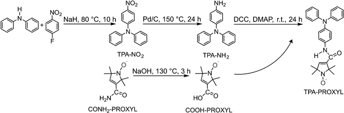
The synthesis of TPA-PROXYL was accomplished according to the previously reported procedure (Fig. 2) [24,25]. Briefly, TPA-PROXYL was synthesized via amide coupling between the carboxy group of carboxyl-2,2,5,5-tetramethyl-3-pyrrolin-1-oxyl (COOH-PROXYL) and the amino group of 4-aminotriphenylamine (TPA-NH2). TPA-NH2 was obtained by catalytic reduction of 4-nitrotriphenylamine (TPA-NO2), which itself was prepared through a nucleophilic aromatic substitution between diphenylamine and 1-fluoro-4-nitrobenzene. Carboxyl-2,2,5,5-tetramethyl-3-pyrrolin-1-oxyl (COOH-PROXYL) was generated by alkaline hydrolysis of carbamoyl-2,2,5,5-tetramethyl-3-pyrrolin-1-oxyl (CONH2-PROXYL). The synthetic details and characterizations are described in Figs. S1–S3 (Supporting information). This architecture is intended to facilitate photoinduced electron transfer (PET), effectively merging the distinct characteristics of both units within a single molecular framework.
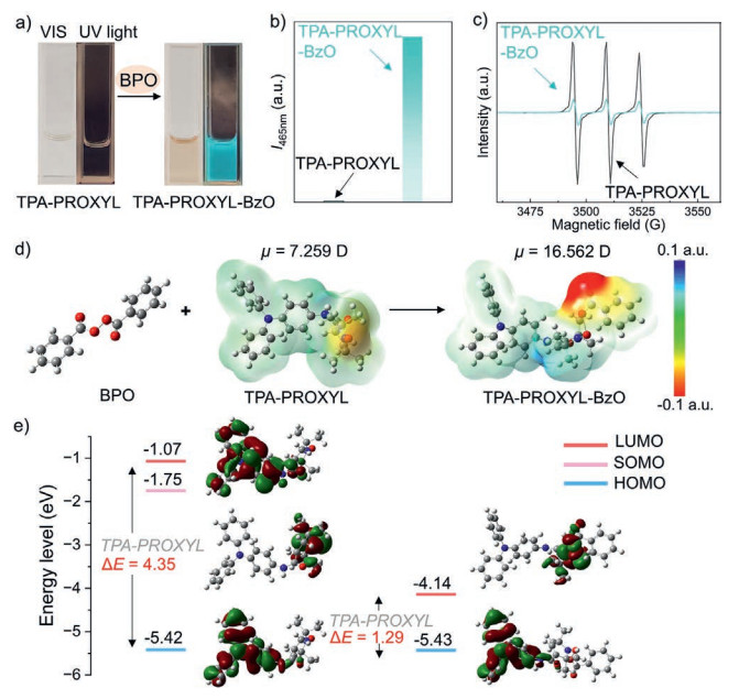
TPA-PROXYL is non-fluorescent due to its intermolecular electron exchange interactions [26,27], as shown in Figs. 3a and b. Upon reaction with BPO, an obvious color change of the system from colorless to brownish yellow can be observed by the naked eye and a strong emission band at 465 nm under the optimal excitation wavelength of 365 nm (Fig. S5 in Supporting information) is developed together with fluorescence intensity restored to 127 times, accompanied with a lighting-up visual change from colorless to blueish green under the excitation of 365 nm UV light in the dark. Compared with DMSO solvent as shown in Fig. S4 (Supporting information), the fluorescence probe in DMF solvent shows a significant increase in emission intensity at 465 nm, resulting in superior signal discrimination capability for BPO detection. These changes in fluorescence are due to the conversion of the nitrogen-oxygen radicals in TPA-PROXYL by BPO, which results in the fluorescence on of the triphenylamine moiety. Fig. 3c shows the EPR images of TPA-PROXYL and TPA-PROXYL-BzO after reaction with 5 mmol/L BPO. The triplet signal observed in the EPR spectrum of nitrogen-oxygen radicals in solution primarily arises from hyperfine coupling between the unpaired electron and the nitrogen nucleus (I = 1), which splits the energy levels, combined with the averaging effect of rapid molecular tumbling in solution [5,6]. The g-factor is 2.006, which is characteristic of the PROXYL radical [28]. The results show that the EPR intensity of TPA-PROXYL decreases significantly after reaction with BPO, indicating a decrease in paramagnetic strength and radical concentration, confirming the conversion of the PROXYL radical. To further investigate the sensing mechanism of TPA-PROXYL, density functional theory (DFT) was used to calculate the ESP-mapped electron density, LUMO, HOMO and SOMO. As shown in Fig. 3d, the drastic increase in dipole moment (from 7.259 D to 16.562 D) and the prominent negative electrostatic potential localized at the peroxide bond in TPA-PROXYL-BzO indicate a significant redistribution of electron density. This redistribution disrupts the original PET pathway from the triphenylamine donor to the nitrogen-oxygen radical acceptor, thereby restoring fluorescence emission [29]. Furthermore, as shown in Fig. 3e, DFT calculations reveal that the LUMO localized on the triphenylamine moiety in TPA-PROXYL (−1.07 eV) lies above the SOMO residing on the PROXYL radical unit (−1.75 eV), affording a positive driving force (ΔE = +0.68 eV) for PET. This process dissipates the excitation energy through non-radiative means, leading to fluorescence suppression [30]. After reacting with BPO, the nitrogen-oxygen radical in TPA-PROXYL-BzO converts into an antiferromagnetic adduct, eliminating the SOMO orbital. Therefore, due to the absence of an electron-accepting radical center, the PET pathway is completely blocked. Concurrently, the LUMO energy level significantly shifts downward to −4.14 eV (compared to −1.07 eV in TPA-PROXYL), while the HOMO remains at −5.43 eV, resulting in a narrowed energy gap of 1.29 eV. This electronic structure reorganization significantly enhances the radiative decay efficiency of the TPA excited state, thereby restoring fluorescence emission.
To investigate the sensing properties of TPA-PROXYL towards BPO, the fluorescent response of TPA-PROXYL (0.1 mmol/L) towards concentrations of BPO ranging from 0.5 mmol/L to 2.4 mmol/L was recorded. The detecting behavior of TPA-PROXYL towards BPO is presented in Fig. 4 [31–37] and Fig. S6 (Supporting information). As shown in Fig. 4a, in the addition of BPO, the system exhibits maximum fluorescent emission at 465 nm with the excitation of 365 nm. In addition, with the increase of the BPO concentration, the fluorescence intensity of probe TPA-PROXYL at 465 nm gradually increased. What is more, at the probe concentration of 1 mmol/L and over a BPO concentration range of 1–12 mmol/L, the system exhibited a lighting-up visual change from colorless to blueish green under UV excitation in the dark (Fig. 4c), corresponding to the fluorescence turn-on induced by the reaction between the probe's nitrogen-oxygen radicals and BPO. Meanwhile, an obvious color change of the system from colorless to brownish yellow can be observed by the naked eye, indicating that the TPA-PROXYL can be used as a colorimetric probe for BPO detection.
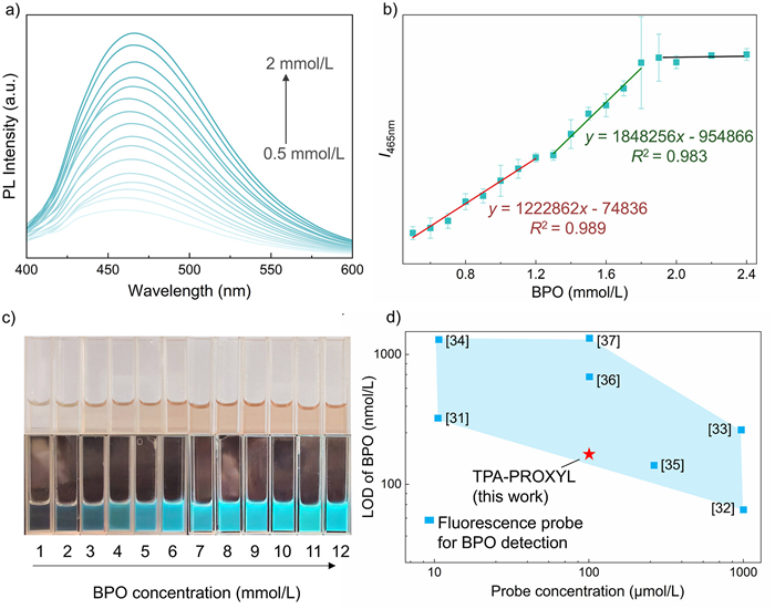
Furthermore, we explored the relationship between the fluorescence intensity and concentrations of BPO to obtain a quantitative analysis of detecting efficiency. Based on the results in Fig. S7 (Supporting information), 0.5 mmol/L BPO was chosen as the lowest detectable concentration, as it provided a clear and measurable fluorescence response, in contrast to the negligible effect observed at 0.1–0.4 mmol/L. Scatter plots of the relative fluorescence intensity with the excitation wavelength at 365 nm and emission wavelength at 465 nm and concentrations of BPO were presented in Fig. 4b. In the concentration range of 0.5 mmol/L to 2.4 mmol/L, the fluorescence intensity of TPA-PROXYL exhibited a three-stage linear relationship with BPO concentration. The fluorescence intensity changes most sharply over 0.5 mmol/L to 1.2 mmol/L, exhibits an increase from 1.3 mmol/L to 1.9 mmol/L, and then plateaus between 2 mmol/L to 2.4 mmol/L. Least square of data fitting was performed and the following equation have established to describes the concentration dependence in the 0.5–1.2 mmol/L and 1.3–1.9 mmol/L regimes:
|
|
(1) |
|
|
(2) |
I1 and I2 denote the fluorescence intensities measured at 465 nm emission under 365 nm excitation for the 0.5–1.2 mmol/L and 1.3–1.9 mmol/L concentration intervals, respectively. They show good linear relationships and the fitting correlation coefficient (R2) are 0.989 and 0.983, respectively. Remarkably, the limit of detection (LOD) was calculated as 211 nmol/L using the 3σ/S method. This value is lower than most of reported BPO detection probes (Fig. 4d). Thus, this probe enables low-concentration detection with a low detection limit.
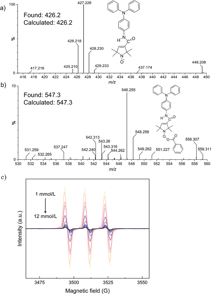
HR-MS analyses and electron paramagnetic resonance (EPR) spectroscopy were performed to further elucidate the sensing mechanism. As shown in Figs. 5a and b, the peaks observed at 427.2 and 548.3 correspond to the protonated adducts of TPA-PROXYL and TPA-PROXYL-BzO, respectively. Stable radical molecules are paramagnetic due to their unique electronic structure and thus electron paramagnetic resonance spectroscopy is one of the most important methods for the detection of stable radicals [1]. When stable radical molecules are subjected to an external magnetic field, unpaired electrons can absorb electromagnetic radiation of a specific frequency under defined conditions, resulting in an EPR signal [7]. As shown in Fig. 5c, the EPR signal intensity decreases with increasing BPO concentration, indicating the consumption of the N–O radical moiety in TPA-PROXYL upon reaction with BPO. This conversion disrupts the photoinduced electron transfer (PET) process from the triphenylamine fluorophore to the nitroxide radical, leading to fluorescence turn-on. A pronounced decrease in EPR intensity is observed within the BPO concentration range of 1–6 mmol/L, suggesting efficient reaction between TPA-PROXYL and BPO in this region. Subsequently, the reaction approaches saturation in higher concentrations of BPO range from 7 mmol/L to 12 mmol/L, resulting in a plateau in EPR signal attenuation as the available nitroxide radicals are progressively consumed. To verify the aggregation-induced emission (AIE) characteristics of TPA-PROXYL-BzO, we investigated the fluorescence intensity of TPA-PROXYL-BzO at different concentrations. As shown in Fig. S8a (Supporting information), the fluorescence intensity at 465 nm remained weak at concentrations below 0.01 mmol/L, with no noticeable emission under UV light (in the inset of Fig. S8a). When the concentration exceeded 0.01 mmol/L, the fluorescence intensity increased significantly, accompanied by distinct blue-green emission under UV light. We further examined the fluorescence behavior of 0.1 mmol/L TPA-PROXYL-BzO in DMF-toluene mixtures with varying toluene fractions [38]. TPA-PROXYL-BzO is highly soluble in DMF, but insoluble in toluene. Therefore, DMF (good solvent) and toluene (poor solvent) were chosen as solvent pair due to their miscibility. As illustrated in Fig. S8b (Supporting information), with an excitation wavelength of 365 nm, the maximum emission intensity exhibited a noticeable blue shift along with the change in solvent polarity, and increased significantly when the toluene content exceeded 20%, which can be attributed to the formation of aggregates, thereby suppressing non-radiative decay [39]. The AIE behavior of TPA-PROXYL-BzO was consistent with that of triphenylamine, confirming a shared emission response to aggregation (Figs. S8c and d in Supporting information). These findings collectively validate that the concentration-dependent reaction of the nitrogen-oxygen radical with BPO triggers the fluorescence turn-on of the TPA-PROXYL probe by suppressing PET, which conclusively establishes the underlying sensing mechanism integrating stable radical and AIE characteristics.
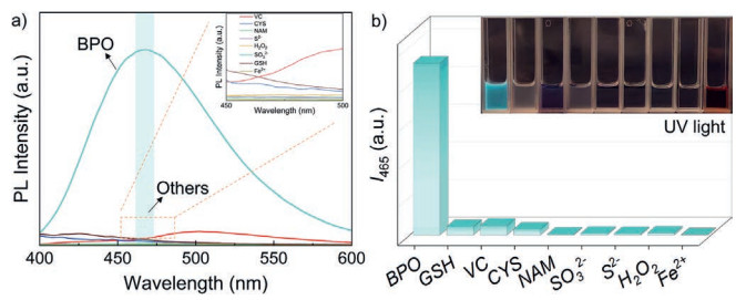
High selectivity is of great importance for the practical deployment of fluorescent probes, as it ensures reliable detection of the target analyte amidst potential interferents. To provide support for the notion that TPA-PROXYL can be used to detect BPO in systems with interference from other substances that may react with radicals, we investigated the selectivity of TPA-PROXYL for BPO and various relevant analytes including GSH, VC, CYS, NAM, SO32-, S2-, H2O2, and Fe2+ (5 mmol/L respectively). As shown in Fig. 6a, under the same conditions, only the addition of BPO can significantly activate the fluorescence of the TPA-PROXYL probe; among the others, the fluorescence recovery of GSH and VC is relatively weak and does not affect the detection of BPO. Fig. 6b demonstrates the fluorescence intensity at an emission intensity of 465 nm, which allows for a more intuitive demonstration of the selectivity of the TPA-PROXYL probe for BPO, and the inset shows that under UV light irradiation, only the addition of BPO allowed for the restoration of fluorescence visible to the naked eye. Additionally, competition experiments were conducted using CYS and GSH, with the results presented in Fig. S9 (Supporting information). After reaction of the TPA-PROXYL probe in DMF solutions containing BPO along with either CYS or GSH at 100 ℃ for 1 h, the presence of CYS or GSH showed negligible impact on the fluorescence recovery intensity, further demonstrating the excellent anti-interference capability of the fluorescent probe. These results confirm the pronounced selectivity of TPA-PROXYL for BPO, evidenced by a distinct fluorescence turn-on response against a range of competing interferents.
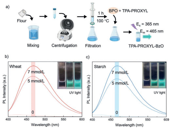
Benzoyl peroxide (BPO) is a common peroxide and strong oxidizer widely used in the chemical, food and pharmaceutical industries [40,41]. Nevertheless, its excessive utilization has been associated with adverse health outcomes in humans [34]. In view of the excellent sensitivity and selectivity for BPO, we further explored the practicality of TPA-PROXYL for detecting BPO in real samples. Two kinds of real samples, edible power and BPO gels, were tested in this assay. As shown in Fig. 7a, we used the standard addition method to detect BPO in wheat flour and starch. Wheat flour and starch were dissolved in DMF, centrifuged for 10 min, and the supernatant was collected. It was then filtered through a 0.22 μm organic filter membrane, and a certain amount of BPO was added. After heating at 100 ℃ for 1 h, the fluorescence curve was measured. As shown in Figs. 7b and c, fluorescence emission spectra (λex = 365 nm. Slits: 2.5/2.5 nm) of the fluorescent probe was detected to be slightly elevated after the addition of the extract without BPO, indicating the presence of trace amounts of BPO in these food samples. 5 and 7 mmol/L of BPO were added to the wheat and starch, respectively, and the fluorescence intensity curve shows a significant increase especially at 465 nm. The detection results are shown in Table 1, with recovery rates above 84%, indicating good detection performance. Based on the results of the measurements and calculations, it can be inferred TPA-PROXYL has a strong capability for detecting BPO in edible power. In addition, two commercially available BPO gel products were acquired for analysis. Each gel was dissolved in DMF, subjected to centrifugation, and subsequently filtered through a 0.22 μm organic membrane filter prior to detection with TPA-PROXYL fluorescent probe and the results are shown in Table 2. The detected BPO content is in line with the official additions of the two gels. Therefore, TPA-PROXYL could quantitatively determine BPO in real samples.
 DownLoad:
CSV
| Sample | Spiked (mmol/L) | Found (mmol/L) | Recovery (%) | RSD (%, n = 3) |
| Wheat | 5 | 4.89 ± 0.28 | 97.7 | 0.1 |
| 7 | 6.7 ± 1.56 | 95.9 | 0.4 | |
| Starch | 5 | 4.20 ± 0.19 | 84.1 | 0.08 |
| 7 | 6.59 ± 0.72 | 99.2 | 0.18 | |
| Recovery (%) = [(Found value-blank value)/spiked value] × 100%. | ||||
DownLoad:
CSV
| Sample | BPO amount (mg/mL) | Found (mg/mL) | RSD (%, n = 3) |
| Type 1 | 1.21 | 1.07 ± 0.14 | 0.23 |
| 1.69 | 2.15 ± 0.04 | 0.03 | |
| Type 2 | 1.21 | 1.04 ± 0.07 | 0.13 |
| 1.69 | 2.10 ± 0.27 | 0.23 |
In summary, we have reported an "OFF-ON" fluorescent probe TPA-PROXYL engineered from a triphenylamine scaffold tethered to a stable PROXYL radical via photoinduced electron transfer (PET) quenching. This probe exhibits sensitive and specific fluorescence turn-on at 465 nm (λex = 365 nm) upon exposure to benzoyl peroxide (BPO), demonstrating a linear response (R2 > 0.98) relevant concentrations with an ultralow detection limit of 211 nmol/L. Specially, TPA-PROXYL maintains uncompromised selectivity against redox interferents, including glutathione (GSH), ascorbic acid (VC), cysteine (CYS), nicotinamide (NAM), sulfite (SO32-), sulfide (S2-), hydrogen peroxide (H2O2), and ferrous ions (Fe2+), validating its utility for ratiometric BPO detection in complex matrices. This study provides guidance for integrating stable radicals with aggregation-induced emission (AIE) fluorophores to develop high-fidelity small molecule sensing in complex environment.
Xinfang Cui: Writing – original draft, Data curation. Zehong Wang: Writing – review & editing, Conceptualization. Yingjiang Li: Validation, Supervision. Yufeng Xiong: Methodology. Tong Zhou: Validation, Supervision. Xiangdong Peng: Formal analysis. Guanghui Zhao: Investigation. Lijie Dong: Project administration, Funding acquisition.
The authors declare that they have no known competing financial interests or personal relationships that could have appeared to influence the work reported in this paper.
This work was supported by the Joint Funds of the National Natural Science Foundation of China (No. U24A2075), the National Natural Science Foundation of China (No. 52303243) and the Fundamental Research Funds for the Central Universities (Nos. 104972025RSCbs0030 and 104972024KFYf0004).
Supplementary material associated with this article can be found, in the online version, at doi:
D. Griller, K.U. Ingold, Acc. Chem. Res. 9 (1976) 13–19. doi: 10.1021/ar50097a003
G.I. Likhtenstein, K. Ishii, S. Nakatsuji, Photochem. Photobiol. 83 (2007) 871–881. doi: 10.1111/j.1751-1097.2007.00141.x
Y.F. Xiong, Y.J. Li, X.F. Cui, et al., Adv. Funct. Mater. 35 (2025) 2419661. doi: 10.1002/adfm.202419661
J.P. Blinco, K.E. Fairfull-Smith, B.J. Morrow, S.E. Bottle, Aust. J. Chem. 64 (2011) 373–389. doi: 10.1071/CH10442
N.V. Blough, D.J. Simpson, J. Am. Chem. Soc. 110 (1988) 1915–1917. doi: 10.1021/ja00214a041
S.A. Green, D.J. Simpson, G. Zhou, P.S. Ho, N.V. Blough, J. Am. Chem. Soc. 112 (1990) 7337–7346. doi: 10.1021/ja00176a038
M.M. Haugland, J.E. Lovett, E.A. Anderson, Chem. Soc. Rev. 47 (2018) 668–680. doi: 10.1039/c6cs00550k
A. Kaur, J.L. Kolanowski, E.J. New, Angew. Chem. Int. Ed. 55 (2016) 1602–1613. doi: 10.1002/anie.201506353
O.G. Ballesteros, L. Maretti, R. Sastre, J.C. Scaiano, Macromolecules 34 (2001) 6184–6187. doi: 10.1021/ma0103831
J.P. Allen, M.C. Pfrunder, J.C. Mcmurtrie, et al., Eur. J. Org. Chem. 2017 (2017) 476–483. doi: 10.1002/ejoc.201601280
K. Ishii, K. Kubo, T. Sakurada, K. Komori, Y. Sakai, Chem. Commun. 47 (2011) 4932–4934. doi: 10.1039/c1cc10817d
T. Maki, N. Soh, K. Nakano, T. Imato, Talanta 85 (2011) 1730–1733. doi: 10.1016/j.talanta.2011.06.054
M. Lewandowski, K. Gwozdzinski, Int. J. Mol. Sci. 18 (2017) 2490. doi: 10.3390/ijms18112490
Y. Kinoshita, K.I. Yamada, T. Yamasaki, et al., Free Rad. Res. 43 (2009) 565–571. doi: 10.1080/10715760902914575
E. Yamaguchi, C. Wang, A. Fukazawa, et al., Angew. Chem. Int. Ed. 54 (2015) 4539–4543. doi: 10.1002/anie.201500229
Z. Jiang, Y. Chen, C. Yang, et al., Org. Lett. 11 (2009) 1503–1506. doi: 10.1021/ol9001152
Z. Shang, S. Tian, Y. Wang, et al., Sens. Actuators B: Chem. 398 (2024) 134725. doi: 10.1016/j.snb.2023.134725
W.N. Cai, T.D. Xiao, H.J. Niu, et al., Sens. Actuators B: Chem. 252 (2017) 330–339. doi: 10.1016/j.snb.2017.06.022
X.D. Yang, X.L. Chen, X.D. Lu, et al., J. Mater. Chem. C 4 (2016) 383–390. doi: 10.1039/C5TC02865E
Z.J. Ruan, H.X. Zheng, C.Y. Deng, et al., Dyes Pigm. 204 (2022) 110476. doi: 10.1016/j.dyepig.2022.110476
Q.Y. Liu, D.X. Wu, X.D. Huang, et al., Dyes Pigm. 241 (2025) 112914. doi: 10.1016/j.dyepig.2025.112914
H.J. Ma, Y.B. An, Y.Y. Han, et al., Adv. Sci. 12 (2025) e07956. doi: 10.1002/advs.202507956
M. Baharfar, A.C. Hillier, G.Z. Mao, Adv. Mater. 36 (2024) 2406083. doi: 10.1002/adma.202406083
J.Q. Xiong, Z. Wei, T. Xu, et al., Polymer 130 (2017) 135–142. doi: 10.1016/j.polymer.2017.10.004
Y.F. Xiong, Y.J. Li, Z.F. Hu, et al., Surf. Interf. 46 (2024) 104126. doi: 10.1016/j.surfin.2024.104126
Y. Xia, Y.J. Li, A.O. Burts, et al., J. Am. Chem. Soc. 133 (2011) 19953–19959. doi: 10.1021/ja2085349
R.A. Caldwell, R.E. Schwerzel, J. Am. Chem. Soc. 94 (1972) 1035–1037. doi: 10.1021/ja00758a076
H. Togashi, H. Shinzawa, T. Matsuo, et al., Free Radical Biol. Med. 28 (2000) 846–853. doi: 10.1016/S0891-5849(99)00280-4
C.D. Lindstrom, X.Y. Zhu, Chem. Rev. 106 (2006) 4281–4300. doi: 10.1021/cr0501689
Y. Matsuoka, M. Yamato, K. Yamada, J. Clin. Biochem. Nutr. 58 (2016) 16–22. doi: 10.3164/jcbn.15-105
L. Zeng, X. Wu, Q. Hu, H.Q. Yuan, G.M. Bao, Sens. Actuators B: Chem. 299 (2019) 126994. doi: 10.1016/j.snb.2019.126994
H.L. Zhang, W.N. Wang, C. Xie, K. Luo, L.Y. Zhou, Food Chem. 452 (2024) 139534. doi: 10.1016/j.foodchem.2024.139534
Y. Sun, H. Ye, Y. Yang, et al., Food Contr. 178 (2025) 111524. doi: 10.1016/j.foodcont.2025.111524
Y. Yang, H. Ye, T. Lu, M. Lan, L. Zeng, Food Chem. 461 (2024) 140822. doi: 10.1016/j.foodchem.2024.140822
L. Zhang, Y.M. Zhu, F.M. Li, et al., Chemosensors 9 (2021) 319. doi: 10.3390/chemosensors9110319
T.R. Lin, M.Q. Zhang, F.H. Xu, et al., Sens. Actuators B: Chem. 261 (2018) 379–384. doi: 10.1016/j.snb.2018.01.172
J. Hu, Y.L. Dong, H.J. Zhang, et al., RSC Adv. 3 (2013) 26307–26312. doi: 10.1039/c3ra44664f
J.D. Luo, Z.L. Xie, J.W.Y. Lam, et al., Chem. Commun. 18 (2001) 1740–1741.
Y.Y. Sun, W.J. Neary, Z.P. Burke, et al., J. Am. Chem. Soc. 144 (2022) 1125–1129. doi: 10.1021/jacs.1c12108
D. Sudhakar, K.S.V. Srinivasan, K.T. Joseph, M. Santappa, Polymer 22 (1981) 491–493. doi: 10.1016/0032-3861(81)90167-1
X. Jia, Y. Wu, P. Liu, Environ. Toxicol. Pharmacol. 31 (2011) 479–484. doi: 10.1016/j.etap.2011.03.009

Figure 1 Schematic illustration of the TPA-PROXYL fluorescence probe for the detection of BPO and the PET molecular orbital transition mechanism of TPA-PROXYL.

Figure 3 (a) The visual color and fluorescence images under 365 nm UV light, (b) fluorescence intensity and (c) EPR spectra of TPA-PROXYL (1 mmol/L) and TPA-PROXYL reacted with 5 mmol/L BPO in DMF (λex = 365 nm, λem = 465 nm. Slits: 2.5/2.5 nm). (d) ESP-mapped electron density of TPA-PROXYL and TPA-PROXYL-BzO. (e) LUMOs, HOMOs, and SOMO of optimized ground-state geometries of TPA-PROXYL and TPA-PROXYL-BzO determined by the B3LYP/6–31+G(d, p).

Figure 4 (a) Fluorescence emission spectra of TPA-PROXYL (0.1 mmol/L) reacted with varied concentrations of BPO (0.5–2 mmol/L, respectively. λex = 365 nm, λem = 465 nm. Slits: 5/5 nm), (b) three-stage linear relationship between fluorescence intensity and concentrations of BPO and (c) the visual color and fluorescence images under 365 nm UV light of TPA-PROXYL (1 mmol/L) reacted with varied concentrations of BPO (1–12 mmol/L, respectively). (d) Comparison of LOD in fluorescent probes for detecting BPO.

Figure 5 Mass spectrum of (a) TPA-PROXYL and (b) TPA-PROXYL reacted with 10 mmol/L BPO in DMF. (c) EPR spectra of TPA-PROXYL (1 mmol/L) reacted with varied concentrations of BPO (1–12 mmol/L, respectively).

Figure 6 (a) Fluorescence emission spectra (λex = 365 nm. Slits: 2.5/2.5 nm) of TPA-PROXYL reacted with varied analytes: BPO, GSH, VC, CYS, NAM, SO32-, S2-, H2O2, Fe2+. (b) Fluorescence intensity (λem = 465 nm) of TPA-PROXYL reacted with varied analytes. Insert: The fluorescence images under 365 nm UV light of TPA-PROXYL.

Figure 7 (a) Standard addition method for detecting BPO concentration in flour and starch: Operational flow chart. The Fluorescence emission spectra (λex = 365 nm. Slits: 2.5/2.5 nm) of TPA-PROXYL after reaction with (b) wheat and (c) starch to which 0, 5 and 7 mmol/L of BPO was added using the standard addition method. Insert: the fluorescence images under 365 nm UV light, respectively.
Table 1. The detection of the spiked BPO in wheat and starch samples by TPA-PROXYL.
| Sample | Spiked (mmol/L) | Found (mmol/L) | Recovery (%) | RSD (%, n = 3) |
| Wheat | 5 | 4.89 ± 0.28 | 97.7 | 0.1 |
| 7 | 6.7 ± 1.56 | 95.9 | 0.4 | |
| Starch | 5 | 4.20 ± 0.19 | 84.1 | 0.08 |
| 7 | 6.59 ± 0.72 | 99.2 | 0.18 | |
| Recovery (%) = [(Found value-blank value)/spiked value] × 100%. | ||||
 下载: 导出CSV
Table 2. The detection of two types of commercially available BPO gels by TPA-PROXYL.
| Sample | BPO amount (mg/mL) | Found (mg/mL) | RSD (%, n = 3) |
| Type 1 | 1.21 | 1.07 ± 0.14 | 0.23 |
| 1.69 | 2.15 ± 0.04 | 0.03 | |
| Type 2 | 1.21 | 1.04 ± 0.07 | 0.13 |
| 1.69 | 2.10 ± 0.27 | 0.23 |
下载: 导出CSV

 扫一扫看文章


扫一扫关注我们

 下载: