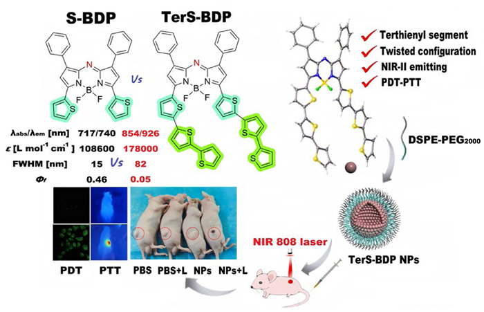
Figure 1.
Chemical structures and optical properties of S-BDP and TerS-BDP, and self-assembling and theranostic mechanisms of TerS-BDP NPs.

Terthienyl dependence of twisted conjugate in NIR-Ⅱ aza-BODIPY for photodynamic-photothermal therapy of cervical cancer
Ji Ai , Hankun Zhang , Yanru Huang , Peijun Yang , Xin-Dong Jiang , Gaowu Qin , Xiangzhi Song


Near-infrared (NIR) functional dyes, especially those operating in the second NIR window (NIR-Ⅱ, 900–1700 nm), exhibit unique advantages in the biomedical field, owing to their capacity to achieve deep-tissue penetration (5–20 mm) and a high signal-to-noise ratio [1–7]. In contrast to visible (400–700 nm) and NIR-I (700–900 nm) dyes [8–11], NIR-Ⅱ dyes demonstrate superior photothermal conversion efficiency (PCE) because of their narrow energy gap between the highest occupied molecular orbital (HOMO) and the lowest unoccupied molecular orbital (LUMO) [12–14]. These properties make them ideal candidates for clinical treatments of deep-tissue tumors such as cervical cancer [15–17]. To date, a variety of NIR-Ⅱ dyes, including quantum dots, carbon nanotubes, gold nanoparticles (NPs) and semiconductor polymers, have been developed [18,19]; and their long-term toxicity and excretion times in cancer treatment remain unknown. Consequently, it is of great significance to develop NIR-Ⅱ organic dyes with superior properties for phototherapeutic applications, including photodynamic therapy (PDT) and photothermal therapy (PTT).
PDT utilizes photosensitizers (PSs) to generate reactive oxygen species (ROS) upon light irradiation, thereby eliminating cancer cells [20–28], and has emerged as a promising approach for cancer treatment due to its precise spatiotemporal control, non-invasiveness, and excellent selectivity [29–34]. Nevertheless, the absorption of most clinically used PSs, such as porphyrins and phthalocyanines, are predominantly in the visible region [35], which significantly limits their further application in diagnosing and treating deep-tissue tumors. PTT transforms light energy into heat to selectively destroy tumor cells with minimal damage to surrounding healthy tissues, while displaying drug negligible resistance [36–39]. However, during PTT, the introduction of heat shock protein hinders the therapeutic efficacy. Therefore, the synergistic integration of PDT and PTT offers a promising strategy to overcome their respective inherent clinical limitations, thereby enhancing the effectiveness of phototherapy.
Among functional organic dyes in biological applications, classical aza-boron-dipyrromethene (aza-BODIPY) dyes have been used as promising PSs, owing to their long-wavelength absorption and high molar extinction coefficient [40–42]. Recently, our group has been devoted to the creation and functional enhancement of aza-BODIPYs in NIR-Ⅱ spectral region [43–45]. In donor-acceptor organic dyes, terthienyl segment has been used not only to extend π-conjugation system, inducing optical wavelength red-shifts and electron-donating capacity, but also form an extremely twisted configuration facilitating intersystem crossing (ISC) [46,47].
We herein introduced terthienyl groups at 3,5-sites of the aza-BODIPY framework to develop a novel NIR-Ⅱ dye, TerS-BDP, thoroughly studied the PDT-PTT characteristics of TerS-BDP using S-BDP as a benchmark for comparison (Fig. 1) [48–50]. Moreover, we prepared the corresponding NPs, TerS-BDP NPs, and assessed their in vivo anti-tumor efficacy. TerS-BDP NPs possess outstanding deep-tissue penetration capabilities in the NIR-Ⅱ region, real-time NIR-Ⅱ imaging potential, and a robust anti-tumor effect for cervical cancer (Fig. 1).
The rational design of aza-BODIPY derivatives to achieve strong absorption within the NIR-Ⅱ window (900–1700 nm) holds significant importance in biomedical imaging and phototherapeutic applications. The electronic effects at 3,5-sites play a pivotal role in the reduced energy gap between HOMO and LUMO [51–54], with the thiophene group serving as an exemplary electron donor [55,56]. The synthetic route for TerS-BDP is depicted in Scheme 1. In aza-BODIPY framework, the terthienyl group at 3,5-sites acts as a potent electron donor, reinforcing the aza-BODIPY core and establishing a robust donor-acceptor (D-A) structure [57–60]. The synthesis of TerS-BDP commenced with terthiophene as the starting material, from which the corresponding ketone was derived. The classical O'Shea method was then employed to smoothly prepare the black solid TerS-BDP through complexation with boron trifluoride etherate [61–63]. The structure of TerS-BDP was meticulously characterized, using nuclear magnetic resonance (NMR) and high-resolution mass spectrometry (HRMS).
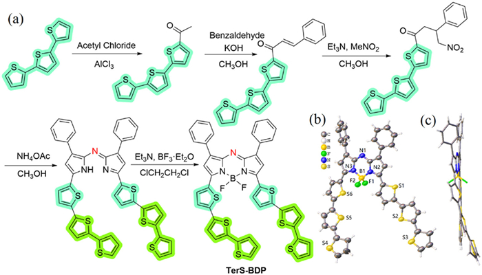
Furthermore, the solid-state structure of TerS-BDP was confirmed by X-ray single-crystal analysis, providing a comprehensive understanding of its molecular configuration (Scheme 1b). Upon examining the angles of the four bonds around the central boron atom, it is found that the deviation from the ideal value (109.5°) resulted in a relatively distorted sp3 hybridization configuration of the B atom [64–69]. Since the introduction of the flexible chain of the terthienyl group, the single crystal structure breaks our understanding of the symmetrical molecular structure, and TerS-BDP was found to adopt an asymmetric configuration with co-directional distortion for the first time (Scheme 1b). According to the side view (Scheme 1c), this terthienyl-substituted molecule is an extremely twisted configuration, which is very beneficial for ISC to produce ROS [46,47].
Further, we delved into the optical characteristics of TerS-BDP. Attaching dithiophene into 3,5-sites on the basis of S-BDP, their spectra undergo dramatic changes. TerS-BDP absorbs at 854 nm and emits at 926 nm respectively, compared to those of S-BDP (λabs/λem = 717/740 nm) (Fig. 2a and Table S1 in Supporting information) [46]. Due to the flexible chain of the terthienyl group, which has greater degrees of freedom to lead to internal conversion, TerS-BDP possesses a broader full width at half maximum (FWHM, 82 nm) and higher molar extinction coefficient (ε = 178,000 L mol−1 cm−1) than those (FWHM, 15 nm; ε = 108,600 L mol−1 cm−1) of S-BDP. Furthermore, the fluorescence band of TerS-BDP is situated within the range of 900–1300 nm (Fig. 2b). Due to intramolecular charge transfer (ICT) of terthiophene [70,71], the fluorescence quantum yield of TerS-BDP is found to be so low (Φf = 5%), compared to that (Φf = 46%) of S-BDP [48–50]. To better understand the optical properties, density functional theory (DFT) calculations of TerS-BDP were then carried out. As shown in Fig. 2c, due to the strong electron-donating nature of the terthienyl group, the HOMO in TerS-BDP is mainly located on the aza-BODIPY core, including the terthienyl segment, whereas the LUMO is mostly distributed over the aza-BODIPY core and the phenyl group at 1,7-sites. TerS-BDP showed gradually decreased LUMOs and increased HOMOs along with the terthienyl groups, and the tapering HOMO-LUMO energy gap (ΔEg = 1.52 eV) matched well with the absorption bathochromic shift trend (Fig. 2c).
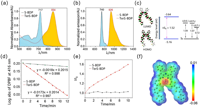
Next, the generation of ROS induced by TerS-BDP was investigated. By employing 1,3-diphenylisobenzofuran (DPBF) as a probe for 1O2 detection [72], DPBF was utilized to assess the 1O2 production in toluene upon laser irradiation of TerS-BDP. The efficiency of 1O2 generation was quantified by monitoring the reduction in the absorbance of the DPBF indicator at 416 nm. As depicted in Fig. 2d and Fig. S1 (Supporting information), the decline in absorbance at 416 nm confirms the 1O2 production. Based on the slope coefficient of the decay lines (Fig. 2d), the 1O2 yield (Φ∆) of TerS-BDP was calculated to be 0.17. Moreover, the unchanged maximum absorption peak at 854 nm under laser illumination suggests that TerS-BDP exhibited excellent photostability (Fig. S2 in Supporting information). Moreover, utilizing dihydrorhodamine 123 (DHR123) as an indicator of O2•− [73], the ability of TerS-BDP to produce ROS was explored (Fig. 2e). The change in fluorescence intensity (1.6 times) of DHR123 was found when TerS-BDP was in dimethyl sulfoxide (DMSO) under the irradiation (Fig. S3 in Supporting information). Due to the twisted structure (Scheme 1c) enhancing the spin-orbit charge transfer intersystem crossing (SOCT-ISC) (Fig. S4 in Supporting information) [74,75], TerS-BDP could also induce a little type-Ⅰ ROS production. In striking contrast, S-BDP could not induce any ROS in the same condition (Fig. 2d and e).
Besides the ISC from S1 to T1, the nearly isoenergetic S1 and T2 levels revealed another feasibility of an efficient ISC path from S1 to T2 [76,77]. Herein, TerS-BDP shared higher T2 energies than the threshold for oxygen sensitization (0.98 eV) (Fig. 2c, Tables S2–S4 in Supporting information) [78], theoretically and experimentally supporting it as type-Ⅱ photosensitizer. More significantly, TerS-BDP exhibited relatively small ES1-T2 (0.02 eV), which was very favorable for SOCT-ISC. Moreover, after excited electrons reaching the T1 orbit through T2, a small amount of type I ROS should be induced because the T1 energy is < 0.98 eV. Additionally, according to the electrostatic potential (ESP) map of TerS-BDP in the gas phase (Fig. 2f), the uneven charge distribution in the twisted π-conjugation framework, the negative charges (red color) and the positive charges (blue color), is indeed beneficial for ISC. Collectively, theoretical calculation results positively predicted that TerS-BDP held potential for NIR-Ⅱ light-triggered ROS, which is in agreement with the experimental result.
To improve the biocompatibility and water solubility of TerS-BDP in biological systems, TerS-BDP NPs were self-assembled by DSPE-PEG2000 and TerS-BDP [79]. The maximum absorption (λabs = 872 nm) of TerS-BDP NPs in aqueous solution was red-shifted by 18 nm compared with that of TerS-BDP in CH2Cl2 (Fig. 3a), with a broader absorption belt (800–1050 nm). Additionally, to gain insight into the aggregation of TerS-BDP, it did not show remarkable aggregation when TerS-BDP was in a THF-H2O mixed solvent (Fig. S5 in Supporting information). Further characterization by transmission electron microscopy (TEM) revealed that the TerS-BDP NPs were spherical and exhibited uniform dispersion in water (Fig. 3b). The average diameter of TerS-BDP NPs was 116.2 nm measured by dynamic light scattering (DLS) experiment, with a polydispersity index (PDI) of 0.2633 (Fig. 3b). Additionally, the zeta potential measurement yielded a value of −19.26 mV. In order to accurately evaluate its stability, we performed zeta potential tests on TerS-BDP NPs on days 3, 6, and 9 (Fig. S6 in Supporting information), which is indicative of the colloidal stability of the TerS-BDP NPs in suspension. This finding is critical for ensuring the consistency and stability of the NPs in biological applications.
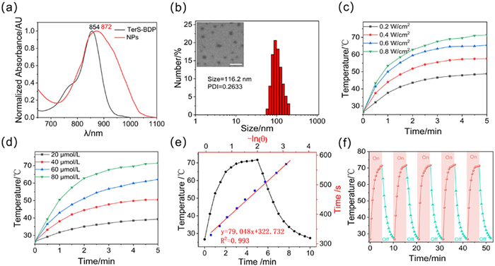
Next, we conducted a thorough assessment of the PCE of TerS-BDP NPs (Fig. S7 in Supporting information). Specifically, when treated with 80 µmol/L TerS-BDP NPs under the different laser conditions, the solution promptly reached 71 ℃ within 5 min (Fig. 3c). Upon exposure to 808 nm laser irradiation at a power density of 0.8 W/cm2 for 5 min, the temperature of the aqueous solutions containing TerS-BDP NPs at concentrations of 20, 40, 60, and 80 µmol/L rose progressively. Notably, the thermal effect was directly proportional to the concentration of TerS-BDP NPs. The PCE of TerS-BDP NPs exhibited the positive correlation with concentration and laser power, suggesting a high degree of controllability in their photothermal performance (Fig. 3d). After the 5-min illumination period, the solution was allowed to cool naturally for an additional 5 min, during which time the temperature almost entirely returned to room temperature, as depicted in Fig. 3e. Based on the temperature response of the heating and cooling curves (Fig. 3e), its PCE is calculated to be 62%. Moreover, we evaluated the photothermal stability of TerS-BDP NPs by subjecting them to five consecutive heating-cooling cycles. The consistent temperature changes observed across these cycles demonstrated the robust photothermal stability of TerS-BDP NPs (Fig. 3f). All data illustrated that TerS-BDP NPs possessed excellent photothermal properties to serve as an efficient phototherapy for PTT.
Given the remarkable ROS generation capacity of TerS-BDP NPs in solution, we further evaluated their PDT potential under NIR irradiation [80]. The 2′,7′-dichlorodihydrofluorescein diacetate (DCFH-DA) probe undergoes enzymatic hydrolysis by cellular esterases to form DCFH, which is subsequently oxidized to the highly fluorescent DCF in the presence of ROS, emitting bright green fluorescence (Fig. 4a). Control experiments demonstrated that HeLa cells incubated solely with DCFH-DA and subjected to 10 min of light irradiation exhibited no detectable green fluorescence. In contrast, cells co-incubated with both DCFH-DA and TerS-BDP NPs under identical irradiation conditions displayed intense green fluorescence. This significant fluorescence enhancement clearly demonstrated the efficient intracellular ROS generation by TerS-BDP NPs and their ability to oxidize DCFH, thereby highlighting their potential for achieving synergistic effects in PDT.
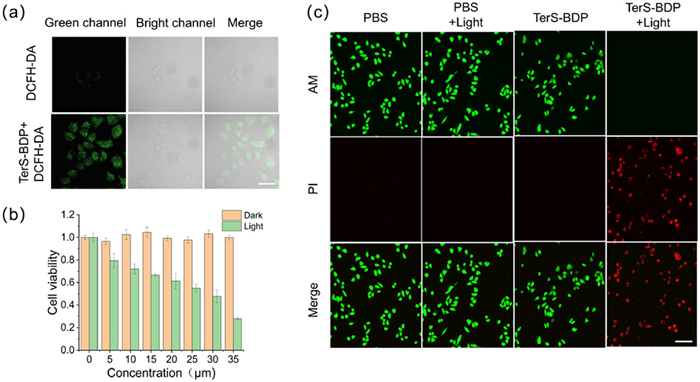
To further explore the cytotoxic effects of TerS-BDP NPs on cancer cells, in vitro phototoxicity was assessed using the MTT assay. As depicted in Fig. 4b, in the absence of light exposure, the cell survival rate remained above 99.8% at a concentration of 35 µmol/L TerS-BDP NPs. However, after a 10-min irradiation with an 808 nm laser, the cell survival rate significantly dropped to 27.9%. Complementing these findings, the live/dead cell staining experiment utilizing calcein-acetoxymethyl ester/propidium iodide (calcein-AM/PI) further validated the remarkable biocompatibility and potent phototoxicity of TerS-BDP NPs. In the phosphate-buffered saline (PBS) control group, as shown in Fig. 4c, green fluorescence was consistently observed irrespective of light exposure, confirming that the laser alone did not induce cytotoxic effects. Similarly, cells treated with TerS-BDP NPs exhibited green fluorescence, indicating that no apoptosis occurred. However, following a 10-min exposure to the 808 nm laser, a morphological transformation from spindle-shaped to round cells was noted, accompanied by the exclusive presence of red fluorescence. This suggests that TerS-BDP NPs, upon light activation, generated substantial heat and ROS, culminating in cell death. Collectively, these results underscored the favorable biocompatibility and high phototoxicity of TerS-BDP NPs, highlighting their potential as effective agents in phototherapy. We have performed a hemolysis assay to further evaluate the hemocompatibility and biosafety of TerS-BDP NPs. The results showed negligible hemolysis (< 5%) at all tested concentrations (20–160 µmol/L). A hemolysis level of 180 µmol/L indicates a 5% hemolysis rate, demonstrating that TerS-BDP NPs do not cause red blood cell damage under physiological conditions. The detailed experimental procedure and corresponding data have been added to the Fig. S8 (Supporting information). These new results further confirm the excellent biosafety of TerS-BDP NPs in agreement with the histopathological findings.
To assess the in vivo therapeutic efficacy of TerS-BDP NPs, we employed a HeLa tumor-bearing mouse model. The animals were monitored continuously for 14 days, and subsequent euthanasia was executed. All related animal experiments were performed according to guidelines approved by the Ethics Committee of Hunan Normal University (No. SYXK (Xiang) 2020–0012). Their organs were harvested for histological analysis. As depicted in Figs. 5a–e, during a continuous 10-min illumination process, the temperature within the tumor tissue of the TerS-BDP NPs+L group escalated to a peak of 71.9 ℃, while the surrounding normal tissues did not show high temperatures. In stark contrast to the PBS group, which only attained a maximum temperature of 34.2 ℃ post-illumination. Previous studies have established that temperatures surpassing 42 ℃ can inflict irreversible damage to biological tissues [81,82], implying that the hyperthermia induced by TerS-BDP NPs under light exposure is potent enough to induce cancer cell necrosis. Over the 14-day monitoring period, it was observed that tumors in the TerS-BDP NPs+L group were nearly obliterated by 6 days and completely eradicated by 8 days. Conversely, tumors in the control groups exhibited continued growth. Importantly, there was no significant alteration in the body weight of the mice throughout the 14-day treatment course, suggesting that TerS-BDP NPs did not detrimentally impact other normal physiological functions.
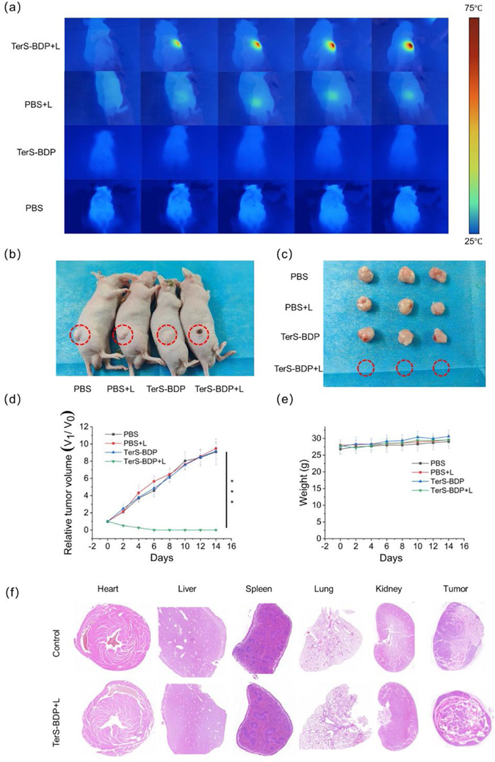
Finally, histopathological analysis was conducted on the heart, liver, spleen, lung, kidney and tumor tissues from both the treatment and control groups using hematoxylin and eosin (H&E) staining. As illustrated in Fig. 5f, the results revealed no significant morphological differences in the heart, liver, spleen, lung and kidney tissues between the control and experimental groups, indicating that 160 µmol/L TerS-BDP NPs did not induce detectable toxicity in these major organs. In contrast, the tumor tissue underwent a notable transformation, transitioning from densely packed and highly cellular structures in the control group to sparsely distributed and reduced cellularity in the treatment group. This observation suggests that TerS-BDP NPs effectively induced structural damage to the tumor tissue and promoted cancer cell apoptosis, further supporting the therapeutic potential.
In summary, TerS-BDP by attaching the terthienyl segment at 3,5-sites was synthesized and its solid-state structure was confirmed by X-ray crystallography for the first time. TerS-BDP has high molar extinction coefficient and wide FWHM. The self-assembled TerS-BDP NPs emitted fluorescence (λem = 926 nm, Φf = 5%) in NIR-Ⅱ region, and the emission band covered the NIR-Ⅱ range of 900–1300 nm. TerS-BDP is an extremely twisted configuration, which is beneficial for producing ROS based on the favorable S1→T2→T1 ISC pathway. TerS-BDP NPs displayed excellent photothermal conversion efficiency (η = 62%). TerS-BDP NPs effectively induced structural damage to the tumor tissue and promoted cancer cell apoptosis, further supporting its therapeutic potential. It can be seen that TerS-BDP has a good anti-tumor effect and can be combined with PTT-PDT therapy of cervical cancer.
Ji Ai: Methodology, Formal analysis, Data curation, Conceptualization. Hankun Zhang: Methodology. Yanru Huang: Validation, Methodology. Peijun Yang: Software. Xin-Dong Jiang: Writing – review & editing, Supervision, Conceptualization. Gaowu Qin: Writing – review & editing. Xiangzhi Song: Writing – review & editing.
The authors declare that they have no known competing financial interests or personal relationships that could have appeared to influence the work reported in this paper.
This work was supported by the National Natural Science Foundation of China (Nos. 22578280, 22078201, 22278447, U1908202), and Liaoning & Shenyang Key Laboratory of Functional Dye and Pigment (Nos. 2021JH13/10200018, 21104023).
Supplementary material associated with this article can be found, in the online version, at doi:
Y. Sun, P. Sun, Z. Li, et al., Chem. Soc. Rev. 514 (2022) 7170–7205. doi: 10.1039/d2cs00179a
H. Lu, J. Mack, Y. Yang, et al., Chem. Soc. Rev. 43 (2014) 4778–4823. doi: 10.1039/C4CS00030G
Z. Qin, T.B. Ren, H. Zhou, et al., Angew. Chem. Int. Ed. 61 (2022) e202201541. doi: 10.1002/anie.202201541
L. Wu, J. Hu, Q. Zou, et al., Nanoscale 12 (2020) 14180–14187. doi: 10.1039/d0nr03269g
S. Luo, E. Zhang, Y. Su, et al., Biomaterials 32 (2011) 7127–7138. doi: 10.1016/j.biomaterials.2011.06.024
C. Yan, Z. Zhu, Y. Yao, et al., Acc. Mater. Res. 5 (2024) 64–75. doi: 10.1021/accountsmr.3c00196
J. Shi, X. Sun, S. Zheng, et al., Biomaterials 152 (2018) 15–23. doi: 10.1016/j.biomaterials.2017.10.032
X. Gao, J. Li, X. Li, et al., ACS Appl. Mater. Interfaces 17 (2025) 35205–35220. doi: 10.1021/acsami.5c05572
Y.Z. Liu, X.Y. Ran, D.H. Zhou, et al., Adv. Funct. Mater. 34 (2024) 2311365. doi: 10.1002/adfm.202311365
C. Yan, Y. Zhang, Z. Guo, Coord. Chem. Rev. 427 (2021) 213556. doi: 10.1016/j.ccr.2020.213556
Y.Y. Duo, L. Zhao, Z.G. Wang, et al., J. Anal. Test. 7 (2023) 245–259. doi: 10.1007/s41664-023-00254-2
J. Li, X. Su, N. Liu, Sens. Actuators B: Chem. 408 (2024) 135535. doi: 10.1016/j.snb.2024.135535
X. Liu, B. Chen, M. Wang, et al., Angew. Chem. Int. Ed. 64 (2025) e202416160. doi: 10.1002/anie.202416160
T. Sun, J.H. Dou, S. Liu, et al., ACS Appl. Mater. Interfaces 10 (2018) 7919–7926. doi: 10.1021/acsami.8b01458
X. Zeng, L. Tang, W. Zhang, et al., Small 21 (2025) 2412296. doi: 10.1002/smll.202412296
Y. Tang, J. Feng, S. Li, et al., J. Mater. Sci. Technol. 213 (2025) 196–212. doi: 10.1016/j.jmst.2024.07.002
S.W. Kim, M.J. Kang, Y. Kim, et al., J. Control. Release 384 (2025) 113946. doi: 10.1016/j.jconrel.2025.113946
Z. Wang, X. Wang, J.B. Wan, et al., Small 17 (2021) 2103780. doi: 10.1002/smll.202103780
Y. Cai, Z. Wei, C. Song, et al., Chem. Soc. Rev. 48 (2019) 22–37. doi: 10.1039/c8cs00494c
L. Sun, Y. Zhao, H. Peng, et al., J. Nanobiotechnol. 22 (2024) 210. doi: 10.1186/s12951-024-02479-4
W. Pang, P. Jiang, S. Ding, et al., Adv. Healthc. Mater. 9 (2020) 2000607. doi: 10.1002/adhm.202000607
Y.Y. Zhao, L. Lu, H. Jeong, et al., Chem. Soc. Rev. 54 (2025) 7749–7768. doi: 10.1039/d5cs00054h
X. Li, S. Lee, J. Yoon, Chem. Soc. Rev. 47 (2018) 1174–1188. doi: 10.1039/c7cs00594f
A. Roshanzadeh, H.C.D. Medeiros, C.K. Herrera, et al., Angew. Chem. Int. Ed. 64 (2025) e202419759. doi: 10.1002/anie.202419759
X. Li, J.F. Lovell, J. Yoon, et al., Nat. Rev. Clin. Oncol. 17 (2020) 657–674. doi: 10.1038/s41571-020-0410-2
K.W.K. Lam, J.H.C. Chau, E.Y. Yu, et al., ACS Nano 17 (2023) 7145–7156. doi: 10.1021/acsnano.2c08855
Y.Y. Zhao, Y. Xu, X. Zhang, et al., Angew. Chem. Int. Ed. 64 (2025) e202506412. doi: 10.1002/anie.202506412
D. Zhang, L. Liu, X.D. Jiang, et al., Res. Chem. Mater. 3 (2024) 103–122.
S.Y. Wang, Y.H. Pan, Y.C. Qu, et al., Smart Mol. 2 (2024) e20230024. doi: 10.1002/smo.20230024
Y. Wu, S. Li, Y. Chen, et al., Chem. Sci. 13 (2022) 5085–5106. doi: 10.1039/d1sc05478c
H. Liu, Z. Li, X. Zhang, et al., Nat. Commun. 16 (2025) 326.
Z. Tu, Y. Wan, J. Ge, et al., Rare Met. 43 (2024) 3220–3231. doi: 10.1007/s12598-024-02626-8
Y.Y. Zhao, X. Zhang, Y. Xu, et al., Angew. Chem. Int. Ed. 63 (2024) e202411514. doi: 10.1002/anie.202411514
H. Zhao, X. Wang, X. Wu, et al., Res. Chem. Mater. 2 (2023) 111–116.
J. Rak, P. Pouckova, J. Benes, et al., Anticancer Res. 39 (2019) 3323–3339. doi: 10.21873/anticanres.13475
K. Yang, S. Zhao, B. Li, et al., Coord. Chem. Rev. 454 (2022) 214330. doi: 10.1016/j.ccr.2021.214330
A. Konieva, V. Deineka, K. Diedkova, et al., ACS Appl. Mater. Interfaces 16 (2024) 43302–43316. doi: 10.1021/acsami.4c08129
D. Hu, Z. Sheng, M. Zhu, et al., Theranostics 8 (2018) 410–422. doi: 10.7150/thno.22226
B. Li, Z. Jiang, D. Xie, et al., Int. J. Nanomedicine 13 (2018) 7289–7302. doi: 10.2147/ijn.s175334
H. Zhang, Q. Sha, G. Li, et al., Mater. Today Chem. 37 (2024) 102018. doi: 10.1016/j.mtchem.2024.102018
Y. Zhang, D. Ma, Y. Guo, et al., ACS Mater. Lett. 7 (2025) 3844–3852. doi: 10.1021/acsmaterialslett.5c01221
Z. Shi, X. Han, W. Hu, et al., Chem. Soc. Rev. 49 (2020) 7533–7567. doi: 10.1039/d0cs00234h
L. Cao, Z. Cui, D. Zhang, et al., ACS Mater. Lett. 6 (2024) 4765–4773. doi: 10.1021/acsmaterialslett.4c01447
Y. Shao, W. Jia, G. Qin, et al., Coord. Chem. Rev. 543 (2025) 216944. doi: 10.1016/j.ccr.2025.216944
X. Ren, C. Wang, X. Wu, et al., J. Am. Chem. Soc. 146 (2024) 6566–6579. doi: 10.1021/jacs.3c11823
W. Sun, X. Wang, Z. Cheng, et al., Biomed. Pharmacother. 158 (2023) 114071. doi: 10.1016/j.biopha.2022.114071
W. Hu, M. Liu, X.F. Zhang, et al., J. Phys. Chem. C 124 (2020) 23558–23566. doi: 10.1021/acs.jpcc.0c07679
X. Zhang, H. Yu, Y. Xiao, J. Org. Chem. 77 (2012) 669–673. doi: 10.1021/jo201413b
X.D. Jiang, S. Li, B.L. Guennic, et al., Phys. Chem. Chem. Phys. 18 (2016) 32686–32690. doi: 10.1039/C6CP05705E
S. Zeng, C. Chen, Z. Guo, et al., Angew. Chem. Int. Ed. 64 (2025) e202513815. doi: 10.1002/anie.202513815
X. Guo, W. Sheng, H. Pan, et al., Angew. Chem. Int. Ed. 63 (2024) e202319875. doi: 10.1002/anie.202319875
D. Chen, Z. Zhong, Q. Ma, et al., ACS Appl. Mater. Interfaces 12 (2020) 26914–26925. doi: 10.1021/acsami.0c05021
D. Zhang, G. Liu, X.D. Jiang, J. Mater. Chem. C 11 (2023) 1668–1677. doi: 10.1039/d2tc05048j
L. Liu, Y. Yuan, Y. Yang, et al., Chem. Commun. 55 (2019) 5851–5854. doi: 10.1039/c9cc01253b
M. Cui, Z. Chai, Y. Lu, et al., Res. Chem. Mater. 2 (2023) 262–276.
Z. Chen, Z. Xu, T. Qin, et al., Sens. Actuators B: Chem. 398 (2024) 134687. doi: 10.1016/j.snb.2023.134687
S. Wu, W. Zhang, C. Li, et al., Chem. Sci. 15 (2024) 5973–5979. doi: 10.1039/d3sc06976a
Z. Wang, M. She, J. Chen, et al., Adv. Opt. Mater. 10 (2022) 2101634. doi: 10.1002/adom.202101634
J. Min, T. Ameri, R. Gresser, et al., ACS Appl. Mater. Interfaces 5 (2013) 5609–5616. doi: 10.1021/am400923b
F. Li, X. Dai, L. Zhang, et al., Fuel 370 (2024) 131812. doi: 10.1016/j.fuel.2024.131812
J. Killoran, L. Allen, J.F. Gallagher, et al., Chem. Commun. (2002) 1862–1863.
D.O. Frimannsson, M. Grossi, J. Murtagh, et al., J. Med. Chem. 53 (2010) 7337–7343. doi: 10.1021/jm100585j
K. Flavin, K. Lawrence, J. Bartelmess, et al., ACS Nano 5 (2011) 1198–1206. doi: 10.1021/nn102831x
Z. Ye, C.Y. Kwok, S.L. Lam, et al., J. Am. Chem. Soc. 147 (2025) 14915–14923. doi: 10.1021/jacs.5c00974
S. Bairagi, S. Giri, G. Joshi, et al., Angew. Chem. Int. Ed. 64 (2025) e202417170. doi: 10.1002/anie.202417170
Z. Liu, B. Tang, S. Zhang, Ind. Eng. Chem. Res. 61 (2022) 1596–1605. doi: 10.1021/acs.iecr.1c04058
W. Jin, Z. Chen, Y. Wang, et al., Chin. Chem. Lett. 35 (2024) 109328. doi: 10.1016/j.cclet.2023.109328
K. Zhao, G. Shen, Y. Liu, et al., ACS Appl. Mater. Interfaces 16 (2024) 67532–67544. doi: 10.1021/acsami.4c18320
L. Wang, A. Mei, N. Li, et al., Chin. Chem. Lett. 35 (2024) 108974. doi: 10.1016/j.cclet.2023.108974
C. Wang, W. Chi, Q. Qiao, et al., Chem. Soc. Rev. 50 (2021) 12656–12678. doi: 10.1039/d1cs00239b
K.R. Anupriya, J. Thomas, M.R. Nagar, et al., Dyes Pigm. 206 (2022) 110658. doi: 10.1016/j.dyepig.2022.110658
J. Ping, H. Peng, J. Qin, et al., Microchim. Acta 185 (2018) 2189.
H. Schweikl, M. Godula, C. Petzel, et al., Dent. Mater. 33 (2017) 110–118. doi: 10.1016/j.dental.2016.11.003
T. Mikulchyk, S. Karuthedath, C.S.P. De Castro, et al., J. Mater. Chem. C 10 (2022) 11588–11597. doi: 10.1039/d2tc02263j
Y. Zhu, J. Liu, M. Lv, et al., Chin. Chem. Lett. 35 (2024) 109446. doi: 10.1016/j.cclet.2023.109446
Y.G. Fang, C. Zhu, L. Shen, et al., J. Am. Chem. Soc. 146 (2024) 28845–28855. doi: 10.1021/jacs.4c08578
M.A. Filatov, T. Mikulchyk, M. Hodée, et al., J. Mater. Chem. C 13 (2025) 6993–7003. doi: 10.1039/d4tc04850d
V.N. da Rocha, M.H. Köhler, K. Nagata, et al., Spectrochim. Acta A 293 (2023) 122500. doi: 10.1016/j.saa.2023.122500
L. Yang, J. Tang, H. Yin, et al., ACS Biomater. Sci. Eng. 8 (2022) 880–892. doi: 10.1021/acsbiomaterials.1c01418
M.J. Reiniers, R.F. van Golen, S. Bonnet, et al., Anal. Chem. 89 (2017) 3853–3857. doi: 10.1021/acs.analchem.7b00043
J. Wang, B. Hao, K. Xue, et al., Adv. Mater. 34 (2022) 2205653. doi: 10.1002/adma.202205653
X. Zhu, W. Feng, J. Chang, et al., Nat. Commun. 7 (2016) 10437. doi: 10.1038/ncomms10437

Figure 1 Chemical structures and optical properties of S-BDP and TerS-BDP, and self-assembling and theranostic mechanisms of TerS-BDP NPs.

Scheme 1 (a) Synthetic route of 3,5-trithienyl aza-BODIPY TerS-BDP, and oak ridge thermal ellipsoid plot (ORTEP) views of TerS-BDP (CCDC: 2468204) (displacement ellipsoids at the 30% probability level). (b) Front and (c) side views of the molecular structure of TerS-BDP.

Figure 2 (a) Absorbance spectra of S-BDP and TerS-BDP. (b) Fluorescence spectra of S-BDP and TerS-BDP. (c) Spatial distribution and TerS-BDP orbital calculations. (d) DPBF degradation rate curves with S-BDP or TerS-BDP in toluene. (e) ROS generation of S-BDP or TerS-BDP (5 µmol/L) in DMSO under continuous 808 nm laser (0.3 W/cm2) irradiation for 10 min using dihydrorhodamine 123 (DHR123) (10 µmol/L) as an indicator. (f) ESP distribution diagram of TerS-BDP.

Figure 3 (a) Comparison of absorption spectra between TerS-BDP in CH2Cl2 and TerS-BDP NPs in aqueous solution. (b) Transmission electron microscopy of TerS-BDP NPs and DLS in aqueous solution. Scale bar: 150 nm. (c) Heating curve of TerS-BDP NPs (80 µmol/L) when the power density increased. (d) Heating curve of different concentrations of TerS-BDP NPs under laser irradiation (0.8 W/cm2). (e) Heating curve of TerS-BDP NPs (80 µmol/L) during laser switching, plot of cooling time of TerS-BDP NPs versus negative natural logarithm of the temperature from the cooling test. (f) Five circles of heating-cooling processes of TerS-BDP NPs (80 µmol/L) under laser irradiations (0.8 W/cm2).

Figure 4 (a) After 10 min of 808 nm laser (0.8 W/cm2) irradiation, the in vivo ROS map of each group were washed three times with PBS buffer; Scale bar: 30 µm. (b) MTT assay was performed after treatment with different concentrations of TerS-BDP NPs under 808 nm laser (0.8 W/cm2) irradiation. Data are presented as mean ± standard deviation (SD) (n = 3). (c) AM/PI co-staining in HeLa cells in absence/presence of 808 nm laser (0.8 W/cm2) irradiation for 10 min. Scale bar: 150 µm. 35 µmol/L TerS-BDP NPs are abbreviated as TerS-BDP.

Figure 5 (a) Whole-body thermal images of HeLa tumor-bearing mice. (b) Photo comparison of mice before and after treatment. (c) Tumor contrast map of mice before and after treatment. (d) Changes in tumor volume in mice during treatment. Data are expressed as mean ± SD (n = 4). ***P < 0.001. (e) Changes in body weight of mice with days during treatment. (f) Heart, liver, spleen, lung, kidney and tumor sections of mice before and after treatment. For clarity, 160 µmol/L TerS-BDP NPs are abbreviated as TerS-BDP.

 扫一扫看文章


扫一扫关注我们
 DownLoad:
 下载:
 下载: