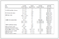
Table 1.
Long neoantigen peptide compared to short neoantigen.

Advances in neoantigen peptide-based nanovaccines for targeted tumor immunotherapy: Multifaceted strategies, challenges, and future perspectives
Sameena Noor Khattak , Kodzo Prosper Adzavon , Weijian Zhao , Wang Sheng


In recent decades, immunotherapy has emerged as a prominent field in cancer treatment and has undergone significant advancements [1-3]. Immunotherapeutic approaches such as immune checkpoint inhibitors, chimeric antigen receptor T cell therapy, and vaccines are the focal points [4-8].
The cancer vaccine is an immunological approach that activates immune cells to target cancer cells and facilitates the establishment of long-term immune memory. The vaccine accomplishes this by targeting lymph nodes (LNs) and delivering antigen to antigen-presenting cells (APCs), mainly dendritic cells (DCs). APCs then present the antigen to adaptive immune cells, such as T cells, which induce potent antitumor immunity and long-term immune memory, thereby inhibiting tumor growth, recurrence, and metastasis [9-12].
Tumor-associated antigens (TAAs), which are typically expressed in healthy cells but exhibit increased expression in cancer cells, were the main focus of earlier vaccination campaigns. Because of their limited tumor specificity, which increases the risk of non-specific toxicity and autoimmunity, TAAs have had limited success. In response to this issue, novel vaccination approaches known as personalized cancer vaccines have surfaced, which target antigens that are specific to cancer cells. These antigens are highly personalized tumor-specific antigens (TSAs), also known as neoantigens, which appear during the tumorigenesis process and are not present in healthy cells. Personalized cancer vaccines induce anti-tumor immune responses to specific epitopes in cancer cells. This increases the targeted efficacy of the anti-cancer immune response and reduces off-target toxicity [13-17].
There are several vaccine platforms based on the type of neoantigen used. These include nucleic acid antigens such as DNA and RNA, cell membrane antigens derived from tumor cells, protein antigens, and peptide antigens [18-22]. Peptide-based vaccines have attracted a lot of interest due to their many advantages. These include the lower cost, ease of modification, and lack of adverse effects of peptide antigens [23]. Despite their safety, peptide vaccines have not been as effective due to poor targeting, low immunogenicity, and reduced accumulation in draining LNs [24]. The primary cause is the absence of antigen (peptide) accumulation in the draining LNs, where DC cross-presentation triggers local immune responses. Several strategies have been used to address these issues. One way to enhance the peptide's immunogenicity is to modify the peptide ligand at the amino acid level. Another is the use of long, synthetic, multi-epitope peptides to enhance the immune response by promoting antigen cross-presentation and multi-targeting. Additional tactics include the use of immunological adjuvants to stimulate APCs for improved antigen presentation and combination therapy [25-28].
Nanotherapy has shown great promise in the therapeutic effectiveness of cancer vaccines [29-32]. The characteristics of nanomaterials used in nanovaccine formulation significantly enhance the immunogenicity of the vaccine [33]. Nanoparticles (NPs) improve the controlled release of vaccine ingredients and protection against degradation. Because they can be flexibly adapted or modified for various purposes, NPs offer several ways to enhance the effectiveness of vaccines. Their size can be controlled, favoring nanovaccine delivery to LNs. Their surface can be modified to target APCs and deliver vaccine components (antigens, adjuvants) with high efficiency [34-39]. In addition to their protective and delivery properties, nanomaterials may have an adjuvant effect that boosts the efficacy of the vaccine. The aim of this review is therefore to explore the advantages of NPs in the development of peptide neoantigen vaccines, the advantages of combining the neoantigen peptide nanovaccine with other therapies, such as immunotherapy and conventional therapy, while noting the challenges, strategies for improving them, and the avenues to be explored.
Researchers have become interested in therapeutic peptides in recent years due to their numerous benefits. Some advantages are low molecular weight, ease of synthesis, ease of modification, and low toxicity [40-43]. Numerous vaccines have been made using peptides as antigens, offering an immunotherapy approach to cancer treatment [44-48]. TSAs, also known as neoantigens, and TAAs, designed to activate T cell response in vivo, are the two types of antigens used to produce peptide vaccines [49]. Neoantigens have several benefits over TAAs in immunotherapy. Among these are their tumor-specificity and the fact that they are absent from healthy cells, which lowers the possibility of autoimmune toxicity. By avoiding the consequences of negative selection, T cells generated through stimulation with neoantigens can evade central tolerance [14,50,51].
Vaccines based on peptides can activate T cells through the presentation of the antigen by APCs. Peptide antigens are recognized by the CD4+ T and CD8+ T cell receptors after being taken up and displayed on the surface of APCs on human leukocyte antigen (HLA) molecules (Fig. S1 in Supporting information) [52-56].
Neoantigen-based peptide vaccines are predicted antigens derived from somatic mutations, including single-nucleotide variations (SNVs), insertions and deletions (INDELs), and gene fusions. Antigen selection remains one of the most important points in the peptide vaccine manufacturing process. The selected antigen must satisfy several requirements with a high degree of satisfaction. This requires that it be recognized by the patient's T lymphocytes and that it appears very frequently and exclusively on the surface of tumor cells. In neoantigen-based peptide vaccines, epitopes are often short immunogenic amino acid sequences that adaptive immune receptors like T cell receptors (TCRs) and B cell receptors (BCRs) can recognize. These epitopes are chosen as potential vaccine candidates by established methods that employ empirical methods and cutting-edge bioinformatics [24,56-60].
Two different kinds of peptides are typically used in peptide neoantigen-based vaccines. These include the long peptides, which have 20 amino acids or more, and the short peptides, which are normally 8–10 amino acids in size [11,61].
The half-life of short peptides in serum is extremely short. They are easily degraded. APCs do not even need to process and present them before they can bind to HLA class Ⅰ molecules. They bind to non-professional APCs that are unable to generate costimulatory signals, like fibroblasts. This could, however, lead to either short-term tolerance or the induction of CD8+ T cells without the simultaneous activation of CD4+ helper T cells. Shorter peptides are typically limited to HLA type because size limits the variety required for high HLA polymorphisms in the general population [61,62].
The inability to induce CD8+ T memory cells and prevent immunological tolerance to immunizing antigens underscores two reasons why short peptide vaccination is far from ideal.
For a peptide vaccine to be most effective, it must have the capacity to activate both CD8+ T cells and CD4+ T cells. To accomplish this, the peptide must contain CD8+ epitopes that are presented on major histocompatibility complex (MHC) I molecules to activate CTL-mediated antitumor immunity, and CD4+ epitopes that are presented on MHC Ⅰ molecules to activate T-helper cells, preserving CTL effector function. Compared to short peptides, synthetic long peptides (SLPs) are more immunogenic and stable. They are initially taken up by APCs, processed, and presented by HLA class Ⅰ and Ⅱ molecules to T cells. This leads to the recruitment and activation of CD4+ T cells and CD8+ T cells and antibody production by B cells. SLPs thus induce potent and persistent antitumor immune responses. In this way, SLPs induce a powerful and persistent anti-tumor immune response, superior to the full antigen from which they are derived (Table 1) [61,63,64].
 DownLoad:
CSV
| Feature | Long neoantigen peptide | Short neoantigen peptide |
| Immune response | CD8+ T and CD4+ T cells | Primarily CD8+ T cells |
| MHC presentation | MHC Ⅰ and MHC Ⅱ (via APCs) | Direct MHC Ⅰ binding |
| Stability | Higher | Lower (rapid clearance) |
| APC dependence | Required | Not required |
| Epitope coverage | Multi-epitope possible | Single epitope |
| Risk of tolerance | Lower (avoids direct MHC binding) | Higher (can induce tolerance) |
| Manufacturing complexity (epitope selection) | Higher (need processing prediction) | Simpler (direct epitope selection) |
Although peptide neoantigen platforms elicit T-cell-mediated immune responses against cancer cells, their efficacy in clinical trials remains a challenge. Low immunogenicity, immune escape, and the tumor immunosuppressive environment are the most important of these challenges. Researchers have therefore found ways to increase the immunogenicity of vaccines based on the use of peptide neoantigens. This involves using strong but nontoxic immunological adjuvants, modified peptides, multi-epitope peptides, and a combination therapy strategy.
The ability of an antigen to be recognized by a T cell with maximum activation of these cells is what makes cancer vaccines effective. Unfortunately, peptide neoantigens often fail in this task when tested in vivo. Peptides can be altered to improve their affinity and binding to the presenting MHC because MHC molecules are crucial for antigen recognition and presentation. These modifications involve substituting amino acids in the peptide chain [65-67]. These include amino acid substitutions at the anchor positions of the MHC bond to increase the peptide's binding affinity with MHC Ⅰ. Amino acid substitutions can also be made at the TCR-contacting residues in the peptide's T-cell contact interface to improve the T-cell response [28,68,69].
In several studies, multiple epitopes combined to create a long synthetic peptide have significantly increased the vaccine's immune response. The purpose of multi-epitope peptide antigen vaccines is to elicit immune responses against multiple important epitopes at the same time. This strategy increases the recruitment of a diverse range of T cells, which improves vaccine efficacy. Clinical trials are underway for several of these multi-epitope peptide vaccines, and the results are encouraging [70-73].
In addition to the possibility of modifying the peptide to enhance the immunogenicity of the neoantigens, adding potent adjuvants is a strategy that improves antigen uptake and directs intracellular trafficking, thereby promoting antigen presentation [52]. The purpose of adjuvants is to facilitate the recognition by APCs, ensure efficient uptake by APCs, and promote their appropriate and complete activation. More specifically, adjuvants stimulate the generation of antigen-presenting signals and co-stimulatory signals by activating APCs [74].
Several adjuvants have been tested with convincing results both in vitro and in vivo. Toll-like receptor (TLR) agonists like poly-ICLC (TLR3 agonist) and CpG (TLR9 agonist) are among them [75]. Another promising adjuvant repertoire consists of STING agonists. Clinical trials have examined other adjuvants, including GM-CSF and Montanide ISA [49,64,76].
To overcome low immunogenicity, one of the main obstacles to neoantigenic peptide vaccination against cancer, researchers have chosen a combination therapy.
According to several studies, when cancer cells undergo traditional treatments like chemotherapy, phototherapy, or radiation therapy, they release antigens known as damage-associated molecular patterns (DAMPs) [77-80]. These antigens can induce DC maturation and T cell activation [81-84]. Considering these facts, it is evident that this would enhance antigen presentation and T cell recruitment. In this regard, peptide neoantigen vaccines have been used in several clinical trials in combination with traditional treatments. Compared to when the vaccine was administered alone, the results indicated a noticeably greater immune response in combination therapy [85-90].
A challenge for nearly all T-cell-based immunotherapies is the immunosuppressive microenvironment of cancers, especially solid cancers [52,91]. Combinatorial methods using direct or indirect tumor microenvironment modifiers, such as B-Raf proto-oncogene (BRAF), mitogen-activated protein kinase (MEK), or poly(ADP-ribose) polymerases (PARP) inhibitors, or treatments based on vascular endothelial growth factor (VEGF) inhibitors and immune checkpoint inhibitors have already been tested to address this issue (NCT04713514, NCT02382549, NCT01551745) [52,92,93].
In clinical trials, all of the methods mentioned earlier for increasing the immunogenicity of the peptide neoantigen have shown improved results. However, to maximize the immune response, several aspects still need refinement. One issue is adjuvant toxicity, which can affect nearby cells [94]. Another is the degradation of injected peptides in systemic circulation, due to exposure to enzymes [40]. A major challenge is achieving the right dose and optimal bioavailability of vaccine components in the immune organs simultaneously. To address these issues, nanotherapy presents a promising alternative. Nanotherapy allows vaccine components to be combined into a single molecule, enabling the linkage of antigen and adjuvant. It also protects the components from potential degradation and enhances targeting of immune organs and cells [95].
Recent years have seen a significant advancement in the use of nanotherapy in medicine [96-103]. Nanomaterials are adaptable and can be changed for many different uses. Therefore, the introduction of nanomaterials as cancer vaccine cargo represents a significant advancement in the field of cancer therapy. They increase the duration of in vivo circulation and boost vaccine immunogenicity by efficiently preventing the degradation of vaccine constituents (peptides, adjuvants, and others). Apart from their protective function, nanovaccines can be produced to target immune organs (spleen, lymphatic nodules), APCs, or the tumor microenvironment with high doses locally. Additionally, nanovaccines facilitate antigen cross-presentation, which improves T cell recruitment. In lymphoid tissues, they can also interact with cells like macrophages to enhance humoral and cell-mediated immune responses. Some nanomaterials even act as adjuvants, enhancing antigen uptake and presentation by APCs [104-108].
For a vaccine to be effective, it must be able to reach the LNs and reach APCs (DCs). One of the most important details in targeting LNs with nanovaccines is their size [109]. NPs with a diameter of <10 nm can enter lymphatic capillaries and blood vessels straight from injection sites. However, due to the fast blood flow, blood vessels primarily eliminate them because of their minuscule size. In contrast, interstitial flow readily transports NPs with sizes ranging from 10 nm to 100 nm to lymphatic capillaries and lymph nodes. Small lymphatic channels prevent NPs larger than 100 nm from efficiently reaching LNs. Nonetheless, DCs in peripheral tissues can uptake NPs larger than 100 nm, which are then transported to lymphatic capillaries and drained into LNs [106,107]. Consequently, a nanovaccine should be between 10 nm and 100 nm in size to reach LNs efficiently.
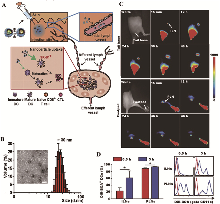
The significance of nanovaccine size in targeting LNs is illustrated by the work of Zhang and associates. They created a nanovaccine based on an HDL receptor (SR-B1)-coupled peptide antigen named α-AP. With a diameter of ~30 nm, this nanovaccine, called α-Ap-FNPs, was able to enter LNs and enhance the antigen presentation through DCs (Fig. 1) [110].
The LNs can also be reached by receptors in the lymphatic system. Among them is the lymphatic vessel endothelial hyaluronan receptor 1 (LYVE-1). Hyaluronic acid (HA)-coated nanovaccines can enter LNs by attaching to the LYVE-1 receptor (Fig. S2 in Supporting information) [111,112].
The surface appearance of NPs is an important factor in targeting LNs. Macrophages frequently capture globular-surfaced NPs, which decreases their interaction with LNs. NP shape is also important, as NPs must migrate through blood vessels to the LNs. Choosing non-spherical NPs, like disks and nanodroplets, is still one of the best strategies to promote their migration to the LNs [107,113].
LNs' ability to target vaccines is also influenced by the surface charge of nanomaterials. Negatively charged NPs have been shown to accumulate easily in lymphatic vessels, while positively charged NPs do not. Conversely, nanomaterials that are positively charged have a higher chance of interacting with the cell membrane and being absorbed by DCs [114]. The surface charge of nanovaccines in targeting LNs and DCs remains a subject for further study, as there are several conflicting studies.
APCs are responsible for processing and presenting the vaccine antigen to T cells. The most effective method of maximizing antigen cross-presentation and enhancing nanovaccine efficacy is to create a nanovaccine that can target APCs specifically. Therefore, combining molecules or ligands that can target APCs to create NPs is a great approach.
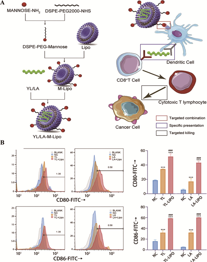
Mostly found on the cell membrane surface of DCs and macrophages, the mannose (Man) receptor is an endocytosis receptor that belongs to the C-type lectin superfamily. Therefore, Man is used to make NPs that target macrophages or DCs [115-120]. The M-YL/LA-Lipo nanovaccine, which is designed to transfer YL or LA peptide epitopes to the DCs for cross-presentation, is one example (Fig. 2) [121].
The most common APCs targeted in the production of a nanovaccine are DCs because they are the APCs with the best antigen presentation. Man receptors are not the only C-type lectin receptors that are targeted on DCs for this purpose [122]. These include, among others, Clec 9A, DC-SIGN, and DEC-205 [123-125]. Apart from C-type leptin receptors, other receptors like CD40, tumor necrosis factor-alpha (TNF-α) family receptor, and CD11c, an integrin receptor, are also targeted on DCs [107,126]. A nanovaccine called PLGA-(OVA + poly I:C + R848) NP has been formulated to coat monoclonal antibodies CD40, DEC-205, and CD11c on its surface. DCs were successfully targeted by this nanovaccine through CD40, DEC-205, and CD11c receptors. As a result, the vaccine's immunological capacity was enhanced, and its T-cell response was noticeably higher than that of the vaccine without monoclonal antibodies (Fig. S3 in Supporting information) [127].
In another study, targeting the class B1 scavenger receptor, SR-B1 (natural HDL receptor), at the DCs' membrane enabled delivery of the neoantigen peptide (Ap) to both mature and immature DCs. To achieve this, the nanovaccine α-Ap-FNPs is formulated by employing an amphipathic α-helical peptide known as α-peptide. As a result, DCs uptook the nanovaccine through the α-peptide/SR-B1 targeting with high efficiency (Fig. S3 in Supporting information) [110].
Peptide nanovaccines are made from a variety of NP types. These include polymer-based NPs, lipid-based NPs, virus-like NPs, ferritin (FN) NPs, and inorganic NPs [95].
The selection of materials is crucial when creating nanovaccines. Nanomaterials are selected to optimize vaccine immunogenicity. These include the size, shape, and surface charge of the nanovaccine, its capacity for antigen control release and biocompatibility, its capacity to target LNs and immune cells (APCs in general), the adjuvant effect of specific materials, limiting the targeting of neighboring cells, minimizing the toxicity that certain adjuvants can cause, and protecting vaccine components from potential degradation (Table 2) [128-142].
DownLoad:
CSV
| Material | NP type | Immunological advantage | Example of nanovaccine | Ref. |
| DOTAP/DOTMA | Cationic liposome NPs | APCs targeting surface charge dependent; Adjuvant effect (via ROS generation) | DOTAP/Trp2 | [133] |
| PEG | Polymer NPs | LN targeting by reducing extra cellular matrix stroma targeting | PEG-PMA-OVA | [134] |
| HA | Polymer NPs | LN (via LYVE-1 receptor) and APCs (via CD44 receptors) targeting, Adjuvant (DC’s maturation, cytokine release) | HANG-V | [135] |
| CS | Polymer NPs | Adjuvant (PAMPs, DCs maturation) | CS gel-OVA | [136] |
| Mn, Zn, Ca | Metall NPs | Adjuvant (trigger STING signaling) | OVA@Mn-DAP, SZCP/APs-CpG, | [137,138] |
| Gold NPs | Metall NPs | Adjuvant (DCs maturationand T cells activation), size and shape dependent LN targeting | AuNP-OVA | [139] |
| Virus-like particles | Virus-like NPs | Adjuvant (PAMPs); LN targeting size dependent | P22-OVAT/B | [140] |
| FN NPs | Protein NPs | LN targeting size dependent | OVAT-FNs and M30-FNs | [141] |
| Baterial cytoplasmic membrane | Cellular membrane coated NPs | Adjuvants (PAMPs) | OVA@MMSNs@BM-Man | [142] |
Adjuvant nanomaterials serve as immunostimulants, often acting as danger signal molecules and functioning as pathogen-associated molecular patterns (PAMPs), DAMPs, or their mimics. They can target APCs through TLRs, cyclic GMP-AMP synthase-interferon gene stimulator (cGAS-STING), C-type lectin receptors (CLRs), or other pattern recognition receptors (PRRs) to induce APCs activation [143-145]. Cell membrane-coated NPs (cancer cells, bacteria, yeast) are a good example [146,147].
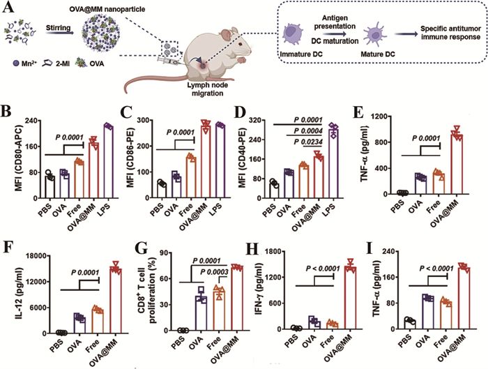
Studies have identified several metal ions, including Mn2+, Zn2+, and Ca2+, as novel immunomodulators for cancer metalloimmunotherapy. They trigger cellular cyclic GMP-AMP synthase (cGAS)-interferon gene stimulator (STING) signaling [148-153]. Numerous metallic nanovaccines have been developed based on these immunomodulating characteristics. One example is the OVA@MM nanovaccine, formulated with Mn2+, 2-methylimidazole, and the neoantigen ovalbumin (OVA). OVA@MM effectively induced DC maturation and elicited a specific antitumor immune response (Fig. 3). Results indicated that OVA-free particles could activate several signaling pathways to enhance BMDC maturation, cross-presentation, and the production of various cytokines [154]. In addition to these metals, gold NPs (AuNPs) can also trigger an immune response by maturing DCs and, depending on their form (cages, rods, and stars), activating CD4+ or CD8+ T cells [29,155].
Other nanomaterials, in addition to metallic nanomaterials, can boost the immune response and produce the adjuvant effect. Certain cationic liposome materials, like 1,2-dioleoyl-3-trimethylammonium propane (DOTAP) and N-[1-(2,3-dioleyloxy)propyl]-N,N,N-trimethylammonium chloride (DOTMA), can be effective immunologic adjuvants by promoting DC maturation through reactive oxygen species (ROS) generation [156-159]. Several studies have already used these materials in the manufacture of peptide neoantigen vaccines [133,159-162]. DOTAP/E7 lipopeptide vaccine is one such example (Fig. S4 in Supporting information). In the animal model of human papillomavirus (HPV)-positive TC-1 tumor mice [163], the injected nanovaccine demonstrated a better immunogenic response than neoantigen peptide E7 alone [163].
Moreover, some polymers have adjuvant properties. Chitosan (CS), a natural polymer, is one example of this. It has been shown that CS and its derivatives can induce DCs’ maturation [164,165]. An example of a neoantigen peptide nanovaccine made from CS is nanovaccine CS/pIC/1338aa-PADRE [166]. Apart from its capacity to target LNs, HA also acts as an immunological adjuvant by enhancing DCs’ maturation, antigen uptake, and cytokine secretion in a molecular weight-dependent manner [111,112,167].
Virus-like particles are also a good material with an immunological adjuvant effect [168]. Virus-like particles can be recognized and taken up by DCs, initiating DC maturation [169,170]. It is also employed in the formulation of a neoantigen peptide nanovaccine [171-173]. This is the case with neoVLPs, nanovaccines, designed to deliver a variety of neoantigen peptides [174].
It is crucial to produce anti-cancer nanovaccines using materials that maximize the targeting of LNs and DCs, because the primary goal of the vaccine is to deliver the antigen to APCs (DCs in particular) in LNs. Targeting LNs and DCs with NPs depends on several factors. Electrical charge, NP size, shape, and surface appearance or modification are factors that we have already covered [175].
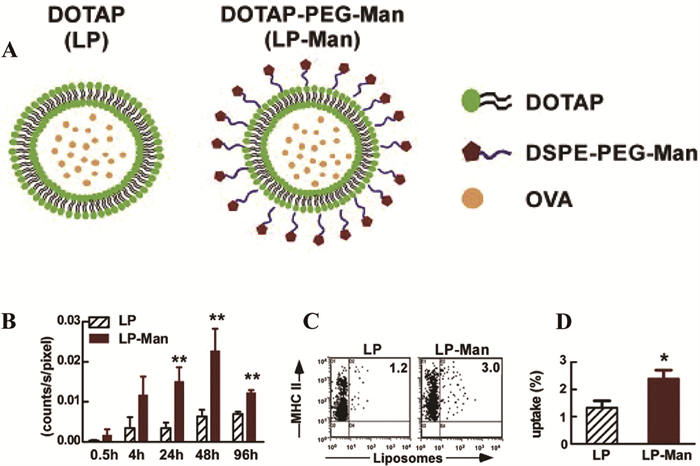
Because cationic liposomes can be preferentially absorbed by APCs, they are utilized far more frequently in the production of peptide antigen vaccines than anionic or neutral liposomes. Therefore, positively charged nanomaterials like DOTAP are the preferred option [107]. Since negative nanomaterials are the most effective option for targeting LNs, the best method is still to add a ligand to the cationic liposomes to improve LN targeting or modify their surface with polyethylene glycol (PEG). Targeting LNs can also be accomplished with PEGylation, a modification of the nanovaccine that incorporates a PEG polymer material. This promotes the nanovaccine's accumulation in LNs by lowering its interaction with extracellular matrix cells [113,134,176]. An example is the study that formulated the LP-Man nanovaccine, which was made using DOTAP, PEG materials, and Man ligand to deliver the OVA peptide antigen. The modification of the cationic liposome by Man ligand improved the accumulation of the nanovaccine in LNs and increased OVA uptake by BMDCs by two to three times (Fig. 4) [177].
We previously talked about how HA polymer materials can target LNs through the LYVE-1 receptor. Additionally, through CD44 receptors, HA can also target APCs (Fig. S2) [111].
Controlling size through the selection of nanomaterials is crucial because size plays a significant role in LN targeting. Nanomaterials like virus-like particles, exosomes, ferritin NPs, and metal particles have shown good size for LN targeting [108,168,178].
In recent years, combining two or more adjuvants to form nanovaccines to improve the antigen’s immune response has become a key area of research [179-182]. According to a recent study, antitumor efficacy in syngeneic and humanized mouse models was considerably increased when combining STING agonists and TLR2 agonist pretreatment [183]. Several adjuvant combinations have been used in various studies in the formulation of cancer neoantigen peptide vaccines, and the results have shown a synergistic effect that has improved the immune response [138,147,184-187].
Chen and colleagues created a nanovaccine by combining the murine peptide neoantigen AdpgK with the TLR7/8 agonist adjuvant R848 and the TLR9 agonist adjuvant CpG ODN. This nanovaccine, BanNVs, increased adjuvant and neoantigen co-delivery to LNs and DC uptake, accompanied by reduced acute systemic toxicity with enhanced neoantigen-specific cytotoxic T-cell responses. BanNVs made T cells more sensitive to the immune checkpoint. When BanNVs and antiprogrammed cell death protein 1 (aPD-1) were combined, a significant therapy response was produced, and 70% of neoantigen-specific tumors completely regressed without recurrence [188].
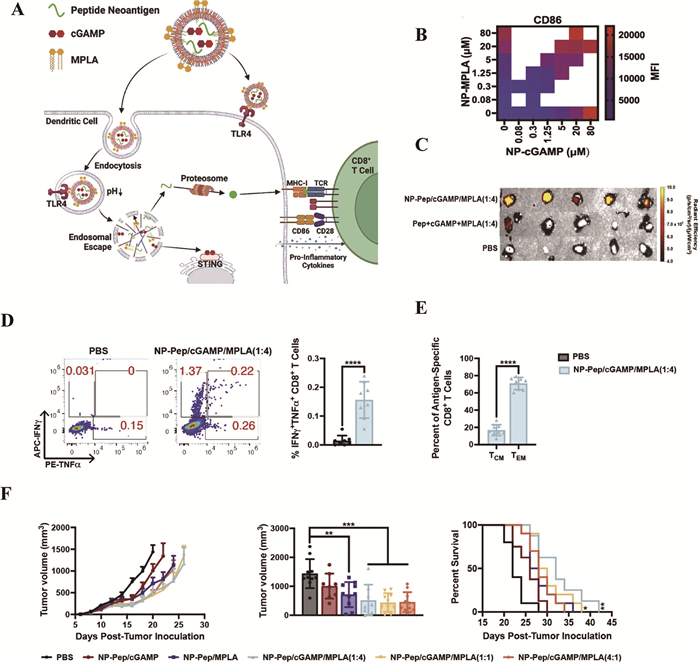
In a different study, Wilson et al. recently developed a peptide neoantigen nanovaccine that encapsulated TLR4 agonist monophosphoryl lipid A (MPLA), the agonist adjuvant STING cGAMP, and the peptide neoantigen OVA. The nanovaccine enhanced DC uptake of peptide antigen and LN accumulation. The co-administration of MPLA, cAMP, and peptide antigen boosted antigen-specific CD8+ T cell responses and slowed tumor growth (Fig. 5) [189].
The NvH loading OVA antigen in vitro is an example of a peptide neoantigen nanovaccine manufactured with more than two adjuvants. The adjuvants are R848, a TLR7/8 agonist; CpG, a TLR9 agonist; and cGAMP, a STING agonist. Combined with the immune checkpoint inhibitor, the combination called NvIH reduced multilevel immunosuppression of the tumor microenvironment, induced both innate and adaptive immune responses with memory, and regressed tumors [190].
The nanovaccine formulation's combination of multiple adjuvants stimulates DCs via multiple pathways, increasing DC activation and resulting in significant T cell recruitment. Apart from the various stimulation pathways, the combination of some adjuvants has been shown to have a synergistic effect that improves the immune response. Consequently, using a multi-adjuvant approach is a smart tactic when creating nanovaccines that contain peptide neoantigens.
Combinatorial therapy is becoming increasingly common due to its multitarget approach, which fosters cross-interactions that enhance each treatment’s efficacy. As previously mentioned, combining the vaccine with conventional treatments and other therapies improves the immune response to the vaccine and, as a result, promotes a better outcome. Combining peptide neoantigen nanovaccines with other therapies is still a better option because the nanovaccine offers improved protection and other benefits in terms of vaccine efficacy.
Although antiprogrammed death-ligand 1 (anti-PD-L1) antibody treatments typically prevent PD-L1 from interacting with PD-1 on T cells or from being present on tumor cells, recent research has shown that PD-L1 expressed on immune cells, especially APCs, has an immunosuppressive function. A promising but untested therapeutic target for boosting immune responses and altering the tumor microenvironment is selective modulation of PD-L1 on APCs. Several studies have combined immune checkpoint inhibitors and peptide neoantigen nano vaccines. These studies have demonstrated that the nanovaccines significantly enhance the effectiveness of checkpoint inhibitor therapy by sensitizing cancer cells to checkpoint inhibitor therapy. Long-lasting immune responses and immune memory are also encouraged by a reversal of immunosuppression and an increase in immune cell infiltration [188,189,191,192]. One such example is the research conducted by Qin Liu and colleagues. They created a nanovaccine by encapsulating a peptide neoantigen and the adjuvant MontanideTM ISA 51 in an amphiphilic 1,2-distearoyl-sn-glycero-3-phosphoethanolamineis (DSPE)-PEG2000 liposome. The nanovaccine's small size and PEG modification allowed it to be successfully delivered to LNs, where DCs were taken up and matured. This triggered the activation of T cells against the cancer cells. When combined with the checkpoint inhibitor, this immune response was enhanced with long-lasting immunemory (Fig. 6A) [193]. Thus, it is evident that a combination of inhibitor checkpoints and the peptide antigen nanovaccine can enhance the effectiveness of both treatments with a strong immune response. In this case, the vaccine has a stronger impact with long-lasting immune memory, and the resistance to inhibitor checkpoints is decreased.
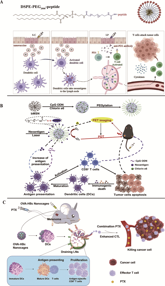
Phototherapy (photodynamic therapy (PDT) and photothermal therapy (PTT)), radiotherapy, and sonodynamic therapy are anti-cancer strategies that enable tumors to be treated non-invasively. They generate ROS that lead to the death of the cancer cell, which releases DAMPs when its membrane ruptures. These DAMPs are an important element in the stimulation of immune cells [194-197]. Their combination with nanovaccines is therefore a good strategy to enhance the immune response in cancer treatment. One study developed a nanosystem (bMSN(CpG/Ce6)-Adpgk) combining neoantigen peptide AdpgK, a CpG adjuvant (TLR9 agonist), and a chlorin e6 photosensitizer, all assembled in mesoporous silica NPs coated with PEG polymer. Treatment with neoantigen, CpG, and PDT elicited a significant immune response from neoantigen-specific CD8+ T cells, compared with PDT alone. Locally, treatment with bMSN (PDT + neoantigen + CpG) increased tumor infiltration by activated T cells and DCs (Fig. 6B) [198].
Similar to phototherapy, when cancer cells are treated with chemotherapeutic agents, they release DAMPs that stimulate immune cells. Numerous studies have shown that chemotherapeutic drugs like doxorubicin, oxaliplatin, cyclophosphamide, and paclitaxel (PTX) cause immunogenic cell death in tumor cells, leading to increased calreticulin (CRT) expression, adenosine triphosphate (ATP) secretion, and high mobility group box 1 (HMGB1) release, which helps DCs capture and process tumor antigens to activate the immune system [82]. An example of the combination of neoantigen peptide nanovaccine and a chemotherapeutic agent is the study by Shan et al., who have developed a nanovaccine called OVA-HBc NCs. This involves a genetic modification aimed at integrating an OVA peptide on the surface of a nanocage of natural hepatitis B core protein. This nanovaccine, combined with a low dose of PTX, had a greater antitumor effect than either treatment alone. In addition, this combination of treatments stopped the spread of established tumors (Fig. 6C) [199].
Shortly, conventional therapies kill cancer cells, causing the release of cellular debris that contains antigens (TAAs and TSAs). These antigens can be captured by DCs, which then present them to T cells, triggering an immune response, even if it is not very strong. When combined with the vaccine, the immune response is increased. The effect of the peptide neoantigen vaccine, along with the immune response from conventional therapy, improves with better recruitment of T cells that are specific to the treated tumor cells.
Peptide neoantigen nanovaccines represent a significant resource in cancer therapy, thanks to their specificity and the promising results obtained in various murine model tests. But many challenges remain. Peptide neoantigen nanovaccines are virtually absent from clinical trials. Among the few exceptions is the DepoVaxTM, which consists of a liposome, a polynucleotide adjuvant, a universal T helper peptide, seven HLA-A2-restricted tumor-specific peptides, and Montanide ISA51 VG. This vaccine stimulated multifunctional effector memory responses to peptide-based TAAs in advanced ovarian cancer (NCT01095848) [200].
Large-scale production of nanovaccines with acceptable batch-to-batch differences remains a major challenge. These challenges are often linked to issues such as the nature of the nanomaterials, which is difficult to reproduce on a large scale to ensure the desired characteristics, such as biodegradability and biocompatibility, and the conform dose of loaded drug, which must be uniform to avoid problems such as toxicity or low bioavailability [201-203]. Some nanomaterials are highly complex, making large-scale production a tricky business. The chemistry, manufacturing, and control (CMC) and good manufacturing practice (GMP) standards necessary to move from preclinical to clinical use and later marketing are incompatible with the escalating complexity of the NP synthesis process. Making this design complexity easier to execute could make them readily available for clinical trials [32,204]. Optimizing the properties of nanovaccines is crucial to encouraging their incorporation into clinical translation. Nevertheless, for medical applications, the stability of nanomaterials in physiological settings is crucial. Developing more effective nanoformulations requires a thorough understanding of the interactions between the immune system and the nanomaterials utilized, as well as their distribution in vivo. Given the potential changes in the physicochemical characteristics of NPs that may arise from their interactions with other biological components in the body, comprehensive research on the toxicity of nanomaterials is imperative [205].
Combining peptide neoantigen nanovaccines with other therapies is a strategy that needs to be promoted much more. Prior to their use, it is essential to understand the adverse effects of combination therapy with neoantigen peptide nanovaccine. Immunocheckpoint inhibitor combinations are frequently the most researched and have produced the best outcomes. Other immunotherapies, like CAR-T therapy, can be used in conjunction with peptide neoantigen nanovaccines in addition to checkpoint inhibitors. Research has demonstrated that combining chimeric antigen receptor T-cell therapy (CAR-T) cells with cancer vaccines improves immune response and increases the rate at which cancer cells are destroyed compared to monotherapies [206-209]. Vaccines can either directly stimulate double or bispecific CAR-T cells at the tumor site or indirectly stimulate CAR-T cells through APCs [210]. In order to test the benefits of this combination, Chan et al. discovered that in immunocompetent mice, combined treatment with OVA-Clec9A-TNE and CAROT cells (CAR-T cells expressing transgenic TCRs specific for OVA peptides) produced some tumor rejection and long-lasting responses. CAROT cell proliferation and infiltration into tumors increased in tandem with tumor regression [211]. Additionally, promoting the combination with conventional therapies would be a smart move. Indeed, clinical trials combining traditional therapies, immunotherapies with peptide neoantigen vaccines often show better results than peptide vaccines alone [85,212-215]. Ferroptosis, pyroptosis, autophagy, and necroptosis are known inducers of immunogenic cell death by releasing DAMPs [216]. It would be beneficial to combine them with neoantigen peptide nanovaccines, and this should be investigated. Nanovaccine-based neoantigen peptide combined with other therapies would therefore bring better results. Eliciting strong immune responses within the tumor microenvironment can be challenging because some tumor microenvironments have higher numbers of suppressor cells (regulatory T lymphocytes, myeloid-derived suppressor cells, and type-2 tumor-associated macrophages) and are more resistant to immunotherapy [217-219]. An effective tactic would be to combine inhibitors of tumor microenvironment suppressor cells with neoantigen peptide nanovaccines.
Choosing nanovaccines that contain multi-epitope neoantigens, along with the inclusion of multiadjuvant agents, could be an effective approach to enhance the immune response against cancer. One of the challenges of vaccine failure against cancer can sometimes be explained by downregulation of HLA or neoantigen mutations, which cause immune escape. The multi-adjuvant approach may address tumor cell immunity evasion. Even if some antigens have already undergone mutations, it would be more likely to activate T cells that can identify tumor cells if multiple types of antigens were presented. Several clinical trials are employing this approach in the development of antitumor vaccines [220-222]. The selection of nanomaterials is also a factor to consider for optimizing the immune response, the biocompatibility of the material in vivo, and the controlled release of drugs to ensure treatment safety. Adding ligands to the nanovaccine to enable precise targeting of APCs (DCs) would significantly improve vaccine efficacy and reduce side effects.
Neoantigen peptide-based anti-cancer nanovaccines represent a revolutionary form of immunotherapy that utilizes the immune system to precisely target tumors and induce long-lasting protective memory by minimizing side effects. Antigen stability, targeted accumulation in LNs, and DC cross-presentation can all be enhanced by nanotechnologies. Nanovaccines can significantly enhance anti-tumor immune responses by utilizing multi-adjuvants, optimizing NP characteristics, and employing multi-epitope designs. Despite these advances, challenges remain regarding manufacturing scalability, biocompatibility, and clinical translation. Ensuring the safety and ease of formulation of the nanovaccines is crucial. Clinical trials of nanovaccines based on peptide neoantigens are virtually nonexistent. It is therefore essential to introduce them into clinical trials to assess their potential. This would also require the inclusion of combinatorial strategies with other therapies to maximize clinical outcomes.
Sameena Noor Khattak: Writing – review & editing, Writing – original draft, Visualization, Validation, Investigation, Conceptualization. Kodzo Prosper Adzavon: Writing – review & editing, Visualization, Validation, Investigation, Conceptualization. Weijian Zhao: Writing – review & editing, Validation, Conceptualization. Wang Sheng: Writing – review & editing, Validation, Supervision, Project administration, Funding acquisition, Conceptualization.
The authors declare that they have no known competing financial interests or personal relationships that could have appeared to influence the work reported in this paper.
This work was supported by the National Natural Science Foundation of China (NSFC, No. 31770999), and Beijing Municipal Natural Science Foundation (No. 7202002).
Supplementary material associated with this article can be found, in the online version, at doi:
O.K. Dagher, R.D. Schwab, S.K. Brookens, A.D. Posey, Cell 186 (2023) 1814-1814.e1. doi: 10.1016/j.cell.2023.02.039
S. Taefehshokr, A. Parhizkar, S. Hayati, et al., Pathol. Res. Pract. 229 (2022) 153723. doi: 10.1016/j.prp.2021.153723
Y. Yang, J. Clin. Invest. 125 (2015) 3335–3337. doi: 10.1172/JCI83871
C. Peterson, N. Denlinger, Y. Yang, Cancers 14 (2022) 3972. doi: 10.3390/cancers14163972
Y. Wang, H. Zhang, C. Liu, et al., J. Hematol. Oncol. 15 (2022) 111. doi: 10.1186/s13045-022-01325-0
C. Liu, M. Yang, D. Zhang, M. Chen, D. Zhu, Front. Immunol. 13 (2022) 961805. doi: 10.3389/fimmu.2022.961805
C. Pilard, M. Ancion, P. Delvenne, et al., J. Cancer 125 (2021) 927–938. doi: 10.1038/s41416-021-01413-x
S. Tan, D. Li, X. Zhu, Biomed. Pharmacother. Biomed. Pharmacother. 124 (2020) 109821. doi: 10.1016/j.biopha.2020.109821
M.J. Lin, J. Svensson-Arvelund, G.S. Lubitz, et al., Brody, Nat. Cancer 3 (2022) 911–926. doi: 10.1038/s43018-022-00418-6
M. Saxena, S.H. van der Burg, C.J.M. Melief, N. Bhardwaj, Nat. Rev. Cancer 21 (2021) 360–378. doi: 10.1038/s41568-021-00346-0
N. Zaidi, E.M. Jaffee, M. Yarchoan, Nat. Rev. Cancer 25 (2025) 517–533. doi: 10.1038/s41568-025-00820-z
T. Fan, M. Zhang, J. Yang, et al., Signal. Transduct. Target. Ther. 8 (2023) 450. doi: 10.1038/s41392-023-01674-3
M. Peng, Y. Mo, Y. Wang, et al., Mol. Cancer 18 (2019) 128. doi: 10.1186/s12943-019-1055-6
N. Xie, G. Shen, W. Gao, Z. Huang, C. Huang, L. Fu, Signal. Transduct. Target. Ther. 8 (2023) 9. doi: 10.1038/s41392-022-01270-x
Z. Hu, P.A. Ott, C.J. Wu, Nat. Rev. Immunol. 18 (2018) 168–182. doi: 10.1038/nri.2017.131
L. Buonaguro, M. Tagliamonte, Vaccines 8 (2020) 615. doi: 10.3390/vaccines8040615
T.N. Schumacher, R.D. Schreiber, Science 1979 348 (2015) 69–74. doi: 10.1126/science.aaa4971
G.M. Lynn, C. Sedlik, F. Baharom, et al., Nat. Biotechnol. 38 (2020) 320–332. doi: 10.1038/s41587-019-0390-x
H.C. Liao, S.J. Liu, J. Biomed. Sci. 32 (2025) 10. doi: 10.1186/s12929-024-01102-w
R. Sarangi, S. Mishra, S. Mahapatra, Indian J. Clin. Biochem. 40 (2025) 191–200. doi: 10.1007/s12291-024-01201-3
J. Liu, M. Fu, M. Wang, et al., J. Hematol. Oncol. 15 (2022) 28. doi: 10.1117/12.2628549
A. Addeo, A. Friedlaender, E. Giovannetti, et al., Curr. Oncol. Rep. 23 (2021) 137. doi: 10.1007/s11912-021-01130-x
W. Xiao, W. Jiang, Z. Chen, et al., Signal. Transduct. Target. Ther. 10 (2025) 74. doi: 10.1038/s41392-024-02107-5
P.D. Katsikis, K.J. Ishii, C. Schliehe, Nat. Rev. Immunol. 24 (2024) 213–227. doi: 10.1038/s41577-023-00937-y
D. Liu, L. Liu, X. Li, et al., Vaccines 12 (2024) 950. doi: 10.3390/vaccines12080950
M.M. Melssen, G.R. Petroni, K.A. Chianese-Bullock, et al., J. ImmunOther Cancer 7 (2019) 163. doi: 10.1186/s40425-019-0625-x
Z. Nafiah, A. Atha, A.I. Rizkita, N.S.O. Ujiantari, A. Hermawan, Vacunas. 26 (2025) 500390. doi: 10.1016/j.vacun.2025.500390
M. Iero, P. Filipazzi, C. Castelli, et al., Cancer Immunol. Immunther. 58 (2009) 1159–1167. doi: 10.1007/s00262-008-0610-6
Z. Sun, H. Zhao, L. Ma, et al., J. Nanobiotechnology. 22 (2024) 61. doi: 10.56578/esm020201
J. Liu, L. Miao, J. Sui, Y. Hao, G. Huang, Asian J. Pharm. Sci. 15 (2020) 576–590.
J. Guo, L. Tang, K. Li, et al., Adv. Nanobiomed. Res. 3 (2023) 2200122. doi: 10.1002/anbr.202200122
J.D. Martin, H. Cabral, T. Stylianopoulos, R.K. Jain, Nat. Rev. Clin. Oncol. 17 (2020) 251–266. doi: 10.1038/s41571-019-0308-z
W. Song, S.N. Musetti, L. Huang, Biomaterials 148 (2017) 16–30. doi: 10.1016/j.biomaterials.2017.09.017
M. Yang, J. Li, P. Gu, X. Fan, Bioact. Mater. 6 (2021) 1973–1987.
L. Jeanbart, M. Ballester, A. de Titta, et al., Cancer Immunol. Res. 2 (2014) 436–447. doi: 10.1158/2326-6066.CIR-14-0019-T
Y. Yi, M. Yu, W. Li, D. Zhu, L. Mei, M. Ou, J. Control. Release 355 (2023) 760–778. doi: 10.1016/j.jconrel.2023.02.015
J.M. Silva, E. Zupancic, G. Vandermeulen, et al., J. Control. Release 198 (2015) 91–103. doi: 10.1016/j.jconrel.2014.11.033
S. Xu, Z. Feng, Y. Zhang, et al., Int. J. Biol. Macromol. 222 (2022) 1936–1947. doi: 10.1016/j.ijbiomac.2022.09.283
G. Zhu, F. Zhang, Q. Ni, G. Niu, X. Chen, ACS Nano 11 (2017) 2387–2392. doi: 10.1021/acsnano.7b00978
K.P. Adzavon, W. Zhao, S.N. Khattak, W. Sheng, Int. J. Biol. Macromol. 311 (2025) 143456. doi: 10.1016/j.ijbiomac.2025.143456
L. Wang, N. Wang, W. Zhang, et al., Signal. Transduct. Target. Ther. 7 (2022) 48. doi: 10.56028/aemr.1.1.48
E. Rosson, F. Lux, L. David, Y. Godfrin, O. Tillement, E. Thomas, Int. J. Pharm. 675 (2025) 125555. doi: 10.1016/j.ijpharm.2025.125555
S. Marqus, E. Pirogova, T.J. Piva, J. Biomed. Sci. 24 (2017) 21. doi: 10.1186/s12929-017-0328-x
U. Toh, S. Sakurai, S. Saku, et al., Cancer Sci. 111 (2020) 2760–2769. doi: 10.1111/cas.14510
R. Sundar, S.Y. Rha, H. Yamaue, et al., BMC Cancer 18 (2018) 332. doi: 10.1186/s12885-018-4234-8
O. Adotévi, D. Vernerey, P. Jacoulet, et al., J. Clin. Oncol. 41 (2023) 373–384. doi: 10.1200/jco.22.00096
M. Platten, L. Bunse, A. Wick, et al., Nature 592 (2021) 463–468. doi: 10.1038/s41586-021-03363-z
S. Kondo, T. Shimizu, T. Koyama, J, et al., Cancer Sci. 112 (2021) 1514–1523. doi: 10.1111/cas.14765
W. Liu, H. Tang, L. Li, X. Wang, Z. Yu, J. Li, Cell Prolif. 54 (2021) e13025. doi: 10.1111/cpr.13025
C.C. Smith, S.R. Selitsky, S. Chai, P, et al., Nat. Rev. Cancer 19 (2019) 465–478. doi: 10.1038/s41568-019-0162-4
T. Jiang, T. Shi, H. Zhang, et al., J. Hematol. Oncol. 12 (2019) 93. doi: 10.1186/s13045-019-0787-5
A. Nelde, H.G. Rammensee, J.S. Walz, Mol. Cell Proteomics 20 (2021) 100022. doi: 10.1074/mcp.R120.002309
C.J.M. Melief, T. van Hall, R. Arens, F. Ossendorp, S.H. van der Burg, J. Clin. Invest. 125 (2015) 3401–3412. doi: 10.1172/JCI80009
M.C. Sellars, C.J. Wu, E.F. Fritsch, Cell 185 (2022) 2770–2788. doi: 10.1016/j.cell.2022.06.035
M. Kaczmarek, J. Poznańska, F. Fechner, et al., Cells 12 (2023) 2159. doi: 10.3390/cells12172159
F. Lang, B. Schrörs, M. Löwer, Ö. Türeci, U. Sahin, Nat. Rev. Drug Discov. 21 (2022) 261–282. doi: 10.1038/s41573-021-00387-y
M. Shah, S. Rafiq, H.G. Woo, Trends. Pharmacol. Sci. 45 (2024) 1104–1118. doi: 10.1016/j.tips.2024.10.013
E. Blass, P.A. Ott, Nat. Rev. Clin. Oncol. 18 (2021) 215–229. doi: 10.1038/s41571-020-00460-2
N. Biswas, S. Chakrabarti, V. Padul, L.D. Jones, S. Ashili, Front. Immunol. 14 (2023) 1105420. doi: 10.3389/fimmu.2023.1105420
Z. Chen, S. Zhang, N. Han, et al., Front. Immunol. 12 (2021) 691605. doi: 10.3389/fimmu.2021.691605
P.M. Dillon, C.L. Slingluff, Peptide vaccine: overview, in: J.L. Marshall (Ed.), Cancer Therapeutic Targets, Springer, New York, 2017, pp. 427–439.
B.A. Shah, J.A. Holden, J.C. Lenzo, S. Hadjigol, N.M. O’Brien-Simpson, NPJ Vaccines 10 (2025) 68. doi: 10.1038/s41541-025-01118-9
N. Abd-Aziz, C.L. Poh, J. Oncol. 2022 (2022) 9749363.
C.J.M. Melief, S.H. van der Burg, Nat. Rev. Cancer 8 (2008) 351–360. doi: 10.1038/nrc2373
J.E. Slansky, M. Nakayama, Semin. Immunol. 47 (2020) 101395. doi: 10.1016/j.smim.2020.101395
M. Candia, B. Kratzer, W.F. Pickl, Int. Arch. Allergy Immunol. 170 (2016) 211–233. doi: 10.1159/000448756
L. Buonaguro, M. Tagliamonte, Front. Immunol. 14 (2023) 1210044. doi: 10.3389/fimmu.2023.1210044
M.R. Parkhurst, M.L. Salgaller, S. Southwood, et al., J. Immunol. 157 (1996) 2539–2548. doi: 10.4049/jimmunol.157.6.2539
O.Y. Borbulevych, T.K. Baxter, Z. Yu, N.P. Restifo, B.M. Baker, J. Immunol. 174 (2005) 4812–4820. doi: 10.4049/jimmunol.174.8.4812
A. Gupta, R.E. O’Cearbhaill, M.S. Block, et al., Gynecol. Oncol. 189 (2024) 90–97. doi: 10.1016/j.ygyno.2024.07.675
J.M. Kirkwood, S. Lee, S.J. Moschos, et al., Clin. Cancer Res. 15 (2009) 1443–1451. doi: 10.1158/1078-0432.CCR-08-1231
V. Lennerz, S. Gross, E. Gallerani, et al., Cancer Immunol. Immunther. 63 (2014) 381–394. doi: 10.1007/s00262-013-1516-5
S. Holmberg-Thydén, I.H. Dufva, A.O. Gang, et al., Cancer Immunol. Immunther. 71 (2022) 433–444. doi: 10.1007/s00262-021-02993-6
T. Zhao, Y. Cai, Y. Jiang, et al., Signal. Transduct. Target. Ther. 8 (2023) 283. doi: 10.1038/s41392-023-01557-7
Y. Li, Y. Chen, Y. Tang, et al., J. Control. Release 382 (2025) 113667. doi: 10.1016/j.jconrel.2025.113667
F. Zahedipour, K. Jamialahmadi, P. Zamani, M.R Jaafari, Int. Immunopharmacol. 123 (2023) 110721. doi: 10.1016/j.intimp.2023.110721
B. Zhang, S. Zhou, S. Lu, et al., ACS Nano 18 (2024) 26690–26703. doi: 10.1021/acsnano.4c06399
C. Ménard, F. Martin, L. Apetoh, F. Bouyer, F. Ghiringhelli, Cancer Immunol. Immunther. 57 (2008) 1579–1587. doi: 10.1007/s00262-008-0505-6
M. Wang, J. Song, F. Zhou, et al., Adv. Sci. 6 (2019) 1802157. doi: 10.1002/advs.201802157
L. Galluzzi, M.J. Aryankalayil, C.N. Coleman, S.C. Formenti, Nat. Rev. Clin. Oncol. 20 (2023) 543–557. doi: 10.1038/s41571-023-00782-x
Y.J. Wang, R. Fletcher, J. Yu, L. Zhang, Genes Dis. 5 (2018) 194–203. doi: 10.1016/j.gendis.2018.05.003
M. Jiang, J. Zeng, L. Zhao, et al., Nanoscale 13 (2021) 17218–17235. doi: 10.1039/d1nr05512g
J. Sprooten, R.S. Laureano, et al., Oncoimmunology 12 (2023) 2219591. doi: 10.1080/2162402X.2023.2219591
Y. Engelen, R. Demuynck, J. Ramon, et al., J. Control. Release 384 (2025) 113721. doi: 10.1016/j.jconrel.2025.113721
M. Noguchi, G. Arai, S. Egawa, et al., Cancer Immunol. Immunther. 69 (2020) 847–857. doi: 10.1007/s00262-020-02498-8
S. Nishida, S. Koido, Y. Takeda, et al., J. Immunother. 37 (2014) 105. doi: 10.1097/CJI.0000000000000020
S. Koido, J. Taguchi, M. Shimabuku, et al., J. Immunother. Cancer 12 (2024) e009765. doi: 10.1136/jitc-2024-009765
K. Fukuda, T. Funakoshi, T. Sakurai, et al., Melanoma Res. 27 (2017) 326. doi: 10.1097/CMR.0000000000000342
P. Nisticò, I. Capone, B. Palermo, et al., Int. J. Cancer 124 (2009) 130–139. doi: 10.1002/ijc.23886
M. Noguchi, K. Matsumoto, H. Uemura, et al., Clin. Cancer Res. 22 (2016) 54–60.
D. Fukumura, J. Kloepper, Z. Amoozgar, D.G. Duda, R.K. Jain, Nat. Rev. Clin. Oncol. 15 (2018) 325–340. doi: 10.1038/nrclinonc.2018.29
A. Leary, L. Gladieff, R. Sabatier, et al., J. Clin. Oncol. 41 (2023) TPS5618. doi: 10.1200/jco.2023.41.16_suppl.tps5618
M.S. Pelster, R.N. Amaria, Ther. Adv. Med. Oncol. 11 (2019) 1758835919830826. doi: 10.1177/1758835919830826
A. Batista-Duharte, D.T. Martínez, I.Z. Carlos, Biomed. Pharmacother. 105 (2018) 616–624. doi: 10.1016/j.biopha.2018.06.026
F. Xu, Y. Yuan, Y. Wang, Q. Yin, Biomed. Pharmacother. 158 (2023) 114117. doi: 10.1016/j.biopha.2022.114117
R. van der Meel, E. Sulheim, Y. Shi, et al., Nat. Nanotechnol. 14 (2019) 1007–1017. doi: 10.1038/s41565-019-0567-y
A. Haleem, M. Javaid, R.P. Singh, S. Rab, R. Suman, Glob. Health J. 7 (2023) 70–77. doi: 10.1016/j.glohj.2023.02.008
K. Pavelic, S. Kraljevi ´ c Paveli ´ c, A. Bulog, et al., Int. J. Mol. Sci. 24 (2023) 12827. ´ doi: 10.3390/ijms241612827
M. Prasad, U.P. Lambe, B. Brar, et al., Biomed. Pharmacother. 97 (2018) 1521–1537. doi: 10.1016/j.biopha.2017.11.026
O. Veiseh, B.C. Tang, K.A. Whitehead, D.G. Anderson, R. Langer, Nat. Rev. Drug Discov. 14 (2015) 45–57. doi: 10.1038/nrd4477
W. Jiang, C.A. von Roemeling, Y. Chen, et al., Nat. Biomed. Eng. 1 (2017) 0029. doi: 10.1038/s41551-017-0029
Z. Zhou, W. Chen, Y. Cao, R. Abdi, W. Tao, Nat. Rev. Cardiol. 22 (2025) 255–272. doi: 10.1038/s41569-024-01094-y
Y. Xiao, Y. Huang, M. Xie, et al., Nat. Rev. Bioeng. 2 (2024) 244–259.
M.E. Aikins, C. Xu, J.J. Moon, Acc. Chem. Res. 53 (2020) 2094–2105. doi: 10.1021/acs.accounts.0c00456
F. Chen, Y. Wang, J. Gao, et al., Biomaterials 270 (2021) 120709. doi: 10.1016/j.biomaterials.2021.120709
M.A. Farooq, A.P.R. Johnston, N.L. Trevaskis, Acta Biomater. 193 (2025) 65–82. doi: 10.1016/j.actbio.2024.12.039
R. He, J. Zang, Y. Zhao, H. Dong, Y. Li, ACS Biomater. Sci. Eng. 8 (2022) 406–423. doi: 10.1021/acsbiomaterials.1c01274
R. Mahjub, S. Jatana, S.E. Lee, et al., J. Control. Release 288 (2018) 239–263. doi: 10.1016/j.jconrel.2018.09.010
V. Manolova, A. Flace, M. Bauer, et al., Eur. J. Immunol. 38 (2008) 1404–1413. doi: 10.1002/eji.200737984
Y. Qian, H. Jin, S. Qiao, et al., Biomaterials 98 (2016) 171–183. doi: 10.1016/j.biomaterials.2016.05.008
D. Carpanese, A. Dalla Pietà, A. Rosato, Cell Mol. Immunol. 20 (2023) 1251–1253. doi: 10.1038/s41423-023-01024-x
G. Rodella, V. Préat, B. Gallez, A. Malfanti, J. Control. Release 383 (2025) 113784. doi: 10.1016/j.jconrel.2025.113784
Z.Z. Li, N.N. Zhong, L.-M. Cao, et al., Small 20 (2024) 2308731. doi: 10.1002/smll.202308731
T. Nakamura, M. Kawai, Y. Sato, et al., Mol. Pharm. 17 (2020) 944–953. doi: 10.1021/acs.molpharmaceut.9b01182
Y. Nie, L. Shi, Y. Zhang, Y. Guo, H. Gu, Cancers 14 (2022) 5107. doi: 10.3390/cancers14205107
L. Martinez-Pomares, J. Leukoc. Biol. 92 (2012) 1177–1186. doi: 10.1189/jlb.0512231
R. Yang, J. Xu, L. Xu, et al., ACS Nano 12 (2018) 5121–5129. doi: 10.1021/acsnano.7b09041
U.J. Nahar, I. Toth, M. Skwarczynski, J. Control. Release 351 (2022) 284–300. doi: 10.1016/j.jconrel.2022.09.038
C. Lai, S. Duan, F. Ye, et al., Theranostics. 8 (2018) 1723–1739. doi: 10.7150/thno.22056
G. Chen, W. Xiong, Z. Gu, et al., Int. J. Biol. Macromol. 223 (2022) 1485–1494. doi: 10.1016/j.ijbiomac.2022.11.045
J. Yu, S. Wang, J. Qi, et al., Int. Immunopharmacol. 101 (2021) 108148. doi: 10.1016/j.intimp.2021.108148
M. Pei, R. Xu, C. Zhang, et al., Colloids. Surf. B: Biointerfaces 197 (2021) 111378. doi: 10.1016/j.colsurfb.2020.111378
J. Zheng, M. Wang, L. Pang, et al., J. Control. Release 373 (2024) 568–582. doi: 10.1016/j.jconrel.2024.07.056
S. Gou, S. Wang, W. Liu, et al., Theranostics 11 (2021) 7308–7321. doi: 10.7150/thno.56406
A.M. de Haas, D.A. Stolk, S.T.T. Schetters, et al., Pharmaceutics 16 (2024) 581. doi: 10.3390/pharmaceutics16050581
H. Yan, G. Lin, Z. Liu, F. Gu, Y. Zhang, Acta Biomater. 161 (2023) 213–225. doi: 10.1016/j.actbio.2023.02.034
L.J. Cruz, R.A. Rosalia, J.W. Kleinovink, et al., J. Control. Release 192 (2014) 209–218. doi: 10.1016/j.jconrel.2014.07.040
J. Liang, X. Zhao, Cancer Biol. Med. 18 (2021) 352–371. doi: 10.20892/j.issn.2095-3941.2021.0004
B. Fu, X. Huang, J. Deng, et al., Oncol. Rep. 39 (2018) 893–900.
N. Desai, V. Chavda, T.R.R. Singh, N.D. Thorat, L.K. Vora, Small 20 (2024) 2401631. doi: 10.1002/smll.202401631
L. Luo, R. Shu, A. Wu, J. Mater. Chem. B 5 (2017) 5517–5531. doi: 10.1039/C7TB01137G
B. Sun, T. Xia, J. Mater. Chem. B 4 (2016) 5496–5509. doi: 10.1039/C6TB01131D
E.A. Vasievich, S. Ramishetti, Y. Zhang, L. Huang, Mol. Pharm. 9 (2012) 261–268. doi: 10.1021/mp200350n
S. De Koker, J. Cui, N. Vanparijs, et al., Angew. Chem. Int. Ed. 128 (2015) 1356–1361.
H. Shiku, F. Momose, T. Nakai, et al., J. Immunother. Cancer 10 (2022), doi: 10.1136/jitc-2022-SITC2022.0359.
A.J. Highton, A. Girardin, G.M. Bell, S.M. Hook, R.A. Kemp, BMC Immunol. 17 (2016) 39. doi: 10.1186/s12865-016-0178-4
H. Zhao, J. Xu, Y. Li, et al., ACS Nano 13 (2019) 13127–13135. doi: 10.1021/acsnano.9b05974
L. Huang, X. Li, H. Zhang, et al., ACS Nano 19 (2025) 13906–13926. doi: 10.1021/acsnano.4c17491
J.P.M. Almeida, A.Y. Lin, E.R. Figueroa, A.E. Foster, R.A. Drezek, Small 11 (2015) 1453–1459. doi: 10.1002/smll.201402179
W. Li, Z. Jing, S. Wang, et al., Biomaterials 271 (2021) 120726. doi: 10.1016/j.biomaterials.2021.120726
W. Zheng, S. Li, Z. Shi, et al., J. Nanobiotechnology 22 (2024) 562. doi: 10.1186/s12951-024-02837-2
P. Zhao, Y. Fan, Z. Wang, et al., Int. J. Nanomedicine 2025 (2025) 2289–2304. doi: 10.2147/ijn.s496873
A. Garg, H.K. Dewangan, Crit. Rev. Ther. Drug Carr. Syst. 37 (2020) 183–204. doi: 10.1615/critrevtherdrugcarriersyst.2020033273
L.M. Cao, Y.F. Yu, Z.Z. Li, et al., J. Nanobiotechnology 22 (2024) 308. doi: 10.1186/s12951-024-02590-6
B. Filipic, I. Panteli ´ c, I. Nikoli ´ c, et al., Vaccines 11 (2023) 1172. ´ doi: 10.3390/vaccines11071172
N. Desai, V. Tambe, P. Pofali, L.K. Vora, Adv. Nanobiomed. Res. 4 (2024) 2400012. doi: 10.1002/anbr.202400012
L. Zheng, Z. Wang, H. Liu, et al., ACS Nano 19 (2025) 17900–17916. doi: 10.1021/acsnano.5c04365
B. Chaigne-Delalande, M.J. Lenardo, Trends. Immunol. 35 (2014) 332–344. doi: 10.1016/j.it.2014.05.001
X. Sun, Y. Zhang, J. Li, et al., Nat. Nanotechnol. 16 (2021) 1260–1270. doi: 10.1038/s41565-021-00962-9
L. Zhang, J. Zhao, X. Hu, et al., Adv. Mater. 34 (2022) 2206915. doi: 10.1002/adma.202206915
C. Chen, Y. Tong, Y. Zheng, et al., Small 17 (2021) 2006970. doi: 10.1002/smll.202006970
M. Lv, M. Chen, R. Zhang, et al., Cell Res. 30 (2020) 966–979. doi: 10.1038/s41422-020-00395-4
K. Yuan, C. Zhang, X. Pan, et al., J. Control. Release 375 (2024) 249–268. doi: 10.1016/j.jconrel.2024.09.008
X. Lv, J. Huang, J. Min, et al., J. Control. Release 357 (2023) 109–119. doi: 10.1016/j.jconrel.2023.01.078
H. Zhao, Y. Li, B. Zhao, et al., Acta Pharm. Sin. B 13 (2023) 3892–3905. doi: 10.1016/j.apsb.2023.02.003
Y. Ma, Y. Zhuang, X. Xie, et al., Nanoscale 3 (2011) 2307–2314. doi: 10.1039/c1nr10166h
R. Bei, V. Guptill, L. Masuelli, et al., J. ImmunOther 21 (1998) 159. doi: 10.1097/00002371-199805000-00001
D.P. Vangasseri, Z. Cui, W. Chen, et al., Mol. Membr. Biol. 23 (2006) 385–395. doi: 10.1080/09687860600790537
W. Chen, W. Yan, L. Huang, Cancer Immunol. Immunther. 57 (2008) 517–530. doi: 10.1007/s00262-007-0390-4
G. Alipour Talesh, Z. Ebrahimi, A. Badiee, et al., Immunol. Lett. 176 (2016) 57–64. doi: 10.1016/j.imlet.2016.05.016
L. Naghibi, M. Yazdani, A.A. Momtazi-Borojeni, et al., PLoS One 15 (2020) e0243550. doi: 10.1371/journal.pone.0243550
M. Mansourian, A. Badiee, S.A. Jalali, et al., Immunol. Lett. 162 (2014) 87–93. doi: 10.1016/j.imlet.2014.07.008
W. Chen, L. Huang, Mol. Pharm. 5 (2008) 464–471. doi: 10.1021/mp700126c
X. Gong, Y. Gao, J. Shu, C. Zhang, K. Zhao, Vaccines 10 (2022) 1906. doi: 10.3390/vaccines10111906
G. Kumar, T. Virmani, S.K. Misra, et al., Carbohydr. Polym. Technol. Appl. 8 (2024) 100537.
J.F. Correia-Pinto, N. Csaba, J.T. Schiller, M.J. Alonso, Vaccines 3 (2015) 730–750. doi: 10.3390/vaccines3030730
B.C. Quartey, J. Sapudom, M. ElGindi, A. Alatoom, J. Teo, Adv. Healthc. Mater. 13 (2024) 2303125. doi: 10.1002/adhm.202303125
M.O. Mohsen, D.E. Speiser, A. Knuth, M.F. Bachmann, WIREs Nanomed. Nanobiotechnol. 12 (2020) e1579. doi: 10.1002/wnan.1579
S. Nooraei, H. Bahrulolum, Z.S. Hoseini, et al., J. Nanobiotechnology 19 (2021) 59. doi: 10.1186/s12951-021-00806-7
R. Noad, P. Roy, Trends. Microbiol. 11 (2003) 438–444. doi: 10.1016/S0966-842X(03)00208-7
H. Hu, N.F. Steinmetz, Cancers 13 (2021) 2909. doi: 10.3390/cancers13122909
B. Donaldson, F. Al-Barwani, S.J. Pelham, et al., J. Immunother. Cancer 5 (2017) 69. doi: 10.1186/s40425-017-0270-1
S. Zhang, L.K. Yong, D. Li, et al., PLoS One 8 (2013) e68303. doi: 10.1371/journal.pone.0068303
A. Barajas, P. Amengual-Rigo, A. Pons-Grífols, et al., J. Transl. Med. 22 (2024) 14. doi: 10.1186/s12967-023-04843-8
J.C. Hsu, P. Liu, Y. Song, et al., Chem. Soc. Rev. 53 (2024) 7657–7680. doi: 10.1039/d4cs00421c
D.A. Rao, M.L. Forrest, A.W.G. Alani, G.S. Kwon, J.R. Robinson, J. Pharm. Sci. 99 (2010) 2018–2031. doi: 10.1002/jps.21970
C. Wang, P. Liu, Y. Zhuang, et al., Vaccine 32 (2014) 5475–5483. doi: 10.1016/j.vaccine.2014.07.081
W. Wang, Z. Liu, X. Zhou, et al., Nanomed. Nanotechnol. Biol. Med. 16 (2019) 69–78. doi: 10.1016/j.nano.2018.11.009
Y. Zhang, L. Liu, H. He, Y. Sun, Z. Zhong, Mater. Today 80 (2024) 406–428. doi: 10.1016/j.mattod.2024.08.006
D. Liu, S. Liang, K. Ma, et al., Adv. Mater. 36 (2024) 2304845. doi: 10.1002/adma.202304845
S. Bhatnagar, V. Revuri, M. Shah, et al., Cancers 14 (2022) 6091. doi: 10.3390/cancers14246091
Y. Pan, S. Liu, H. Zhao, Z. Yu, Y. Qi, Y. Huang, Int. J. Biol. Macromol. 310 (2025) 143268. doi: 10.1016/j.ijbiomac.2025.143268
H. Song, L. Chen, X. Pan, et al., Cancer Cell 43 (2025) 503–518 e10. doi: 10.1016/j.ccell.2025.02.014
B. Bayyurt Kocabas, K. Almacioglu, E.A. Bulut, et al., J. Control. Release 328 (2020) 587–595. doi: 10.1016/j.jconrel.2020.09.040
T.L. Nguyen, B.G. Cha, Y. Choi, J. Im, J. Kim, Biomaterials 239 (2020) 119859. doi: 10.1016/j.biomaterials.2020.119859
T. Su, X. Liu, S. Lin, F. Cheng, G. Zhu, Bioact. Mater. 26 (2023) 169–180.
M. Zhu, X. Ding, R. Zhao, et al., J. Control. Release 272 (2018) 72–82. doi: 10.1016/j.jconrel.2018.01.004
Q. Ni, F. Zhang, Y. Liu, et al., Sci. Adv. 6 (2020) eaaw6071. doi: 10.1126/sciadv.aaw6071
J.J. Baljon, A.J. Kwiatkowski, H.M. Pagendarm, et al., ACS Nano 18 (2024) 6845–6862. doi: 10.1021/acsnano.3c04471
F. Cheng, T. Su, S. Zhou, et al., Sci. Adv. 9 (2023) eade6257. doi: 10.1126/sciadv.ade6257
Y. Kim, S. Kang, H. Shin, et al., Angew. Chem. 132 (2020) 14736–14746. doi: 10.1002/ange.202006117
Y. Yin, J. Guo, S. Zhang, et al., Chin. Chem. Lett. 36 (2025) 110771. doi: 10.1016/j.cclet.2024.110771
Y. Chu, L. Qian, Y. Ke, X. F, et al., J. Nanobiotechnology 20 (2022) 190. doi: 10.1186/s12951-022-01397-7
W. Li, J. Yang, L. Luo, et al., Nat. Commun. 10 (2019) 3349. doi: 10.1038/s41467-019-11269-8
W. Xiao, M. Gao, B. Mo, et al., Pharmaceuticals 18 (2025) 491. doi: 10.3390/ph18040491
S. Zhang, J. Wang, Z. Kong, et al., Biomaterials 282 (2022) 121433. doi: 10.1016/j.biomaterials.2022.121433
L. Qin, W. Wei, K. Wang, et al., Chin. Chem. Lett. 36 (2025) 110777. doi: 10.1016/j.cclet.2024.110777
C. Xu, J. Nam, H. Hong, Y. Xu, J.J. Moon, ACS Nano 13 (2019) 12148–12161. doi: 10.1021/acsnano.9b06691
W. Shan, H. Zheng, G. Fu, et al., Nano Lett. 19 (2019) 1719–1727. doi: 10.1021/acs.nanolett.8b04722
N.L. Berinstein, M. Karkada, M.A. Morse, et al., J. Transl. Med. 10 (2012) 156. doi: 10.1186/1479-5876-10-156
T. Feng, J. Hu, J. Wen, et al., J. Hematol. Oncol.J. Hematol. Oncol. 17 (2024) 115. doi: 10.1186/s13045-024-01628-4
J. Qiu, C. Wu, Front. Nanotechnol. 7 (2025) 1564267. doi: 10.3389/fnano.2025.1564267
J.M. Metselaar, T. Lammers, Drug Deliv. Transl. Res. 10 (2020) 721–725. doi: 10.1007/s13346-020-00740-5
C. Zheng, M. Li, J. Ding, BIO Integr. 2 (2021) 57.
P. Zhang, Y. Xiao, X. Sun, et al., Med. 4 (2023) 147–167. doi: 10.1016/j.medj.2022.12.001
L. Liu, Y. Liu, Y. Xia, et al., J. Cancer 12 (2021) 6629–6639. doi: 10.7150/jca.62123
M. Wu, L. Zhang, H. Zhang, et al., Cytotherapy. 21 (2019) 659–670. doi: 10.1016/j.jcyt.2019.03.313
I. Caruana, G. Weber, B.C. Ballard, et al., Clin. Cancer Res. 21 (2015) 2952–2962. doi: 10.1158/1078-0432.CCR-14-2998
X. Zhang, Q. Hu, X. He, et al., J. Nanobiotechnology. 21 (2023) 159. doi: 10.1186/s12951-023-01900-8
M. Al-Haideri, S.B. Tondok, S.H. Safa, et al., Cancer Cell Int. 22 (2022) 365. doi: 10.1186/s12935-022-02778-6
J.D. Chan, B. von Scheidt, B. Zeng, et al., Clin. Transl. Immunol. 9 (2020) e1157. doi: 10.1002/cti2.1157
M. Murahashi, Y. Hijikata, K. Yamada, et al., Clin. Immunol. 166–167 (2016) 48–58.
H. Iinuma, R. Fukushima, T. Inaba, J, et al., J. Transl. Med. 12 (2014) 84. doi: 10.1186/1479-5876-12-84
Y. Sawada, T. Yoshikawa, M. Shimomura, et al., Int. J. Oncol. 46 (2015) 28–36. doi: 10.3892/ijo.2014.2737
M. Ishihara, Y. Nishida, S. Kitano, et al., Int. J. Cancer 152 (2023) 2554–2566. doi: 10.1002/ijc.34453
W. Gao, X. Wang, Y. Zhou, X. Wang, Y. Yu, Signal. Transduct. Target. Ther. 7 (2022) 196. doi: 10.1038/s41392-022-01046-3
S.I. Kim, C.R. Cassella, K.T. Byrne, Front. Immunol. 11 (2021) 629722. doi: 10.3389/fimmu.2020.629722
V. Kumar, S. Patel, E. Tcyganov, D.I. Gabrilovich, Trends. Immunol. 37 (2016) 208–220. doi: 10.1016/j.it.2016.01.004
D.H. Munn, V. Bronte, Curr. Opin. Immunol. 39 (2016) 1–6.
J.M. Hubbard, D.H. Ahn, J.C. Jones, et al., J. Clin. Oncol. 42 (2024) 3594–3594. doi: 10.1200/jco.2024.42.16_suppl.3594
E. Blass, D.B. Keskin, C.R. Tu, et al., Cell 188 (2025) 5125–5141 e27. doi: 10.1016/j.cell.2025.06.019
S. Jung, A. Nelde, Y. Maringer, et al., Blood 144 (2024) 1504 1. doi: 10.1182/blood-2024-198082

Figure 1 LN and DCs targeting efficiency by α-Ap-FNP. (A) Schematic of α-Ap-FNP targeting LNs and DCs. (B) α-Ap-FNP size distribution. (C) Biodistribution of α-Ap-FNP after injection at the base of the tail (inguinal LNs, ILN) or the footpad (popliteal LNs, PLN). (D) Percentage of DCs in LNs after injections. Reproduced with permission [110]. Copyright 2016, Elsevier.

Figure 2 Ability of M-YL/LA-Lipo to target and improve DCs activation. (A) Schematic illustration of M-YL/LA-Lipo nanovaccine and its ability to target DCs through the Man receptor on DCs with antigen presentation and T cell activation for cancer treatment. (B) M-YL/LA-Lipo improved the expression of CD80 and CD86 markers in DCs more effectively than YL/LA. Reproduced with permission [121]. Copyright 2021, Elsevier.

Figure 3 The ability of OVA@MM to trigger an immune response against tumors. (A) Schematic illustration of OVA@MM NPs formulation and its abilities to induce an immune response. The maturation of DCs induced by OVA@MM led to elevated levels of CD80 (B), CD86 (C), and CD40 (D), as well as the secretion of TNF-α (E) and interleukin-12 (IL-12, F). Activation of CD8+ T cells (G) and release of TNF-α (H) and interferon gamma (IFN-γ, I). Reproduced with permission [154]. Copyright 2023, Elsevier.

Figure 4 Man-based nanovaccine for LNs targeting. (A) LP-Man nanovaccine formulation illustration. (B) LN targeting abilities of LP-Man. (C, D) The uptake rate of LP/LP-Man by APCs in draining LNs. Reproduced with permission [177]. Copyright 2014, Elsevier.

Figure 5 Bi-adjuvant nanovaccine for cancer therapy. (A) Schematic illustration of the bi-adjuvant nanovaccine promoting delivery of MPLA to the cell surface and endosome, as well as delivery of cAMP and peptide antigens to the cytosol. (B) CD86 expression by BMDCs treated with the indicated doses of NP-cGAMP, NP-MPLA, or NP-cGAMP/MPLA, indicating DC differentiation and maturation. (C) LNs targeting abilities of the nanovaccine. (D) Quantification of CD8+ T cells, IFNγ+, TNFα+ after ex vivo restimulation of splenocytes with the SIINFEKL peptide. (E) Percentage of central memory and effector antigen-specific CD8+ T cells in the spleen after vaccination. (F) Tumor volume and survival rate after vaccination. Reproduced with permission [189]. Copyright 2024, American Chemical Society.

Figure 6 Combination strategies to improve cancer nanovaccine immune response. (A) Schematic diagram of the combined therapy of the neoantigenic nanovaccine DSPE-PEG2000-peptide combined with an anti-PD1 antibody. Reproduced with permission [193], Copyright 2022, Springer Nature. (B) Schematic illustration of bMSN(CpG/Ce6) neoantigen manufacturing and the combinatorial effect of bMSN(CpG/Ce6) neoantigen nanovaccines with PDT to enhance cancer immunotherapy. Reproduced with permission [198], Copyright 2019, American Chemical Society. (C) Schematic illustration of OVA-HBc NCs-mediated antitumor Immunity in combination with PTX. Reproduced with permission [200]. Copyright 2019, American Chemical Society.
Table 1. Long neoantigen peptide compared to short neoantigen.
| Feature | Long neoantigen peptide | Short neoantigen peptide |
| Immune response | CD8+ T and CD4+ T cells | Primarily CD8+ T cells |
| MHC presentation | MHC Ⅰ and MHC Ⅱ (via APCs) | Direct MHC Ⅰ binding |
| Stability | Higher | Lower (rapid clearance) |
| APC dependence | Required | Not required |
| Epitope coverage | Multi-epitope possible | Single epitope |
| Risk of tolerance | Lower (avoids direct MHC binding) | Higher (can induce tolerance) |
| Manufacturing complexity (epitope selection) | Higher (need processing prediction) | Simpler (direct epitope selection) |
 下载: 导出CSV
Table 2. List of some materials with their immunological advantages in neoantigen peptide nanovaccine formulation.
| Material | NP type | Immunological advantage | Example of nanovaccine | Ref. |
| DOTAP/DOTMA | Cationic liposome NPs | APCs targeting surface charge dependent; Adjuvant effect (via ROS generation) | DOTAP/Trp2 | [133] |
| PEG | Polymer NPs | LN targeting by reducing extra cellular matrix stroma targeting | PEG-PMA-OVA | [134] |
| HA | Polymer NPs | LN (via LYVE-1 receptor) and APCs (via CD44 receptors) targeting, Adjuvant (DC’s maturation, cytokine release) | HANG-V | [135] |
| CS | Polymer NPs | Adjuvant (PAMPs, DCs maturation) | CS gel-OVA | [136] |
| Mn, Zn, Ca | Metall NPs | Adjuvant (trigger STING signaling) | OVA@Mn-DAP, SZCP/APs-CpG, | [137,138] |
| Gold NPs | Metall NPs | Adjuvant (DCs maturationand T cells activation), size and shape dependent LN targeting | AuNP-OVA | [139] |
| Virus-like particles | Virus-like NPs | Adjuvant (PAMPs); LN targeting size dependent | P22-OVAT/B | [140] |
| FN NPs | Protein NPs | LN targeting size dependent | OVAT-FNs and M30-FNs | [141] |
| Baterial cytoplasmic membrane | Cellular membrane coated NPs | Adjuvants (PAMPs) | OVA@MMSNs@BM-Man | [142] |
下载: 导出CSV

 扫一扫看文章


扫一扫关注我们
 下载: