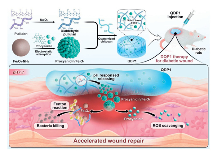
Scheme 1.
Fabrication and use of QDP1 in managing diabetic wounds.

A procyanidin/Fe3O4 nanoparticle-reinforced pullulan/chitosan hydrogel for treating diabetic wounds
Hao Yu , Xiaoliang Qi , Yuxi Chen , Youjian Chen , Jinghui Zhang , Wei Dong , Jianliang Shen , Tian Jiang


Diabetic wounds represent a serious complication of diabetes mellitus, placing a substantial economic strain upon healthcare infrastructures and individuals [1]. These expenses are linked to comprehensive wound management protocols, which encompass recurrent hospital admissions and regular replacement of dressings [2]. Foot and leg ulcers constitute the most frequently encountered type of wound among diabetic patients [3]. Remarkably, reports indicate that 84% of individuals suffering from diabetic foot ulcers eventually undergo lower limb amputation, with a cumulative mortality rate that surpasses 50% within three years [4]. Across the globe, the yearly incidence of such ulcers is estimated to span from 9.1 million to 26.1 million cases [5]. Managing wounds in diabetic individuals poses distinct difficulties owing to complex pathophysiologies such as microcirculation impairment and immune dysregulation [6]. These factors disrupt natural healing pathways, emphasizing a pressing demand for novel therapeutic strategies to enhance patient outcomes [7].
Wound repair in diabetic patients involves complex contributing elements [8]. Persistent inflammation markedly slows tissue restoration. Under such circumstances, activated immunological cells secrete elevated quantities of signaling molecules, including interleukin-1 beta (IL-1β), IL-6, and tumor necrosis factor-alpha. These signaling molecules gather within injuries, inducing notable damage. Oxidative load critically affects sluggish recovery in hyperglycemic tissue [9]. Such stress reflects an imbalance involving reactive oxygen species (ROS) and the body's antioxidant defenses [6]. Elevated periwound glucose concentrations amplify mitochondrial ROS formation. Excessive ROS can impair mitochondrial function while simultaneously impeding blood vessel-associated pathways, thus restricting angiogenesis and reducing endothelial cell movement and renewal [10].
Wounds that become chronic in diabetic patients showcase a persistent gathering of pro-inflammatory compounds combined with increased evidence of oxidative injury. Such a pathological state is often correlated with microbial colonization present at the lesion site [11]. Pathogenic microbes form robust colonies embedded in protective matrices on wound surfaces. These biofilms efficiently protect bacteria against host defense mechanisms while reducing antimicrobial treatment effectiveness [12]. Complex pathogen-host dynamics drive marked tissue inflammation and extended recovery periods, typical of many chronic diabetic injuries. Invading microbes, especially Staphylococcus aureus (S. aureus), associate with lower quantities of essential structural proteins, including collagen and oxyproline, greatly hindering tissue repair mechanisms [13,14].
Addressing critical hurdles in diabetic tissue management, we created an innovative hydrogel named QDP1 (Scheme 1). This design combines quaternized chitosan, dialdehyde pullulan, and procyanidin/Fe3O4 nanoparticles, formulated for the comprehensive treatment of ulcerations in diabetic individuals. After application to diabetic wounds, the QDP1 hydrogel exhibits the following functions: (1) In the pathological low-pH environment caused by bacterial infections (such as S. aureus), the dynamic imine bonds in the hydrogel responsively dissociate, enabling the targeted release of procyanidin/Fe3O4 nanoparticles. The Fe3O4 nanoparticles then catalyze endogenous H2O2 to generate high concentrations of ROS, achieving early-stage, highly efficient sterilization without relying on antibiotics [15]. (2) As the infection alleviates and the wound microenvironment gradually returns to a pH ≥ 6.5, the peroxidase-like activity of Fe3O4 nanoparticles is inhibited, halting ROS production [16]. At this stage, the procyanidin in the procyanidin/Fe3O4 nanoparticles exerts its antioxidant properties, thereby ameliorating local hypoxic stress. (3) Additionally, the QDP1 hydrogel features a 3D porous architecture sourced entirely from natural materials. This network acts as a superior reservoir, delivering essential mechanical support plus nutritional cues to cells. Such an environment fosters cellular proliferation and encourages ensuing angiogenesis for treating diabetic wounds. Based on these functions, QDP1 can dynamically adapt to changes in the wound microenvironment, progressively and orderly accelerating the transition from the inflammatory phase to the proliferative and remodeling phases, ultimately achieving rapid and high-quality repair of bacterially infected diabetic wounds.
Scanning electron microscopy (SEM, Fig. 1A) and transmission electron microscopy (TEM, Fig. 1B) micrographs reveal morphological characteristics of procyanidin/Fe3O4 nanoparticles. Micrographs depict consistently sized nanospheres showing minimal particle clustering. Dynamic light scattering analysis (Fig. 1C) demonstrated procyanidin/Fe3O4's mean hydrodynamic diameter approaching 162 nm. Supplementary measurements determined an approximate –30.2 mV zeta potential for procyanidin/Fe3O4 (Fig. S1 in Supporting information), attributable to phenolic hydroxyl moieties on particle surfaces.
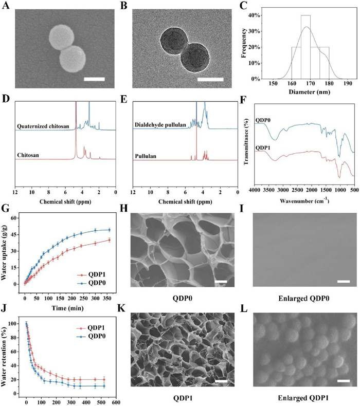
QDP1, targeted at wound management, was generated via a Schiff base mechanism using dialdehyde-modified pullulan, procyanidin/Fe3O4 nanoparticles, and quaternized chitosan [17]. Synthesis began with the uniform distribution of procyanidin/Fe3O4 nanoparticles within a solution of quaternized chitosan and oxidized pullulan. Following vigorous vortex mixing lasting 30 s, the QDP1 material promptly solidified. Celebrated for simplicity and rapidity, this synthetic strategy avoids reliance upon hazardous crosslinking agents. Combining the polysaccharide matrix with procyanidin/Fe3O4 nanoparticles created a versatile hydrogel dressing. The resultant biomaterial exhibits swift self-healing capability, strong resilience, exceptional fluid uptake, and significant anti-inflammatory effects, fostering regeneration of wound tissues.
Initial characterization utilized Fourier transform infrared (FTIR) alongside proton nuclear magnetic resonance (1H NMR) to investigate hydrogel chemical compositions; findings are presented in Figs. 1D–F. The 1H NMR spectrum of chitosan displays characteristic signals clustered primarily within the 3.0–4.0 ppm region, corresponding to protons from its polysaccharide backbone (e.g., ring CH and CH2 groups) (Fig. 1D). In contrast, the spectrum of quaternized chitosan reveals two distinct new peaks diagnostic of successful modification: A sharp singlet at ~3.3 ppm, assigned to the methyl protons (–N+(CH3)3) of the introduced quaternary ammonium groups, and a peak at ~4.3 ppm, attributed to the methylene protons (–N+–CH2–) linking these groups to the chitosan chain [18]. The persistence of the backbone signals (3.0–4.0 ppm) confirms the core structure remains intact, while the new peaks provide clear evidence of quaternization.
The 1H NMR spectrum of pullulan (bottom) shows characteristic signals clustered between 3 and 4 ppm, typical of polysaccharide backbone protons (e.g., anomeric H-1, ring CH/CH2 groups) (Fig. 1E). In contrast, dialdehyde pullulan (top) exhibits a distinct new peak at ~9–10 ppm, confirming the presence of aldehyde groups (–CHO) formed via periodate oxidation. This peak is absent in native pullulan. The persistence of signals around 3–4 ppm indicates the polysaccharide backbone remains largely intact, while the aldehyde peak at 9–10 ppm provides clear evidence of successful oxidation, validating the conversion to dialdehyde pullulan.
For FTIR, both hydrogels show broad peaks in the 3000–3500 cm–1 region, typical of N–H and O–H stretching vibrations from the dialdehyde pullulan and quaternized chitosan, confirming their polymeric backbone (Fig. 1F). The key difference is the presence of additional peaks/features in the QDP1 spectrum, likely attributable to the procyanidin/Fe3O4 nanoparticles. These may appear as specific absorptions between ~1500 cm–1 and 1700 cm–1 (potentially C=C, C=N stretching, or carboxylate vibrations influenced by the nanoparticle components or their interaction with the polymers). The absence of these features in QDP0 confirms its composition lacks these nanoparticles. Overall, the spectra verify the presence of the core polymers in both gels, while QDP1 exhibits distinct features confirming the successful incorporation of the procyanidin/Fe3O4 nanoparticles.
Surface charge analysis validated effective incorporation of procyanidin/Fe3O4 nanoparticles into the QDP0 hydrogel network. Fig. S2 (Supporting information) depicts the baseline QDP0 formulation without procyanidin/Fe3O4 nanoparticles, exhibiting a positive zeta potential measuring +27.1 mV, ascribed to structural cationic functional groups. Gradual incorporation of anionic procyanidin/Fe3O4 nanomaterials progressively lowered the hybrid material's surface charge. Formulations QDP0.5, QDP1, and QDP2, containing 0.5, 1, and 2 mg/mL of procyanidin/Fe3O4 nanomaterials, respectively, displayed measured zeta potentials of 23.8, 18.7, and 14.7 mV.
Hydration absorption and retention properties were evaluated for QDP1, supporting suitability as a dressing under hyperosmotic conditions characteristic of diabetic wounds [19]. Time-dependent monitoring of swelling capacity for QDP0 and QDP1 appears in Fig. 1G. Significant improvement in liquid uptake occurred throughout the initial 90 min. Equilibrium swelling states were attained after 360 min. Maximum hydration values reached were 49.3 for QDP0 and 40.1 for QDP1. As illustrated in Fig. 1J, adding procyanidin/Fe3O4 nanocomponents to QDP0 improved moisture retention capability. Collectively, the data demonstrate enhanced liquid uptake and retention features for the constructed polysaccharide networks, emphasizing therapeutic relevance within wound care [20].
SEM evaluated the microstructure and surface morphology of dried QDP0 and QDP1 samples. Images presented in Fig. 1H (QDP0) and Fig. 1K (QDP1) indicate that both biomaterials feature interconnected three-dimensional porous architectures [10]. Imaging revealed QDP0 possessed higher architectural homogeneity internally (Fig. 1I), while QDP1 exhibited a rougher surface morphology characterized by uniformly distributed procyanidin/Fe3O4 nanoparticles (Fig. 1L). Besides, the mean pore diameter for QDP1, containing 1 mg/mL procyanidin/Fe3O4 nanomaterial, was substantially decreased to 29.3 ± 4.7 µm vs. 60.4 ± 6.1 µm measured in QDP0, implying structural densification during cross-linking [21].
Subsequent analysis focused on assessing the material characteristics of the QDP1 sample, including its performance in laboratory tests for combating bacteria and inhibiting oxidation. Initial strain amplitude tests determined the material's linear viscoelastic zone, shown in Fig. 2A. The gelation transition for QDP1 occurred near 130% strain, indicating the sol-gel transformation point. Rheological behavior was further examined within a 0.1–15 Hz frequency spectrum. Over this entire range, the storage modulus G′ (Fig. 2B) maintained stability and persistently surpassed the loss modulus G″ (Fig. 2C), indicating that solid-like elastic behavior dominates these hydrogel systems [22]. QDP1 hydrogel durability was evaluated via sequential cyclic loading experiments. Strain amplitudes incrementally rose from 5% toward 500%, then returned to initial conditions. Fig. 2D shows that applying over 500% strain induced a transition in QDP1 to a sol phase. Subsequent reduction of strain to 5% promptly returned its G′ and G″ to initial readings, confirming structural recovery [23]. Significantly, after two full cycles, swift, reversible sol-gel conversion persisted, confirming strong autonomous structural recovery. Viscosity characteristics for QDP0 and QDP1 were assessed under escalating shear rates. Elevated shear rates induced considerable viscosity decline through shear-thinning performance, arising from rearrangement of reversible bonds throughout the QDP1 (Fig. 2E) [24]. Besides, we found that QDP1 material was effortlessly extruded employing a 2 mL syringe, forming a continuous "SCI" pattern (Fig. 2F). Collective observations highlight outstanding injectability and inherent self-restoration properties characterizing this QDP1-based biomaterial, validating applicable value for managing wound healing.
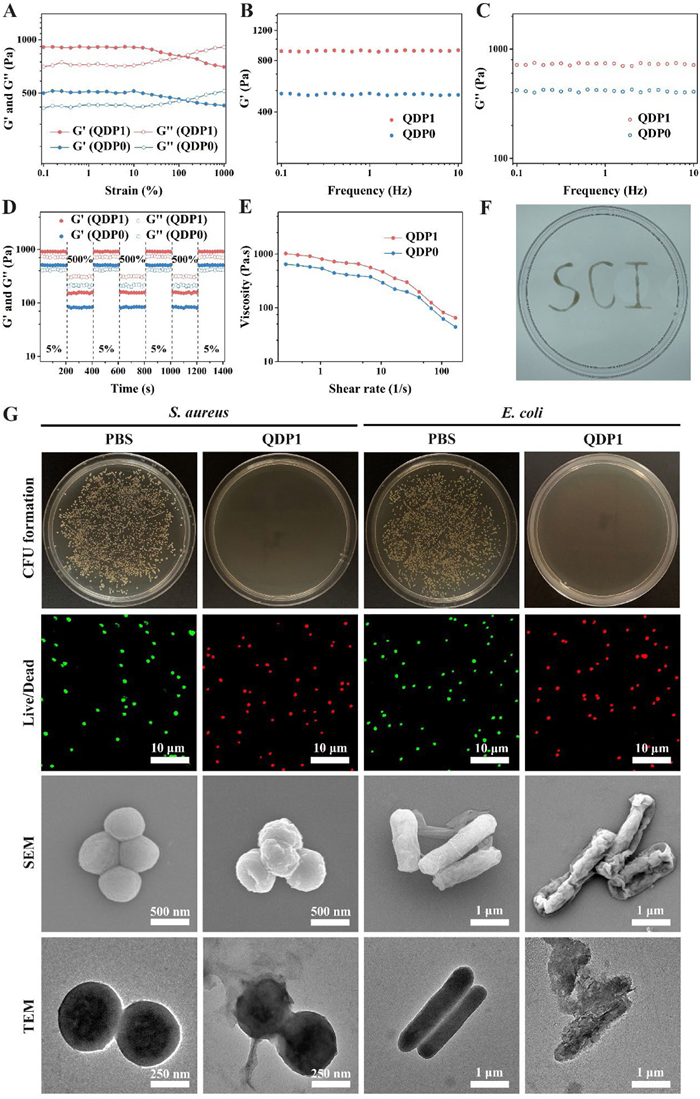
Following this, the in vitro antimicrobial efficacy of the QDP1 formulation was thoroughly assessed [25]. As indicated in Fig. 2G, QDP1 attained antibacterial efficiencies approaching 99.2% toward S. aureus (Fig. S6 in Supporting information) and 99.8% toward Escherichia coli (E. coli) (Fig. S7 in Supporting information). These antimicrobial outcomes primarily originate from chitosan's protonated amino functionalities. Microbial viability evaluations utilized dual-fluorescence labeling: SYTO9 marked metabolically active pathogens green, whereas propidium iodide detected membrane-damaged microorganisms through red emission [26]. Untreated control samples emitted predominantly green signals, while QDP1-exposed specimens exhibited intense red fluorescence, consistent with cationic disruption of pathogen envelopes (Figs. S8 and S9 in Supporting information).
Additional ultrastructural SEM studies examined morphological alterations in treated pathogens [27]. Control microorganisms preserved characteristic morphologies: S. aureus maintained spherical configurations, while E. coli displayed intact rod-shaped forms featuring smooth surfaces. In stark contrast, specimens incubated with the QDP1 polymeric system exhibited profoundly deformed architectures showing irregular, collapsed topographies. These morphological disruptions correspond to electrostatic attraction forces between the hydrogel's quaternized chitosan elements and negatively charged pathogen surfaces [28]. Such molecular interaction compromises membrane integrity through destabilization, ultimately leading to cellular fragmentation and microbe eradication.
Complementary TEM analysis documented alterations in bacterial internal architecture (Fig. 2G). Following exposure to the treatment, S. aureus and E. coli specimens were morphologically examined using TEM. Control microbes treated with PBS retained spherical shapes, uniform nucleoid dispersal, and discernible flagella. Conversely, bacteria subjected to QDP1 exhibited severe structural compromise. Their cell walls substantially attenuated or disintegrated completely, plasma membranes displayed pronounced folding, and cellular integrity was profoundly disrupted. Additionally, pathogens showed evidence of either contraction or pathological enlargement, concurrent with envelope distortion, rupture, and consequent leakage of cytoplasmic material.
Thereafter, the capacity for neutralizing oxidative radicals was assessed to clarify the essential role of ROS in wound recovery [29]. Standard markers, 2,2-diphenyl-1-picrylhydrazyl (DPPH), 2,2′-azinobis(3-ethylbenzothiazoline-6-sulfonic acid) (ABTS) and superoxide anion radical (O2•−), were methodologically employed [30]. Findings from the DPPH assay (Fig. S3 in Supporting information) indicated that following thirty minutes of incubation, mixtures incorporating hydrogel specimens underwent a color shift from deep violet toward light purple for QDP0, while QDP1 treatment induced a yellow coloration. This visual transformation signifies substantial DPPH radical reduction facilitated by integrated procyanidin/Fe3O4 nanoparticles. Quantification of optical density validated these observations, revealing scavenging efficiencies attaining 27.1% with QDP0 and 65.6% utilizing QDP1. Evaluations using ABTS demonstrated that QDP1 exhibited almost twice the radical removal capacity compared to QDP0 (Fig. S4 in Supporting information). Suppression of O2•−, critically examined because such radicals impede diabetic wound resolution, received further attention (Fig. S5 in Supporting information). The QDP1 composite yielded diminished blue formazan product relative to QDP0, confirming its enhanced scavenging performance. Cumulatively, the experimental outcomes highlight significant antioxidant contributions originating from procyanidin/Fe3O4 nanoparticle constituents embedded within the hydrogel architecture, confirming QDP1′s potential as a promising enhancer for wound regeneration.
For biomedical hydrogels, biocompatibility represents a fundamental requirement [31]. To evaluate QDP1 cytocompatibility, RS1 fibroblasts were cultured and analyzed through Live/Dead staining. This technique employed calcein acetoxymethyl ester (calcein-AM) and propidium iodide (PI) to distinguish living cells displaying green fluorescence from nonviable counterparts exhibiting red fluorescence [32]. Results illustrated in Fig. 3A indicate high fibroblast survival rates coupled with negligible cytotoxic effects. Exposure to the hydrogel formulation did not compromise cellular viability or hinder proliferation. Assessment of cell morphology via fluorescein isothiocyanate-phalloidin (FITC-phalloidin) and 4′, 6-diamidino-2-phenylindole (DAPI) co-staining (Fig. 3B) demonstrated enhanced spreading alongside a structured F-actin cytoskeletal network within treated samples, closely resembling that of untreated counterparts.
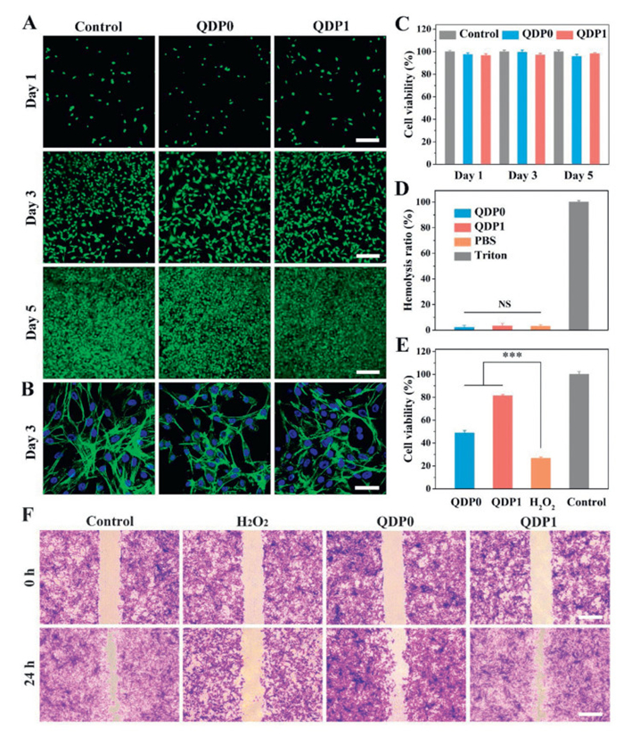
Further rat skin 1 (RS1) fibroblast examination using the cell counting kit-8 (CCK-8) assay substantiated earlier fluorescent staining observations (Fig. 3C). Viability following exposure to QDP1 extracts exceeded 94.1% across multiple continuous incubation periods, paralleling negative control groups containing only culture medium. These data confirm the material's outstanding cellular compatibility. Subsequent analysis focused on blood compatibility, an essential consideration for biomaterials that contact vascular systems at injury locations (Fig. 3D). Data were analyzed using a two-tailed Student's t-test [33]. Supernatant clarity for all hydrogel specimens corresponded to phosphate-buffered saline (PBS) negative controls; hemolysis rates for QDP0 and QDP1 remained beneath 5%, demonstrating blood compatibility appropriate for wound-contact applications. Collectively, these findings strongly endorse QDP1's potential as a tissue engineering dressing candidate.
To assess biological ROS neutralization capacity, fibroblast viability was quantified under induced oxidative stress. RS1 cultures exposed to QDP1 hydrogel and hydrogen peroxide (H2O2) underwent CCK-8 survival profiling. As Fig. 3E indicates, positive control groups treated exclusively with 680 µmol/L H2O2 exhibited only 26.7% survival. QDP0 hydrogel administration significantly enhanced cellular survival to 48.8% under equivalent oxidative challenge, a viability improvement attributed to intrinsic ROS-reducing characteristics from bioactive moieties and porous architecture. Integrating procyanidin/Fe3O4 nanoconstituents (recognized for potent free radical scavenging capabilities) into QDP0 markedly amplified antioxidant performance within the QDP1 composite, achieving 81.3% viability. These results confirm that QDP1, augmented via 1 mg/mL procyanidin/Fe3O4 nanomaterial incorporation, offers dual advantages: Exceptional biocompatibility and robust ROS suppression, indicating substantial promise for managing diabetic wounds clinically.
Cellular migration and proliferation dynamics during oxidative insult were further examined via in vitro scratch testing [34]. RS1 monolayers received standardized linear scratches using pipette tips. After applying H2O2 and hydrogel treatments, wound closure progression was assessed following 12- and 24-h incubation intervals. Fig. 3F reveals that untreated fibroblasts displayed anticipated migration behavior, substantially reducing the wound zone within 24 h. Conversely, cultures subjected solely to H2O2 showed minimal movement into the injury site, retaining nearly the initial scratch measurements. The QDP1 hydrogel cohort, however, presented accelerated motility in contrast to the oxidative stress condition, fostering wound recovery. These observations indicate that QDP1 fortifies cellular antioxidant defenses by effectively scavenging externally administered ROS [35].
After evaluating QDP1′s physicochemical properties, the investigation focused on evaluating the therapeutic influence of the engineered QDP1 hydrogel on wounds in a diabetic model. The Animal Care and Use Committee at Nanjing University of Science & Technology authorized every animal study protocol. All conducted work strictly followed regulations concerning facility conditions and housing for laboratory animals. Wound sites received applications of different materials, with each sample weighing 90 mg. The tissue regeneration process was carefully documented via photographic evidence captured on post-treatment days 3, 7 and 14 (Fig. 4A). A marked reduction in wound dimensions was observable after only 72 h for subjects treated with either QDP1 or QDP0, especially when contrasted with the PBS control cohort. Furthermore, microbial specimens harvested from the injuries on day three underwent quantification utilizing an agar plate-based technique, detailed in Fig. 4B [36]. Consistent with earlier findings, noticeable bacterial colonization was evident in wounds treated with QDP0 and PBS. In stark contrast, the QDP1 treatment cohort demonstrated an almost complete absence of such colonies. Upon reaching the fourteenth day, significantly larger impaired regions persisted in the PBS and QDP0 cohorts, retaining 85.4% and 73.7% of their original area, respectively. The novel QDP1 formulation, however, yielded the most substantial regenerative outcome, achieving a remarkable reduction in the wound area to just 9.8% (Fig. 4C). Histological analysis with hematoxylin and eosin (H&E, Fig. 4D) revealed that specimens from control and QDP0 groups exhibited epidermal necrosis and desquamation, together with substantial infiltration by inflammatory cells. Conversely, tissue from the QDP1 treatment demonstrated a notable reduction in inflammatory cell presence and a marked rise in fibroblast numbers [37]. Evaluation of collagen deposition through Masson staining (Fig. 4E) indicated that QDP1 wounds possessed the most robust and well-organized architecture, characterized by densely packed fibrous structures [38].
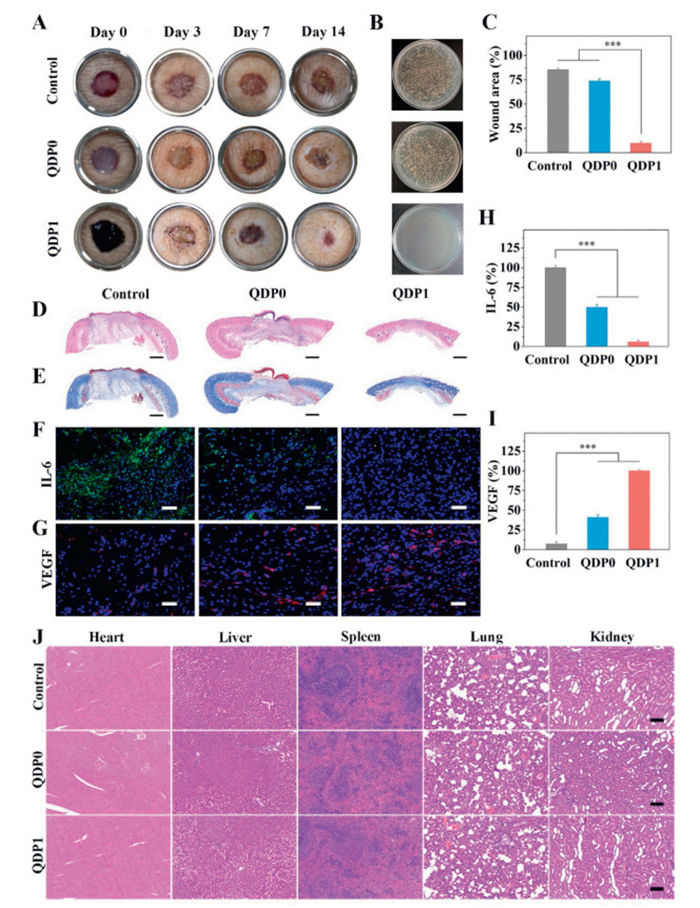
Motivated by these wound closure outcomes, subsequent analysis explored the potential mechanism behind tissue repair through immunofluorescence staining. As displayed in Fig. 4F, tissues treated with PBS exhibited heightened IL-6 expression, which moderately decreased after QDP0 administration and demonstrated marked attenuation under QDP1 conditions; corresponding quantification appears in Fig. 4H. These collective findings substantiate QDP1′s locally potent anti-inflammatory efficacy [39]. Analysis of angiogenesis employed vascular endothelial growth factor (VEGF) immunostaining, presented visually in Fig. 4G. Substantial microvascular activity, elaborated upon within Fig. 4I, emerged specifically across subjects receiving QDP1 hydrogel. Developing new microvasculature proves critical for delivering essential oxygen and nutrients to traumatized areas, thereby promoting regenerative matrix formation [40]. Concurrently, the QDP1 formulation powerfully inhibits inflammatory processes while actively encouraging angiogenic growth, synergistically accelerating wound repair.
Furthermore, an exhaustive biosafety evaluation of QDP1 hydrogel, crucial for wound healing applications, was conducted [41]. Fig. S10 depicts consistent body weight gains across both non-treated and experimental cohorts, implying a lack of systemic toxicity [42]. Systemic consequences were appraised via blood biochemistry analysis. Outcomes revealed comparable profiles for vital hematological parameters (red blood cells, platelet count, white blood cells, hematocrit, hemoglobin, and mean cell volume) between hydrogel-treated rodents and control counterparts, confirming QDP1's hemocompatibility (Fig. S11). Moreover, H&E staining assessed potential organ damage [43]. Tissue sections from diabetic animals administered QDP1 displayed minimal inflammatory cell presence relative to PBS-treated subjects, as Fig. 4J demonstrates. Collectively, these observations indicate QDP1 possesses negligible biotoxicity towards healthy tissues in vivo, underlining its advantageous safety profile and supporting translational potential for wound management.
In sum, our study successfully developed QDP1 hydrogel, a multifunctional biomaterial incorporating dialdehyde pullulan, quaternized chitosan, and procyanidin/Fe3O4 nanoparticles, specifically designed for bacterially infected diabetic wounds. This engineered formulation displayed significant ROS quenching capacity (achieving 65.6% DPPH reduction and enhanced ABTS radical clearance), powerful antibacterial efficacy (eliminating 99.2% of S. aureus and 99.8% of E. coli), alongside notable angiogenesis-promoting functionality, dramatically accelerating wound closure within diabetic rats. By post-operative day 14, wound defects managed using QDP1 attained almost complete re-epithelialization, decreased interleukin-6 levels, and increased VEGF marker density, indicating suppressed inflammatory responses and enhanced microvascular formation. Combined with outstanding biocompatibility, proven hemocompatibility (< 5% hemolysis), and lack of systemic toxic effects, the QDP1 hydrogel represents a safe and effective therapeutic strategy for managing diabetic wounds, exhibiting considerable potential for clinical translation prospects.
Hao Yu: Writing – review & editing. Xiaoliang Qi: Investigation. Yuxi Chen: Methodology. Youjian Chen: Software. Jinghui Zhang: Software. Wei Dong: Investigation. Jianliang Shen: Supervision. Tian Jiang: Validation.
We declare that there are no known conflicts of interest, either financial or personal, which might have been perceived as affecting the work presented in the manuscript.
This research was supported by the National Natural Science Foundation of China (No. 32501180).
Supplementary material associated with this article can be found, in the online version, at doi:
Y. Yang, Q. Fang, J. Wang, et al., Adv. Mater. 37 (2025) 2419158. doi: 10.1002/adma.202419158
Y. Zhang, C. Xue, Y. Zhang, et al., Chin. Chem. Lett. 35 (2024) 109196. doi: 10.1016/j.cclet.2023.109196
J. Du, C. Xian, X. Liang, et al., MedComm Biomater. Appl. 4 (2025) e70019.
X. Qi, Y. Lan, J. Chen, et al., Adv. Healthc. Mater. 14 (2025) 2500688. doi: 10.1002/adhm.202500688
H. Gong, L. Yang, Y. Li, et al., Biomaterials 322 (2025) 123414. doi: 10.1016/j.biomaterials.2025.123414
A. Bassam, M. Du, Y. Li, et al., Respon. Mater. 3 (2025) e20240031.
J. Wang, X. Ge, Y. Xiang, et al., Chin. Chem. Lett. 36 (2025) 109819. doi: 10.1016/j.cclet.2024.109819
J. Luo, Z. Liang, X. Zhao, et al., Natl. Sci. Rev. 12 (2025) nwaf118. doi: 10.1093/nsr/nwaf118
P. Xin, S. Han, J. Huang, et al., Chin. Chem. Lett. 34 (2023) 108125. doi: 10.1016/j.cclet.2022.108125
Y. Sun, Y. Zhu, J. Si, et al., Chin. Chem. Lett. 36 (2025) 110012. doi: 10.1016/j.cclet.2024.110012
Z. Li, S. Guan, X. Duan, et al., Int. J. Biol. Macromol. 318 (2025) 144944. doi: 10.1016/j.ijbiomac.2025.144944
X. He, S. Koo, E. Obeng, et al., Coordin. Chem. Rev. 492 (2023) 215275. doi: 10.1016/j.ccr.2023.215275
Z. Li, D. Xu, Z. Deng, et al., Chem. Eng. J. 452 (2023) 139587. doi: 10.1016/j.cej.2022.139587
M. Liu, W. Zhao, D. Li, et al., Langmuir 41 (2025) 17104–17116. doi: 10.1021/acs.langmuir.5c01761
X. Pan, S. Cheng, C. Zhang, et al., Chem. Eng. J. 409 (2021) 128203. doi: 10.1016/j.cej.2020.128203
S. Cheng, C. Zhang, J. Li, et al., Carbohydr. Polym. 262 (2021) 117951. doi: 10.1016/j.carbpol.2021.117951
M. Li, L. Li, X. Zhang, et al., J. Med. Chem. 68 (2025) 17808–17822. doi: 10.1021/acs.jmedchem.5c01536
Y. Shan, F. Cao, X. Zhao, et al., Biomaterials 315 (2024) 122936.
M. Chen, Q. Zhou, T. Qin, et al., Chin. Chem. Lett. 36 (2025) 110441. doi: 10.1016/j.cclet.2024.110441
Z.-P. Zhou, X. Wei, M. Yan, et al., Chin. Chem. Lett. 36 (2025) 111400. doi: 10.1016/j.cclet.2025.111400
J. Luo, Z. Wu, X. Zhao, et al., Adv. Funct. Mater. 35 (2025) 2419037. doi: 10.1002/adfm.202419037
C. Zhang, E. Cai, X. Qi, et al., Chem. Eng. J. 487 (2024) 150756. doi: 10.1016/j.cej.2024.150756
J. Chen, X. Zhao, L. Qiao, et al., Adv. Healthc. Mater. 13 (2024) 2303157. doi: 10.1002/adhm.202303157
P. Shan, J. Liao, J. Li, et al., Chin. Chem. Lett. 35 (2024) 108545. doi: 10.1016/j.cclet.2023.108545
X. He, Z. Wang, B. Mao, et al., Bioact. Mater. 51 (2025) 841–857.
X. Zheng, L. Pang, Y. Wang, et al., Mater. Today Bio 34 (2025) 102170. doi: 10.1016/j.mtbio.2025.102170
B. Zeng, Z. Mu, T. Shen, et al., Chin. Chem. Lett. 36 (2025) 110350. doi: 10.1016/j.cclet.2024.110350
X. Qi, Y. Xiang, E. Cai, et al., Chem. Eng. J. 439 (2022) 135691. doi: 10.1016/j.cej.2022.135691
J. Wang, Z. Gao, H. Liang, et al., Chin. Chem. Lett. 36 (2025) 110569. doi: 10.1016/j.cclet.2024.110569
Y. Xiang, X. Qi, E. Cai, et al., Chem. Eng. J. 460 (2023) 141852. doi: 10.1016/j.cej.2023.141852
J. Chen, L. Qiao, D. Cheng, et al., J. Control. Release 385 (2025) 113983. doi: 10.1016/j.jconrel.2025.113983
C. Liu, L. Li, J. Gao, et al., Chin. Chem. Letters. 36 (2025) 110118. doi: 10.1016/j.cclet.2024.110118
X. He, X. Liao, H. Li, et al., Inorgan. Chem. 56 (2017) 15041–15048. doi: 10.1021/acs.inorgchem.7b02380
Z. Hu, J. Shan, X. Jin, et al., ACS Nano 18 (2024) 24327–24349. doi: 10.1021/acsnano.4c06565
M. Cheng, Y. Fan, C. Wang, et al., J. Colloid Interface Sci. 702 (2025) 138785.
W. Su, X. Luo, P. Li, et al., Chin. Chem. Lett. 35 (2024) 109522. doi: 10.1016/j.cclet.2024.109522
L. Xie, X. Zhang, X. Wang, et al., Chin. Chem. Lett. 36 (2025) 110848. doi: 10.1016/j.cclet.2025.110848
Y. Fang, T. Nie, G. Li, et al., Chem. Eng. J. 480 (2024) 147930. doi: 10.1016/j.cej.2023.147930
X. He, Y. Lv, Y. Lin, et al., Adv. Mater. 36 (2024) 2400366. doi: 10.1002/adma.202400366
J. Wang, J. He, Y. Yang, et al., Chem. Eng. J. 479 (2024) 147577. doi: 10.1016/j.cej.2023.147577
Y. Zang, W. Zhang, W. Wang, et al., Cell Biomater. 2 (2026) 100217. doi: 10.1016/j.celbio.2025.100217
C. Liu, X. Xiao, Y. Chen, et al., Chem. Mater. 37 (2025) 224–237. doi: 10.1021/acs.chemmater.4c02308
X. Sun, D. Wang, S. Ma, et al., Chem. Eng. J. 512 (2025) 162627. doi: 10.1016/j.cej.2025.162627

Figure 1 Physicochemical analysis of QDP1. (A) Procyanidin/Fe3O4 nanoparticle morphology visualized via SEM (scale bar: 150 nm). (B) Procyanidin/Fe3O4 nanoparticle features observed through TEM (scale bar: 150 nm). (C) Distribution of procyanidin/Fe3O4 nanoparticle sizes. (D) 1H NMR spectral profiles comparing chitosan derivatives. (E) 1H NMR analysis of pullulan modification. (F) FTIR spectra contrasting QDP0 and QDP1. (G) Kinetics of hydration uptake (comparative). (H) QDP0 microstructure (scale bar: 30 µm). (I) High-resolution QDP0 morphology (scale bar: 150 nm). (J) Comparison of water retention. (K) Composite architecture of QDP1 (scale bar: 30 µm). (L) High-magnification features of QDP1 (scale bar: 150 nm).

Figure 2 Rheological properties and in vitro antibacterial performance of the QDP1. (A) Viscoelastic response dependent on strain. (B) G′ across varied amplitudes (1.5% strain). (C) G″ under diverse deformations (1% strain). (D) Strain sweep assay at 5% and 500% deformation (0.5 Hz). (E) Illustration of shear-thinning behavior. (F) Demonstration of syringe extrusion. (G) Antimicrobial evaluation of QDP1 toward S. aureus and E. coli.

Figure 3 In vitro cytocompatibility assessment and QDP1-driven ROS alleviation. (A) RS1 viability staining across different treatment intervals (scale bar: 150 µm). (B) Fluorescent photos demonstrating the morphology of RS1 (actin: green; nuclei: blue; scale bar: 20 µm). (C) RS1 cell activity over time. (D) Assessment of hemolysis. (E) Survival rates of RS1 under H2O2. (F) Representative images of migration at 0/24 h (scale bar: 50 µm). Error bars: mean ± SD (n = 3). ***P < 0.001. NS: non-significant.

Figure 4 Therapeutic effectiveness of QDP1 in diabetic wounds. (A) Healing progression across 14 days in QDP0, QDP1, and control groups. (B) Visual documentation of microbial growth on agar plates, cultured from wound sites of treated rats. (C) Quantitative analysis of wound area reduction (n = 6). (D) H&E-stained wound tissue section (scale bar: 500 µm). (E) Collagen deposition visualized by Masson (scale bar: 500 µm). (F) IL-6 immunolocalization within wound tissues (scale bar: 100 µm). (G) VEGF immunodetection within wound beds (scale bar: 100 µm). (H) Measurement of IL-6 levels (n = 6). (I) Measurement of VEGF-positive areas (n = 6). (J) Histopathology of major organs in rats after therapy (scale bar: 100 µm). Error bars: mean ± SD. ***P < 0.001.

 扫一扫看文章


扫一扫关注我们
 DownLoad:

 下载:
 下载: