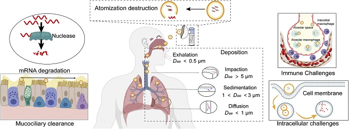
Figure 1.
Challenges in pulmonary mRNA delivery for therapeutic applications.

Pulmonary mRNA delivery systems for the treatment of respiratory diseases: Current advances and challenges
Kaiqing Zhang , Yue Zhou , Guanlin Wang , Bing Zhu , Ziyu Zhao , Xi Kong , Yihong Gao , Xin Pan , Zhengwei Huang , Chuanbin Wu , Xuejuan Zhang


Respiratory diseases constitute a major global health burden, adversely impacting both human health and socioeconomic systems. Rapid urbanization and demographic aging have contributed to escalating pulmonary disease mortality, with three respiratory conditions currently ranking among the top ten causes of death globally, namely chronic obstructive pulmonary disease (COPD), pulmonary infections, and lung cancer [1]. Furthermore, significant unmet clinical needs persist in the treatment of respiratory diseases, particularly for highly prevalent conditions including lung cancer and pulmonary infections. Furthermore, certain respiratory diseases (e.g., inherited pulmonary conditions), while exhibiting relatively low prevalence, present high clinical value and research imperative and thus warrant dedicated efforts. Most inherited pulmonary conditions originate from single-gene defects that disrupt essential pulmonary homeostasis mechanisms, particularly cystic fibrosis (CF) and α−1 antitrypsin deficiency [2–4]. Present therapeutic approaches predominantly offer symptomatic relief and disease progression mitigation rather than curative outcomes.
Fortunately, delivering messenger ribonucleic acid (mRNA) has brought a novel turnaround for respiratory diseases, which present a variety of therapeutically challenging but offer attractive targets. mRNA has demonstrated its potential to tackle the issues posed by the coronavirus disease 2019 (COVID-19) pandemic, including 1273-mRNA (Moderna) and 162b2-mRNA (Pfizer/BioNTech), which showcase their potential to elicit strong and specific immune responses and support their practical applications and efficacy observed in clinical settings. Besides, > 800 clinical trials, including pathogen- or tumor-derived vaccines and antigen-encoding mRNA drugs, had been registered (
Inhalation therapy represents a non-invasive administration in which medications are administered via inhalation, facilitating direct therapeutic delivery to the lesions of respiratory diseases. Inhaled mRNA enables targeted pulmonary deposition, avoids potential off-target effects in other organs, minimizes systemic exposure, and offers distinct pharmacological advantages over oral and parenteral administration routes [11]. These attributes are advantageous for chronic pulmonary diseases such as CF, lung cancer, α−1 antitrypsin deficiency, and pulmonary infections. Inhaled mRNA therapies include mRNA immunotherapy, protein replacement therapy, and gene editing. Among them, mRNA immunotherapy is mainly used in lung cancer and pulmonary infections, which leverages pivotal mediators of immune responses antigen-presenting cells (APCs) serve as an efficient mucosal immune network, including dendritic cells (DCs), B cells, and alveolar macrophages [12]. Then, cellular immune responses are activated through major histocompatibility complex class I (MHC I) and class II (MHC II) pathways to clear pathogens or kill tumors [13]. On the other hand, mRNA promotes the differentiation of B cells into plasma cells in the lung mucosa, produces mucosal secretory IgA (sIgA), establishing an immune barrier by neutralizing viruses and blocking pathogen mucosal adhesion [14]. The successful implementation of mRNA-based COVID-19 vaccines has provided mechanistic proof-of-concept for protein-encoding mRNA therapeutics. Protein replacement therapy is a representative therapeutic strategy for CF treatment, and several mRNA inhalation formulations leveraging this approach are already in the clinical study phase [15]. Notably, as a representative gene-editing technology, the clustered regularly interspaced short palindromic repeats (CRISPR) and CRISPR-associated proteins (CRISPR/Cas) system delivered via mRNA encoding the Cas nuclease is widely recognized as a powerful and safe platform for precise mutation correction and has received considerable attention in clinical practice [16]. Despite the promise, the use of pulmonary mRNA delivery systems in the treatment of respiratory disease is limited by four challenges largely including complex airway tract structure, mucus cilia clearance system, immune system, and the "final hurdle" intracellular delivery barrier [17–19]. These challenges are directly related to the effectiveness and safety of treatment. The following details and discusses the current challenges of promising inhalable formulations applicable to the delivery of mRNA therapeutics treating respiratory diseases.
This paper provides an overview of the current challenges, strategies, and advances in pulmonary mRNA delivery for the treatment of respiratory diseases, as there are currently few published studies on the development of inhaled therapies for respiratory diseases. This review will offer forward-looking insights outlining the present development of pulmonary mRNA delivery via inhalation, with an emphasis on their design and critical experimental results for mRNA therapies. This review begins with the therapeutic mechanisms of mRNA-based therapies from pharmacological and physiological perspectives. Then, pulmonary drug delivery systems are categorized by formulation characteristics, pharmacological benefits, and inherent limitations. It is followed by a detailed discussion on current challenges, strategies, and advances of pulmonary mRNA delivery.
Human physiological functions rely on correct proteins being synthesized through the processes of DNA transcription, mRNA translation, and subsequent post-translational modifications, including antibodies, hormones, enzymes, cytokines. The success of COVID-19 mRNA vaccines has opened new avenues for applying mRNA technology to the treatment of diseases, where it holds promise for creating personalized mRNA drugs that can encode therapeutic proteins, target specific antigens, enhance the immune response, and modulate gene expression. mRNA-based therapeutics can be categorized into three major fields: Immunotherapy (including preventive and therapeutic vaccines), protein replacement therapy, and gene editing therapy (Fig. S1 in Supporting information) [20].
mRNA immunotherapy provides a therapeutic approach to stimulate the immune system that utilizes synthesized therapeutic mRNAs translated into target proteins in humans, potentially for the treatment and prevention of disease, including lung infections [21,22] and lung cancers [23]. mRNA can induce an immune response by encoding pathogen- or tumor-derived antigens, eliminating pathogen-infected cells and tumor cells expressing these antigens, to achieve the effect of preventing viral infection and treating tumors [24,25]. mRNA immunotherapy has demonstrated its ability to solve the issues posed by the COVID-19 pandemic, which contributed to the renaissance of research on mRNA and their nano-formulations as potential therapeutic modalities for various diseases. For mRNA immunotherapy of pulmonary infections, the expressed pathogen-derived antigenic proteins are presented on cell surfaces via MHC I/II molecules, initiating adaptive immune responses that generate neutralizing antibodies and establish memory immune cells [26,27]. For mRNA immunotherapy of lung cancer, the high-quality tumor antigen delivery to APCs, the maturation of APCs promotion and CD4+ T helper cell activation, and CD8+ T cytotoxic cell response and immune cell infiltration into the tumor microenvironment (TME) are the vital circle to generate immunologic memory for sustainable therapeutic effects [28,29].
Human physiological functions rely on correct proteins being synthesized through the processes of DNA transcription and mRNA translation and subsequent post-translational modifications, including hormones, enzymes, cytokines, etc. mRNA-directed protein replacement has emerged as a potential therapeutic strategy for certain refractory diseases. mRNA-mediated protein replacement therapy represents a transformative therapeutic platform with multifaceted advantages: (1) Programmable synthesis of therapeutic proteins/peptides including undruggable targets [30]; (2) minimal genomic integration risk [30]; (3) sustained therapeutic protein production [31]; (4) combinatorial therapeutic protein production capabilities [30]; and (5) streamlined development timelines [32]. This technology platform demonstrates potential for two critical scenarios involving rapid-response drugs (e.g., antibodies) development against emerging pathogens and cancers, and treatment of refractory disorders such as CF and alpha-1 antitrypsin deficiency (AATD) [33]. mRNA therapeutics for pathogen infections and tumors differ fundamentally from mRNA vaccines, which require only low-dose, localized transient expression of prophylactic or therapeutic antigens. In contrast, mRNA-based protein replacement therapy typically necessitates sustained expression of therapeutic protein in specific tissues to achieve significant efficacy. When applied to refractory diseases stemming from genetic defects or mutations, these therapeutics employ mRNA encoding the required functional proteins, demanding targeted delivery to relevant cell types (e.g., epithelial cells and fibroblasts) for biological activity.
Gene editing has attracted considerable interest across the scientific community for its therapeutic and biotechnological promise. Gene editing technologies include meganucleases, zinc finger nucleases (ZFNs), followed by transcription activator-like effector nucleases (TALENs), and CRISPR/Cas systems [34]. Among these, the mRNA-based CRISPR/Cas system has emerged as the preferred gene-editing platform due to its high efficiency, favorable safety profile, absence of integration risks, and broad therapeutic applicability. Clinically, mRNA-encoded CRISPR/Cas system holds great potential for cancer treatment due to the ability to precisely inactivate or repair cancer-related genes, and it offers safe and efficient targeted gene editing capabilities for genetic respiratory diseases, including CF [35], AATD [2], and surfactant protein dysfunction syndrome [36], supporting personalized therapeutic strategies. CRISPR/Cas systems function via gRNA-mediated targeting: DNA editing (e.g., Cas9) requires nuclear delivery and risks off-target effects [37], while RNA editing (e.g., Cas13a) enables transient, cytoplasm-localized regulation without genomic changes [38]. CASGEVY™, a CRISPR/Cas-based gene editing therapy, has demonstrated curative potential for monogenic disorders and received conditional marketing authorization as the first approved CRISPR/Cas therapeutic platform. This regulatory milestone demonstrates the therapeutic potential of gene editing therapy in genetic respiratory diseases, exemplified by CF (CF transmembrane conductance regulator (CFTR) mutations).
Pulmonary drug delivery systems are a highly attractive choice for the treatment of respiratory diseases, a non-invasive mRNA-based delivery strategy with significant advantages including reducing dosage requirements, off-target effects, and thereby mitigating systemic toxicity. Pulmonary drug delivery systems primarily comprise three categories, namely the nebulizers (NEBs), the metered-dose inhalers (MDIs), and the dry powder inhalers (DPIs) (Table S1 in Supporting information). Each of them utilizes specialized devices to generate respirable particles for lung deposition with distinct advantages and limitations. The following details the differences, advantages, and limitations between those three pulmonary drug delivery systems.
The NEBs (inhalation solution, suspension or powder for inhalation) refer to the dispersion of drugs into small droplets through specialized nebulization device. They provide advantages of large delivery doses, user-friendly, high flexibility in formulation development and process optimization [39,40]. Given these advantages, nebulizers have become the delivery platform of choice for mRNA therapy research. For example, Liu et al. employed a nebulizer to deliver charge-assisted stable LNPs loaded with mRNA vaccines, achieving efficient lung transfection and robust mucosal immune activation in murine, miniature pig, and canine models [41]. These results underscore the significant potential of nebulization for pulmonary mRNA delivery. The inherent aqueous instability of nucleic acids represents the primary challenge for mRNA nebulization. Although lyophilization enables storage stability, reconstitution often induces significant nanoparticle size alterations. Furthermore, different types of nebulization devices exhibit distinct characteristics in terms of shear forces and aerosol properties, which can significantly impact nanoparticle integrity and mRNA stability during pulmonary delivery, thereby affecting the effective delivery of mRNA drugs [42,43]. The MRT5005 Phase I trial showed no clinical benefit despite stable lung function, primarily due to insufficient lung deposition below preclinical expectations [44]. Generally, inadequate pulmonary mRNA deposition, shear stress during nebulization, storage instability, and accessibility challenges in cold-chain distribution significantly limit the translational potential of NEBs for pulmonary mRNA delivery. Optimizations are typically directed toward device design and formulation. The co-optimization of devices and formulations ensures the effectiveness of the delivery systems. After selecting the nebulization device, the delivery systems must be customized to the selected device by adjusting formulation composition, buffer systems, and other essential excipients [45]. Simultaneously, formulation properties such as viscosity and surface tension must be carefully designed, as they directly influence droplet size and efficiency of pulmonary drug delivery. For example, viscosity impacts nebulization performance, while surface tension plays a critical role in determining aerosol droplet size and stability, which affects lung deposition efficiency [46]. Thus, the co-optimization of both device and formulation is crucial for developing effective inhalation therapies.
The MDIs refer to the propellant-drug mixture dispersed by plastic actuators into aerosols with an inhalable size. Compared to NEBs, they offer the advantages of compact size and portability, making them particularly suitable for chronic disease management [47]. While previous studies have confirmed negligible effects of propellants on the bioactivity of certain nucleic acid drugs [48,49], mRNA-propellant compatibility has not yet been clarified and urgently needs systematic reevaluation. However, shear stress of high-pressure and hand-mouth coordination are the main obstacles that limit the translational potential of MDIs for pulmonary mRNA delivery. Furthermore, to lower the carbon footprints, the hydrofluoroalkane (HFA), as the second generation of propellant, is undergoing phasing out. The transition to next-generation propellants (e.g., HFO-1234ze and HFA 152a) forces require key variables including reformulation and actuator retooling, thereby posing temporary uncertainties for the MDI-based mRNA development.
The DPIs refer to carrier-free active pharmaceutical ingredients (APIs) or mixtures of carrier and micronized APIs aerosolized by inhaler devices into respirable particles. They exhibit the advantages of much better storage stability, flexibility and lower shear stress compared to NEBs, and avoid hand-mouth coordination, high-pressure shear stress of propellants and no prospective deleterious effects on the environment compared to MDIs. Additionally, formulation optimization enables precise management of DPIs particles' characteristics, ensuring alveolar deposition critical for pulmonary drug efficacy. Formulations of DPIs are categorized into two primary types: Carrier-based systems (typically lactose-bound) and carrier-free formulations [50]. For mRNA therapeutics, carrier-free DPIs offer flexible batch customization and diverse processing options, including spray drying, freeze drying, thin-film freeze drying, and spray freeze drying, enabling more efficient and scalable production [51]. DPIs have been successfully adapted for pulmonary mRNA delivery, maintaining mRNA integrity and bioactivity during powder production and storage [52,53]. Developing effective mRNA DPIs requires co-optimizing both formulation processes and device design to balance mRNA stability with aerosol performance under thermal and mechanical stresses [54,55]. Key strategies of formulation stability and aerosolization performance improvement include protectants (e.g., fucoidan, mannitol) that preserve mRNA integrity through water replacement and modulate powder cohesiveness, and surfactants that stabilize LNPs while tailoring powder surface energy to device shear forces [54]. Moreover, properties of dry powder inhalations (e.g., particle size, moisture, flowability) and device characteristics (e.g., airflow resistance, dispersion mechanics) must be optimized. For instance, if powder adhesion is improper or airflow dynamics are mismatched, particle dispersion may be compromised [56–58]. Successful pulmonary mRNA delivery thus demands integrated design of both the powder formulation and inhalation device to achieve optimal lung deposition while maintaining nucleic acid stability.
The MDIs are suitable for delivering fine particles (~1 µm) and nucleic acid drugs directly to the lungs. However, phase-out mandates for environmentally hazardous propellants, compounded by uncertainties surrounding next-generation alternatives, have constrained research focus on this platform. NEBs and DPIs now represent the most intensively investigated pulmonary mRNA delivery systems. Among these, NEBs constitute the most widely utilized inhalation platform for pulmonary mRNA delivery, primarily owing to their broad device compatibility. Nevertheless, given the inherent hydrolysis sensitivity and aqueous instability of NEBs, DPIs hold greater promise as the optimal delivery format for pulmonary mRNA applications. Exemplified by BMD006 (NCT06928922), mRNA DPIs demonstrate superior long-term stability, ultra-low temperature storage and transport logistics. Such advantages substantially facilitate the translational clinical outcome and progress of pulmonary mRNA therapeutics.
Despite the promise, the use of pulmonary mRNA delivery system in respiratory diseases treatments is limited by four challenges largely including inadequate pulmonary deposition caused by the complex airway tract structure, mucociliary clearance (MCC) barrier, immune response, and the "last hurdle" intracellular delivery barrier (Fig. 1). These challenges are directly related to the effectiveness and safety of the treatment. To address these barriers, advanced nanocarriers have been widely employed (Table S2 in Supporting information), including LNPs, polymeric, and exosomes (Exos). Among these, LNPs lead in clinical translation due to high encapsulation efficiency, yet require optimization for lung distribution and nebulization stability [59,60]. Polymer carriers provide flexibility and cost benefits, with newer designs reducing cytotoxicity [61]. Cell-penetrating peptides (CPPs) excel in cellular uptake but face off-target effects [62]. While Exos, though biocompatible and inherently targeted, face challenges in standardized production and drug loading [63]. Hybrid systems such as LNP-CPP composites improve delivery efficiency [64,65]. Wu's team showed that nanocarrier surface properties and size affect pulmonary retention, cellular uptake, and endosomal escape [66–68]. The following reviews the key challenges in pulmonary mRNA delivery while analyzing how the physicochemical properties of nanoparticle carriers can be engineered to overcome these challenges. Importantly, these barriers are interconnected, requiring balanced optimization as strategies addressing one challenge may compromise another.
The airway tract for oral inhalation includes the pharynx, larynx and trachea, branching into 24 generations from the bronchioles to the alveoli. This continuous branching reduces airway diameter and lowers the deposition efficiency of inhaled drugs [69]. As a result, only a small percentage of particles reach the deep lungs, while the majority are deposited in the upper airways. Thus, the efficacy of pulmonary mRNA delivery depends critically on the fraction of mRNA that can bypass the upper airways and reach the deep lung regions. Aerodynamic diameter (Dae) is deposited within specific regions through three primary mechanisms: Inertial impaction, gravitational settling, and Brownian diffusion. Aerosol distribution is related to the device design and characterizations of formulations, such as particle size, velocity, viscosity and hygroscopic properties. Inertial deposition is mainly associated with particle sizes of inhaled drugs. Particle size greater than 5 µm would deposit in the upper airways, which are not the targeted sites of respiratory diseases. On the contrary, particles smaller than 1 µm are easily exhaled through Brownian motion [70]. Only formulations with Dae between 1 µm and 5 µm can deposit at the sites of action in respiratory diseases. mRNA therapeutics are commonly delivered via nanoplatforms with hydrated particle sizes ranging from 1 nm to 100 nm. However, the Dae of such particles is < 1 µm, increasing their susceptibility to exhalation and significantly decreasing pulmonary deposition efficiency.
To balance pulmonary drug delivery efficiency with nanoparticle integrity in inhaled formulations, NEBs optimization represents a critical strategy through selection and design of device types. This approach focuses on reducing aerosol droplet sizes post-nebulization while maintaining nanocarrier stability, hence enabling efficient nanoparticle deposition in the lungs. For example, vibrating mesh nebulizer can effectively generate inhalable droplets smaller than 5 µm, boosting the lung deposition efficiency of mRNA LNPs to 60% while maintaining mRNA encapsulation efficiency over 90% [60]. Further optimization can be achieved through low-shear microfluidic nebulization systems, which offer precise control over aerosol generation, producing monodisperse particles with tailored size distributions while preserving exceptional mRNA encapsulation efficiency approaching 100% [71]. In addition, optimized mRNA delivery systems require comprehensive formulation strategies to enhance stability and efficiency (a more detailed discussion is provided in Section S1 in Supporting information). Though NEBs currently represent the predominant delivery method for clinical-stage nucleic acid drugs owing to their broad device compatibility, DPIs remain the optimal form for mRNA delivery. However, technological constraints in developing formulation-specific DPI devices limit their implementation. For DPIs, particle drying techniques, including freeze drying, spray drying, spray freeze-drying, and thin-film freezing facilitate nano-to-micro conversion. These processes engineer micron-scale nanoparticle aggregates that dissociate post-deposition to reconstitute functional nanoparticles with preserved characteristics. For instance, Zimmermann et al. spray-dried a nucleic acid within a 5% lactose solution. The prepared inhalable particles showed uniform and slightly rough spherical morphology. Median mass aerodynamic diameter (MMAD) of inhalable nucleic acid DPIs is 2.85 µm, resulting in approximately 80% drug deposition in the lower respiratory tract (a more detailed discussion is provided in Section S1) [72].
The MCC comprises a periciliary layer, mucus layer, ciliated epithelium, goblet cells, and serous glands, functioning through airway surface liquid (ASL) that maintains ciliary hydration [73]. Ion channels (e.g., CFTR) critically regulate ASL osmolality and viscosity. While MCC serves as a primary defense against inhaled pathogens, it concurrently impedes mRNA delivery through three mechanisms [74–76] (1) Electrostatic adhesion by negatively charged mucins hinders cationic nanoparticle penetration; (2) nucleases within the mucus degrade nucleic acid therapeutics; and (3) coordinated ciliary action clears particulates, eliminating up to 50% of deposited drugs [77]. These intricate mechanisms have the potential to significantly augment the clearance rate of mRNA nanoparticles within pulmonary tissues.
To overcome MCC barriers, researchers have adopted various strategies, including surface modification and biomimetics of nanocarriers. Surface modification of nanoparticles can improve mucus penetration by reducing interaction between nanoparticles and mucus. A prime example employing hydrophilic coatings, such as polyethylene glycol (PEG), which shield electrostatic and hydrophobic interactions to enhance penetration [78]. However, a dense PEGylation is often associated with low uptake by the cells [79]. Alternative polymers like N-(2-hydroxypropyl)methacrylamide (HPMA) and chondroitin sulfate A show comparable permeability while potentially improving cellular uptake [80]. In addition, the coupling of mucus diluents (e.g., mannitol) can be directly carried out on the surface of the carrier, thereby achieving the delivery effect of mRNA drugs [80,81]. Beyond steric shielding, precise modulation of surface charge through methods such as acidic dialysis to neutralize conventional LNPs or by formulating intrinsically anionic LNPs further minimizes electrostatic adhesion to mucins [41,59]. For active penetration, surface-functionalized CPPs have demonstrated a superior ability to facilitate mucus penetration compared to traditional PEGylation [82]. Biomimetic engineering offers another avenue by mimicking native biological entities. Incorporating pulmonary surfactants (e.g., dipalmitoylphosphatidylcholine (DPPC)) into LNPs can reduce mucosal surface tension and viscosity, promoting nanoparticle spreading and penetration [83]. More profoundly, natural nanocarriers like lung-derived exosomes (Lung-Exos) exhibit innate superior mucus (a more detailed discussion is provided in Section S2 in Supporting information).
Pulmonary alveolar macrophages and DCs are innate immune sentinels in the distal alveoli, forming a crucial barrier against pathogens [84,85]. Innate immune cells possess a repertoire of pattern-recognition receptors (PRRs) that detect foreign RNA, triggering potent type I interferon (IFN-I) and inflammatory responses, a major hurdle for non-immunotherapeutic mRNA applications [86,87]. Consequently, suppressing this innate immune recognition is essential for effective mRNA delivery. Therefore, how to avoid triggering immune reactions when delivering mRNA to target cells is also a key issue to be considered.
The immunogenicity of exogenous mRNA, mediated through PRR recognition, can be effectively mitigated using key chemical modification approaches: (1) Strategic replacement of uridine residues with modified nucleotides, like pseudouridine (
mRNA must first navigate through complex bronchial structures, mucus, and lung fluids to reach target cells. After internalization, a process influenced by vector size and surface properties, the cargo is encapsulated within endosomal vesicles. Successful escape from these vesicles is essential to prevent lysosomal degradation and ensure mRNA functionality [91]. While multiple escape mechanisms have been proposed, such as the proton sponge effect and membrane fusion, their precise mechanisms remain poorly understood [92]. Endosomal escape efficiency remains strikingly low; studies indicate that only about 2% of RNA delivered via liposomes reaches the cytoplasm [93]. Therefore, optimizing the efficiency of endosomal escape is pivotal to enhancing mRNA efficacy. To address the bottleneck of low endosomal escape efficiency in mRNA delivery, researchers have developed the following innovative solutions, including optimization of carrier composition (e.g., ionizable lipids in LNPs) [59] and functionalization with membrane-penetrating agents (e.g., CPP conjugation) [94].
LNP delivery systems are composed of four essential components: Ionizable lipids, cholesterol, helper lipids, and PEG-lipids [95]. Optimized lipid composition critically enhances mRNA delivery efficiency. A pivotal strategy involves replacing conventional cationic lipids with ionizable lipids, whose multivalent polar head groups not only improve mRNA binding stability but also facilitate endosomal escape through pH-responsive conformational changes [96,97]. The selection of helper lipids is equally crucial. 1,2-Dioleoyl-sn‑glycero-3-phosphoethanolamine (DOPE) induces membrane-destabilizing phase transitions, as shown in Fig. S2 (Supporting information), while β-sitosterol enhances membrane fluidity and fusion, significantly improving escape efficiency over conventional components (Fig. S2A) [98,99]. Beyond conventional lipid modifications, chemical functionalization strategies such as fluorination have emerged as powerful tools. Fluorinated lipid components (e.g., fluorinated 1,2-distearoyl-sn‑glycero-3-phosphoethanolamine-poly(ethylene glycol)-2000 (PEG-DSPE)) leverage the high electronegativity and hydrophobic-lipophobic balance of fluorine atoms to further destabilize endosomal membranes (Fig. S2B). This approach can enhance endosomal escape efficiency, achieving 3-fold higher mRNA expression compared to non-fluorinated LNP [100]. While LNPs rely on membrane fusion, polymeric carriers typically facilitate endosomal escape through either the proton sponge effect (mediated by cationic polymers such as polyethylenimine (PEI)) or pH-responsive conformational changes [101]. The positively charged surface of cationic polymers promotes electrostatic interaction between nanoparticles and lung epithelial cells, thereby enhancing clathrin-mediated endocytosis. Following endosomal entry, pH-sensitive polymers undergo protonation in the acidic environment, triggering the proton sponge effect that increases osmotic pressure and causes endosomal membrane rupture, significantly improving the endosomal escape efficiency of mRNA [101]. Polymer performance can be optimized by adjusting the ratio of cationic groups to PEG chains. For instance, Suberi et al. developed a PEGylated poly(amine-co-ester) (10% PEG-PACE, or E14), which achieved 99.7% transfection efficiency in A549 cells (Fig. S2C) [102]. Additionally, fluorination of cationic polymers enhances their membrane penetration due to strong phase separation, further promoting endosomal escape (Fig. S2D) [103]. CPPs and other functional molecules such as sialic acid (SA) enhance endosomal escape efficiency through membrane interaction and disruption mechanisms [94,104]. For example, the SA-functionalized lipid nanoparticle system (SAP@LNPs) was developed through co-assembly of SA-cholesterol conjugate with ionizable lipids and PEGylated phospholipids. In vitro characterization demonstrated the platform's exceptional endosomal escape capability, with quantitative imaging analysis revealing that beyond 90% of SA-LNPs successfully evaded endosomal entrapment within 2 h post-treatment (a more detailed discussion is provided in Section S3 in Supporting information) [104].
Notably, the four major physiological barriers are interconnected systems, and strategies targeting one barrier may compromise others. For instance, PEGylation can enhance carrier stability and mucus penetration, but excessive PEGylation may hinder cellular uptake efficiency [79,105]. Similarly, while cholesterol enhances LNP structural integrity, it may compromise mRNA encapsulation and transfection efficiency [106]. Effective pulmonary delivery requires multifunctional designs to reconcile trade-offs, achieved by either refining existing systems (e.g., the "LOOP" platform, a four-step workflow to obtain LNPs) [59] or developing hybrid carriers [82,107]. However, the inherent complexity of these systems often hinders clinical translation. Future development should focus on redesigning components for intrinsic multifunctionality, enabling simpler yet effective formulations with improved safety and translational potential. In addition, the carrier design must be tailored to disease-specific pathology, which dictates device selection and barriers to overcome, thus guiding mRNA delivery system optimization. In summary, effective pulmonary mRNA delivery requires integrated consideration of key factors, including delivery device, physiological barriers, and cargo properties. Only through coordinated optimization of these elements can current limitations be effectively overcome and the clinical translation of inhaled mRNA therapies advanced.
In recent years, significant advances in pulmonary mRNA delivery have addressed previous bottlenecks, enabling successful clinical applications such as the COVID-19 vaccines ComirnatyⓇ and SpikevaxⓇ. These mRNA-based therapies have demonstrated significant clinical benefits (Table S3 in Supporting information), spurring increased research into their use for other respiratory diseases. The following details and discussion of the current advances of pulmonary mRNA delivery systems in treating respiratory diseases, including CF, idiopathic pulmonary fibrosis (IPF), lung cancer, and pulmonary viral infection, are presented below. It aims to provide a foundational reference for further research in pulmonary mRNA delivery systems.
CF is a typical monogenic disease caused by mutations in the CFTR gene, which encodes the CFTR protein for ion transport across epithelial surface [108]. CFTR regulates electrolyte and fluid balance in secretions and influences other channels including Na+ channels and Cl− channels [109]. CFTR mutations lead to reduced CFTR function, disrupting ASL homeostasis and resulting in thickened mucus, impaired MCC, chronic inflammation, and progressive lung damage such as bronchiectasis. Severe cases may develop right heart hypertrophy or failure. For patients unresponsive to CFTR modulators, mRNA-based therapies delivering functional CFTR mRNA via pulmonary route offer a promising alternative.
Multiple studies have confirmed that mRNA lung delivery systems have good safety and therapeutic efficacy. Kim et al. developed LNP carriers with a dense PEG layer on the surface, significantly improving the delivery efficiency of mRNA [110]. Histopathological analysis in BALB/c mice confirmed that inhaled LNPs induced no pathological alterations. Stable luciferase expression post-repeated dosing and consistent animal weight further demonstrated their biocompatibility and repeatability. CFTR gene knockout mouse model studies have shown that inhaled LNPs can effectively deliver hCFTR mRNA to the lungs, and immunoprecipitation detection has confirmed that lung tissue can express 170 kDa functional hCFTR protein. This study demonstrates that inhaled LNPs can effectively deliver therapeutic mRNA to the lungs, providing a new strategy for the treatment of CF [110]. Another study developed a self-assembly mRNA nanoplatform using a ternary synthetic peptide (peptide 9) and poloxamer T407 [59]. In a CF mouse model, the ternary complex showed significantly enhanced mRNA expression in lung tissue compared to binary complexes (T407/mRNA), as demonstrated by quantitative bioluminescence imaging [59]. Therefore, it introduced a new concept for the clinical translation of inhaled mRNA therapy for CF [111].
In clinical research, inhaled mRNA therapy for CF has seen both progress and challenges. While the first Food and Drug Administration (FDA) approved nebulized CFTR mRNA therapy (MRT5005) was shown to be safe and well-tolerated, interim results indicated no significant functional benefit despite stable lung function [44]. Nevertheless, other LNP-based mRNA drugs, such as Vertex Pharmaceuticals' VX-522 currently in Phase 2 trials (NCT05668741), have shown preliminary efficacy signals. To date, five CFTR mRNA therapeutics for pulmonary delivery have received FDA clinical trial authorization. Overall, inhaled mRNA therapy represents a promising approach for restoring CFTR function, though clinical outcomes remain variable and further optimization is needed.
IPF affects approximately 3 million people worldwide and has an elevated mortality rate compared to many cancers; it is one of the most prevalent forms of fibrotic-predominant interstitial respiratory disease [112]. Generally, IPF develops as impaired epithelial cells repair abnormally, and further scars form in the gas-exchange sites of the lungs [113]. Currently, only two FDA-approved drugs, pirfenidone and nintedanib, are clinically used for IPF treatment. However, their therapeutic efficacy is limited to slowing disease progression, with no demonstrated disease-modifying effects or significant clinical benefit [113,114]. mRNA-based protein replacement therapy has emerged as a powerful strategy for IPF therapy.
Generally, the lung pathology of IPF is characterized by abnormal epithelial cells and excessive extracellular matrix (ECM) deposition. This injury-repair dysregulation creates a vicious cycle: Injured epithelial cells persistently release profibrotic mediators (e.g., transforming growth factor-β (TGF-β)), activating fibroblasts and driving aberrant ECM synthesis. The resulting ECM accumulation leads to largely irreversible fibrotic scarring and alveolar architecture collapse. Therapeutically, targeted ECM clearance and functional alveolar epithelial regeneration represent critical strategies to halt IPF progression. Currently, LNP-based mRNA delivery systems have demonstrated efficient targeted delivery to fibrotic lung tissue. In bleomycin-induced mouse pulmonary fibrosis models, intratracheally administered luciferase mRNA-LNP complexes exhibited high delivery efficiency, with strong bioluminescence signals detectable in lungs within 2 h post-administration and sustained protein expression lasting over 48 h. This study not only confirmed the efficient delivery capability of mRNA-LNP systems to fibrotic lungs but also provided critical experimental support for developing mRNA-based therapies against IPF [115]. Building on the efficiency of LNP-mediated mRNA delivery to fibrotic lungs demonstrated in [115], recent studies have developed targeted therapeutic strategies addressing both ECM remodeling and epithelial regeneration. Wang et al. showed that co-delivery of CYB5R3 and BMP4 mRNAs via LNPs significantly reduced fibrotic markers and improved survival by promoting alveolar epithelial repair [116]. Complementing this approach, Zhang et al. designed a bifunctional mRNA nanoplatform that simultaneously enabled matrix metalloproteinase-13 (MMP13)-mediated ECM degradation and keratinocyte growth factor (KGF)-driven epithelial regeneration, effectively restoring alveolar architecture [117]. To ensure the clinical translation safety of mRNA therapies, researchers have adopted a dual optimization strategy combining mRNA molecular modifications with low-immunogenicity delivery vehicles (e.g., optimized iLNP-HP08LOOP and P76 polymeric NPs) (Fig. S3 in Supporting information). This approach has demonstrated excellent safety and tolerability in cross-species studies (e.g., rodents, livestock, and non-human primates), with efficacy independent of mRNA molecular characteristics, thereby laying the foundation for clinical applications (detailed case studies are provided in Section S4 in Supporting information) [59,118].
Lung cancer is the most common cancer worldwide, with cancer mortality up to 18.7%. Based on the Global Cancer Observatory 2022 published by the International Agency for Research on Cancer, lung cancer was responsible for an estimated 1.8 million deaths globally in 2022 [119]. Lung cancer treatment faces key limitations due to rapid tumor proliferation and metastasis. mRNA-based immunotherapy represents a promising approach, exemplified by the Moderna/Merck melanoma vaccine (mRNA-4157). Phase IIb data of mRNA-4157 showed significant survival benefits. These positive results demonstrate that the mRNA vaccines hold great promise in treating lung cancer, though delivering mRNA encoding tumor-associated antigens (TAAs) to express therapeutic antibodies in vivo, activating systemic innate and adaptive immunity. Nevertheless, the efficacy of TAAs is significantly hindered by the immunosuppressive TME. To overcome this limitation, a variety of adjuvant strategies have been developed, including advanced delivery systems that intrinsically enhance immunogenicity and molecular immunomodulators such as cytokines, TLR agonists [120]. Recent studies have demonstrated that cytokines (e.g., interleukin-12 (IL-12), IFN-α) play multifaceted regulatory roles in tumor immunotherapy. These immunomodulators can promote systemic antigen-specific T cell expansion, enhance cytotoxic T lymphocyte infiltration, and induce long-lasting immunological memory, thereby significantly improving antitumor efficacy. However, the systemic off-target toxicity of cytokines may cause severe adverse effects, substantially limiting their therapeutic potential [121,122].
To enhance the immunogenicity of mRNA vaccines, mRNA-encoding antigens are usually delivered to the body together with adjuvants to achieve tumor suppression effects. For example, co-delivery of TLR4 agonists (e.g., monophosphoryl lipid A (mPLA)) and tumor antigen mRNA synergistically enhances both innate and adaptive immune responses, leading to significant tumor growth inhibition (Fig. S4 in Supporting information) [23,123]. In addition, targeted delivery systems (e.g., CD44 or mannose receptor-mediated nanoparticles [124,125] and natural exosome carriers (e.g., IL-12 mRNA Exo) can reduce off-target toxicity and enhance tumor specific accumulation (detailed case studies are provided in Section S5 in Supporting information) [122].
The global impact of pulmonary viral infections, amplified by the COVID-19 pandemic's mortality burden, remains a critical public health focus. According to the World Health Organization statistics, COVID-19 alone has claimed over 6.9 million deaths since 2019 [126]. Effective prevention and treatment of pulmonary viral infections constitute an urgent public health imperative. Inhaled mRNA delivery demonstrates unique suitability for respiratory viral infections owing to its: Rapid induction of mucosal immunity, precise targeting of pulmonary tract pathogens, and inherent flexibility to accommodate viral mutations [17]. mRNA vaccines have emerged as a powerful platform for both prophylactic and therapeutic interventions against viral infections, utilizing advanced delivery systems including polymeric nanoparticles (e.g., PACE, a poly(β-amino ester) (PBAE)) [22,102] and exosome-based carriers (Fig. S5 in Supporting information) [52]. When administered via intranasal or pulmonary routes, these vaccines induce potent cellular immune responses along with mucosal antibody production (IgG and IgA), conferring enhanced protection against respiratory viruses such as SARS-CoV-2 in animal models while effectively controlling viral replication in lung tissues. For therapeutic applications, mRNA technology offers innovative antiviral strategies through two distinct mechanisms: Encoding competitive inhibitors like soluble angiotensin-converting enzyme 2 (ACE2) to block viral entry, and employing CRISPR-Cas13 systems for precise viral RNA degradation (detailed case studies are provided in Section S6 in Supporting information) [127,128].
Inhaled mRNA therapies have also demonstrated potential for other pulmonary diseases. For example, a DNAI1 mRNA therapy for primary ciliary dyskinesia (PCD) has entered Phase I clinical trials (NCT06633757), while preclinical studies on alpha-1 antitrypsin deficiency (AATD) and acute respiratory distress syndrome (ARDS) have confirmed the therapeutic value of mRNA in protein replacement and immunomodulation (for further details, see Section S7 in Supporting information) [129–131].
This review examined the therapeutic mechanisms of mRNA-based therapies, detailed the pulmonary delivery systems, and highlighted current challenges, strategies and advances. The remarkable success of COVID-19 vaccines (Moderna-1273, BioNTech-162b2) has validated the capacity of mRNA to induce robust, specific immunity while demonstrating excellent clinical efficacy. Inhaled mRNA therapy represents a non-invasive administration in which medications are administered via inhalation, facilitating direct delivery to the lesions of respiratory diseases, reducing off-target effects. However, it is critical to determine therapeutic safety and effectiveness by considering anatomical complexity, MCC, immune responses, intracellular delivery limitations, and suboptimal transfection efficiency in pulmonary mRNA delivery. To overcome these delivery challenges, targeted strategies have been developed: (1) Particle engineering optimizes aerodynamic size and device design to enhance lung deposition; (2) surface modification and biomimetic nanoparticles improve mucus penetration by reducing mucus adhesion; (3) nucleoside modification (e.g., pseudouridine) minimizes mRNA immunogenicity; and (4) carrier optimization with membrane-penetrating agents (e.g., CPP) enhances endosomal escape. Future efforts should prioritize integrating and substantially developing current strategies to promote clinical translation of inhaled mRNA therapeutics.
While research and development in the laboratories are advancing rapidly, undesirable translational clinical outcome and progress still faces unresolved issues, including mismatch in animal models, long-term safety of mRNA, and short storage period of mRNA. These hurdles are further compounded by our incomplete understanding of the in vivo fate of nanoparticles after pulmonary delivery, as well as the insufficient predictability of existing disease models. We look forward to the emergence of novel in vivo fate research methods and theories to assist in the development of pulmonary mRNA delivery systems for the treatment of respiratory diseases. mRNA is susceptible to degradation caused by carrier leakage and thermodynamic instability, imposing substantial cold chain requirements, leading to the high costs and logistical complexity that limit broad clinical deployment. In general, it is necessary to research further the in vivo fate of inhaled mRNA formulations and establish pathologically relevant disease models, optimize carriers with enhanced biostability and delivery performance, and improve long-term safety and stability of inhaled mRNA formulations. Implementing such solutions will bridge the translational gap for mRNA therapeutics, enabling next-generation pulmonary mRNA delivery systems with improved efficacy and safety and expanded clinical applicability.
The authors declare that they have no known competing financial interests or personal relationships that could have appeared to influence the work reported in this paper.
Kaiqing Zhang: Writing – original draft, Investigation. Yue Zhou: Writing – review & editing, Writing – original draft. Guanlin Wang: Visualization, Conceptualization. Bing Zhu: Writing – review & editing. Ziyu Zhao: Writing – review & editing. Xi Kong: Visualization. Yihong Gao: Validation. Xin Pan: Supervision. Zhengwei Huang: Writing – review & editing, Supervision. Chuanbin Wu: Resources, Funding acquisition. Xuejuan Zhang: Writing – review & editing, Funding acquisition.
This work was supported by the Guangdong Basic and Applied Basic Research Foundation (Nos. 2025A1515010639, 2024A1515010896).
Supplementary material associated with this article can be found, in the online version, at doi:
F. Shahbazi, S. Moslehi, Z. Mirzaei, Y. Mohammadi, Int. Health 7 (2025) 517–526.
D. Alapati, E.E. Morrisey, Am. J. Respir. Cell Mol. Biol. 56 (2017) 283–290. doi: 10.1165/rcmb.2016-0301PS
M. Bisserier, X.Q. Sun, S. Fazal, et al., Cells 11 (2022) 984. doi: 10.3390/cells11060984
B.A. Pletcher, N.L. Turcios, Pediatr. Clin. North Am. 68 (2021) 1–24.
L.R. Baden, H.M. El Sahly, B. Essink, et al., Nat. Commun. 15 (2024) 7469. doi: 10.1038/s41467-024-50376-z
Y.N. Lamb, Drugs 81 (2021) 495–501. doi: 10.1007/s40265-021-01480-7
Y. Ren, L. Lin, M. Abdallah, et al., J. Control. Release 384 (2025) 113945. doi: 10.1016/j.jconrel.2025.113945
Q. Cheng, T. Wei, L. Farbiak, et al., Nat. Nanotechnol. 15 (2020) 313–320. doi: 10.1038/s41565-020-0669-6
A. Costa, B. Sarmento, V. Seabra, Eur. J. Pharm. Sci. 114 (2018) 103–113. doi: 10.1016/j.ejps.2017.12.006
M. Aschner, A.V. Skalny, R. Lu, et al., Nanotoxicology 19 (2025) 413–452. doi: 10.1080/17435390.2025.2511694
Y. Zhou, J. Huang, G. Wang, et al., Sci. Bull. 68 (2023) 3225–3239. doi: 10.1016/j.scib.2023.11.004
K.E. Hargrave, M.K.L. MacLeod, J.C. Worrell, Int. J. Biochem. Cell Biol. 153 (2022) 106331. doi: 10.1016/j.biocel.2022.106331
Q. Cao, H. Fang, H. Tian, Biomaterials 310 (2024) 122628. doi: 10.1016/j.biomaterials.2024.122628
K. Sano, D. Bhavsar, G. Singh, et al., Nat. Commun. 13 (2022) 5135. doi: 10.1038/s41467-022-32389-8
M.Y.T. Chow, Y. Qiu, J.K.W. Lam, Trends Pharmacol. Sci. 41 (2020) 715–729. doi: 10.1016/j.tips.2020.08.002
A.A.A. Aljabali, M. El-Tanani, M.M. Tambuwala, J. Drug Deliv. Sci. Technol. 92 (2024) 105338. doi: 10.1016/j.jddst.2024.105338
E.H. Roh, C.A. Fromen, M.O. Sullivan, Curr. Opin. Biotechnol. 74 (2022) 104–109. doi: 10.1016/j.copbio.2021.10.017
M. Zhang, H. Lu, L. Xie, et al., Adv. Drug Deliv. Rev. 203 (2023) 115144. doi: 10.1016/j.addr.2023.115144
C. Huang, H. Li, X. Duan, et al., Acta Pharm. Sin. B 15 (2025) 2402–2430. doi: 10.1016/j.apsb.2025.03.033
M.M. Zak, L. Zangi, Mol. Ther. 33 (2025) 2583–2609. · doi: 10.1016/j.ymthe.2025.03.034
G. Zhang, T. Tang, Y. Chen, X. Huang, T. Liang, Signal Transduct. Target. Ther. 8 (2023) 365. doi: 10.1038/s41392-023-01579-1
D. Vanover, C. Zurla, H.E. Peck, et al., Adv. Sci. 9 (2022) e2202771. doi: 10.1002/advs.202202771
S. Ma, X. Li, Y. Mai, et al., Acta Biomater. 169 (2023) 489–499. doi: 10.1016/j.actbio.2023.07.059
D. Haghmorad, M. Eslami, N. Orooji, et al., Front. Bioeng. Biotechnol. 13 (2025) 1547025. doi: 10.3389/fbioe.2025.1547025
J.A. Meshanni, E.R. Stevenson, D. Zhang, et al., Nat. Commun. 16 (2025) 1806. doi: 10.1038/s41467-025-56448-y
A. Kowalczyk, F. Doener, K. Zanzinger, et al., Vaccine 34 (2016) 3882–3893. doi: 10.1016/j.vaccine.2016.05.046
P.A. Roche, K. Furuta, Nat. Rev. Immunol. 15 (2015) 203–216. doi: 10.1038/nri3818
A. Bishani, E.L. Chernolovskaya, Int. J. Mol. Sci. 22 (2021) 13360. doi: 10.3390/ijms222413360
J. Zhu, W.E. Paul, Blood 112 (2008) 1557–1569.
J.Y. Zhou, Y. Yang, Life Med. 3 (2024) lnae035. doi: 10.1093/lifemedi/lnae035
D. Cao, M. Tian, Z. Liu, et al., J. Control. Release 383 (2025) 113837. doi: 10.1016/j.jconrel.2025.113837
H. Kwon, M. Kim, Y. Seo, et al., Biomaterials 156 (2018) 172–193. doi: 10.1016/j.biomaterials.2017.11.034
I. Sahu, A. Haque, B. Weidensee, P. Weinmann, M.S.D. Kormann, Mol. Ther. 27 (2019) 803–823. doi: 10.1016/j.ymthe.2019.02.019
M. Pacesa, O. Pelea, M. Jinek, Cell 187 (2024) 1076–1100. doi: 10.1016/j.cell.2024.01.042
S.B. Shaikh, Y.P. Bhandary, Curr. Gene Ther. 20 (2020) 333–346. doi: 10.2174/1566523220666201012145731
D. Alapati, W.J. Zacharias, H.A. Hartman, et al., Sci. Transl. Med. 11 (2019) eaav8375. doi: 10.1126/scitranslmed.aav8375
J.A. Doudna, Nature 578 (2020) 229–236. doi: 10.1038/s41586-020-1978-5
Y. Zhang, S. Li, R. Li, et al., Front. Cell. Infect. Microbiol. 14 (2024) 1291557. doi: 10.3389/fcimb.2024.1291557
R. Yan, C. Zou, X. Yang, et al., J. Mater. Chem. B 13 (2025) 821–843. doi: 10.1039/D4TB01938E
F. Fu, W. Wang, L. Wu, W. Wang, Z. Huang, et al., ACS Nano 17 (2023) 5486–5502. doi: 10.1021/acsnano.2c10830
S. Liu, Y. Wen, X. Shan, et al., Nat. Commun. 15 (2024) 9471. doi: 10.1038/s41467-024-53914-x
A. Ari, A.D. de Andrade, M. Sheard, B. AlHamad, J.B. Fink, J. Aerosol Med. Pulm. Drug Deliv. 28 (2015) 281–289. doi: 10.1089/jamp.2014.1149
A.L. Sidler-Moix, E.R. Di Paolo, U. Dolci, et al., Respir. Care 60 (2015) 38–46. doi: 10.4187/respcare.02490
S.M. Rowe, J.B. Zuckerman, D. Dorgan, et al., J. Cyst. Fibros. 22 (2023) 656–664. doi: 10.1016/j.jcf.2023.04.008
U. Chheda, S. Pradeepan, E. Esposito, et al., J. Pharm. Sci. 113 (2024) 377–385. doi: 10.1016/j.xphs.2023.11.023
T. Ghazanfari, A.M. Elhissi, Z. Ding, K.M. Taylor, Int. J. Pharm. 339 (2007) 103–111. doi: 10.1016/j.ijpharm.2007.02.035
G.T. Ferguson, A.J. Hickey, S. Dwivedi, Respir. Med. 134 (2018) 16–23. doi: 10.1016/j.rmed.2017.09.012
E. Bielski, Q. Zhong, H. Mirza, et al., Int. J. Pharm. 527 (2017) 171–183. doi: 10.1016/j.ijpharm.2017.05.046
D.S. Conti, D. Brewer, J. Grashik, S. Avasarala, S.R. da Rocha, Mol. Pharm. 11 (2014) 1808–1822. doi: 10.1021/mp4006358
A.M. Healy, M.I. Amaro, K.J. Paluch, L. Tajber, Adv. Drug Deliv. Rev. 75 (2014) 32–52. doi: 10.1016/j.addr.2014.04.005
B. Chaurasiya, Y.Y. Zhao, Pharmaceutics 13 (2020) 31.
K.D. Popowski, A. Moatti, G. Scull, et al., Matter 5 (2022) 2960–2974. doi: 10.1016/j.matt.2022.06.012
A. Sarode, P. Patel, N. Vargas-Montoya, et al., Drug Deliv. Transl. Res. 14 (2024) 360–372. doi: 10.1007/s13346-023-01402-y
E. Trenkenschuh, W. Friess, Eur. J. Pharm. Biopharm. 165 (2021) 345–360. doi: 10.1016/j.ejpb.2021.05.024
J. Wu, L. Wu, F. Wan, et al., Int. J. Pharm. 566 (2019) 32–39. doi: 10.1016/j.ijpharm.2019.05.019
X. Zhang, Z. Zhao, Y. Cui, et al., Powder Technol. 358 (2019) 46–54. doi: 10.1016/j.powtec.2018.08.058
T. Peng, X. Zhang, Y. Huang, et al., Sci. Rep. 7 (2017) 46517. doi: 10.1038/srep46517
Y. Cui, X. Zhang, W. Wang, et al., Pharmaceutics 10 (2018) 252. doi: 10.3390/pharmaceutics10040252
X. Bai, Q. Chen, F. Li, et al., Nat. Commun. 15 (2024) 6844. doi: 10.1038/s41467-024-51056-8
H.Y. Li, L. Granger, B.T. Raimi-Abraham, et al., Int. J. Pharm. 680 (2025) 125796. doi: 10.1016/j.ijpharm.2025.125796
Y. Liu, Y. Li, D. Keskin, L. Shi, Adv. Healthc. Mater. 8 (2019) e1801359. doi: 10.1002/adhm.201801359
A. Gori, G. Lodigiani, S.G. Colombarolli, G. Bergamaschi, A. Vitali, ChemMedChem 18 (2023) e202300236. doi: 10.1002/cmdc.202300236
J. Li, J. Wang, Z. Chen, Mol. Cancer 24 (2025) 13. doi: 10.1186/s12943-024-02215-4
J.C. Kaczmarek, A.K. Patel, L.H. Rhym, et al., Biomaterials 275 (2021) 120966. doi: 10.1016/j.biomaterials.2021.120966
Y. Cao, Z. He, Q. Chen, X. He, L. Su, et al., Nano Lett. 22 (2022) 6580–6589. doi: 10.1021/acs.nanolett.2c01784
H. He, L. Wang, Y. Ma, et al., J. Control. Release 327 (2020) 725–736. doi: 10.1016/j.jconrel.2020.09.024
Y. Lu, J. Qi, X. Dong, W. Zhao, W. Wu, Drug Discov. Today 22 (2017) 744–750. doi: 10.1016/j.drudis.2017.01.003
Y. Cai, J. Qi, Y. Lu, H. He, W. Wu, Adv. Drug Deliv. Rev. 188 (2022) 114463. doi: 10.1016/j.addr.2022.114463
P. Yue, W. Zhou, G. Huang, et al., Drug Deliv. 29 (2022) 637–651. doi: 10.1080/10717544.2022.2039809
E.R. Sudduth, M. Trautmann-Rodriguez, N. Gill, K. Bomb, C.A. Fromen, Adv. Drug Deliv. Rev. 199 (2023) 114831. doi: 10.1016/j.addr.2023.114831
J. Kim, A. Jozic, E. Bloom, et al., ACS Nano 18 (2024) 11335–11348. ´ doi: 10.1021/acsnano.4c00768
C.M. Zimmermann, D. Baldassi, K. Chan, et al., J. Control. Release 351 (2022) 137–150. doi: 10.1016/j.jconrel.2022.09.021
X.M. Bustamante-Marin, L.E. Ostrowski, Cold Spring Harb. Perspect. Biol. 9 (2017) a028241. doi: 10.1101/cshperspect.a028241
U. Griesenbach, E.W. Alton, Adv. Drug Deliv. Rev. 61 (2009) 128–139. doi: 10.1016/j.addr.2008.09.010
R. Bansil, B.S. Turner, Curr. Opin. Colloid Interface Sci. 11 (2006) 164–170. doi: 10.1016/j.cocis.2005.11.001
N. Kim, G.A. Duncan, J. Hanes, J.S. Suk, J. Control. Release 240 (2016) 465–488. doi: 10.1016/j.jconrel.2016.05.031
W. Möller, K. Häußinger, L. Ziegler-Heitbrock, J. Heyder, Respir. Res. 7 (2006) 10. doi: 10.1186/1465-9921-7-10
J.T. Huckaby, S.K. Lai, Adv. Drug Deliv. Rev. 124 (2018) 125–139. doi: 10.1016/j.addr.2017.08.010
J. Qi, J. Zhuang, Y. Lu, et al., Drug Discov. Today 22 (2017) 166–172. doi: 10.1016/j.drudis.2016.09.024
A. Kumari, S. Pal, B.R. G, et al., Mol. Pharm. 19 (2022) 1309–1324. doi: 10.1021/acs.molpharmaceut.1c00770
W. Tu, X. Hu, R. Wan, et al., J. Control. Release 365 (2024) 602–616. doi: 10.1016/j.jconrel.2023.11.034
M.R. Soto, M.M. Lewis, J. Leal, et al., Mol. Ther. Nucleic Acids 35 (2024) 102375. doi: 10.1016/j.omtn.2024.102375
A. Hidalgo, A. Cruz, J. Pérez-Gil, Biochim. Biophys. Acta Biomembr. 1859 (2017) 1740–1748. doi: 10.1016/j.bbamem.2017.04.019
T. Kawasaki, M. Ikegawa, T. Kawai, Front. Immunol. 13 (2022) 860915. doi: 10.3389/fimmu.2022.860915
N. Joshi, J.M. Walter, A.V. Misharin, Cell. Immunol. 330 (2018) 86–90. doi: 10.1016/j.cellimm.2018.01.005
S.W. Brubaker, K.S. Bonham, I. Zanoni, J.C. Kagan, Annu. Rev. Immunol. 33 (2015) 257–290. doi: 10.1146/annurev-immunol-032414-112240
M. Brisse, H. Ly, Front. Immunol. 10 (2019) 1586. doi: 10.3389/fimmu.2019.01586
T.H. Chen, V. Potapov, N. Dai, J.L. Ong, B. Roy, Sci. Rep. 12 (2022) 13017. doi: 10.1038/s41598-022-17249-1
V. Hornung, J. Ellegast, S. Kim, et al., Science 314 (2006) 994–997. doi: 10.1126/science.1132505
J.T. Granados-Riveron, G. Aquino-Jarquin, Biomed. Pharmacother. 142 (2021) 111953. doi: 10.1016/j.biopha.2021.111953
Y. Jiang, Q. Lu, Y. Wang, et al., Nano Lett. 20 (2020) 1117–1123. doi: 10.1021/acs.nanolett.9b04426
E. Xu, W.M. Saltzman, A.S. Piotrowski-Daspit, J. Control. Release 335 (2021) 465–480. doi: 10.1016/j.jconrel.2021.05.038
D.J. Brock, L. Kustigian, M. Jiang, et al., Traffic 19 (2018) 421–435. doi: 10.1111/tra.12566
N. Tateshita, N. Miura, H. Tanaka, et al., J. Control. Release 310 (2019) 36–46. doi: 10.1016/j.jconrel.2019.08.002
X. Jiao, X. He, S. Qin, X. Yin, T. Song, et al., Wiley Interdiscip Rev. Nanomed. Nanobiotechnol. 16 (2024) e1992. doi: 10.1002/wnan.1992
F. Ponti, M. Campolungo, C. Melchiori, N. Bono, G. Candiani, Chem. Phys. Lipids. 235 (2021) 105032. doi: 10.1016/j.chemphyslip.2020.105032
H. Xu, J. Fan, J. Liu, et al., Chin. Chem. Lett. 36 (2025) 110833. doi: 10.1016/j.cclet.2025.110833
B.D. Barbieri, D.J. Peeler, et al., J. Control. Release 374 (2024) 280–292. doi: 10.1016/j.jconrel.2024.08.016
S. Patel, N. Ashwanikumar, E. Robinson, et al., Nat. Commun. 11 (2020) 983. doi: 10.1038/s41467-020-14527-2
H. Zhang, C. Meng, X. Yi, et al., ACS Nano 18 (2024) 7825–7836. doi: 10.1021/acsnano.3c04507
B. Rathore, K. Sunwoo, P. Jangili, et al., Biomaterials 211 (2019) 25–47. doi: 10.1016/j.biomaterials.2019.05.002
A. Suberi, M.K. Grun, T. Mao, et al., Sci. Transl. Med. 15 (2023) eabq0603. doi: 10.1126/scitranslmed.abq0603
A. Vasukutty, S. Chahal, K.H. Lee, et al., Adv. Healthc. Mater. 14 (2025) e2403374. doi: 10.1002/adhm.202403374
X. Tang, J. Zhang, Y. Sun, Z. Xu, T. Huang, et al., J. Control. Release 382 (2025) 113647. doi: 10.1016/j.jconrel.2025.113647
J. Guo, M. Gu, Y. Chen, et al., Chin. Chem. Lett. 36 (2025) 110849. doi: 10.1016/j.cclet.2025.110849
C. Luo, Y. Li, H. Liu, et al., Int. J. Pharm. 671 (2025) 125240. doi: 10.1016/j.ijpharm.2025.125240
A.L. Coolen, C. Lacroix, P. Mercier-Gouy, et al., Biomaterials 195 (2019) 23–37. doi: 10.1016/j.biomaterials.2018.12.019
C.M. Farinha, I. Callebaut, Biosci. Rep. 42 (2022) BSR20212006. doi: 10.1042/BSR20212006
A. Ghigo, C. De Santi, M. Hart, N. Mitash, A. Swiatecka-Urban, J. Cyst. Fibros. 22 (2023) S12–S16.
J. Kim, A. Jozic, Y. Lin, et al., ACS Nano 16 (2022) 14792–14806. doi: 10.1021/acsnano.2c05647
S. Guan, A. Munder, S. Hedtfeld, et al., Nat. Nanotechnol. 14 (2019) 287–297. doi: 10.1038/s41565-018-0358-x
L. Richeldi, H.R. Collard, M.G. Jones, Lancet 389 (2017) 1941–1952. doi: 10.1016/S0140-6736(17)30866-8
J. Sauleda, B. Núñez, E. Sala, J.B. Soriano, Med. Sci. 6 (2018) 110.
Y. Yu, C. Gan, K. Shi, et al., Chin. Chem. Lett. 37 (2026) 111596. doi: 10.1016/j.cclet.2025.111596
M. Massaro, S. Wu, G. Baudo, et al., Eur. J. Pharm. Sci. 183 (2023) 106370. doi: 10.1016/j.ejps.2023.106370
Y. Wang, J. Zhang, Y. Liu, et al., Sci. Adv. 10 (2024) eado4791. doi: 10.1126/sciadv.ado4791
R. Zhang, W. Jing, C. Chen, et al., Adv. Mater. 34 (2022) e2107506. doi: 10.1002/adma.202107506
L. Rotolo, D. Vanover, N.C. Bruno, et al., Nat. Mater. 22 (2023) 369–379. doi: 10.1038/s41563-022-01404-0
F. Bray, M. Laversanne, H. Sung, J. Ferlay, R.L. Siegel, et al., CA Cancer J. Clin. 74 (2024) 229–263.
J. Guo, Y. Xie, M.W. Hasan, et al., Chin. Chem. Lett. 37 (2026) 111213. doi: 10.1016/j.cclet.2025.111213
C. Hotz, T.R. Wagenaar, F. Gieseke, et al., Sci. Transl. Med 13 (2021) eabc7804. doi: 10.1126/scitranslmed.abc7804
M. Liu, S. Hu, N. Yan, K.D. Popowski, K. Cheng, Nat. Nanotechnol. 19 (2024) 565–575. doi: 10.1038/s41565-023-01580-3
Y. Mai, J. Guo, Y. Zhao, et al., Cell. Immunol. 354 (2020) 104143. doi: 10.1016/j.cellimm.2020.104143
Z. Luo, Y. Dai, H. Gao, Acta Pharm. Sin. B 9 (2019) 1099–1112. doi: 10.1016/j.apsb.2019.06.004
W. Xiao, F. Wang, Y. Gu, et al., Chin. Chem. Lett. 35 (2024) 108755. doi: 10.1016/j.cclet.2023.108755
World Health Organization, Coronavirus disease (COVID-19), 2023.
J. Kim, A. Jozic, A. Mukherjee, et al., Adv. Sci. 9 (2022) e2202556. doi: 10.1002/advs.202202556
E.L. Blanchard, D. Vanover, S.S. Bawage, et al., Nat. Biotechnol. 39 (2021) 717–726. doi: 10.1038/s41587-021-00822-w
B. Connolly, C. Isaacs, L. Cheng, K.H. Asrani, R.R. Subramanian, J. Nucleic. Acids 2018 (2018) 8247935.
S. Guan, M. Darmstädter, C. Xu, J. Rosenecker, Pharmaceutics 13 (2021) 1281. doi: 10.3390/pharmaceutics13081281
H. Sun, Y. Zhang, J. Wang, et al., Theranostics 13 (2023) 4974–4992. doi: 10.7150/thno.86466

 扫一扫看文章


扫一扫关注我们
 DownLoad:

 下载:
下载:
 下载:
下载: