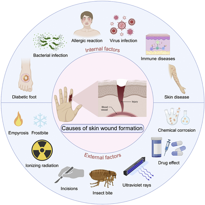
Figure 1.
Causes of skin wound formation. The formation of skin wounds can primarily be categorized into two major factors: external and internal.

Biocompatible mesoporous silica: A novel nanomaterial for skin wound healing
Yang Hong , Qianwei Su , Xiang Wu , Qin Zhang , Jianhua Sun , Yu Wang , Jiacan Su


The skin is the largest organ of the human body, serving as the first line of physiological defense, playing critical roles in preventing the invasion of external pathogens, maintaining fluid balance, and regulating body temperature [1]. However, once the skin barrier is damaged, the body faces a series of risks, including infection, chronic inflammation, and impaired tissue repair [2]. With the aging global population, the incidence of skin wounds continues to rise, putting significant pressure on medical systems and leading to high medical costs. Therefore, accelerating wound healing and reducing complications have become urgent global issues that need to be addressed effectively. Although traditional wound healing methods such as antibiotic therapy, surgical debridement, and conventional dressings can improve wound conditions to some extent, they still face significant limitations when dealing with chronic wounds and multi-drug-resistant bacterial infections. The growing problem of bacterial resistance due to antibiotic overuse is becoming increasingly severe, while conventional dressings lack specificity and are unable to address the multiple needs of local inflammation regulation, promoting cell proliferation, and tissue regeneration [3,4]. Therefore, it is of great importance and interest to develop new materials and technologies that can promote wound healing in a more efficient and sustainable manner.
In recent years, biocompatible mesoporous materials have gradually become a research focus in the field of wound healing [5]. Mesoporous materials, with pore sizes ranging from 2 nm to 50 nm, highly ordered pore structures, and high specific surface areas, not only allow for efficient loading of drugs, small-molecule antibiotics, or growth factors but also enable optimization of drug release rates through pore size control, meeting different therapeutic needs [6]. The preparation methods of mesoporous materials allow for precise control over pore size, pore morphology, and surface chemistry, providing extensive space and flexibility for the functional design of the materials. For instance, Lu et al. loaded Ag+ and clarithromycin (CLR) into periodic mesoporous organosilicas, then coated them with hyaluronic acid and outer membrane vesicles, forming a multifunctional nanocarrier. Through acid-responsive release of Ag+ and CLR, they inhibited urease activity and improved the treatment of drug-resistant Helicobacter pylori, exploring its therapeutic potential in drug-resistant infections [7]. Additionally, copper-containing mesoporous bioactive glass nanoparticles have demonstrated significant antibacterial and angiogenic effects, effectively inhibiting bacterial biofilm formation and promoting new blood vessel formation, while also exhibiting excellent biocompatibility and sustained copper ion release, showing great potential for use in chronic wound treatment [8].
Within the mesoporous family, mesoporous silica (MS) shows strong promise for tissue repair because of its biocompatibility, biodegradability, and tunable surface functionalization. Liu et al. enhanced the degradation performance of mesoporous silica nanoparticles (MSNs) by doping with magnesium ions, which facilitated the release of magnesium ions and silicic acid with antioxidant and tissue regeneration functions, ultimately achieving repair of various complex tissues such as periodontal tissue [9]. Due to its tunable pore structure, surface functionalization properties, and smart responsive release capabilities, MS has demonstrated multi-dimensional advantages in overcoming the traditional limitations of wound healing treatments. MS not only achieves a synergistic effect of antibacterial, anti-inflammatory, and wound-healing through precise drug-controlled release, but its porous network can also mimic the extracellular matrix microenvironment, providing innovative solutions for complex wound management. However, to our knowledge, there are few comprehensive reviews specifically focusing on the use of MS in skin wound healing. This review synthesizes current applications, advantages, and challenges of biocompatible mesoporous materials in wound healing, focusing on advanced MS platforms for drug delivery, dressings, and tissue engineering scaffolds, and sets out a framework for next generation materials.
As the largest organ in the human body, the skin plays critical physiological roles in protecting the body, sensing the external environment, regulating body temperature, and secreting various substances [10]. Histologically, the skin is architecturally stratified into three concentric layers: the epidermis, dermis and hypodermis [11,12]. The structure and function of these layers work in coordination to maintain the skin's barrier function and its ability to repair damage [13]. The formation of skin wounds is a complex process that can be broadly categorized into external injuries and internal pathological factors (Fig. 1). External factors include mechanical, chemical, thermal, and radiation-induced injuries, all of which can directly damage the integrity of the skin tissue. Healthcare professionals are often at risk of radiation exposure, which is why metallic bismuth powder is used as a radiation protection material, typically applied to the hands or medical gloves, to effectively prevent radiation-induced damage to the skin [14]. Internal pathological factors involve conditions such as skin infections, diabetes-related complications, pressure-induced damage, and microcirculatory disturbances, which are often chronic processes. Diabetic skin ulcers are a common and challenging complication of diabetes [15,16]. Current treatment methods, including debridement, infection control, and microcirculation improvement, remain suboptimal, leading to risks of poor healing, scarring, recurrence, and even amputation [17]. Diabetic skin ulcers are characterized by chronic inflammatory infiltration, and multiple angiogenic factors participate in the formation of granulation tissue during the healing process [18,19]. It is important to note that these pathogenic factors can act independently or synergistically, collectively influencing the onset, development, and prognosis of wounds.
Wound healing is a complex and highly coordinated physiological process, typically divided into four stages: hemostasis, inflammation, proliferation, and remodeling (Fig. 2) [20,21]. The successful completion of each stage is crucial for the final healing, and each stage is finely regulated by various cellular and molecular mechanisms.
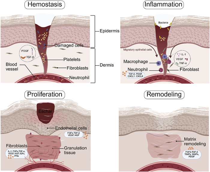
Hemostasis is the first stage of wound healing. Platelets aggregate at the wound, forming a clot to stop bleeding while releasing platelet-derived growth factor (PDGF) and transforming growth factor-beta (TGF-β) to initiate healing signals [22]. Key during this stage is rapid closure of the wound in order to establish a sound platform for repair that will occur later. Following hemostasis, the inflammatory stage occurs for several days. During this stage, pathogen removal and removal of dead cellular material occurs, infection is prevented, and preparation for proliferation takes place. Neutrophils engulf cellular debris via phagocytosis, while macrophages secrete interleukin-1 (IL-1), tumor necrosis factor alpha, and vascular endothelial growth factor (VEGF) for tissue restoration [23]. Concurrently, medium-chain fatty acids activate the receptor GPR84, increasing macrophage migration and promoting initial healing [24]. Not only do they destroy bacteria but they also prepare the site for cells and tissue regeneration. As the healing progresses with decreased inflammation, the wound transitions into the proliferative stage, marked by neovascularization, proliferation of the epithelial cells, and collagen synthesis. VEGF induces angiogenesis, providing oxygen and nutrients to the wound [25,26]. Fibroblasts congregate at the site of injury, synthesizing and depositing collagen to build the extracellular matrix, supporting tissue repair and cell migration [27,28]. The successful outcome of this stage sets up the structural framework for wound repair. It takes weeks, and in some cases, much later, for the remodeling phase, where redundant collagen is broken down and rearranged, resulting in a network of collagen fibrils that improves tissue strength and flexibility. As blood vessels regress, the wound tissue gradually restores its structure and function to resemble normal skin [23].
Wound healing is a multi-stage, complex process that is influenced by various factors [29]. Inflammation regulation, infection management, and nutritional supply are critical factors in wound healing, directly determining the speed and effectiveness of wound repair. Any imbalance in these factors can delay the healing process or lead to complications (Fig. 3) [30].
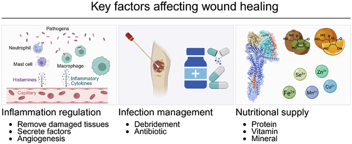
Inflammation is an early stage of wound healing, and a normal inflammatory response helps clear damaged tissue and pathogens, initiating the repair process. However, excessive or prolonged inflammation can lead to chronic inflammation, suppress cell proliferation and angiogenesis, thus affecting the subsequent healing process [23,31]. Clinically, effective control of local and systemic inflammation can significantly improve wound healing efficiency [32-34]. Therefore, maintaining a balance in the inflammatory response is crucial for promoting early wound healing. Wound infection is one of the most common and serious complications during the wound healing process. Infection not only prolongs the healing time but may also lead to systemic infections and sepsis, which threaten the patient's life [35]. Factors such as medications, complications, and microorganisms are often the primary causes of wound infections. To effectively prevent infection, clinical practice first requires active local debridement and disinfection measures to ensure wound cleanliness. Second, the rational use of antibiotics is crucial for the prevention and treatment of local infections [36]. Nutritional supply is another critical factor in wound healing, as adequate nutrition provides the necessary foundation for tissue repair, particularly proteins, vitamins, and minerals [37,38]. Proteins are essential for cell proliferation, matrix synthesis, and repair processes, and a lack of proteins can delay wound healing. Additionally, vitamin C plays a key role in collagen synthesis, and its deficiency can lead to insufficient collagen production, thereby affecting wound closure and repair [39,40]. Trace elements such as zinc also play an important role in wound repair, as zinc is involved in cell division, immune responses, and protein synthesis [41,42].
Mesoporous materials are a class of porous materials with a characteristic pore size distribution ranging from 2 nm to 50 nm and highly ordered mesoscopic pore structures [43]. There are various types of mesoporous materials, with the most common being MS, mesoporous carbon, and mesoporous metal oxides. Among them, MS, with its excellent biocompatibility, three-dimensional interconnected pore structure, modifiable surface chemistry, and high surface area and tunable pore size, has become a cutting-edge research system in the fields of wound healing and controlled drug delivery [44].
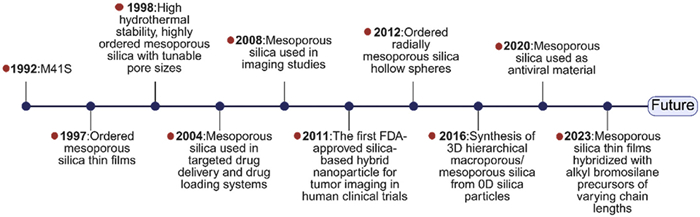
A milestone in mesoporous material research was achieved in 1992 when Beck's research team first used long-chain alkyl quaternary ammonium salt surfactants as templates to successfully synthesize M41S series mesoporous materials with a regular pore structure [45]. As a significant breakthrough in the synthesis methodology of mesoporous materials, Zhao et al. innovatively used nonionic triblock copolymers and other surfactants as templates in 1998, successfully synthesizing MS materials with highly ordered and tunable pore sizes, significantly improving their hydrothermal stability and expanding their range of applications [46,47]. In 2012, Lu et al. synthesized MS hollow spheres with ordered radial intermediate channels using a self-assembly method, which was simple, efficient, and avoided the complexity of traditional templating methods. This approach enabled the preparation of mesoporous hollow spheres with tunable pore sizes, uniform pore structures, and adjustable shell thickness [48]. In 2016, Cheng et al. achieved the synthesis of 3D hierarchical macro-/mesoporous silicon from 0D silica particles [49]. In 2023, Parola et al. synthesized MS films using different chain-length bromoalkyl silane as hybrid precursors via a co-condensation method [50]. Since then, MS has been widely studied and applied in biomedical fields such as drug delivery, tissue engineering, immune modulation, and cancer therapy (Fig. 4) [51-54].
MS materials can have different porous structures, namely pore symmetries [55,56]. By selecting proper template agents, optimizing synthesis parameters, the precise control of the pore structure of MS can be achieved, thereby optimizing its drug loading capacity, release kinetics, and local microenvironment regulation capabilities [57].
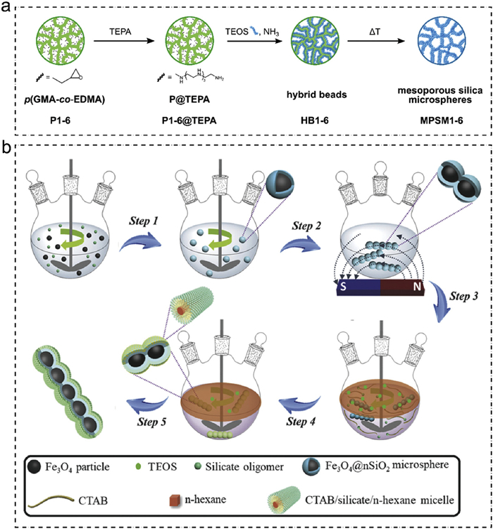
The synthesis approaches for mesoporous materials are generally classified into two categories: Soft-templating and hard-templating methods [58-61]. The hard-templating method employs pre-synthesized mesoporous materials as rigid templates. The precursors of target species are introduced into the pore channels by impregnation, followed by their in situ conversion within the confined space of the template. Finally, selective removal of the templates via chemical dissolution or thermal combustion leads to replica mesoporous structures with pore morphologies precisely matching those of the original templates [62]. Using glycidyl methacrylate-ethylene glycol dimethacrylate (GMA-co-EDMA) copolymers as hard templates, along with surface functionalization by tetraethylene pentamine, precise control over pore size (6–35 nm), specific surface area (15–270 m2/g), and surface charge can be achieved. The degree of amination determines the affinity of the templates toward silica nanoparticles, influencing their deposition and size within the pores. After template removal, the resulting MS microspheres exhibit uniform particle size and pore structures that closely replicate those of the original templates. Upon further functionalization, these materials demonstrate excellent protein separation performance in high-performance liquid chromatography (Fig. 5a) [63]. In contrast, the soft-templating method utilizes flexible templates, such as surfactants or amphiphilic block copolymers, as structure-directing agents. Typically, a silicon precursor (tetraethyl orthosilicate, TEOS) undergoes controlled hydrolysis and condensation catalyzed by acids or bases, interacting cooperatively with organic surfactants like cetyltrimethylammonium bromide (CTAB). These components self-assemble to form ordered organic-inorganic composites. Subsequent removal of the organic templates through calcination or solvent extraction yields mesoporous materials with uniform pore structures [64]. The pore size and structure can be finely tuned by adjusting synthesis conditions such as temperature, pH, and the choice of silicon precursor [65,66]. After annealing or solvent washing to remove the structure-directing agent, the desired MS materials with different pore size and pore symmetries can be obtained [67,68]. Wan et al. synthesized magnetic MS nanochains (Fe3O4@nSiO2@mSiO2) with a core-shell-shell structure via a magnetic-field-guided interfacial co-assembly method using CTAB as the soft template at a water/n-hexane interface, precisely controlling pore size, shell thickness, and specific surface area (Fig. 5b) [69]. The advantages of this method include its mature process, low equipment requirements, and wide control over pore size and specific surface area. In addition, the one-dimensional, field-addressable architecture supports shear-enhanced cell interaction and externally guided release. Potential limitations include chain-length dispersion and orientation stability in physiological media, CTAB removal and interfacial solvent residues, and limited evidence on in vivo degradability and batch-to-batch reproducibility, which warrant further validation. Gluns et al. combined the soft-templating method with 3D printing technology, using light-induced sol-gel chemistry to achieve 3D printing of ordered MS. The printed objects exhibited mesopore sizes of approximately 5 nm and a specific surface area of about 400 m2/g [70]. This method successfully achieved precise structural control from nanoscale mesoporous structures to micron and macroscopic scales, and, due to the synchronous execution of all steps, it avoided time-consuming post-processing procedures.
The soft-templating strategy excels in controlling pore architecture and dimensions, making it suitable for precise tuning of structural characteristics. Meanwhile, the hard-templating method enables exact replication of specific pore sizes, morphologies, and ordered arrangements predefined by the template. Both approaches thus possess significant value and versatility in the controllable synthesis of mesoporous materials. Each synthesis method has its unique advantages and disadvantages, and selecting the appropriate method requires a balance based on application needs [71].
In terms of biocompatibility, MS has been shown to have low toxicity, and its biocompatibility is influenced by various factors, particularly its physicochemical properties such as particle size, surface characteristics, and morphology [72,73]. By adjusting specific parameters, the biocompatibility of MSNs can be significantly improved, reducing their potential harmful effects on cells and tissues [44,74]. Clinical evidence has further confirmed the biosafety and compatibility of MS. In a randomized crossover study using fenofibrate as a model drug, MS formulations in healthy volunteers significantly enhanced oral absorption and systemic exposure without serious adverse events, demonstrating good tolerability and translational potential [75].
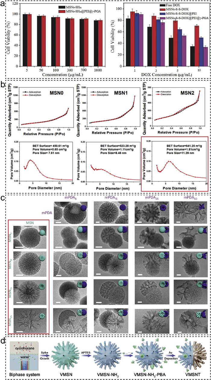
For the uptake kinetics of different cell lines, MSNs demonstrate good cellular uptake ability without causing significant cytotoxicity. Research by Mou et al. has shown that MSNs, even at high concentrations (100 mg/mL), do not significantly affect cell proliferation. Additionally, their surface charge, which is near neutral at physiological pH, enhances stability in biological systems and reduces non-specific interactions with cell membranes [76]. Notably, 50 nm MSNs exhibit the best cellular uptake efficiency, indicating that their tunable size contributes to precise control over biocompatibility (Fig. 6a). Qiao et al. utilized γ-glutamic acid (γ-PGA) coated MSNs, covalently linking anticancer drugs such as doxorubicin to the MS via disulfide bonds, while γ-PGA was applied to the particle surface via charge interactions [77]. This design enhances MSNs uptake within cells and enables targeted drug release.
Furthermore, the distribution, metabolism, degradation, and excretion of MS in vivo have been widely studied, showing that MS possesses good stability and degradability within the body. Research indicates that MSNs can distribute through the liver-spleen reticuloendothelial system and are cleared via renal or hepatic biliary pathways [78]. Degradation rate of MSNs largely relies upon particle size, pore size, and surface functionalization. By manipulating these factors, the biocompatibility and clearance rate of in vivo MSNs can be significantly improved. Chen et al. found the rate of MS degradation in simulated body fluids occurs via three stages: initial rapid volume degradation, silicon concentration reduction, and slow diffusive phase. The rate of MS degradation largely relies upon factors such as initial concentration, surface area, and deposition of calcium/magnesium ions. Higher concentrations and lower surface areas are likely to slow the rate of degradation [79]. The high biocompatibility of MS provides a stable support system biologically, which can support its clinical application.
MS has demonstrated significant advantages in the biomedical field due to its high specific surface area [80]. This property enhances the performance of MS in drug delivery systems by increasing the drug loading capacity and enabling controlled release.
MS can be utilized for targeted delivery of anticancer drugs, and by adjusting its specific surface area, efficient accumulation of drugs at tumor sites can be achieved, thereby improving therapeutic efficacy and reducing systemic side effects [81]. Shao et al. developed diselenide-bridged MSNs exhibiting a large surface area (641.3 m2/g), significantly enhancing protein loading capacity and interfacial electrostatic interactions (Fig. 6b). These features enabled dual-responsive protein release triggered by oxidative (ROS) and reductive (GSH) environments. Additionally, the synergistic effect of high surface area and cancer-cell membrane coating prolonged circulation time and improved tumor targeting and therapeutic efficacy in vivo. This study provides valuable insights into interfacial design strategies for advancing mesoporous materials in drug delivery applications [82]. Additionally, in gene delivery, the high specific surface area of MS provides ample space for nucleic acid molecules, enhancing their stability and delivery efficiency [83,84]. In the field of tissue engineering, MSNs are widely used to construct scaffold materials due to their high specific surface area, which promotes cell attachment and proliferation [85]. The high specific surface area of MSNs also facilitates the biomineralization of bone tissue, promoting osteocyte growth and accelerating fracture healing [86]. These materials support tissue repair and regeneration, including the regeneration of bone, cartilage, and skin. Zhang et al. designed a hybrid MSNs system, integrating polyethylene glycol chains as "strings" and polydopamine (PDA) as binding nodes (MS-PDA). This adhesive system achieved strong wound closure, inflammation suppression, and angiogenesis when applied to skin wound treatment [87]. Additionally, high specific surface area MSNs have played a significant role in diagnostic imaging [88,89]. In molecular imaging, MSNs can effectively carry imaging agents such as fluorescent probes or magnetic nanoparticles, providing high-resolution imaging results. Recent research by Magzoub et al. indicated that pH-responsive upconversion MSNs used as imaging carriers could effectively load fluorescent probes and provide high-resolution MRI, thermal imaging, and fluorescence imaging. Their upconversion properties make them highly effective in tumor imaging, and when combined with near-infrared (NIR) excitation, they offer precise tumor localization and imaging capabilities, showing broad potential for multimodal imaging applications [90]. These diagnostic imaging technologies can be used for complex diseases, particularly in the innovation of diagnostic and therapeutic approaches for cancer and cardiovascular diseases.
The tunable pore size of MS is one of its key distinctions from traditional porous materials. Pore size adjustment not only enhances the performance of MS in drug delivery systems but also expands its applications in fields such as biomedicine, catalysis, and environmental remediation, demonstrating significant flexibility and adaptability [91]. Precisely controlling the template agent type, synthesis conditions, and post-processing techniques enables controlled design of pore sizes [92,93]. This characteristic allows MSNs to be designed according to the size specificity of the drug molecule, increasing the drug loading, release rate of the drug, and targeting. In addition, the pore size can be tuned to allow MS for precise molecule capture and delivery during gene delivery and cell imaging, resulting in higher therapeutic effectiveness and sensitivity of diagnostics.
Recent research has established a continuous flow micro-reactor process for the preparation of mesoporous silica particles of varied sizes with narrow sizes [94]. Kim et al. in 2017 presented a facile route for synthesizing uniform extra-large pore MSNs with a pore size of 30 nm. The XL-MSNs showed much improved capacities for model proteins of varied molecular weights and IL-4 over traditional small-pore MSNs [95]. The research also determined that the XL-MSNs had the ability to effectively load IL-4 and deliver IL-4 to macrophages, which caused the polarization of macrophages towards anti-inflammatory M2 macrophages. This indicates that XL-MSNs with their cytokine-targeting delivery have the ability to regulate the immune system, presenting new avenues for therapy. In the Janus dual-core nanoparticles developed by Zhao et al., the MS segment has tunable pore sizes between 3 nm and 25 nm with selective loading of drug molecules. Concurrently, the mesoporous polydopamine (mPDA) segment, characterized by larger pore dimensions of 5–50 nm, facilitates the encapsulation of enzymes and other biomacromolecules. This dual-functional system enhances its flexibility and precision in biomedical applications (Fig. 6c) [96]. The tunable pore size feature provides significant advantages in responding to specific biological signals and achieving targeted drug release. In the field of drug loading, Ju et al. developed a ROS-responsive MSNs, which achieved precise drug loading and release through surface functionalization and pore size regulation. The innovative structural design enables drug-controlled release [97]. This system contains ultra-small cerium oxide nanoparticles, which are rapidly released in ROS environments, exhibiting antioxidant effects. Through controlling the pore size and functional groups on its surface, its drug loading capacity was maximized and ROS-responsive drug release was enabled, exhibiting its application in targeted drug delivery and antioxidant therapy.
Tunable pore size of MS through the choice of template agents, adjustment of synthesis conditions, and post-modification methods enables total control over pore size. This has acted as a central benefit in its functional design and has made it applicable on a broad spectrum for drug delivery and targeted therapy. The tunable pore size also allows MSNs to be controlled by specific physiological signals, enabling them to deliver drugs with precision. This exhibits their application in targeted therapy and antioxidant treatment, thus making them a perfect multifunctional drug delivery system.
MS possesses great functionalization and modification capability, making it exhibit great promise for many applications in the field of biomedicine. Owing to its ordered pore structure and dense surface silanol groups, MSNs can be subjected to specific chemical modification and functional combination by exploiting numerous surface engineering methods. MSN surface can be functionalized with a variety of functional groups, such as amino, carboxyl, and hydroxyl groups, in order to optimize their biocompatibility, toxicity, targeting, and responsiveness (Fig. 6d) [98-100]. These surface modifications not only significantly improve the biocompatibility of MSNs and reduce cytotoxicity, but also bestow them with special functions, such as antibacterial, antioxidant, immunomodulatory, and gas sensing capabilities. This functional versatility makes MSNs highly adaptable for diverse biomedical applications, including drug delivery, tissue engineering, disease diagnosis, and therapeutic interventions [101].
Through surface functionalization, MSNs can specifically interact with biomolecules, enhancing their interactions with cells or tissues, thereby improving the targeting and controlled release properties of drug delivery systems. Xie et al. functionalized MSNs by combining them with COOH-PEG-phenylboronic acid and gold nanoshells, demonstrating excellent bacterial targeting and self-heat-induced propulsion properties [100]. The functionalized MSNs can selectively recognize and bind to bacterial surfaces, and with the aid of NIR laser irradiation, generate thermal effects that disrupt bacterial membranes, allowing for deep penetration and exerting antibacterial activity. Furthermore, this functionalization not only improves targeting and therapeutic effectiveness but also enhances the efficacy against drug-resistant bacteria through their unique virus-like structure, showcasing significant potential in the treatment of drug-resistant bacterial infections. Similarly, Chen et al. functionalized MSNs to effectively load photothermal agents, which respond to pH, enzymes, and NIR laser stimuli in the tumor microenvironment, achieving precise drug release. Moreover, the functionalized MSNs triggered dendritic cell maturation and macrophage polarization, augmented immune responses, enhanced tumor therapy effectiveness, and inhibited metastasis. Chen's research further exposed the receptor-ligand-free mesoporous silica nanoparticles (RMSNs) drug targeting delivery advantages in penetrating through the blood-brain barrier (BBB) [81]. Through PEGylation and adjustment of surface charge, drug accumulation in the brain was greatly improved by RMSNs with exceptional BBB penetration abilities.
Additionally, the functionalization of MSNs significantly enhances their strength, making them have exceptional structural stability when utilized as scaffold material for tissue engineering and thus facilitating cell attachment and proliferation [102]. Therefore, surface functionalization of MSNs not only possesses great promise for their application in the context of biomedical engineering but also serves as a robust basis for the creation of intelligent nanomaterials.
MS, with its highly ordered pore structure, excellent specific surface area, and precisely tunable physicochemical properties, has garnered significant attention in the field of wound healing in recent years. Not only does MS, as a new delivery platform, facilitate targeted loading and controlled drug and bioactive molecule release via pore size control, but also dynamically reformulates the wound microenvironment via surface functionalization, greatly promoting tissue repair results. Notably, by constructing pH, ROS, or enzyme-responsive multifunctional composite systems and smart-responsive platforms, MS has overcome the passive treatment model of traditional dressings. This enables real-time monitoring of wound status, on-demand drug release, and dynamic regulation of therapeutic effects, providing crucial theoretical and technological support for wound treatment strategies in the era of precision medicine.
MS, with its highly ordered pore structure and excellent specific surface area, has become an ideal carrier in drug delivery systems. Furthermore, by integrating MS with microneedle (MN) technology and other functional materials, the therapeutic efficacy is significantly enhanced, while side effects are effectively reduced (Fig. 7a) [103,104].
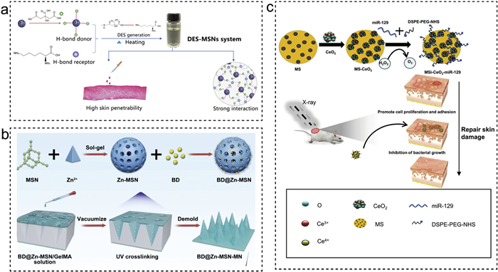
Gao et al. successfully developed a drug long-acting delivery system combining zinc-doped MSNs with MN, designed to load betamethasone dipropionate (BD) for transdermal sustained-release drug delivery and continuous anti-inflammatory and anti-pruritic treatment in imiquimod-induced psoriasis mouse models (Fig. 7b) [105]. Similarly, researchers encapsulated methotrexate (MTX) in chitosan-coated hollow MSNs (MTX@HMSN/CS) and combined this with MN-assisted delivery technology, allowing for precise penetration of the stratum corneum and achieving efficient local drug release [106]. In the psoriasis mouse model, MTX@HMSN/CS demonstrated significant anti-proliferative and anti-inflammatory effects, maintaining drug concentrations for longer periods compared to traditional drug delivery methods, thus reducing the frequency of treatment. Furthermore, up to 95% of cancer patients undergoing radiation therapy experience radiation-induced skin injury (RISI), with nearly 10% suffering severe skin damage. Li et al. designed multifunctional MS-cerium oxide nanomaterials (MS-CeO2) to deliver miR129 and promote the healing of radiation-induced skin damage (Fig. 7c) [107]. This system improves wound healing by inhibiting ROS and the activation of HIF-1α, and by targeting the RAD17 gene to regulate the Chk2 pathway, reducing cell apoptosis induced by DNA damage.
MS has demonstrated its tremendous potential as a precise delivery platform in the specific application of skin wound healing, laying the foundation for the development of future personalized treatment and smart healthcare systems. It holds vast clinical translation prospects, offering innovative solutions for enhancing therapeutic efficacy and advancing personalized medicine.
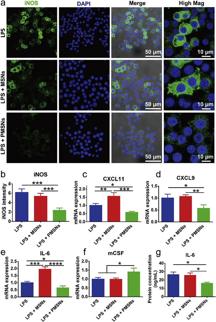
Inflammation is a crucial step in the wound healing process, where moderate inflammation helps clear pathogenic factors from the injury site and initiates the repair mechanisms. However, excessive or prolonged inflammation can delay healing and lead to complications. MS, through surface functionalization and pore size regulation, plays a significant role in inflammation regulation during wound healing [55]. Topography-engineered, pollen-like mesoporous silica nanoparticles modulate inflammation via contact-dependent signaling. Macrophages sense rough surfaces, upregulate CD28, and suppress ERK signaling, which attenuates lipopolysaccharide (LPS)-induced M1 activation. Compared with smooth MSNs, these particles reduce iNOS and IL-6 while elevating colony-stimulating factor (mCSF), promoting a pro-repair immune profile independent of internalization (Fig. 8) [108]. Research by Babu et al. demonstrated that MS, as a caffeine delivery system, has a significant inhibitory effect on the inflammatory response [109]. Moreover, MS not only regulates local immune responses but also promotes the activation and polarization of immune cells, accelerating wound healing and reducing adverse reactions [110]. Therefore, MS provides a new strategy for inflammation regulation and immune modulation in wound repair.
Effective inflammation induction and timely inflammation resolution are crucial for wound healing. Studies have shown that MS, as a tissue adhesive, can activate appropriate inflammatory responses and promote wound healing [111]. The excellent adhesion properties and tunable degradation rates of MS not only aid in connecting wound edges but also accelerate healing by modulating inflammation, while simultaneously improving the tensile strength of the wound. Bacterial infection is a major factor contributing to wound deterioration. Chen et al. developed a smart drug delivery system based on MS, utilizing thioketal gatekeepers to achieve ROS-sensitive drug release, enhancing the targeting of antibiotics and boosting antibacterial effects while reducing the required dosage [112]. In terms of immune modulation, Zheng et al. explored the immunomodulatory role of MS in wound healing [113]. The research team combined MS with polydopamine and iron oxide nanoparticles (Fe/PDA@GOx@HA) to construct a MN patch system that integrates antibacterial and immune modulation functions. Specifically, this system regulates the immune microenvironment at the wound site, promoting the polarization of macrophages to the M2 phenotype and inhibiting excessive inflammatory responses, thereby effectively improving the healing process of the wound.
Angiogenesis and tissue regeneration are crucial processes in wound healing, directly influencing the speed and quality of wound closure. Effective angiogenesis provides oxygen and nutrients to the wound area, promoting the growth and repair of new tissue. MS, with its tunable drug delivery capacity and topology regulation features, can target the delivery of angiogenic factors such as VEGF, regulating endothelial cell migration and adhesion behaviors [114]. Through surface functionalization, MS can also modulate the pH balance and oxidative stress within the tissue microenvironment, breaking the vicious cycle of ischemia and hypoxia, thereby providing dynamic support for efficient vascular network reconstruction and functional tissue regeneration [115,116].
Diabetic wounds (DW) often result in local ulcers, wound necrosis, and even amputation due to peripheral nerve damage and vascular dysfunction. Traditional drug treatments are often limited in efficacy as they struggle to effectively reach deeper tissues and promote wound healing. Li et al. designed a multifunctional MS mesoporous MN patch that can induce the formation of multiple regenerative sites at different locations. This patch exhibits antibacterial properties, reduces reactive oxygen species, regulates macrophage niches, and promotes vascular regeneration and collagen deposition, significantly accelerating the healing process of infectious diabetic wounds [117].
In the wound healing process, oxidative stress is a key factor influencing the healing outcome. During wound healing, damaged tissues release a large amount of ROS. These free radicals damage cell membranes, DNA, proteins, and lipids, activate inflammatory responses, delay healing, and lead to tissue necrosis. Antioxidants can scavenge ROS, reduce oxidative stress damage to cells, decrease inflammation, promote cell proliferation and migration, and improve the wound healing microenvironment. The synergistic antioxidant effect when combined with drugs or other materials can significantly enhance the quality and speed of wound healing, with important clinical applications, especially in chronic wounds and severe burns [118].
Studies have shown that MS can load metformin and cerium oxide, regulating ROS levels in the wound microenvironment, reducing oxidative stress damage to cells, and promoting wound healing [119]. Additionally, MS modified with thiomethyl chains can respond to ROS, precisely release drugs, further enhance antioxidant effects, increase antibacterial activity, and accelerate the wound healing process [112]. Wu et al. successfully attached ultrafine cerium oxide nanoparticles to the surface of MS, creating an effective functionalized ROS-responsive tissue adhesive nanocomposite [120]. This system exhibited excellent tissue adhesion properties, aiding in rapid wound closure, and significantly inhibiting scar formation by scavenging excess ROS at the wound site.
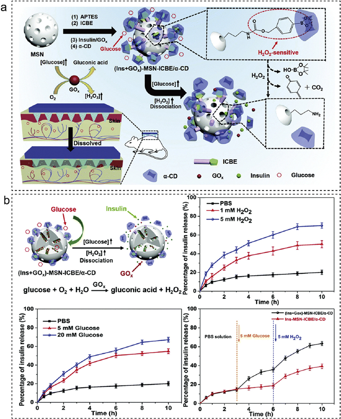
The integration of MS with smart responsive materials marks a significant leap towards dynamic and precise wound healing strategies. By incorporating light, pH, temperature, or enzyme-responsive components, MS can form multifunctional composite systems to achieve on-demand drug release, targeted accumulation, and adaptive regulation of the microenvironment [121-123]. H2O2-responsive mesoporous silica enables programmable, on-demand release by translating glucose-derived H2O2 into pore uncapping via phenylboronate-ester cleavage and α-cyclodextrin gate removal. Embedded in dissolvable MN, MSNs co-load insulin and glucose oxidase, couple dermal delivery with stimulus intensity, and prolong glycemic control, exemplifying MS-centered, triggerable therapy (Fig. 9) [124]. Wang et al. developed a self-responsive MN patch that releases hydrogen gas, using MS loaded with aminoborane (AB), which intelligently releases hydrogen gas in the diabetic wound microenvironment, effectively scavenging excess ROS and alleviating oxidative stress [125]. This multifunctional smart response system, by modulating the microenvironment, demonstrates the significant application potential of MS combined with other materials in intelligent response and wound repair. Such systems require precise spatiotemporal "smart switch" characteristics, providing a modular technological platform for personalized intervention in complex wounds. The composite system of MS with smart responsive materials, through dynamic and precise regulation mechanisms, significantly enhances wound healing efficacy, offering a new paradigm of multimodal collaborative intervention for complex wound treatment, highlighting its broad prospects in precision medicine.
Despite its excellent drug loading capacity, tissue scaffold functionality, and smart-responsive properties in skin wound healing, the biocompatibility and biodegradability of MS remain major challenges in clinical applications [78]. Therefore, optimizing the biocompatibility of MS, reducing immune reactions, and improving its long-term safety for extended use have become key factors for its successful application in the field of wound healing.
Surface modification is one of the common strategies to optimize the biocompatibility of mesoporous materials. By functionalizing the surface of the material, the interaction between the material and cells and tissues in the biological system can be improved, while reducing immune system rejection responses [126]. Most surface modifications include the most widely used hydrophilic and biomolecule modifications. For example, through the incorporation of hydrophilic groups of molecules like polyethylene glycol (PEG), the material's biological stability significantly increases with reduced serum protein adsorption and subsequent activation of the immune response [127]. PEG modification not only increases the circulation time of mesoporous material in the body but also reduces the opportunity for phagocytosis by macrophages, thus reducing the clearance of the material. Additionally, through their inclusion of cell adhesive molecules like RGD peptides on the surface, cell adhesion and proliferation of the surface-modified material are ensured, thus making the material function optimally for tissue regeneration.
Size and shape are also critical factors that influence the biocompatibility of mesoporous materials. Researchers have established that the size, shape, and surface charge of nanoparticles have a direct impact on the strength of immune responses [128]. Materials with sizes less than 100 nm are usually more likely to cross cell membranes and avoid macrophage phagocytosis, hence decreasing immune reactions [129]. However, particles with sizes smaller than these may be cytotoxic or damage tissues. Hence, setting an appropriate size and surface charge is one of the most important considerations when enhancing biocompatibility.
Additionally, enhancing material degradability is one of the key strategies for promoting biocompatibility. Material residues in the body for long periods of time can induce chronic immune responses, and these should be of concern for their safety. It has been indicated by studies that the degradation of MS materials usually occurs over several days, with full elimination from the body perhaps requiring several weeks. Even if degradation products are not toxic, their accumulation in the body will inevitably have an effect on tissue function [130,131]. Thus, material design with higher degradation speeds, or the addition of degradable organic units in order to enhance degradation, has become a critical research goal for maximizing biocompatibility.
In summary, improving the long-term safety of MS can be achieved through personalized strategies such as surface modification, size optimization, degradability design, and the introduction of immune-modulating functions. With the development of new technologies, the long-term safety of MS will continue to be optimized, providing more reliable support for its widespread application in disease treatment and further enhancing its clinical therapeutic potential.
In the application of MS for wound healing, despite its tremendous potential in drug delivery, tissue regeneration, and multifunctional therapies, achieving compatibility and integration of multifunctional modules remains a key challenge when addressing the repair of complex wounds.
Firstly, when co-loading bioactive molecules such as drugs, imaging agents, and growth factors, the pores of MS may become blocked or affected by chemical reactions [132]. Wound healing typically requires the simultaneous release of drugs to promote healing, imaging agents for real-time monitoring of the wound status, and growth factors to support cell proliferation and angiogenesis. However, interactions between different components may lead to pore blockage or unintended reactions between drugs and imaging agents, which not only affects the therapeutic outcome but could also reduce the activity of the drugs or other components. Therefore, optimizing the pore structure of MS to prevent these adverse effects has become a critical issue that needs to be addressed [133].
Secondly, the timing of drug release during the wound healing process also faces complex coordination challenges [134]. The release timing of drugs needs to align with the different stages of wound healing, while the release schedules and rates of various therapeutic components such as antibiotics, anti-inflammatory drugs, and healing factors often differ. If the release timing is not well coordinated, it could affect the effectiveness of wound healing and may even hinder the final quality of the healing process.
Another limitation of mesoporous silica is its excessive rigidity and brittleness, which restricts its direct use as a wound dressing. In contrast, hydrogels are soft, flexible, and injectable, but lack sufficient strength. Recent studies have therefore focused on integrating MS into hydrogel matrices, aiming to combine mechanical stability with compliance while enabling controlled release of therapeutic agents. Examples include GelMA/SFMA hydrogels incorporating RES-loaded MSNs and platelet vesicles to modulate inflammation and angiogenesis, and boronate-linked, glucose/pH-responsive hybrids embedding MSN@PDA-antibiotic and metformin for antibacterial and metabolic regulation [135-137]. These findings highlight that addressing the mechanical limitations of MS through composite design remains an important challenge for future applications
While MS has achieved significant results in laboratory settings, scaling up its production and effectively controlling costs remain major challenges for its widespread application [138]. Therefore, meeting the demand for large-scale production has become a critical issue. Improving production efficiency and reducing costs are core concerns in large-scale manufacturing, and many researchers are focused on increasing yield and minimizing solvent consumption. By carefully controlling the production process, resource waste can be significantly reduced, thereby lowering costs [94]. Additionally, the introduction of automated production lines can significantly enhance production efficiency, reduce human error, and ensure product consistency. With increasing environmental protection requirements, green synthesis techniques have become an important direction for the production of MS [139]. Techniques of green synthesis reduce processes of production by sparing the usage of harmful solvents and reduced energy input, correlated to reduced ecological footprint. Usability of aqueous solvents or biodegradable chemicals as the template and the auxiliaries has emerged as a successful method of keeping the ecological footprint low and maximizing the production processes.
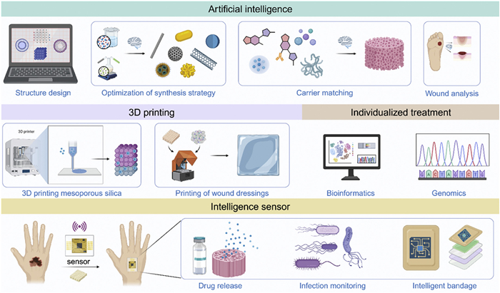
To realize the extensive clinical application of MS in the field of wound healing, not only do its intrinsic properties need to be optimized, but also it should be integrated with other novel technologies to facilitate the improvement of therapeutic outcomes and the realization of precision medicine targets. Kang et al. developed ChatMOF, an artificial intelligence system based on large language models, aimed at predicting and generating metal-organic frameworks (MOF) through natural language processing [140]. The system combines machine learning models and databases, enabling efficient MOF property prediction, data retrieval, and material inverse design. In the analysis of MOF structures and properties, ChatMOF significantly improves design efficiency and accuracy, achieving accuracies of 96.9%, 95.7%, and 87.5% in retrieval, prediction, and generation tasks, respectively. This system provides a new tool for MOF research, demonstrating the potential of artificial intelligence (AI) in materials design. While developed for MOFs, this agent-tool-evaluator workflow suggests a transferable route for MS by coupling pore-structure generators and predictors to inverse-design pore size, connectivity, and surface chemistry under constraints, followed by synthesis-pathway retrieval. Such pipelines could guide MS architectures optimized for loading, release kinetics, and immune modulation. AI, leveraging deep learning technologies, accelerates biomaterial design, optimizes drug delivery systems, and enhances disease diagnostics by efficiently predicting biocompatibility and material-tissue interactions, enabling precise and personalized integration of biomaterials with life science research [141]. Looking ahead, the integration of AI technology has brought new breakthroughs in the application of MS in skin wound healing [142].
AI, through machine learning, can optimize the design and preparation process of MS, driving its personalized application in wound healing [143]. Specifically, AI can accurately predict the optimal healing path for different types of wounds and tailor MS structural synthesis strategies based on these predictions. Additionally, AI can analyze multidimensional data such as the synthesis methods, surface functionalization, and biocompatibility of MS to quickly identify key features related to wound repair, thus optimizing material design [144]. In terms of drug and bioactive molecule loading, AI can match potential payload information by analyzing the pore structure and size data of MS, and predict the efficacy and toxicity of the delivery system in different patients. This not only speeds up the development of new delivery systems but also improves the accuracy and safety of treatment. It also assists in the structural compatibility analysis of MS under different conditions of experiments, the optimization of conditions of synthesis aiming to improve the reproducibility and stability of the experiment. In general, the synergy of AI and MS has allowed for the achievement of more accurate wound healing and the reinforcement of the mechanisms of wound healing. Using the deep learning of MS datasets, AI can potentially identify the mechanisms of degradation and metabolism of MS in vivo and in vitro and suggest more accurate strategies of structural optimization. In addition, AI can potentially perform real-time monitoring of the course of the wound healing, analyze the data of the wounds, assist the clinicians in the measurement of the effectiveness of the treatment, and adjust the schemes of the treatment using the real-time feedback.
MS has demonstrated great compatibility with precision medicine tools in individualized treatment [145]. With advances in precision medicine and genomics, individualized treatment has emerged as the central direction for the healthcare of the future [146]. Using personal biomarkers or genomics, doctors can customize treatment for patients. With the application of MS in drug delivery systems, treatment becomes customizable according to the needs of the patient. For instance, MS optimizes drug discharge rates according to wound type and drug metabolism rate, making treatment more tailored [147]. Also, with advances in genomics and bioinformatics, the functional design of MS becomes more specific, such that it responds according to disease features or physiological conditions.
The integration of MS with 3D printing technology in tissue engineering has provided a new perspective for its clinical translation [148]. 3D printing allows for precise control over the microscopic structure and porosity of scaffolds, providing efficient support for tissue engineering. By incorporating growth factors, drugs, or smart sensors into 3D printed MS materials, more complex therapeutic scaffolds can be developed [149]. For example, in bone tissue repair, MS scaffolds manufactured using 3D printing technology can mimic the structure of bone tissue and promote bone regeneration by slowly releasing growth factors [150]. This multifunctional material combines the advantages of drug release, cell support, and tissue regeneration, showcasing significant clinical translation potential.
Moreover, the combination of smart sensors with mesoporous materials enhances drug delivery systems in terms of intelligence and precision. For example, in the treatment of wounds, smart sensors are able to track the conditions of the wound in real time and adjust drug release dynamically to sustain the constant delivery of antibiotics and other drugs to the wound site, as well as optimize the dosage [151]. Smart drug release by this approach facilitates control of the drug release by precision, as per the actual conditions of the wound, hence facilitating better therapeutic performance and minimization of the side effects of the drug.
Ultimately, the clinical translation of MS not only exists in its capability of delivering drugs and repairing tissues but also in its combination with the latest technologies. Through multidisciplinary collaboration, the combined application of MS with technologies such as AI, personalized treatment, and 3D printing will provide more efficient, safe, and personalized treatment methods for skin wound healing (Fig. 10). This integration will provide more powerful tools for future biomedical research, advancing the application of MS in disease diagnosis, therapeutic strategy development, and tissue engineering to new heights.
MS has demonstrated substantial potential in fundamental research and is now approaching clinical translation, notably in wound healing applications [147]. Successful transition into clinical practice requires standardized synthesis and characterization protocols, strengthened interdisciplinary collaboration among researchers, clinicians, and regulatory agencies, and enhanced translational infrastructure. Furthermore, clear regulatory frameworks and clinician-oriented training programs will accelerate the practical implementation of MS-based technologies. Systematic attention to these areas will ensure MS effectively contributes to advancements in regenerative medicine and personalized healthcare [152-154].
MS, with its unique structural characteristics, excellent biocompatibility, and tunable physicochemical properties, has demonstrated tremendous potential in the field of wound healing. As a novel multifunctional nanomaterial, it not only possesses exceptional drug loading capacity and controlled release properties but also enables precise modulation of the wound microenvironment through surface functionalization and pore size regulation. With ongoing research into its synthesis methods, structural design, and functional modifications, the applications of MS in drug delivery, inflammation regulation, tissue regeneration, and other areas continue to expand, providing new solutions for the treatment of chronic wounds, diabetic foot ulcers, and other refractory skin injuries.
However, despite the broad application prospects of MS in wound healing, several challenges remain. Safety of long-term application, possible risks of immune responses, and translatability into the clinic remain key challenges that must be resolved. To maximize the benefits of MS in wound healing, the following should be addressed in the future: optimization of its biodegradability, immunological compatibility, and its efficacy in a variety of patient populations in the clinic.
At the same time, with ongoing advances in intelligent technology and precision medicine, the combination of MS with sophisticated tools like 3D printing and AI offers a revolutionary change towards more individualized, dynamic, and highly adaptive wound healing. 3D printing provides the accurate fabrication of MS-based scaffolds with customized geometry and porosity, adapting the scaffolds to the specificity of the wound and supporting higher tissue regeneration. Through the creation of patient-specific wound therapy platforms, 3D printing provides the incorporation of functional MS material with optimized structure for tailored drug delivery, cellular interaction, and regenerative treatment. At the same time, AI increases the predictive function of material design, providing deep learning optimization of MS features, enabling real-time monitoring and individualized treatment protocols. Using data from AI, the most efficient MS formulation may be simplified by predictive assessment of their interactions with the wound microenvironment, guiding the design of intelligent drug delivery. Constructing multifunctional composite systems and incorporating smart-responsive platforms, these advances enable the creation of dynamic on-demand wound therapy that adjusts according to the dynamics of the wound healing.
In essence, MS research on wound healing is still in its infancy, but its vision of reshaping the regenerative medicines field becomes more transparent. As material science continues to grow and converge rapidly with interdisciplines such as 3D printing, AI, and precision medicines, MS sits on the threshold of not only reinventing wound healing but also reequilibrating more comprehensive therapy mechanisms for tissue regeneration. The vision of MS for the future holds its ability for harmonization with cutting-edge technology, thereby enabling prospects of customized, dynamic therapies. With the realization of MS's unmatched tunability and biocompatibility, research in the future will establish new standards of wound healing, with the ability to provide extremely specific, reactive, and on-demand therapy responses tailored uniquely for individual patients.
The authors declare that they have no known competing financial interests or personal relationships that could have appeared to influence the work reported in this paper.
Yang Hong: Writing – review & editing, Writing – original draft, Conceptualization. Qianwei Su: Writing – review & editing. Xiang Wu: Writing – review & editing. Qin Zhang: Writing – review & editing, Supervision, Project administration, Funding acquisition, Conceptualization. Jianhua Sun: Writing – review & editing. Yu Wang: Writing – review & editing, Supervision, Project administration, Funding acquisition, Conceptualization. Jiacan Su: Writing – review & editing, Supervision, Project administration, Funding acquisition, Conceptualization.
This work was financially supported by Key program of the National Natural Science Foundation of China (No. 82230071), General program of the National Natural Science Foundation of China (Nos. 82172098, 32471420), Shanghai Committee of Science and Technology Laboratory Animal Research Project (No. 23141900600), Natural Science Foundation of Shanghai (No. 22ZR1424900).
S. Knox, N.M. O'Boyle, Chem. Phys. Lipids 236 (2021) 105055. doi: 10.1016/j.chemphyslip.2021.105055
M. Schmuth, S. Eckmann, V. Moosbrugger-Martinz, et al., J. Invest. Dermatol. 144 (2024) 989-1000. e1. doi: 10.1016/j.jid.2024.03.006
Z.P. Bulman, S.G. Wicha, E.I. Nielsen, et al., Lancet Microbe 3 (2022) e795–e802. doi: 10.1016/S2666-5247(22)00121-5
S. Pickles, E. McAllister, G. McCullagh, et al., Int. J. Orthop. Trauma. Nurs. 45 (2022) 100922. doi: 10.1016/j.ijotn.2022.100922
W.N. Zeng, D. Wang, Q.P. Yu, et al., ACS Appl. Mater. Interfaces 14 (2022) 2534–2550. doi: 10.1021/acsami.1c19209
Z. Li, J. Yang, B. Ren, et al., Adv. Mater. 36 (2024) e2313212. doi: 10.1002/adma.202313212
H. Fan, K.I. Wong, Y. Ma, et al., Adv. Funct. Mater. 35 (2024) 2412893.
T.E. Paterson, A. Bari, A.J. Bullock, et al., Front. Bioeng. Biotechnol. 8 (2020) 246. doi: 10.3389/fbioe.2020.00246
G. Liu, R. Xia, M. Gui, et al., ACS Nano 18 (2024) 32932–32949. doi: 10.1021/acsnano.4c12519
R.B. Ansaf, R. Ziebart, H. Gudapati, et al., Regen. Biomater. 10 (2023) rbad060. doi: 10.1093/rb/rbad060
L.Y. Daikuara, X. Chen, Z. Yue, et al., Adv. Funct. Mater. 32 (2021) 2105080.
D. Chouhan, N. Dey, N. Bhardwaj, et al., Biomaterials 216 (2019) 119267. doi: 10.1016/j.biomaterials.2019.119267
R.M. Sarate, J. Hochstetter, M. Valet, et al., Cell 187 (2024) 5298-5315. e5219. doi: 10.1016/j.cell.2024.07.031
S. Subramanian, B.R. Waller, N. Winders, et al., Catheter. Cardiovasc. Interv. 89 (2017) 709–716.
J. Wang, X. Ge, Y. Xiang, et al., Chin. Chem. Lett. 36 (2025) 109819. doi: 10.1016/j.cclet.2024.109819
X. Qi, Y. Li, Y. Xiang, et al., Cell Biomater. 1 (2025) 100020. doi: 10.1016/j.celbio.2025.100020
D.G. Armstrong, T.W. Tan, A.J.M. Boulton, et al., JAMA 330 (2023) 62–75. doi: 10.1001/jama.2023.10578
E. Mahdipour, A. Sahebkar, J. Diabetes. Res. 2020 (2020) 6320514.
R.W. Kartika, I. Alwi, F.D. Suyatna, et al., Heliyon 7 (2021) e07934. doi: 10.1016/j.heliyon.2021.e07934
M. Rodrigues, N. Kosaric, C.A. Bonham, et al., Physiol. Rev. 99 (2019) 665–706. doi: 10.1152/physrev.00067.2017
B.K. Sun, Z. Siprashvili, P.A. Khavari, Science 346 (2014) 941–945. doi: 10.1126/science.1253836
S. Liarte, A. Bernabe-Garcia, F.J. Nicolas, Cells 9 (2020) 306. doi: 10.3390/cells9020306
O.A. Pena, P. Martin, Nat. Rev. Mol. Cell. Biol. 25 (2024) 599–616. doi: 10.1038/s41580-024-00715-1
P.O. Cooper, S.S. Kleb, S.K. Noonepalle, et al., Cell. Rep. 43 (2024) 114288. doi: 10.1016/j.celrep.2024.114288
L.M. Wise, G.S. Stuart, N.C. Jones, et al., J. Clin. Med. 9 (2020) 1085. doi: 10.3390/jcm9041085
F. Shams, H. Moravvej, S. Hosseinzadeh, et al., Sci. Rep. 12 (2022) 18529. doi: 10.1038/s41598-022-23304-8
J. Li, Q. Zhao, X. Gao, et al., Wound. Repair. Regen. 32 (2024) 55–66. doi: 10.1111/wrr.13144
F.Z. Gharbia, A.S. Abouhashem, Y.A. Moqidem, et al., Sci. Rep. 13 (2023) 886. doi: 10.1038/s41598-022-27152-4
S. Guo, L.A. Dipietro, J. Dent. Res. 89 (2010) 219–229. doi: 10.1177/0022034509359125
L. Deng, C. Du, P. Song, et al., Oxid. Med. Cell Longev. 2021 (2021) 8852759. doi: 10.1155/2021/8852759
D. Lv, X. Cao, L. Zhong, et al., Cell Rep. Med. 4 (2023) 101129. doi: 10.1016/j.xcrm.2023.101129
S. Bindu, S. Mazumder, U. Bandyopadhyay, Biochem. Pharmacol. 180 (2020) 114147. doi: 10.1016/j.bcp.2020.114147
S. Lea, A. Higham, A. Beech, et al., Eur. Respir. Rev. 32 (2023) 230084. doi: 10.1183/16000617.0084-2023
Y. Oishi, I. Manabe, Int. Immunol. 30 (2018) 511–528. doi: 10.1093/intimm/dxy054
A. Uberoi, A. McCready-Vangi, E.A. Grice, Nat. Rev. Microbiol. 22 (2024) 507–521. doi: 10.1038/s41579-024-01035-z
D. Baltzis, I. Eleftheriadou, A. Veves, Adv. Ther. 31 (2014) 817–836. doi: 10.1007/s12325-014-0140-x
E. Arribas-Lopez, N. Zand, O. Ojo, et al., Nutrients 13 (2021) 2498. doi: 10.3390/nu13082498
P. Ghaly, J. Iliopoulos, M. Ahmad, Br. J. Nurs. 30 (2021) S38–S42. doi: 10.12968/bjon.2021.30.5.S38
R. Gref, C. Delomenie, A. Maksimenko, et al., Sci. Rep. 10 (2020) 16883. doi: 10.1038/s41598-020-72704-1
H.C. Gemalmaz, K. Sariyilmaz, O. Ozkunt, et al., Acta Orthop. Traumatol. Turc. 52 (2018) 452–458.
P.H. Lin, M. Sermersheim, H. Li, et al., Nutrients 10 (2018) 16.
S. Yao, J. Chi, Y. Wang, et al., Adv. Healthc. Mater. 10 (2021) e2100056. doi: 10.1002/adhm.202100056
T. Zhao, A. Elzatahry, X. Li, et al., Nat. Rev. Mater. 4 (2019) 775–791. doi: 10.1038/s41578-019-0144-x
B. Xu, S. Li, R. Shi, et al., Signal Transduct. Target. Ther. 8 (2023) 435. doi: 10.1038/s41392-023-01654-7
C.T. Kresge, M.E. Leonowicz, W.J. Roth, et al., Nature 359 (1992) 710–712. doi: 10.1038/359710a0
D. Zhao, Q. Huo, J. Feng, et al., J. Am. Chem. Soc. 120 (1998) 6024–6036. doi: 10.1021/ja974025i
D. Zhao, J. Feng, Q. Huo, et al., Science 279 (1998) 548–552. doi: 10.1126/science.279.5350.548
Z. Teng, X. Su, Y. Zheng, et al., Chem. Mater. 25 (2012) 98–105.
X. Zuo, Y. Xia, Q. Ji, et al., ACS Nano 11 (2017) 889–899. doi: 10.1021/acsnano.6b07450
J. Kusz, C. Boissiere, D. Ihiawakrim, et al., Chem. Mater. 35 (2023) 7671–7682. doi: 10.1021/acs.chemmater.3c01438
M. Vallet-Regi, A. Rámila, R.P. del Real, et al., Chem. Mater. 13 (2000) 308–311.
G.K. Pouroutzidou, L. Liverani, A. Theocharidou, et al., Int. J. Mol. Sci. 22 (2021) 577. doi: 10.3390/ijms22020577
M. Sun, P. Gu, Y. Yang, et al., J. Immunother. Cancer 9 (2021) e002508. doi: 10.1136/jitc-2021-002508
M.K.M. Esfahani, N. Islam, P.J. Cabot, et al., ACS Biomater. Sci. Eng. 8 (2022) 4153–4162. doi: 10.1021/acsbiomaterials.1c00066
B.S. Sivamaruthi, S. Thangaleela, P. Kesika, et al., Pharmaceutics 15 (2023) 439. doi: 10.3390/pharmaceutics15020439
S. Li, J. Zhu, D. Shi, et al., Next Mater. 2 (2024) 100144. doi: 10.1016/j.nxmate.2024.100144
A. Wang, Y. Ma, D. Zhao, ACS Nano 18 (2024) 22829–22854. doi: 10.1021/acsnano.4c08708
H. Ghaedi, M. Zhao, Energy Fuels 36 (2022) 2424–2446. doi: 10.1021/acs.energyfuels.1c04435
Z. Li, L. Liu, Z. Wang, et al., Energy Fuels 37 (2023) 3413–3427. doi: 10.1021/acs.energyfuels.2c03882
J. Wei, H. Wang, Y. Deng, et al., J. Am. Chem. Soc. 133 (2011) 20369–20377. doi: 10.1021/ja207525e
Y. Deng, Y. Cai, Z. Sun, et al., Chem. Phys. Lett. 510 (2011) 1–13.
D. Koo, Y. Choi, U. Kim, et al., Nat. Nanotechnol. 20 (2025) 75–82. doi: 10.1038/s41565-024-01799-8
F. Fait, J.C. Steinbach, A. Kandelbauer, et al., Micropor. Mesopor. Mat. 351 (2023) 112482. doi: 10.1016/j.micromeso.2023.112482
L. Peng, H. Peng, W. Li, et al., Nat. Protoc. 18 (2023) 1155–1178. doi: 10.1038/s41596-022-00784-6
A. Marjani, R. Soltani, M. Pishnamazi, et al., Micropor. Mesopor. Mat. 310 (2021) 110531. doi: 10.1016/j.micromeso.2020.110531
J. Wang, X. Fan, X. Han, et al., Adv. Mater. 36 (2024) e2312374. doi: 10.1002/adma.202312374
A. Erigoni, U. Diaz, Catalysts 11 (2021) 79. doi: 10.3390/catal11010079
Z. Zhou, Y. Gao, X. Chen, et al., ACS Appl. Mater. Interfaces 13 (2021) 39066–39075. doi: 10.1021/acsami.1c10135
L. Wan, H. Song, X. Chen, et al., Adv. Mater. 30 (2018) e1707515. doi: 10.1002/adma.201707515
J. Gluns, L. Zhao, D. Spiehl, et al., Adv. Funct. Mater. 34 (2024) 2405511. doi: 10.1002/adfm.202405511
L. Duan, C. Wang, W. Zhang, et al., Chem. Rev. 121 (2021) 14349–14429. doi: 10.1021/acs.chemrev.1c00236
F. Tang, L. Li, D. Chen, Adv. Mater. 24 (2012) 1504–1534. doi: 10.1002/adma.201104763
M. Ye, Q. Xu, H. Jian, et al., Chin. Chem. Lett. 36 (2025) 110221. doi: 10.1016/j.cclet.2024.110221
M.A. Shahbazi, M. Hamidi, E.M. Makila, et al., Biomaterials 34 (2013) 7776–7789. doi: 10.1016/j.biomaterials.2013.06.052
K. Bukara, L. Schueller, J. Rosier, et al., Eur. J. Pharm. Biopharm. 108 (2016) 220–225. doi: 10.1016/j.ejpb.2016.08.020
F. Lu, S.H. Wu, Y. Hung, et al., Small 5 (2009) 1408–1413. doi: 10.1002/smll.200900005
X. Du, L. Xiong, S. Dai, et al., Adv. Healthc. Mater. 4 (2015) 771–781. doi: 10.1002/adhm.201400726
Y. Zhang, X. Lin, X. Chen, et al., Int. J. Nanomedicine 19 (2024) 5859–5878. doi: 10.2147/IJN.S451919
Q. He, J. Shi, M. Zhu, et al., Micropor. Mesopor. Mat. 131 (2010) 314–320. doi: 10.1016/j.micromeso.2010.01.009
D. Tarn, C.E. Ashley, M. Xue, et al., Acc. Chem. Res. 46 (2013) 792–801. doi: 10.1021/ar3000986
Z.A. Chen, C.H. Wu, S.H. Wu, et al., ACS Nano 18 (2024) 12716–12736. doi: 10.1021/acsnano.3c08993
D. Shao, M. Li, Z. Wang, et al., Adv. Mater. 30 (2018) 1801198. doi: 10.1002/adma.201801198
J.L. Paris, M. Vallet-Regi, Pharmaceutics 12 (2020) 526. doi: 10.3390/pharmaceutics12060526
W. Cha, R. Fan, Y. Miao, et al., Molecules 22 (2017) 782. doi: 10.3390/molecules22050782
X. Zhou, Y. Qian, L. Chen, et al., ACS Nano 17 (2023) 5140–5156. doi: 10.1021/acsnano.3c00598
A. Pinna, M. Torki Baghbaderani, V. Vigil Hernandez, et al., Acta Biomater. 122 (2021) 365–376. doi: 10.1016/j.actbio.2020.12.029
J. Huang, S. Wang, X. Wang, et al., Acta Biomater. 152 (2022) 171–185. doi: 10.1016/j.actbio.2022.08.069
S. Zheng, Y. Wang, S. Pan, et al., Adv. Funct. Mater. 31 (2021) 2100936. doi: 10.1002/adfm.202100936
X. Li, Y. Li, C. Yu, et al., ACS Nano 17 (2023) 6387–6399. doi: 10.1021/acsnano.2c11112
L. Palanikumar, M. Kalmouni, T. Houhou, et al., ACS Nano 17 (2023) 18979–18999. doi: 10.1021/acsnano.3c04564
H. Guo, X. Zhao, Y. Duan, et al., Colloids Surf. B: Biointerfaces 249 (2025) 114534. doi: 10.1016/j.colsurfb.2025.114534
T. Xu, K. Cui, S. Jin, Materials 17 (2024) 1711. doi: 10.3390/ma17081711
M. Yu, L. Zhou, J. Zhang, et al., J. Colloid Interface Sci. 376 (2012) 67–75. doi: 10.1016/j.jcis.2012.03.014
R.B. Jundale, B.L.V. Prasad, R.N. Devi, et al., Ind. Eng. Chem. Res. 63 (2024) 1843–1852. doi: 10.1021/acs.iecr.3c03304
D. Kwon, B.G. Cha, Y. Cho, et al., Nano Lett. 17 (2017) 2747–2756. doi: 10.1021/acs.nanolett.6b04130
T. Zhao, L. Chen, M. Liu, et al., Nat. Chem. 15 (2023) 832–840. doi: 10.1038/s41557-023-01183-4
O. Purikova, I. Tkachenko, B. Šmíd, et al., Adv. Funct. Mater. 32 (2022) 2208316. doi: 10.1002/adfm.202208316
S. Borcanescu, A. Popa, O. Verdes, et al., Int. J. Mol. Sci. 24 (2023) 10345. doi: 10.3390/ijms241210345
J. Zhao, D.Y. Bu, N. Zhang, et al., Environ. Toxicol. 36 (2021) 1422–1433. doi: 10.1002/tox.23138
H. Zhang, D. Li, H. Ren, et al., Adv. Mater. 37 (2025) e2408221. doi: 10.1002/adma.202408221
X. An, Y. Liu, Y. Sun, et al., Chem. Eng. J. 487 (2024) 150675. doi: 10.1016/j.cej.2024.150675
L. Chen, X. Zhou, C. He, Wiley Interdiscip. Rev. Nanomed. Nanobiotechnol. 11 (2019) e1573. doi: 10.1002/wnan.1573
Z. Zhao, M. Li, L. Zheng, et al., J. Control. Release 343 (2022) 43–56. doi: 10.1016/j.jconrel.2022.01.019
L. Yuan, M. Pan, H. Deng, et al., Chin. Chem. Lett. 36 (2025) 110723. doi: 10.1016/j.cclet.2024.110723
J. Li, Z. Yuan, S. Shi, et al., J. Nanobiotechnol. 22 (2024) 706. doi: 10.1186/s12951-024-02986-4
H. Du, J. Yang, M. Li, et al., J. Mater. Chem. B 12 (2024) 2618–2627. doi: 10.1039/D3TB02643D
D. Zhou, M. Du, H. Luo, et al., J. Nanobiotechnol. 20 (2022) 409. doi: 10.1186/s12951-022-01620-5
Q. Zhang, J.L. Pathak, H. Wu, et al., Acta Biomater. 197 (2025) 339–356. doi: 10.1016/j.actbio.2025.03.014
M. Babu, C. Jerard, B.P. Michael, et al., J. Appl. Pharm. Sci. 8 (2018) 124–131.
H. Vallhov, N. Kupferschmidt, S. Gabrielsson, et al., Small 8 (2012) 2116– 2124. doi: 10.1002/smll.201102620
Z. Pan, K.R. Zhang, H.L. Gao, et al., Nano Res. 13 (2020) 373–379. doi: 10.1007/s12274-020-2619-x
J. Li, Z. Ding, Y. Li, et al., Mater. Des. 195 (2020) 109021. doi: 10.1016/j.matdes.2020.109021
S. Li, X. Wang, Z. Yan, et al., Adv. Sci. 10 (2023) e2300576. doi: 10.1002/advs.202300576
Y. Li, X. Chen, R. Jin, et al., Sci. Adv. 7 (2021) eabd6740. doi: 10.1126/sciadv.abd6740
F.L. Wei, Y. Zhai, T.F. Wang, et al., Sci. Adv. 10 (2024) eadq6700. doi: 10.1126/sciadv.adq6700
Y. Zhao, P. He, J. Yao, et al., Biomaterials 301 (2023) 122237. doi: 10.1016/j.biomaterials.2023.122237
D. Yu, L. Chen, T. Yan, et al., Adv. Healthc. Mater. 13 (2024) e2301985. doi: 10.1002/adhm.202301985
X. Liu, Y. Sun, J. Wang, et al., Bioact. Mater. 34 (2024) 269–281.
M. Li, Y. Dong, Y. Shang, et al., Small 20 (2024) e2407802. doi: 10.1002/smll.202407802
H. Wu, F. Li, S. Wang, et al., Biomaterials 151 (2018) 66–77. doi: 10.1016/j.biomaterials.2017.10.018
S. Zhou, Q. Zhong, Y. Wang, et al., Coordin. Chem. Rev. 452 (2022) 214309. doi: 10.1016/j.ccr.2021.214309
M. Li, C. Wang, Q. Yu, et al., Nat. Commun. 15 (2024) 9380. doi: 10.1038/s41467-024-53579-6
R. Dong, B. Guo, Nano Today 41 (2021) 101290. doi: 10.1016/j.nantod.2021.101290
B. Xu, G. Jiang, W. Yu, et al., J. Mater. Chem. B 5 (2017) 8200–8208. doi: 10.1039/C7TB02082A
H. Tao, Y. Xia, T. Tang, et al., Chem. Eng. J. 489 (2024) 151200. doi: 10.1016/j.cej.2024.151200
R.K. Kankala, Y.H. Han, J. Na, et al., Adv. Mater. 32 (2020) e1907035. doi: 10.1002/adma.201907035
P. Dogra, N.L. Adolphi, Z. Wang, et al., Nat. Commun. 9 (2018) 4551. doi: 10.1038/s41467-018-06730-z
S. Najafi-Hajivar, P. Zakeri-Milani, H. Mohammadi, et al., Biomed. Pharmacother. 83 (2016) 1365–1378. doi: 10.1016/j.biopha.2016.08.060
Y. Wu, S. Wan, S. Yang, et al., J. Nanobiotechnol. 20 (2022) 542. doi: 10.1186/s12951-022-01746-6
T. Liu, L. Li, X. Teng, et al., Biomaterials 32 (2011) 1657–1668. doi: 10.1016/j.biomaterials.2010.10.035
W.M. MacCuaig, A. Samykutty, J. Foote, et al., Pharmaceutics 14 (2022) 969. doi: 10.3390/pharmaceutics14050969
Y. Chu, L. Wang, Y. Ke, et al., NPG Asia Mater. 16 (2024) 40. doi: 10.1038/s41427-024-00560-w
S.G. Kothalawala, J. Jiao, R. Speight, et al., Micropor. Mesopor. Mat. 338 (2022) 111963. doi: 10.1016/j.micromeso.2022.111963
Y. Ma, Y. -L. Zhu, R. Lin, et al., Nat. Synth. 3 (2023) 236–244. doi: 10.1038/s44160-023-00434-z
Y. Jiao, H. Wang, M. Zhao, et al., J. Control. Release 384 (2025) 113893. doi: 10.1016/j.jconrel.2025.113893
W. Zhu, Y. Dong, P. Xu, et al., Acta Biomater. 154 (2022) 212–230. doi: 10.1016/j.actbio.2022.10.038
W. Zhang, Z. Yang, M. Zhang, et al., J. Mater. Chem. B 11 (2023) 4922–4933. doi: 10.1039/D3TB00395G
K. Lan, D. Zhao, Nano Lett. 22 (2022) 3177–3179. doi: 10.1021/acs.nanolett.2c00902
G. Feng, J. Wang, M. Boronat, et al., J. Am. Chem. Soc. 140 (2018) 4770–4773. doi: 10.1021/jacs.8b00093
Y. Kang, J. Kim, Nat. Commun. 15 (2024) 4705. doi: 10.1038/s41467-024-48998-4
M. Luo, W. Yang, L. Bai, et al., Innovat. Life 2 (2024) 100105. doi: 10.59717/j.xinn-life.2024.100105
A. Vishnyakov, Materials 18 (2025) 534. doi: 10.3390/ma18030534
A. Khalvandi, S. Saber-Samandari, M.M. Aghdam, Heliyon 10 (2024) e28995. doi: 10.1016/j.heliyon.2024.e28995
M. Mubashir, T. Ahmad, X. Liu, et al., Chemosphere 338 (2023) 139525. doi: 10.1016/j.chemosphere.2023.139525
H. Wu, M.D. Wang, J.Q. Zhu, et al., Pharmaceutics 14 (2022) 1760. doi: 10.3390/pharmaceutics14091760
K.B. Johnson, W.Q. Wei, D. Weeraratne, et al., Clin. Transl. Sci. 14 (2021) 86–93. doi: 10.1111/cts.12884
M. Vallet-Regi, F. Schuth, D. Lozano, et al., Chem. Soc. Rev. 51 (2022) 5365–5451. doi: 10.1039/D1CS00659B
Y. Gao, J. Lalevée, A. Simon-Masseron, Adv. Mater. Technol. 8 (2023) 2300377. doi: 10.1002/admt.202300377
L.M. Schmidt, J. Dos Santos, T.V. de Oliveira, et al., Int. J. Pharm. 620 (2022) 121750. doi: 10.1016/j.ijpharm.2022.121750
M. Monavari, R. Medhekar, Q. Nawaz, et al., Macromol. Biosci. 22 (2022) e2200113. doi: 10.1002/mabi.202200113
R. Liu, P. Xi, N. Yang, et al., ACS Appl. Mater. Interfaces 16 (2024) 41927–41938. doi: 10.1021/acsami.4c09353
A. Lerida-Viso, A. Estepa-Fernandez, A. Garcia-Fernandez, et al., Adv. Drug Deliv. Rev. 201 (2023) 115049. doi: 10.1016/j.addr.2023.115049
E. Sharifi, A. Bigham, S. Yousefiasl, et al., Adv. Sci. 9 (2022) e2102678. doi: 10.1002/advs.202102678
T.I. Janjua, Y. Cao, C. Yu, Nat. Rev. Mater. 6 (2021) 1072–1074. doi: 10.1038/s41578-021-00385-x

Figure 1 Causes of skin wound formation. The formation of skin wounds can primarily be categorized into two major factors: external and internal.

Figure 2 Stages of wound healing. Wound healing is classically divided into four stages: hemostasis, inflammation, proliferation and remodeling. HGF, hepatocyte growth factor; IFNs, interferons; FGFs, fibroblast growth factors; IGFs, insulin-like growth factors; MMPs, matrix metalloproteinases; TIMPs, tissue inhibitor of metalloproteinases.

Figure 5 Two approaches to synthesis of mesoporous materials: (a) Synthesis of mesoporous silica microspheres with different p(GMA-co-EDMA) polymer templates. Reproduced with permission [63]. Copyright 2023, Elsevier Inc. (b) Synthesis of magnetic mesoporous silica nanochains. Reproduced with permission [69]. Copyright 2018, Wiley-VCH GmbH & Co. KGaA, Weinheim.

Figure 6 Advantages of mesoporous silica. (a) Cell viability of HeLa cells after being incubated with MSNs-SH in and MSNs-SH in @PEI@γ-PGA at different particle concentrations for 24 h. And viability of HeLa cells after being incubated with free DOX, MSNs-S-S-DOX, MSNs-S-S-DOX@PEI, and MSNs-S-S-DOX@PEI@γ-PGA at varied DOX concentrations for 24 h. Reproduced with permission [77]. Copyright 2015, Wiley-VCH GmbH & Co. KGaA, Weinheim. (b) Nitrogen adsorption-desorption isotherms and pore size distribution of MSN. Reproduced with permission [82]. Copyright 2018, Wiley-VCH GmbH & Co. KGaA, Weinheim. (c) Janus double-spherical MSN & mPDA nanoparticles with tunable large mesopores. Reproduced with permission [96]. Copyright 2023, Springer Nature. (d) Steps in producing biomimetic VMSNT with bactericidal mechanical action. Reproduced with permission [98]. Copyright 2025, Wiley-VCH GmbH.

Figure 7 Application of mesoporous silica in drug delivery. (a) Noninvasive transdermal delivery of mesoporous silica nanoparticles using deep eutectic solvent. Reproduced with permission [103]. Copyright 2022, Elsevier B.V. (b) Illustrative overview of BD@Zn-MSN-MN fabrication. Reproduced with permission [105]. Copyright 2024, Springer Nature. (c) Schematic illustration showing the fabrication of MS-CeO2-miR129. Reproduced with permission [107]. Copyright 2022, Springer Nature.

Figure 8 The nanoparticle surface topography modulates the inflammatory response of M1 macrophages. (a) iNOS immunofluorescence staining images of RAW 264.7 that cultured with different groups treated, and the cells were stained for the iNOS (green) and nucleus (blue). (b) The quantitative statistical of iNOS fluorescence intensity. (c–e) The pro-inflammatory genes (CXCL11, CXCL9, and IL-6) and the anti-inflammatory related gene (f) macrophage mCSF in M1 type RAW264.7 cells (LPS induced 2 h) with PBS, MSNs, and PMSNs treated for 24 h. (g) Protein level expression of IL-6 analyzed by ELISA. Data are mean ± SD or SEM (n = 3). *P < 0.05, **P < 0.01, **P < 0.001, ****P < 0.0001. Reproduced with permission [108]. Copyright 2025, Elsevier B.V.

Figure 9 Applications of mesoporous silica in composite systems and intelligent response. (a) Schematic of the H2O2-responsive mesoporous silica nanoparticles integrated with MN patches for the glucose-monitored transdermal delivery of insulin. (b) The glucose-responsive mechanism and a schematic of insulin release, the insulin release profiles against different concentrations of H2O2 and glucose, and the insulin release profiles with different concentrations of H2O2 and glucose. Reproduced with permission [124]. Copyright 2017, Royal Society of Chemistry.

 扫一扫看文章


扫一扫关注我们
 DownLoad:
 下载:
下载:


 下载:
下载: