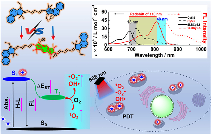
Scheme 1.
Schematic illustration of cyanine structure, photophysical properties, ROS generation mechanism, and application in PDT.
Conjugated di-Cy5.5 derivative achieving strong light-harvesting ability beyond 808 nm for high-efficient antitumor photodynamic therapy
Hua Gu , Juan Zhang , Puan Yuan , Zhongyue Zheng , Wenkai Liu , Xiang Xia , Wen Sun , Jianjun Du , Jiangli Fan , Xiaojun Peng

Cyanine dyes possess outstanding photophysical properties, adjustable wavelength, biocompatibility, and biosafety, which have been widely applied in fields of chemistry, biology, and medicine [1,2]. For example, indocyanine green (ICG) is approved by the Food and Drug Administration (FDA) for optical imaging of vasculature and navigation surgery in clinic, with good tissue penetration capability (excited by 808 nm laser). Up to the present, therefore, various near infrared (NIR) photosensitizers have been developed based on pentamethine cyanine (Cy5) and heptamethine cyanine (Cy7) dyes for photodynamic therapy (PDT) of solid tumor [3-8]. As a kind of safe and noninvasive treatment, PDT exhibits charming characteristic for tumor therapy with good spatiotemporal controllability and without drug resistant [9-16]. For a good photosensitizer which is the key point of PDT, besides NIR light absorption, it should also have high efficiency of intersystem crossing (ISC) and long lifetime of excited states [4,5,11,15,16]. However, there are few efficient tactics to construct Cy7-based NIR photosensitizers, besides introducing heavy atoms (e.g., I, Br, and Se) to enhance ISC process (S1→T1) [17-20]. Nevertheless, the heavy atom strategy usually produces 1O2 as the only product which restricts the PDT effect heavily in hypoxic solid tumors, along with inevitable dark toxicity and the reduced lifetime of excited states [4,5,10]. Recently, the spin–orbit charge transfer intersystem crossing (SOCT-ISC) is introduced to improve the triplet yield [1,3,7,21-23]. For example, the anthracene-substituted Cy5 (at meso–position) is demonstrated to enrich the populations in high vibrational–rotational energy levels and subsequently improve 1O2 generation [5]. However, the large twisted configuration always leads to heavy decline of extinction efficiencies of dyes along with an obvious blue-shift in absorption. Therefore, there are still significant challenges in how to develop an ingenious approach to transform a good NIR cyanine dye into an excellent cyanine photosensitizer while maintaining and even enhancing its photophysical properties.
The ISC efficiency and the NIR absorption of photosensitizers are directly related to ΔES-T (the band gap of singlet-excited and triplet-excited states) and ∆EH-L (the band gap of lowest unoccupied molecular orbital (LUMO) and the highest occupied molecular orbital (HOMO)), respectively [24-27]. Actually, increasing conjugation of dyes is one of the most direct and effective methods to benefit the extinction coefficient and adjust ∆EH-L. Moreover, ΔES-T also decreases along with ∆EH-L. Studies have shown that the introduction of D (electron donor)-π-A (electron acceptor) structure in dyes can change the decay property of the excited state, reduce the radiative transition (fluorescence performance), promote ISC, and prolong the triplet state lifetime effectively [28-31]. Therefore, compared with normal D-π-A structure, cyanine dyes with larger conjugation system (such as D-π-A-π-D structure) could much more effectively red-shift absorption/emission, increase intramolecular electron transfer, and reduce ΔES-T. Most importantly, the linear extension of cyanine dyes could lead to the proper twisted structure, increasing the charge separation that make electron transfer possible with O2, H2O, and other surrounding molecules (Scheme 1) [2,24,24,32-34].
Herein, as a-proof-of-concept, a novel D-π-A-π-D type of di-Cy5.5 derivative (2LBCy5.5) was constructed based on a classical fluorescent dye Cy5.5, with dual-cationic benzo[1,2-b:4,5-b′]dipyrrole (as an electron acceptor) bridging two Cy5.5 molecules (as electron donors). The 2LBCy5.5 exhibits a maximum absorption wavelength of 802 nm, with a redshift of 110 nm compared with Cy5.5 and much greater molar extinction coefficient of 4.2 × 10⁵ L mol−1 cm−1 than that of Cy5.5 (692 nm with molar extinction coefficient of 2.5 × 10⁵ L mol−1 cm−1) and ICG (795 nm with molar extinction coefficient of 3.2 × 10⁵ L mol−1 cm−1). Furthermore, 2LBCy5.5 showed low ∆EH-L and ΔES-T, fast ISC rate, and high triplet-excited state yield, compared to its parent Cy5.5. The 2LBCy5.5 showed a good anti-tumor ability under both normoxic and hypoxic conditions by producing various reactive oxygen species (ROS) under 808 nm light irradiation. This work provides a new approach for designing high-performance NIR photosensitizers with improved light-harvesting ability, photosensitizing efficiency, and stability.
2LBCy5.5 (Fig. 1a) was synthesized with the dual-cationic benzo[1,2-b:4,5-b′]dipyrrole as an acceptor coupled two Cy5.5 molecules, according to the routes provided in Fig. S1 (Supporting information), which was confirmed by 1H nuclear magnetic resonance (NMR), 13C NMR, and high resolution mass spectrometer (HRMS) (Figs. S2–S12 in Supporting information) [35-37].
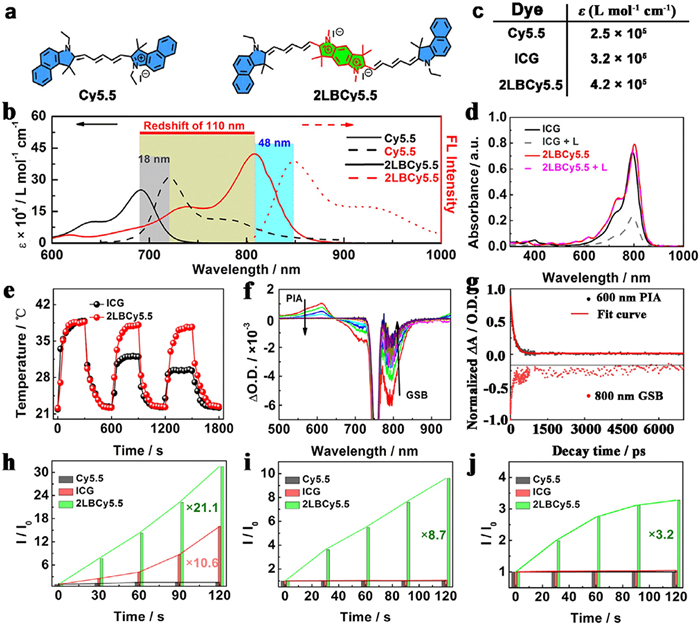
The photophysical properties of dyes were investigated, such as ultraviolet-visible (UV–vis)-NIR absorption and fluorescence emission. As shown in Fig. 1b, 2LBCy5.5 exhibited a maximum absorption at 802 nm in dimethylsulfoxide (DMSO), which redshifted about 110 nm compared to Cy5.5 (692 nm) and even exceeded that of ICG (795 nm). The maximum fluorescence emission of Cy5.5, ICG, and 2LBCy5.5 located at 720, 833, and 850 nm in DMSO, with Stokes shifts of 28, 37, and 48 nm, respectively. Meanwhile, the molar extinction coefficient of 2LBCy5.5 reached 4.2 × 10⁵ L mol−1 cm−1, which was much more than that of Cy5.5 (2.5 × 10⁵ L mol−1 cm−1) and ICG (3.2 × 10⁵ L mol−1 cm−1), suggesting its excellent light-harvesting capability (Fig. 1c and Fig. S13 in Supporting information). The fluorescence quantum yield of 2LBCy5.5 was 0.06, which was lower than that of Cy5.5 (0.33).
Generally, the stability of cyanine dyes is the basis for their functions. Upon 808 nm laser irradiation (10 W/cm2, 10 min), the absorbance of ICG decreased about 62%, whereas there was little change for 2LBCy5.5, demonstrating good stability against photobleaching (Fig. 1d). Furthermore, the stability of 2LBCy5.5 was evaluated by high-power irradiation (0.5 W/cm2) for three heating and cooling cycles. As shown in Fig. 1e, 2LBCy5.5 exhibited a repeatable temperature variation without absorption changes with acceptable photothermal conversion efficiency of 22.3% (Fig. S14 in Supporting information). However, under the same conditions, the maximum temperature of ICG decreased obviously after three cycles. These results indicated excellent photostability of 2LBCy5.5.
In addtion, femtosecond transient absorption spectra were obtained to analyze the excited-state energy dissipation pathways (Fig. 1f). The positive and negative signals signify the S1 to Sn transition (photoinduced absorption (PIA)) and S0 to S1 transition (ground-state bleaching (GSB)), respectively [38,39]. Since the excited-state energy dissipation pathways are mainly composed of radiative transition (kr represents the radiative decay rate), non-radiative transition (knr represents the nonradiative decay rate) and ISC process (kISC represents the ISC decay rate) [7,29,40-42]. According to the GSB remaining/GSB initial ratio and defined as kx/(kISC + knr + kr) (x stands for ISC, nr or r). Thus, by fitting the decay kinetics (Fig. 1g), the total decay rate (kISC + knr + kr) was obtained, 2.89 ns−1. Subsequently, the kr value was 0.25 ns−1 (9.6% proportion), the knr value was 1.43 ns−1 (49.4% proportion) and the kISC value was 1.21 ns−1 (41.0% proportion), which indicated that the balance of excited state decay was more in favor of ISC process for PDT. As a result, the triplet state yield of 2LBCy5.5 was determined to be 26.1%, higher than that of Cy7 derivative (17%–22%) under the same conditions [7,29], implying a greater T1 population and a stronger ROS-generating capacity.
Thus, under 808 nm laser irradiation with 10 W/cm2 power, the ROS generation ability of 1O2, •O2−, and •OH of Cy5.5, ICG and 2LBCy5.5 were measured using singlet oxygen sensor green probe (SOSG), dihydroethidium (DHE), and hydroxyphenyl fluorescein (HPF) as indicators, respectively. As shown in Figs. 1h–j and Figs. S15–S17 (Supporting information), the characteristic fluorescence signals of SOSG (525 nm), DHE (610 nm), and HPF (515 nm) showed no fluorescence changes in the presence of ICG or Cy5.5, while gradually enhanced in the presence of 2LBCy5.5 upon laser irradiation, indicating both type-Ⅰ and type-Ⅱ ROS-generating mechanisms for 2LBCy5.5. More importantly, 2LBCy5.5 showed higher ROS generation yield (about 10.6-/21.1-fold of 1O2, ~8.7-fold of •O2−, and 3.2-fold of •OH) than that of ICG or Cy5.5 (as references), respectively. Therefore, 2LBCy5.5 can be used for both normoxia and hypoxia PDT.
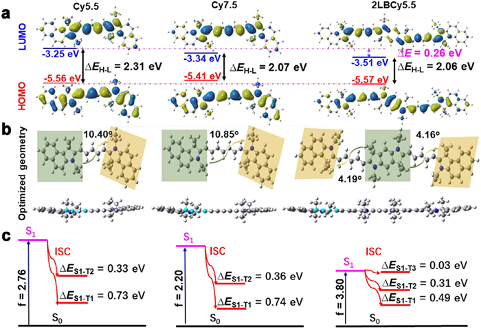
To further interpret the photosensitization mechanism of 2LBCy5.5, the density functional theory (DFT) of Cy5.5, Cy7.5 and 2LBCy5.5 was executed. The LUMO and HOMO energy levels of Cy5.5 were −3.25 and −5.56 eV with ∆EH-L of 2.31 eV (Fig. 2a). As the conjugate structure was extended, the LUMO, HOMO, and ∆EH-L of Cy7.5 were all changed to −3.34, −5.41, and 2.07 eV, respectively. However, 2LBCy5.5 exhibited strong electron-withdrawing ability, resulting in a low LUMO value of −3.51 eV. And ∆EH-L of 2LBCy5.5 was 2.06 eV, lower than that of Cy7.5, which matched well with its red-shifted absorption. In addition, the electron-hole diagram of excited states calculated by time dependent (TD)-DFT also proved that 2LBCy5.5 has higher electron delocalization and charge separation states than those of Cy5.5 and Cy7.5 due to the dual-cation fused indole-induced intramolecular charge transfer (ICT) effect, which was conducive to electron hole transport and promoted excited state electron transfer to generate type-Ⅰ ROS [8,25,29]. Furthermore, the optimized molecular conformations of Cy5.5, Cy7.5, and 2LBCy5.5 were investigated (Fig. 2b). The dihedral angles of the two terminal indole groups in Cy5.5 and Cy7.5 were 10.40° and 10.85°, respectively. In contrast, the dihedral angle between the terminal indole group and the fused indole group in the 2LBCy5.5 was only 4.16° (or 4.19°), indicating good planar structure. Even the distant terminal indole group in 2LBCy5.5 had a dihedral angle of only 8.35°, which might be the main factor leading to the increase in the molar extinction coefficient of 2LBCy5.5. Additionally, the oscillator strength (f) and the ΔES-T values between the lowest singlet (S1) and triplet energy levels (T1) of the photosensitive dyes were also calculated in detail (Fig. 2c). Oscillator strength is an important physical parameter to characterize the absorption or emission of atoms, which is positively correlated to the absorption peak area and the molar extinction coefficient [7,40,43-45]. According to DFT, the f values of Cy5.5, Cy7.5, and 2LBCy5.5 were 2.76, 2.20, and 3.80, respectively, which fit the trend of molar extinction coefficients. The ΔES1-T1 (0.49 eV) of 2LBCy5.5 was apparently decreased compared with that of Cy7.5 (0.74 eV) and Cy5.5 (0.73 eV). What is more, the ΔES1-T3 of 2LBCy5.5 was 0.03 eV, much lower than the values of ΔES1-T1 and ΔES1-T2, indicating a potential ISC process from the S1 state to the T3 state. These theoretical results are consistent with experimental results.
Considering the excellent ROS generation ability, in vivo PDT of 2LBCy5.5 was studied. As shown in Fig. 3a, 2LBCy5.5 was rapidly taken up by MCF-7 cells within 10 min. The fluorescence signals of 2LBCy5.5 were overlapped with those of the Mito-green tracker, indicating an excellent mitochondrial-targeting function (Pearson's correlation coefficient is 0.82). However, a weak fluorescence signal was observed in MCF-10A cells under the same incubation period, and it required 20 min to achieve maximum brightness (i.e., optimal enrichment), which is twice the duration observed in tumor cells (Fig. 3b). The above difference indicated that 2LBCy5.5 could discriminate normal cells from cancer cells to some extent, potentially attributed to its dual cationic structure, which contributes to its preferential targeting of tumor cells with higher electronegativity [29,45-49]. Furthermore, we investigated the ROS generation performance in MCF-7 cells. Commercially available reagents SOSG, DHE, and HPF were used as detection probes for 1O2, •O2−, and •OH, respectively. As shown in Fig. 3c, the significant fluorescent signals of SOSG, DHE, and HPF, were observed under 808 nm laser irradiation, indicating intracellular generation of 1O2, •O2−, and •OH, which contributed to anti-tumor PDT under both hypoxic and normoxic environment.
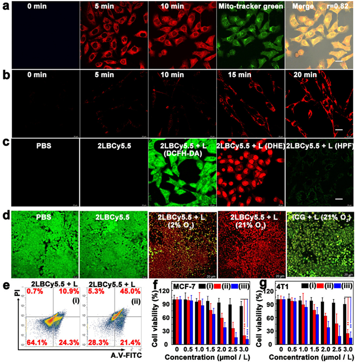
To further verify the antitumor ability of 2LBCy5.5, calcein-AM (green fluorescence for live cells) and propidium iodide (PI, red fluorescence for dead cells) were used for staining experiments. In Fig. 3d and Fig. S18 (Supporting information), a strong green fluorescence signal was observed in the control group treated with phosphate buffer saline (PBS) alone or 2LBCy5.5 (or ICG) alone, indicating their good biocompatibility. ICG cannot kill MCF-7 cells (green fluorescence under laser irradiation) under both normoxia and hypoxic conditions. In contrast, MCF-7 cells treated with 2LBCy5.5 and 808 nm laser irradiation were killed efficiently (bright red fluorescence) under both normoxic and hypoxic conditions. Since 2LBCy5.5 was localized in mitochondria of cells, commercially available JC-1 was employed as a probe to test the integrity of the mitochondrial membrane. As shown in Fig. S19 (Supporting information), under normoxia or hypoxia, JC-1 existed as a monomer in cells (green fluorescence) for the 2LBCy5.5 + L group, which was consistent with the result of the positive control group (CCCP), indicating that 2LBCy5.5-mediated PDT significantly depolarized mitochondria. Next, flow cytometry was employed to assess the mechanism of photoinduced cytotoxicity. As shown in Fig. 3e and Fig. S20 (Supporting information), the MCF-7 cells underwent apoptosis with the pyroptosis index of 66.3% after treatment with 2LBCy5.5 and 808 nm laser irradiation. Lastly, the in vitro tumor PDT effect of 2LBCy5.5 was evaluated by the MTT assay (Figs. 3f and g), and 2LBCy5.5 was found to have negligible toxicity to MCF-7 and 4T1 cells in the absence of laser irradiation, with cell survival up to 95%. However, under laser irradiation, 2LBCy5.5 exhibited superior PDT performance with increasing concentrations in both normoxia and hypoxia condition.
The excellent tumor-targeting properties and PDT performance of 2LBCy5.5 motivated us to explore its anti-tumor effects in vivo. All in vivo studies were approved by the Dalian Medical University Animal Care and Use Committee (approval number: DUTNI230309-01). A tumor-bearing BALB/c mouse model was established with subcutaneous inoculation of 4T1 cells on the upper side of the right lower limb. When the tumor volume reached about 100 mm3, all tumor-bearing BALB/c mice were randomly divided into four groups for in vivo anti-cancer PDT. Before treatment, fluorescence imaging was executed to record after the injection of 2LBCy5.5 through the caudal vein to obtain the optimal time point for PDT. As shown in Fig. 4a, with the extension of time, the fluorescence signal at the tumor site gradually increased and reached the maximum at 3 h, wherein 2LBCy5.5 was mainly enriched in the tumor site, followed by the liver and kidney. Therefore, the best treatment time was 3 h after tail vein injection.
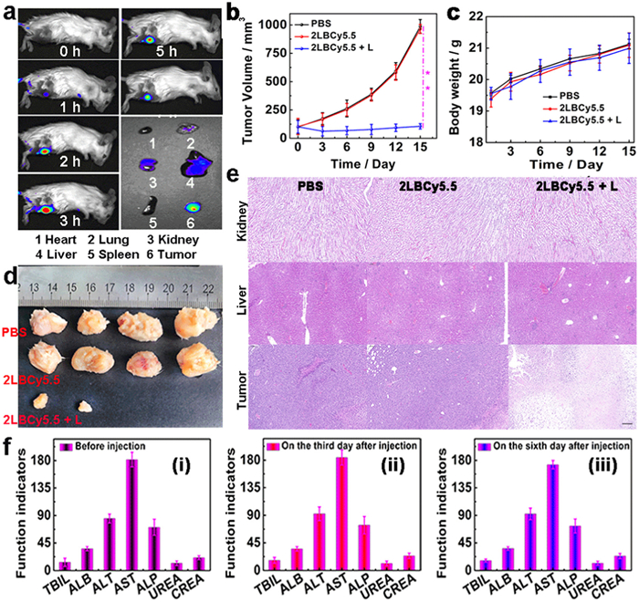
Based on the above results, in vivo PDT was conducted after 2.5 h tail vein injection of 2LBCy5.5 by using 808 nm laser irradiation (100 mW/cm2) for 10 min. After the laser irradiation, the changes in body weight and tumor volume of all the mice were continuously monitored for 15 d to estimate the PDT effect. As illustrated in Fig. 4c, all the mice grew well and exhibited no obvious difference during the treatment, indicating low side effects of 2LBCy5.5. And the tumors in blank group (treated with PBS) and the control group (treated with 2LBCy5.5) increased gradually about 10 times compared to their original volumes (Figs. 4b and d). As a sharp contrast, the tumors treated with 2LBCy5.5 + L (808 nm laser) underwent necrosis and eventually disappeared entirely, which demonstrated the high PDT efficiency of 2LBCy5.5. Hematoxylin and eosin (H & E) staining revealed significant morphological damage to tumor tissue following photosensitizer-mediated PDT, with no observed cell necrosis or inflammation in major organs (such as the heart, liver, spleen, lung, and kidney) (Fig. 4e and Fig. S21 in Supporting information). Simultaneously, there were no abnormal liver and kidney function indexes before and after treatment (Fig. 4f (ⅰ)–(ⅲ)), which showed that 2LBCy5.5 has no negative effect on the functions of the liver and kidney. These findings suggested that 2LBCy5.5 exhibited good anti-tumor effects and biocompatibility.
In summary, the conjugated di-Cy5.5 derivative, 2LBCy5.5, was successfully developed by conjugating two classical fluorescent dyes Cy5.5 with dual-cationic benzo[1,2-b:4,5-b′]dipyrrole group. As a result, 2LBCy5.5 showed a significant red shift in absorption/emission wavelength (802 and 850 nm, respectively), especially with a significantly enhanced molar extinction coefficient as high as 4.2 × 10⁵ L mol−1 cm−1. Because the extend conjugation results in strong ICT effect that endowed 2LBCy5.5 with a narrow ∆EH-L and ∆ES-T, which promoted ISC progress efficiently and following excellent type-Ⅰ and type-Ⅱ ROS-generation capacity. Therefore, 2LBCy5.5 showed good anti-tumor ability under both normoxic and hypoxic conditions. In short, a simple and effective strategy was developed for constructing high-performance photosensitive dyes with excellent NIR light-harvesting ability, photosensitive ability, and photostability.
The authors declare that they have no known competing financial interests or personal relationships that could have appeared to influence the work reported in this paper.
Hua Gu: Writing – original draft, Validation, Software, Resources, Project administration, Investigation, Funding acquisition, Formal analysis, Data curation, Conceptualization. Juan Zhang: Visualization, Validation, Methodology, Formal analysis. Puan Yuan: Software, Methodology, Data curation. Zhongyue Zheng: Validation, Software. Wenkai Liu: Software. Xiang Xia: Software, Data curation. Wen Sun: Funding acquisition. Jianjun Du: Writing – review & editing, Funding acquisition, Conceptualization. Jiangli Fan: Writing – review & editing, Funding acquisition, Conceptualization. Xiaojun Peng: Writing – review & editing, Conceptualization.
This work was financially supported by the National Natural Science Foundation of China (Nos. 21925802, 22308049, 22338005), Liaoning Provincial Science and Technology Joint Fund (Nos. 2023JH2/101800039, 2023JH2/101800037), Liaoning Binhai Laboratory (No. LBLB-2023-03), the Fundamental Research Funds for the Central Universities (No. DUT22LAB601), the China Postdoctoral Science Foundation (No. 2023M740486).
Supplementary material associated with this article can be found, in the online version, at doi:
A. Sharma, P. Verwilst, M. Li, et al., Chem. Rev. 124 (2024) 2699–2804. doi: 10.1021/acs.chemrev.3c00778
J. Yuan, H. Yang, W. Huang, et al., Chem. Soc. Rev. 54 (2025) 341–366. doi: 10.1039/d3cs00585b
X. Zhao, J. Du, W. Sun, et al., Acc. Chem. Res. 57 (2024) 2582–2593. doi: 10.1021/acs.accounts.4c00399
X. Yuan, J. Zhou, L. Yuan, et al., Sci. China Chem. 68 (2025) 826–865. doi: 10.1007/s11426-024-2411-7
X. Zhao, S. Long, M. Li, et al., J. Am. Chem. Soc. 142 (2020) 1510–1517. doi: 10.1021/jacs.9b11800
X. Zhao, Q. Yao, S. Long, et al., J. Am. Chem. Soc. 143 (2021) 12345–12354. doi: 10.1021/jacs.1c06275
X. Zhou, C. Shi, S. Long, et al., ACS Cent. Sci. 9 (2023) 1679–1691. doi: 10.1021/acscentsci.3c00611
F. Han, S. Abedi, S. He, et al., Adv. Sci. 11 (2024) 2305761. doi: 10.1002/advs.202305761
T. Pham, V. Nguyen, Y. Choi, et al., Chem. Rev. 121 (2021) 13454–13619. doi: 10.1021/acs.chemrev.1c00381
W. Fan, P. Huang, X. Chen, Chem. Soc. Rev. 45 (2016) 6488–6519. doi: 10.1039/C6CS00616G
W. Su, X. Luo, P. Li, et al., Chin. Chem. Lett. 35 (2024) 109522. doi: 10.1016/j.cclet.2024.109522
K. Teng, D. Zhang, B. Liu, et al., Angew. Chem. Int. Ed. 63 (2024) e202318783. doi: 10.1002/anie.202318783
X. Liu, J. Li, F. Zhang, et al., Sci. China Chem. 67 (2024) 2314–2324. doi: 10.1007/s11426-024-2086-8
Y. Yang, J. Zhang, Y. Wang, et al., Chin. Chem. Lett. 36 (2025) 110834. doi: 10.1016/j.cclet.2025.110834
Z. Cui, D. Zhang, Y. Huang, et al., Chin. Chem. Lett. 36 (2025) 110460. doi: 10.1016/j.cclet.2024.110460
S. Zhen, Z. Xu, M. Suo, et al., Adv. Mater. 37 (2025) 2411133. doi: 10.1002/adma.202411133
N. Sellet, J. Frey, M. Cormier, et al., Chem. Sci. 15 (2024) 8639–8650. doi: 10.1039/d4sc00814f
J. Yu, Y. Wen, M. Li, Adv. Funct. Mater. 34 (2024) 2402663. doi: 10.1002/adfm.202402663
Y. Yue, B. Li, D. Wang, et al., Adv. Funct. Mater. 35 (2024) 2414542.
H. Wen, Q. Wu, X. Xiang, et al., ACS Appl. Mater. Interfaces 16 (2024) 61739–61750. doi: 10.1021/acsami.4c14754
D. Liu, A. El-Zohry, M. Taddei, et al., Angew. Chem. Int. Ed. 59 (2020) 11591–11599. doi: 10.1002/anie.202003560
Zhao X, S. He, W. Chi, et al., Adv. Sci. 9 (2022) 2202885. doi: 10.1002/advs.202202885
M. Williams, I. Schlesinger, R. Jacobberger, et al., J. Am. Chem. Soc. 144 (2022) 18607–18618. doi: 10.1021/jacs.2c08584
H. Huang, Q. Liu, J. Zhu, et al., Aggregate 6 (2025) e706. doi: 10.1002/agt2.706
W. Liu, S. He, X. Ma, et al., Angew. Chem. Int. Ed. 63 (2024) e202484761. doi: 10.1002/anie.202484761
H. Janeková, S. Fisher, T. Šolomek, et al., Chem. Sci. 16 (2025) 1677–1683. doi: 10.1039/d4sc07165d
C. You, Y. Zhu, J. Zhu, et al., Angew. Chem. Int. Ed. 64 (2024) e202417865.
V. Nguyen, Z. Zhao, B. Tang, et al., Chem. Soc. Rev. 51 (2022) 3324–3340. doi: 10.1039/d1cs00647a
H. Gu, W. Liu, W. Sun, et al., Chem. Sci. 13 (2022) 9719–9726. doi: 10.1039/d2sc02879d
Y. Tang, H. Bisoyi, X. Chen, et al., Adv. Mater. 35 (2023) 2300232. doi: 10.1002/adma.202300232
H. Gu, W. Liu, H. Li, et al., Coord. Chem. Rev. 473 (2022) 214803. doi: 10.1016/j.ccr.2022.214803
G. Gopika, P. Hari Prasad, A. Lekshmi, et al., Mater. Today Proc. 46 (2021) 3102–3108. doi: 10.1016/j.matpr.2021.02.622
X. Ma, Y. Huang, S. Abedi, et al., CCS Chem. 4 (2022) 1961–1976. doi: 10.31635/ccschem.021.202101630
Y. Yue, J. Ai, W. Chi, et al., Adv. Mater. 36 (2024) 2408450. doi: 10.1002/adma.202408450
H. Shindy, Dyes Pigm. 145 (2017) 505–513. doi: 10.1016/j.dyepig.2017.06.029
O. Klochko, I. Fedyunyayeva, S. Khabuseva, et al., Dyes Pigm. 85 (2010) 7–15. doi: 10.1016/j.dyepig.2009.09.007
F. Mihailenko, A. Boguslavskaya, A. Kiprianov, Chem. Heter. Comp. 7 (1971) 578–580. doi: 10.1007/BF00945496
C. Ruckebusch, M. Sliwa, P. Pernot, et al., J. Photoch. Photobio. C: Photoch. Rev. 13 (2012) 1–27.
R. Sension, T. McClain, R. Lamb, et al., J. Am. Chem. Soc. 145 (2023) 14070–14086. doi: 10.1021/jacs.3c04099
X. Xia, C. Shi, S. He, et al., Adv. Funct. Mater. 33 (2023) 2300340. doi: 10.1002/adfm.202300340
B. Dereka, A. Rosspeintner, Z. Li, et al., J. Am. Chem. Soc. 138 (2016) 4643–4649. doi: 10.1021/jacs.6b01362
J. Kim, R. Kishi, E. Kayahara, et al., Angew. Chem. Int. Ed. 59 (2020) 16989–16996. doi: 10.1002/anie.202006066
M. Yang, X. Ou, J. Zhang, et al., Adv. Funct. Mater. 35 (2024) 2411838.
Y. Li, J. Zhang, S. Liu, et al., Adv. Funct. Mater. 31 (2021) 2102213. doi: 10.1002/adfm.202102213
M. Li, T. Xiong, J. Du, et al., J. Am. Chem. Soc. 141 (2019) 2695–2702. doi: 10.1021/jacs.8b13141
M. Wu, M. Gu, J. Leung, et al., Small 17 (2021) 2101770. doi: 10.1002/smll.202101770
M. Li, S. Long, Y. Kang, et al., J. Am. Chem. Soc. 140 (2018) 15820–15826. doi: 10.1021/jacs.8b09117
G. Li, Y. Huang, Q. Feng, et al., Molecules 19 (2014) 12224–12241. doi: 10.3390/molecules190812224
R. Horobin, J. Stocker, F. Rashid-Doubell, Histochem. Cell Biol. 139 (2013) 623–637. doi: 10.1007/s00418-013-1090-0

Scheme 1 Schematic illustration of cyanine structure, photophysical properties, ROS generation mechanism, and application in PDT.

Figure 1 (a) The structures of Cy5.5 (as control) and 2LBCy5.5. (b) UV–vis-NIR spectra and fluorescence emission spectra of Cy5.5, ICG, and 2LBCy5.5 in DMSO, respectively. (c) The molar extinction coefficient of Cy5.5, ICG, and 2LBCy5.5 in DMSO, respectively. (d) Stability of 2LBCy5.5 and ICG against photobleaching. (e) Photothermal stability study of 2LBCy5.5 and ICG (20.0 µmol/L) during repeated heating and cooling cycles under 808 nm laser irradiation with 0.5 W/cm2. (f) Femtosecond transient absorption spectra and (g) the kinetics of PIA and GSB of 2LBCy5.5. The (h) 1O2, (i) •O2−, and (j) •OH measurement of Cy5.5, ICG, and 2LBCy5.5 upon laser irradiations (10 mW/cm2) by using SOSG, DHE, and HPF as probes respectively, where I0 and I are the fluorescence intensity at maximum emission peaks of ROS indicators in the presence of the photosensitive dyes before and after irradiation. ([2LBCy5.5 and ICG] = 4 µmol/L and [SOSG, DHE, and HPF] = 10 µmol/L).

Figure 2 Molecular orbital amplitude plots of Cy5.5, Cy7.5, and 2LBCy5.5, calculated using the Gaussian 16 B3LYP/def2svp program. (a) Calculated HOMO and LUMO geometries, (b) optimized molecular structure, and (c) oscillator strength (f), singlet energy level (S1), triplet energy level (T1), and ΔES-T values.

Figure 3 Confocal images of cellular uptake and colocalization of 2LBCy5.5 (2 µmol/L) in MCF-7 (a) and MCF-10A cells (b) at different times, and (c) the ROS detected of 2LBCy5.5 (2 µmol/L) in MCF-7 cells under 808 nm laser irradiation with SOSG, DHE, and HPF as detection probes for 1O2, •O2−, and •OH, respectively. In vitro PDT of 2LBCy5.5. Confocal images of (d) calcein-AM (green)/PI (red) in MCF-7 cells treated under different experimental conditions. (e) Flow cytometry analysis of MCF-7 cells (i was hypoxic conditions, ii was normoxic conditions) and (f, g) cell viability of MCF-7 and 4T1 cells incubated with different concentrations of 2LBCy5.5 (results are presented as mean ± SD, n = 5). Where, i, ii, and iii are represented the 2LBCy5.5 without laser irradiation, 2LBCy5.5 with laser irradiation under hypoxia condition and 2LBCy5.5 with laser irradiation under normoxia condition ([2LBCy5.5 or ICG] = 2 µmol/L, 808 nm laser irradiation with 30 mW/cm2, 10 min). P-values were calculated using Student's t-test. **P < 0.01, ***P < 0.001. Scale bar: 20 µm.

Figure 4 In vivo PDT of 2LBCy5.5. (a) The real-time fluorescence imaging of tumor-bearing mice and major organs after injection of 2LBCy5.5. The tumor volume (b) and body weights (c) of the mice after different treatments. (d) Images of tumors harvested in mice from different groups at 15 days of post-treatment. (e) HE staining of tumor sections from different treatment groups after 15 days of treatment. (f) Hematological assessment of liver and kidney function indicators of mice at different post-injection times. Where, i, ii and iii are represented the different time of before injection, on the third day after injection and on the sixth day after injection, respectively). Scale bar: 100 µm. Data were expressed as mean ± SD (n = 4). **P < 0.01 determined by Student's t-test.

 扫一扫看文章

扫一扫关注我们
 DownLoad:
 下载:
下载:
 下载:
下载: