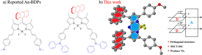
Figure 1.
(a) Design strategies for An-BDP with 1,7-di-anthracyl groups in aza-BODIPY system. (b) Orthogonal structure of An-azaBDP.
Heavy-atom-free orthogonal configurative dye 1,7-di-anthra-aza-BODIPY for singlet oxygen generation
Yan Zhu , Jia Liu , Meiheng Lv , Tingting Wang , Dongxiang Zhang , Rong Shang , Xin-Dong Jiang , Jianjun Du , Guiling Wang

Azadipyrromethene as a star molecule has attracted increasing attention since 1943 [1]. Boron-azadipyrromethene (aza-BODIPY) is widely used in photoelectric materials, molecular recognition and medicine and so on, owing to its excellent spectral properties such as high molar extinction coefficient and long-wavelength absorption [2–10]. In recent years, due to the significant impact of cancer, it is of great significance to develop methods to respond to cancer treatment [11–18]. Especially, photodynamic therapy (PDT) has shown the outstanding effects in responding to tumors and has developed some reagents and drugs [19–27]. Therefore, based on aza-BODIPY platform, its research and development for PDT has attracted the widespread interest.
The introduction of heavy atom is well-known to efficiently improve the efficiency of intersystem crossing (ISC) [28–34], thereby generating reactive oxygen species (ROS) for PDT. However, there is also a risk of disengage of the heavy atom and the potential dark toxicity. Therefore, in recent years, heavy-atom-free BODIPY photosensitizers (PSs) have been sought after. Indeed, dimeric BODIPYs and structurally twisted BODIPYs are able to produce ROS [35–42]. Moreover, spin-orbit, charge-transfer intersystem crossing (SOCT-ISC) can directly overcome the disadvantages of the traditional heavy-atom effect and improve the generation efficiency of ROS [43–48]. The SOCT-ISC mechanism was firstly proposed by Willigen, who designed an orthogonal configuration between naphthalene and acridine [49]. Upon photoexcitation, charge separation and charge compounding processes took place in this molecule, and then ISC process was achieved. It is found that anthracene as a typical segment can promote SOCT-ISC under irradiation. Since the orthogonal molecular orbitals of donor-acceptor (D-A) pairs favor the SOCT-ISC transition, BODIPY with the anthracyl group at meso site was popularly designed, owing to steric hindrance to produce a big dihedral angle between the two molecular orbital (MO) planes. According to the X-ray crystallography, indeed the orthogonal structure between the anthracyl group at meso site and the parent nucleus BODIPY was confirmed by Yang et al. [50]. Subsequently, Sun et al. reported heavy-atom-free nanomaterials based on An-BDP with the methoxy groups, which promotes ISC by SOCT and enhances the generation of ROS, plays an active part in the realization of PDT (Fig. 1a) [29]. By introducing a stronger electron donating diphenylamino group, instead of the methoxy group in An-BDP, the conjugation of anthracene onto the BODIPY moiety at meso site enhances the ISC by SOCT, and acts as storage for the reversible capture and release of ROS to enhance PDT efficacy (Fig. 1a) [51].
To date, the anthracyl group is found not to introduce into the aza-BODIPY system to engage in this SOCT-ISC study. By replacing the C atom at meso site with the N atom, the energy gap between the highest occupied molecular orbital (HOMO) and the lowest unoccupied molecular orbital (LUMO) is reduced, and aza-BODIPY has longer wavelength absorption and higher molar extinction coefficient [52–57], compared to BODIPY. Therefore, we are particularly curious about whether the new molecule aza-BODIPY bearing an anthryl group has SOCT-ISC and efficiently generates ROS. Based on the unique structure of aza-BODIPY core, the smart design is needed to introduce an anthryl group into the BODIPY scaffold. Herein, by the wise use of raw materials, we successfully designed aza-BODIPY with the anthryl groups at 1,7-sites (An-azaBDP) (Fig. 1b). Based on the molecular stereo structure, this molecule should be an orthogonal structure. Indeed, according to our experimental results, this molecule could efficiently produce ROS, and was successfully applied for PDT.
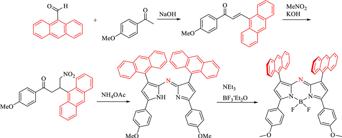
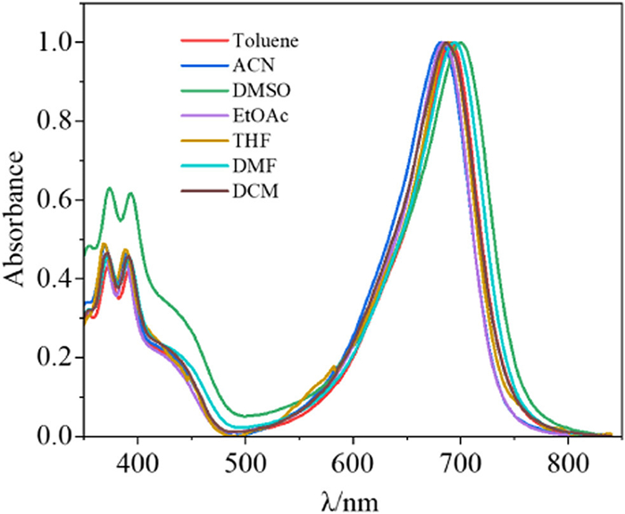
In order to introduce the anthryl groups at 1,7-sites in aza-BODIPY system, 9-anthracenecarboxaldehide was employed as a key starting material. Based on the classic O'Shea synthetic method [58], we successfully prepared An-azaBDP with 1,7-dianthryl groups in 35% yields (Scheme 1). The absorption maximum (688 nm) of An-azaBDP is consistent with that of aza-BODIPY bearing 1,7-diphenyl groups (Ph-azaBDP) [60], indicating that the contribution to absorption wavelength is identical between anthracene and benzene at 1,7-sites in aza-BODIPY system. Absorption of An-azaBDP in different solvents locates at the near infrared region (NIR) (λabs = 681–700 nm), with high molar extinction coefficient (Fig. 2 and Table S1 in Supporting information). Surprisingly, no fluorescence of An-azaBDP was observed, compared to that (Φf = 0.36) of Ph-azaBDP [56]. This is likely due to that the SOCT of the anthryl group enhances the ISC effect and the pathway for releasing fluorescence is maximally restricted.
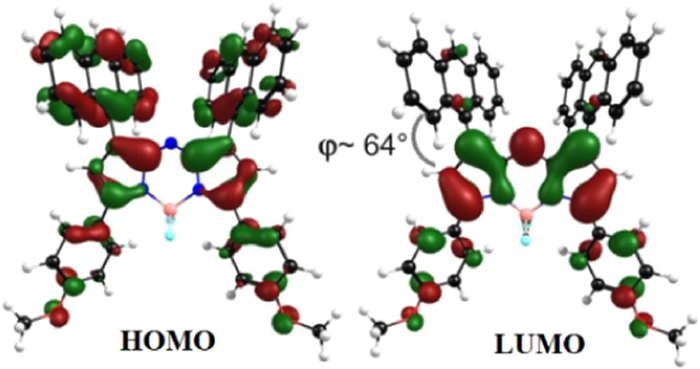
To unravel the optical properties of An-azaBDP, theoretical simulations are conducted based on density functional theory (DFT) and time-dependent density functional theory (TDDFT) methods [60–63]. The primary absorption and emission peaks of An-azaBDP both correspond to the S0-S1 transition, which is composed of the transition between the HOMO and the LUMO. As shown in Fig. 3, the electron density of HOMO mainly distributes on the whole molecule including the anthryl groups at 1,7-sites, whereas the LUMO mainly located on the core of An-azaBDP, which indicates that the S0-S1 transition has an obvious charge-transfer property. In the optimized molecular structure, the dihedral angle between the anthryl group and the parent nucleus is about 64°, which is indeed a nearly orthogonal molecular model. Moreover, the energy difference between the S1-T1 orbitals is 0.67 eV (Fig. S1 in Supporting information), which is such small energy difference and more inclined towards ISC [64].
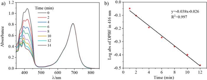
Subsequently, singlet oxygen (1O2) generation capability of An-azaBDP was assessed in toluene under monochromatic light at 690 nm by using a 150 W xenon lamp at 0.5 mW/cm2 [65]. By utilizing 1,3-diphenylisobenzofuran (DPBF) as singlet oxygen indicator, the efficiency of singlet oxygen generation was evaluated by detecting the decrease of DPBF absorbance at 416 nm (Fig. 4a) [66], and methyl blue (MB, ΦΔ = 0.57 in DCM) was used as the reference [37]. According to the linear relationship of the decay curve (Fig. 4b), the 1O2 yields of An-azaBDP (S = 0.038) was high and calculated to be 0.16, comparing to that (ΦΔ = 0.02) of Ph-azaBDP (λabs= 688 nm) (Fig. S2 in Supporting information) [59].
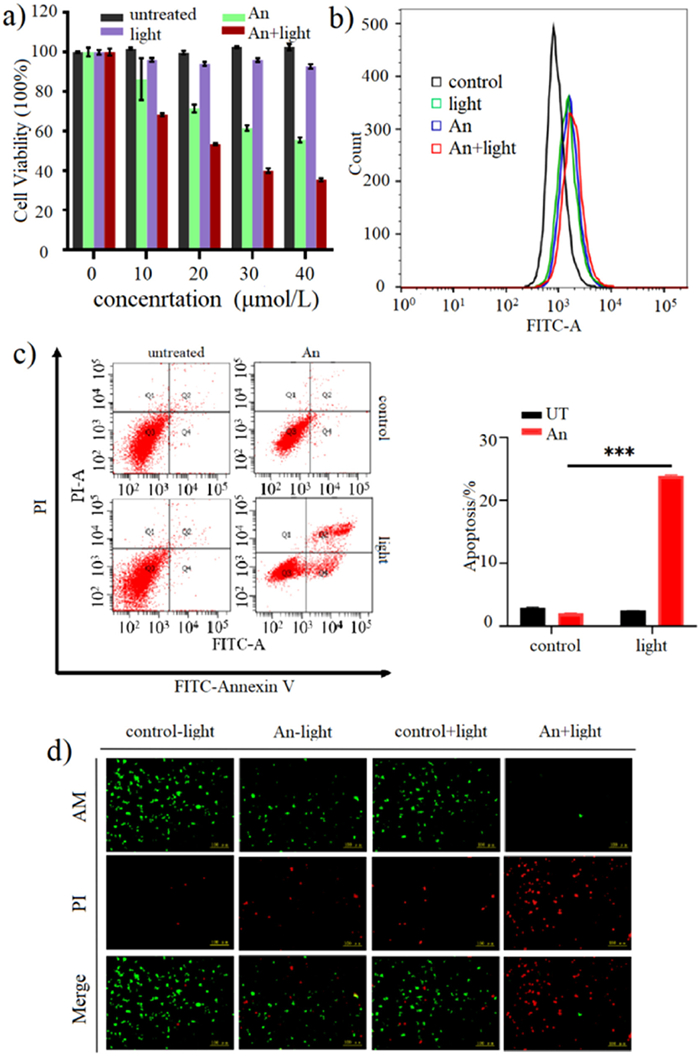
Considering the no fluorescence (Fig. S3 in Supporting information) and the 1O2 yield, PDT of An-azaBDP with light irradiation for human colorectal cancer cells (SW620 cells) was next investigated. To identify the concentration dependence and the best concentration of An-azaBDP, SW620 cells were treated with diverse drug concentrations (0–40 µmol/L) under laser radiation, which subjected to cell counting kit-8 (CCK-8) assay. The results indicated that laser radiation combined with An-azaBDP was the most effective to reduce cell viability compared with the no-treatment group, drug-treatment group and laser radiation group. There was the drug concentration dependence, and the difference was more obvious when the drug concentration was 40 µmol/L (Fig. 5a). Therefore, we chose 40 µmol/L concentration for the subsequent experiments. High ROS are products of cellular metabolism, and ROS levels are important signals for normal physiological cell function and environmental factors leading to cell damage. To analysis the amount of ROS, we applied flow cytometry to detect ROS produced by SW620 cells treated with An-azaBDP alone, light alone or combination of treatments. It was showed that ROS levels increased after treatment with An-azaBDP and laser irradiation [44], and cells treated with An-azaBDP or light alone exhibited lower ROS levels, suggesting that An-azaBDP combined with light treatment can effectively lead to cellular damage (Fig. 5b). In order to further explore the potential effect of An-azaBDP on cancer cells, we performed relevant apoptosis analysis of SW620 cells using flow cytometry. The result reveals that the number of apoptotic cells increased significantly after treatment with An-azaBDP and laser irradiation (~23%), and cells treated with An-azaBDP or light alone showed a lower rate of apoptosis (~3%), demonstrating the effective ability of An-azaBDP combined with laser irradiation to induce apoptosis in cancer cells (Fig. 5c). Meanwhile, a double staining kit of calcein acetoxymethyl ester (AM, staining live cells, showing green fluorescence) and propidium iodide (PI, staining dead cells, showing red fluorescence) was applied to display the viability effectiveness of An-azaBDP with laser irradiation on cancer cells (Fig. 5d). As shown in Fig. 5d, SW620 cells were killed by the co-treatment of An-azaBDP and laser irradiation, showing a markedly red fluorescence. On the contrary, the control group, the laser-treated group alone or the An-azaBDP-treated group alone had clearly green fluorescence, suggesting that laser treatment alone or An-azaBDP treatment alone was almost invalid for killing cancer cells. These results implicated that An-azaBDP treatment followed by laser irradiation (690 nm, 0.3 W/cm2) could kill cancer cells.
In conclusion, using 9-anthracenecarboxaldehide as a key starting material, herein An-azaBDP with 1,7-dianthryl groups successfully prepared. In the optimized molecular structure, the dihedral angle between the anthryl group and the parent nucleus is about 64°, which is indeed a nearly orthogonal molecular model. The energy difference between the S1-T1 orbitals is small and more inclined towards ISC. No fluorescence of An-azaBDP was observed, and this is due to that the SOCT of the anthryl group enhances the ISC effect and the pathway for releasing fluorescence is maximally restricted. The 1O2 yields of An-azaBDP with light irradiation was high and calculated to be 0.16, comparing to that (ΦΔ = 0.02) of aza-BODIPY with 1,7-diphenyl groups. So, the SOCT of the anthryl group enhances the ISC effect and improves the ability of An-azaBDP to generate the singlet oxygen. An-azaBDP with light irradiation can induce apoptosis in SW620 cells, and could serve as a potential candidate for the treatment of cancer cells and tumors in PDT.
The authors declare that they have no known competing financial interests or personal relationships that could have appeared to influence the work reported in this paper.
This work was supported by the National Natural Science Foundation of China (Nos. 22078201, U1908202), Liaoning & Shenyang Key Laboratory of Functional Dye and Pigment (Nos. 2021JH13/10200018, 21–104–0–23).
Supplementary material associated with this article can be found, in the online version, at doi:
M.A.T. Rogers, J. Chem. Soc. (1943) 590–596.
S. Li, M. Lv, J. Wang, et al., Mater. Adv. 3 (2022) 1254–1262. doi: 10.1039/D1MA01052B
R. Li, J. Ren, D. Zhang, et al., Mater. Today Bio 16 (2022) 100446. doi: 10.1016/j.mtbio.2022.100446
X. Jiang, S. Yue, K. Chen, et al., Chin. Chem. Lett. 30 (2019) 2271–2273. doi: 10.1016/j.cclet.2019.07.027
M. Liu, S. Ma, M. She, et al., Chin. Chem. Lett. 30 (2019) 1815–1824. doi: 10.1016/j.cclet.2019.08.028
R. Li, Y. Du, W. Guo, et al., Dyes Pigm. 179 (2020) 108351. doi: 10.1016/j.dyepig.2020.108351
V. -N. Nguyen, Z. Zheng, J. Yoon, et al., Chem. Soc. Rev. 51 (2022) 3324–3340. doi: 10.1039/D1CS00647A
J. Yin, X. Jiang, G. Sui, et al., J. Mater. Chem. B 9 (2021) 7461–7471. doi: 10.1039/D1TB01155C
F.Z. Li, J.F. Yin, G.C. Kuang, Coord. Chem. Rev. 448 (2021) 214157. doi: 10.1016/j.ccr.2021.214157
C. Li, Y. Xu, L. Tu, et al., Chem. Sci. 13 (2022) 6541–6549. doi: 10.1039/D2SC01518H
Outersterp R E van, J. Oosterhout, C.R. Gebhardt, et al., ACS Appl. Bio Mater. 95 (2023) 3406–3413.
D. Chen, Z. Zhong, Q. Ma, et al., ACS Appl. Mater. Interfaces 12 (2020) 26914–26925. doi: 10.1021/acsami.0c05021
H. Luo, S. Zhao, J. Transp. Geogr. 90 (2021) 102920. doi: 10.1016/j.jtrangeo.2020.102920
L.J. Schipper, L.J. Zeverijn, M.J. Garnett, et al., Cancer Discov. 12 (2022) 1634–1641. doi: 10.1158/2159-8290.CD-21-0612
C. Ma, T. Zhang, Z. Xie, J. Mater. Chem. B 9 (2021) 7318–7327. doi: 10.1039/D1TB00855B
B.C. Wilson, R.A. Weersink, Photochem. Photobio. 96 (2020) 219–231. doi: 10.1111/php.13184
R. Zheng, S. Zhang, H. Zeng, et al., J. Natl. Cancer Inst. 2 (2022) 1–9.
D. Ma, S. Hou, C. Bae, et al., Chin. Chem. Lett. 32 (2021) 3886–3889. doi: 10.1016/j.cclet.2021.05.048
C. Liu, X. Ji, Z. Yu, et al., J. Med. Chem. 66 (2023) 7205–7220. doi: 10.1021/acs.jmedchem.2c01653
C. Li, H. Tan, R. Lu, et al., Nanophotonics 11 (2022) 5077–5088. doi: 10.1515/nanoph-2022-0417
J. Yan, T. Gao, Z. Lu, et al., ACS Appl. Mater. Interfaces 13 (2021) 27749–27773. doi: 10.1021/acsami.1c06818
J. Treekoon, T. Pewklang, K. Chansaenpak, et al., Org. Biomol. Chem. 19 (2021) 5867–5875. doi: 10.1039/D1OB00400J
E. Caruso, M.C. Malacarne, E. Marras, et al., Bioorg. Med. Chem. 28 (2020) 115737. doi: 10.1016/j.bmc.2020.115737
Y. Liu, G. Liu, W. Zhou, et al., Angew. Chem. Int. Ed. 135 (2023) e202309786. doi: 10.1002/ange.202309786
Z. Ji, J. Zheng, Y. Ma, et al., Small 19 (2023) 2207888. doi: 10.1002/smll.202207888
L. Wang, A. Mei, N. Li, et al., Chin. Chem. Lett. 35 (2024) 108974. doi: 10.1016/j.cclet.2023.108974
C. Ye, S. Zhang, D. Zhang, et al., Chin. Chem. Lett. 34 (2023) 108223. doi: 10.1016/j.cclet.2023.108223
H. Liang, M. Lu, Z. Mahmood, et al., Angew. Chem. Int. Ed. 62 (2023) e202312600. doi: 10.1002/anie.202312600
C. Li, T. Sun, X. Li, et al., ACS Appl. Nano Mater. 5 (2022) 18691–18696. doi: 10.1021/acsanm.2c04459
J. Piskorz, W. Porolnik, M. Kucinska, et al., Chem. Med. Chem. 16 (2021) 399–411. doi: 10.1002/cmdc.202000529
Z. Shi, X. Han, W. Hu, et al., Chem. Soc. Rev. 49 (2020) 7533–7567. doi: 10.1039/D0CS00234H
W. Hu, Y. Lin, X.F. Zhang, et al., Dyes Pigm. 164 (2019) 139–147. doi: 10.1016/j.dyepig.2019.01.019
Y. Zhang, Z. Yang, X. Zheng, et al., Dyes Pigm. 178 (2020) 108348. doi: 10.1016/j.dyepig.2020.108348
W. Sun, X. Wang, Z. Cheng, et al., Biomed. Pharmacother. 158 (2023) 114071. doi: 10.1016/j.biopha.2022.114071
Y. Yan, A.A. Sukhanov, M.H. Bousquet, et al., J. Phys. Chem. B 125 (2021) 6280–6295. doi: 10.1021/acs.jpcb.1c03189
V.N. Nguyen, J. Ha, C.W. Koh, et al., Chem. Mater. 33 (2021) 7889–7896. doi: 10.1021/acs.chemmater.1c02776
Y. Dong, B. Dick, J. Zhao, Org. Lett. 22 (2020) 5535–5539. doi: 10.1021/acs.orglett.0c01903
J. Li, X. Du, X. Zhou, et al., Adv. Healthc. Mater. 12 (2023) 2301022. doi: 10.1002/adhm.202301022
M.A. Filatov, S. Karuthedath, P.M. Polestshuk, et al., ChemPhotoChem 2 (2018) 606–615. doi: 10.1002/cptc.201800020
S.R. Zarcone, H.J. Yarbrough, M.J. Neal, et al., New J. Chem. 46 (2022) 4483–4496. doi: 10.1039/D1NJ05976A
K. Chen, Y. Dong, X. Zhao, et al., Front. Chem. 7 (2019) 821. doi: 10.3389/fchem.2019.00821
D. Yang, L. Sun, L. Xue, et al., J. Innov. Opt. Health Sci. 15 (2022) 2250004. doi: 10.1142/S1793545822500043
A. Olesund, V. Gray, J. Martensson, et al., J. Am. Chem. Soc. 143 (2021) 5745–5754. doi: 10.1021/jacs.1c00331
Z. Wang, L. Huang, Y. Yan, et al., Angew. Chem. Int. Ed. 132 (2020) 16248–16255. doi: 10.1002/ange.202005269
J.T. Buck, A.M. Boudreau, A. DeCarmine, et al., Chem 5 (2019) 138–155. doi: 10.1016/j.chempr.2018.10.001
J.T. Ly, K.F. Presley, T.M. Cooper, et al., Phys. Chem. Chem. Phys. 23 (2021) 12033–12044. doi: 10.1039/D0CP05904H
M.A. Filatov, S. Karuthedath, P.M. Polestshuk, et al., J. Am. Chem. Soc. 139 (2017) 6282–6285. doi: 10.1021/jacs.7b00551
Y. Luo, K. Zhang, Z. Ding, et al., Nat. Commun. 13 (2022) 6892. doi: 10.1038/s41467-022-34573-2
H. Willigen, J. Phys. Chem. 100 (1996) 3312–3316. doi: 10.1021/jp953176+
L. Yang, Y. Liu, W. Liu, et al., Bio. Med. Chem. Lett. 25 (2015) 5716–5719. doi: 10.1016/j.bmcl.2015.10.091
J. Zou, L. Li, J. Zhu, et al., Adv. Mater. 33 (2021) 2103627. doi: 10.1002/adma.202103627
H. Lu, J. Mack, T. Nyokong, et al., Coord. Chem. Rev. 318 (2016) 1–15. doi: 10.1016/j.ccr.2016.03.015
M. Poddar, P. Gautam, Y. Rout, et al., Dyes Pigm. 146 (2017) 368–373. doi: 10.1016/j.dyepig.2017.07.017
J. Tao, D. Sun, L. Sun, et al., Dyes Pigm. 168 (2019) 166–174. doi: 10.1016/j.dyepig.2019.04.054
G. Tarafdar, U.K. Pandey, S. Sengupta, et al., Sol. Energy 186 (2019) 215–224. doi: 10.1016/j.solener.2019.04.093
P.E. Kesavan, R.N. Behera, S. Mori, et al., J Fluoresc. 27 (2017) 2131–2144. doi: 10.1007/s10895-017-2152-9
A. Kokalj, Corros. Sci. 180 (2021) 109016. doi: 10.1016/j.corsci.2020.109016
J. Killoran, L. Allen, D.F. O'Shea, et al., Chem. Commun. (2002) 1862–1863.
A. Gorman, J. Killoran, D.F. O'Shea, et al., J. Am. Chem. Soc. 126 (2004) 10619–10631. doi: 10.1021/ja047649e
S. Ahmed, D.J. Kalita, J. Mol. Graph. Model. 100 (2020) 107631. doi: 10.1016/j.jmgm.2020.107631
T. Bai, T. Chu, J. Mol. Liq. 309 (2020) 113145. doi: 10.1016/j.molliq.2020.113145
Y. Liu, P. Bhattarai, X. Chen, et al., Chem. Soc. Rev. 48 (2019) 2053–2108. doi: 10.1039/C8CS00618K
Y. Xiao, F. Cai, X. Peng, et al., Chin. Chem. Lett. 32 (2021) 3566–3569. doi: 10.1016/j.cclet.2021.02.066
X. Zhang, A.A. Sukhanov, J. Zhao, et al., Chem. Sci. 14 (2023) 5014–5027. doi: 10.1039/D3SC00854A
L. Huang, X. Cui, J. Zhao, et al., Chem. Eur. J. 19 (2013) 17472–17482. doi: 10.1002/chem.201302492
K. Zamojc, M. Zdrowowicz, P. Niedzialkowski, et al., Free Radic. Res. 51 (2017)38–46. doi: 10.1080/10715762.2016.1262541

Figure 1 (a) Design strategies for An-BDP with 1,7-di-anthracyl groups in aza-BODIPY system. (b) Orthogonal structure of An-azaBDP.

Figure 3 Frontier molecular orbitals of An-azaBDP at the B3LYP/6–31G(d) level with Gaussian 09. LUMO/HOMO (eV) = −3.053/−4.974.

Figure 4 (a) Time-dependent photodegradation of DPBF with An-azaBDP. (b) DPBF degradation rate curves with An-azaBDP (S = 0.038) in toluene.

Figure 5 (a) Cell viability was analyzed by CCK-8 assays in SW620 cells to determine the concentration-dependent effect of An-azaBDP (0–40 µmol/L). (b) SW620 cells were treated with An-azaBDP (40 µmol/L) alone, light alone, or their combination, the intracellular ROS levels were detected by flow cytometry. (c) Apoptosis analysis was performed using flow cytometry in SW620 cells after treatment with An-azaBDP (40 µmol/L) alone, light alone, or their combination. Light irradiation (0.3 W/cm2, 20 min) was conducted after cells were incubated with An-azaBDP. An-azaBDP treatment with laser irradiation was shown to induce more apoptosis. (d) Fluorescence images of co-stained AM and PI on SW620 cells after 40 µmol/L An-azaBDP-treated with or without 690 nm laser irradiation (0.3 W/cm2) for 20 min. Scale bar: 100 µm. For clarity, An-azaBDP is abbreviated as An. Mean ± standard deviation (SD) (n = 3); ***P < 0.001.

 扫一扫看文章

扫一扫关注我们
 DownLoad:
 下载:
下载:


 下载:
下载: