Figure 1.
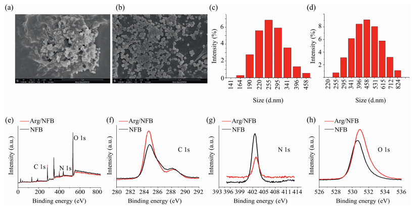
Characterization of Arg/NFB nanocomplex: (a) SEM images of NFB; (b) SEM images of Arg/NFB nanocomplex; (c) DLS analysis of NFB; (d) DLS analysis of Arg/NFB nanocomplex; (e–h) XPS analysis of NFB and Arg/NFB nanocomplex.
L-Arginine/nanofish bone nanocomplex enhances bone regeneration via antioxidant activities and osteoimmunomodulatory properties
Liping Huang , Jianhua Zhang , Xianhu Liu , Tianbao Zhao , Zhipeng Gu , Yiwen Li

Recently, advances of osteoimmunology provide various strategies for the development of bone biomaterials which can regulate bone regenerated microenvironment to promote osteogenesis [1, 2]. As an exogenous substance, a bone biomaterial with favorable osteoimmunomodulatory properties would activate the bone immune system and thus benefit for bone regeneration process [3, 4]. In previous studies, various bone biomaterials with osteoimmunomodulatory properties have been investigated as candidates and exhibited their potential [5, 6]. For instance, Xiao group have demonstrated physicochemical properties of various bone biomaterials including surface and host properties were relevant with the osteoimmunomodulatory properties for bone regeneration [7]. Our previous investigations also found biomass-derived biomaterials such as Tofu and nanofish bone (NFB) also can play osteoimmunomodulatory properties that are great values for bone tissue regeneration [8, 9]. However, in these studies, the bone defect induced reactive oxygen species (ROS) excessive in the early stage would disrupt the intracellular redox status and eventually inducing apoptosis, which might elicit detrimental to the bone regeneration [10-12]. This point is worth noting that excessive ROS is associated with immune response would increase the difficulty of bone biomaterials for modulate the microenvironment [13, 14]. Normally, antioxidant agents with excellent free radical scavenging capacity have been considered as the main strategy against excessive ROS damage [15, 16]. Therefore, it could be hypothesized that designing of bone biomaterials with ROS scavenging activities and suitable osteoimmunomodulatory properties would be a promising way for bone regeneration.
Hence, L-arginine/nanofish bone (Arg/NFB) nanocomplex has been developed to attenuate oxidative stress in the early stage, and then regulate bone immune microenvironment for bone regeneration (Scheme 1). The L-arginine (Arg) has exhibited good biocompatibility property as an antioxidant in previous studies [17, 18]. Nanofish bone with abundant mineral elements such as magnesium (Mg), calcium (Ca), phosphorus (P) is rich in proteins, lipids and other nutrients which could be a potential biomaterial for bone regeneration [19]. Besides, Arg/NFB nanocomplex could be fabricated via a simple method via electrostatic force between the Arg and NFB.
Quantification by dynamic light scattering (DLS) revealed that the negative zeta-potential of NFB and Arg/NFB nanocomplex were -33.9 ± 0.5 mV and -18.4 ± 0.5 mV. These results indicated the Arg and NFB could be successfully prepared the Arg/NFB nanocomplex. As shown in Figs. 1a and b, the Arg/NFB nanocomplex was observed with cuboidal shapes by scanning electron microscopy (SEM) while the NFB with morphologies resembling spheres. The average diameter of the NFB and Arg/NFB nanocomplex was confirmed by DLS measurement (Figs. 1c and d). The results showed that the diameters of the resulting Arg/NFB nanocomplex was around (300~600 nm) bigger than that from NFB (100~300 nm), owing to the additional arginine in the NFB surface. The XPS data of the Arg/NFB nanocomplex are shown in Figs. 1e-h. The survey spectra disclosed that all Arg/NFB nanocomplex composed of C, O and N, whereas the C, O and N identified on the Arg/NFB nanocomplex can be attributed to the Arg successfully binding for the NFB particles. High-resolution XPS spectra of C 1s presented one major peaks at (285.7 eV). N 1s presented one major peak at (399.6 eV) and one peak at (401.1 eV). Additionally, two peaks centered at (530.6 eV) and (531.6 eV) in the high-resolution XPS spectra of O 1s. All these results indicated that the Arg/NFB nanocomplex was successfully fabricated.
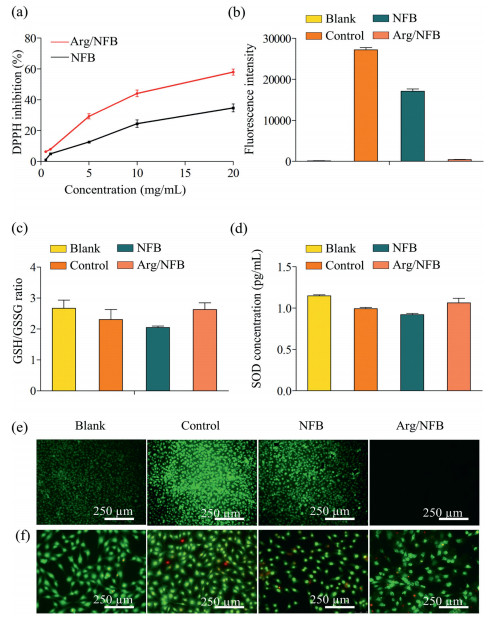
Before in vitro biological evaluations, the cytocompatibility of Arg/NFB nanocomplex cultured with cells was evaluated (Figs. S1a and b in Supporting information). As we know, the generation of ROS is a fundamental aspect of normal human biology, which could influence multiple cell functions such as proliferation, apoptosis [20]. However, uncontrolled ROS production and persistence oxidative stress in the bone defect microenvironment could induce cellular damage, apoptosis and dysfunction thus go against the bone regeneration [21]. To verify the antioxidant ability of Arg/NFB nanocomplex, in vitro DPPH assay was first examined and showed that the Arg/NFB nanocomplex scavenged DPPH radical capacity was increased with the concentration of the Arg/NFB nanocomplex increased (Fig. 2a). Moreover, the intracellular antioxidant ability was invested by using hydrogen peroxide (H2O2) as an exogenous ROS source to assess the oxidative damage suffered by NIH 3T3 cells cultured with samples. As shown in Fig. 2e, the intensity of 2′, 7′-dichlorofluorescein (DCF) fluorescence of H2O2 treated group emitted stronger green fluorescence under oxidative conditions. In contrast, the Arg/NFB nanocomplex group revealed significantly attenuated compared with NFB pretreated group and H2O2 treated group, indicating that Arg/NFB nanocomplex can alleviate oxidative stress induced by H2O2. Furthermore, the cellular ROS production was also quantitatively analyzed by flow cytometry. Fig. 2b indicated that the Arg/NFB nanocomplex can effectively scavenge intracellular ROS levels in NIH 3T3 cells. As revealed in Figs. 2c and d, the levels of SOD and GSH/GSSG were significantly decreased compared with H2O2 treated group and NFB group. These results suggested that the Arg/NFB nanocomplex might protect NIH 3T3 cells against injury induced by H2O2 through increasing the expression of the antioxidant enzymes SOD and GSH/GSSG. To study the cell damage induced by H2O2, cell apoptosis assay was also an important factor. AO/EB staining was used to demonstrate the morphological changes of the nucleolus, which is typical of early apoptosis of NIH 3T3 cells following treatment with H2O2. Fig. 2f revealed that untreated cells were stained with uniform dark green fluorescence which indicated the chromatin evenly distributed in the nucleolus. However, after H2O2 treatment, the Arg/NFB nanocomplex group displayed nucleolus pyknosis and congregated chromatin, which emitting very stronger bright fluorescence, the early phenomena of apoptosis. It suggested that the Arg/NFB nanocomplex had excellent anti-apoptosis capability against ROS damage. All these results indicated that the Arg/NFB nanocomplex has a good antioxidant ability which may be further influence the osteoimmunomodulatory properties.
As mentioned above, the introduction of Arg onto NFB can make the sample have good free radical scavenging ability. To further explore whether the antioxidant properties benefit for immunoregulation in the initial stage after bone defect thus beneficial bone regeneration. Macrophages (RAW264.7 cells) have been used as immune regulate cell line. As revealed in Fig. 3a, macrophages revealed different cell morphologies on the NFB or Arg/NFB nanocomplex surfaces. Macrophages grown on the NFB surfaces showed more rounded with pseudopodia while were slenderer spindle shape and pseudopodia in Arg/NFB nanocomplex group. To further investigate the influence of Arg/NFB nanocomplex on macrophages, the relevant cytokines expression of macrophages on materials was studied by ELISA and RT-qPCR. As showed in Fig. 3b, the concentration of IL-18 had no significant difference in all the groups. The protein concentration of IL-10 on Arg/NFB nanocomplex was increased compared with NFB and control groups, while the concentration of TNF-α and IL-6 was decreased. The change of cytokines level by RT-qPCR in Fig. 3c demonstrated that the Arg/NFB nanocomplex down-regulating pro-inflammatory cytokines TNF-α, IL-18, IL-6 and OSM compared with the NFB group and control group. Otherwise, the IL-1β cytokine and anti-inflammatory cytokine (IL-10) were up-regulating. Altogether, the above results indicated that the Arg/NFB nanocomplex can decrease the overproduction of ROS, down-regulate some disadvantage inflammatory cytokines and up-regulate some advantage cytokines thus might promote bone formation process.
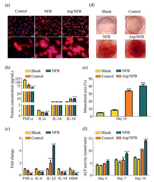
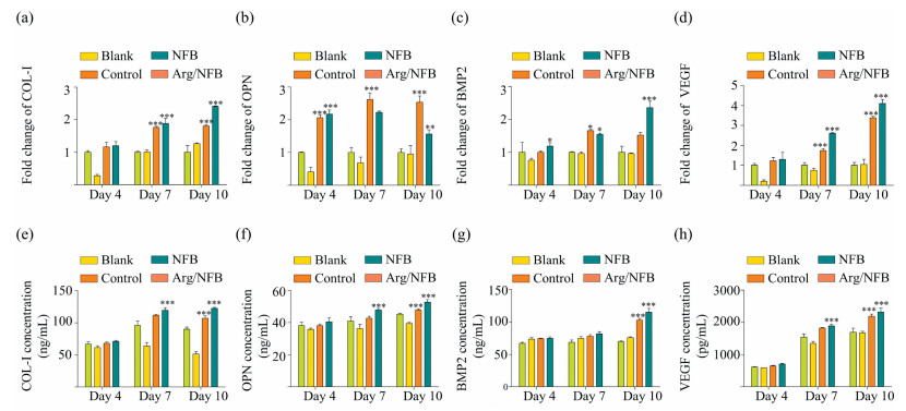
To verify whether the change of immune microenvironment beneficial bone regeneration, the osteogenic differentiation of MC3T3-E1 cells in different immune microenvironments was evaluated. Figs. 3d and e of Alizarin Red S indicated that there was no obvious difference between the blank and control groups during the formation of mineralized nodules after 10 days. However, there were more calcified nodules in the Arg/NFB nanocomplex group than in the NFB group. Fig. 3f revealed that the expression of ALP on the Arg/NFB nanocomplex group was higher than the NFB group or control group at days 4, 7, 10. Besides, the ALP expression in the control groups was lower than in other groups. As is known, ALP is a byproduct of osteoblast activity, which increases during bone formation. Therefore, the higher ALP activity of MC3T3-E1 cells treated with Arg/NFB nanocomplex group suggests higher osteogenic differentiation activity. To deeply study the effect of Arg/NFB nanocomplex on MC3T3-E1 cells, the expression of osteogenic genes (BMP2, COL-I, and OPN) was detected by RT-qPCR on days 4, 7 and 10. As demonstrated in Figs. 4a-c, the expression level of osteogenic genes significantly increased in MC3T3-E1 treated with Arg/NFB nanocomplex as compared with the NFB group after incubation 10 days. Besides, cells in the Arg/NFB nanocomplex group expressed the higher osteogenic relative protein expression (Figs. 4e-g). In conclusion, the immune microenvironment treated with Arg/NFB nanocomplex exerted the greatest influences on osteogenic differentiation, which further proved that the macrophages cultured on Arg/NFB nanocomplex could facilitate the macrophages to produce a number of immune cytokines and demonstrated the optimal immunomodulatory osteogenic effect. Interestingly, the VEGF (a critical factor regulating neovascularization) expression of HUVECs treated with Arg/NFB nanocomplex was higher than other groups (Figs. 4d and h) that also beneficial the angiogenic differentiation of HUVECs to enhance bone regeneration. All these results indicated that the Arg/NFB nanocomplex has good antioxidant and osteoimmunomodulatory capacity, would play a critical role in bone regeneration.
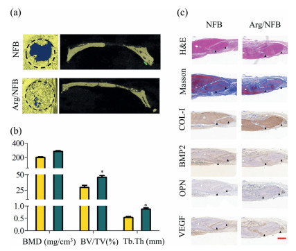
Finally, in vivo study was performed to further confirm the intervention effects of Arg/NFB nanocomplex on bone regeneration. A 5 mm diameter craniotomy defect was created on each side of the rat skull (rat weighing 250~280 g). First, H & E and Masson trichrome staining of the bone defect initial showed the bone regeneration at 4, 8 weeks (Fig. 5 and Fig. S2 in Supporting information). The results displayed in Fig. 5 showed the Arg/NFB nanocomplex groups had a better new bone formation and collagen deposition than NFB groups, which indicated the Arg/NFB nanocomplex have a better promoting effect on bone regeneration. The immunohistochemical analysis next proved the activity of bone formation because of the expression of COL-I, BMP2, OPN and VEGF. Further immunohistochemical analysis was applied to evaluate the microenvironment of Arg/NFB nanocomplex caused. The results demonstrated that the immune results of IL-6 have a milder response in the Arg/NFB nanocomplex group. However, the anti-inflammatory cytokines IL-10 and IL-1β expression were increased with Arg/NFB nanocomplex groups (Fig. S3 in Supporting information). At the same time, the micro-CT images (Fig. 5a) clearly showed that the bone regeneration was significantly better in Arg/NFB nanocomplex group than the NFB group. Further quantitative analysis of the micro-CT data confirmed that the three indexes reflecting new bone formation (bone mineral density (BMD), bone volume fraction (BV/TV), and trabecular thickness (Tb.Th)) in the Arg/NFB nanocomplex group were higher than those in the NFB group (Fig. 5b). All the results demonstrated that Arg/NFB nanocomplex could improve the osteogenesis performance of NFB based on the combination of antioxidant activities with osteoimmunomodulatory properties.
In summary, a Arg/NFB nanocomplex has been developed via electrostatic force between the L-arginine and nanofish bone in aqueous solution. The results revealed that the size of arginine/nanofish bone nanocomplex was around (300~600 nm), and showed cuboidal shapes. In vitro antioxidant experiments demonstrated that the Arg/NFB nanocomplex had antioxidant ability. Notably, the Arg/NFB nanocomplex could modulate the polarization of nonactivated macrophage to different types and induce the secretion of a series of pre-inflammatory, anti-inflammatory and osteogenic cytokines. Additionally, the regulated immune microenvironment can enhance the osteogenic differentiation of MC3T3-E1 cells and improve the angiogenic activity of HUVECs cells, leading to the increase of mineralized nodules, alkaline phosphatase activity and angiogenic. In vivo experiments result further demonstrated that the antioxidant effect of arginine on the Arg/NFB nanocomplex can accelerate the new bone formation and enhance angiogenesis compare to NFB groups. Our results indicated that the Arg/NFB nanocomplex with antioxidant activities and immunomodulatory properties could be a promising bone biomaterial for developing the next generation of bone regeneration.
The authors declare that they have no known competing financial interests or personal relationships that could have appeared to influence the work reported in this paper.
This study was supported by the Science and Technology Planning Project of Shenzhen (No. JCYJ20180307163534533) and the Program of the Science, Technology Department of Guangzhou (No. 201803020039).
H. Takayanagi, Nat. Rev. Immunol. 7 (2007) 292-304. doi: 10.1038/nri2062
V. Domazetovic, G. Marcucci, T. Iantomasi, et al., Clin. Cases Miner. Bone Metab. 14 (2017) 209-216. doi: 10.11138/ccmbm/2017.14.1.209
W. Liu, J. Li, M. Cheng, et al., Adv. Sci. 5 (2018) 749-1800.
K. Okamoto, T. Nakashima, M. Shinohara, et al., Physiol. Rev. 97 (2017) 1295-1349. doi: 10.1152/physrev.00036.2016
Z. Chen, T. Klein, R.Z. Murray, et al., Mater. Today. 658 (2015) 1369-7021.
H. Kang, S. Kim, D.S.H. Wong, et al., Nano Lett. 17 (2017) 6415-6427. doi: 10.1021/acs.nanolett.7b03405
Z. Chen, A. Bachhuka, S. Han, et al., ACS Nano 11 (2017) 4494-4506. doi: 10.1021/acsnano.6b07808
K. Huang, Z. Gu, J. Wu, ACS Biomater. Sci. Eng. 6 (2020) 3037-3045. doi: 10.1021/acsbiomaterials.9b01997
L. Huang, J. Zhang, Z. Gu, ACS Biomater. Sci. Eng. 6 (2020) 3270-3274. doi: 10.1021/acsbiomaterials.0c00443
A. Sandukji, H. Al-Sawaf, A. Mohamadin, et al., Hum. Exp. Toxicol. 30 (2011) 435-442. doi: 10.1177/0960327110374203
Z. Gu, B. Huang, Y. Li, et al., Mater. Sci. Eng. C: Mater. Biol. Appl. 61 (2016) 526-533. doi: 10.1016/j.msec.2015.12.077
A.H.S. Wallner, B. Mayer, T.C. Wascher, Eur. J. Clin. Invest. 31 (2001) 98-102. doi: 10.1046/j.1365-2362.2001.00771.x
L. Cui, J. Zhang, J. Zou, et al., Biomaterials 230 (2020) 119-617.
C. Xian, Z. Gu, G. Liu, et al., Chin. Chem. Lett. 31 (2020) 1612-1615. doi: 10.1016/j.cclet.2019.09.011
Z. Li, H. Li, J. Zhang, et al., Chinese J. Polym. Sci. 31 (2020) 1149-1156.
J. Ding, J. Zhang, J. Li, et al., Prog. Poly. Sci. 90 (2020) 1-34.
T. Zhang, J. Yao, J. Tian, et al., Chin. Chem. Lett. 31 (2020) 1129-1132. doi: 10.1016/j.cclet.2019.07.010
T. Yin, J.W. Park, Food Chem. 180 (2015) 42-47. doi: 10.1016/j.foodchem.2015.02.021
T. Yin, J.W. Park, S. Xiong, LWT-Food Sci. Technol. 64 (2015) 367-373. doi: 10.1016/j.lwt.2015.06.007
M.N. Sack, F.Y. Fyhrquist, O.J. Saijonmaa, et al., J. Am. Coll. Cardiol. 70 (2017) 196-211. doi: 10.1016/j.jacc.2017.05.034
H. Cui, L. Cui, P. Zhang, et al., Macromol. Biosci. 14 (2014) 440-450. doi: 10.1002/mabi.201300366

Figure 1 Characterization of Arg/NFB nanocomplex: (a) SEM images of NFB; (b) SEM images of Arg/NFB nanocomplex; (c) DLS analysis of NFB; (d) DLS analysis of Arg/NFB nanocomplex; (e–h) XPS analysis of NFB and Arg/NFB nanocomplex.

Figure 2 Antioxidant potential characterization: (a) DPPH radical scavenging activity of NFB and Arg/NFB nanocomplex; (b) The fluorescence intensity results of cells treated with H2O2 were determined by flow cytometry analysis; (c) Alterations of GSH/GSSG in NIH 3T3 cells treated with H2O2 after 24 h; (d) SOD activity induced by after 24 h of incubation with 100 μmol/L H2O2; (e) Fluorescence images of intracellular ROS of NIH 3T3 cells grown onto NFB and Arg/NFB nanocomplex, staining with DCFH-DA; (f) AO/EB staining test for the discrimination of apoptosis vs. necrotic cells after treatment with selected fractions.

Figure 3 Inflammatory response of macrophages on NFB or Arg/NFB nanocomplex: (a) The cell morphology of macrophages on NFB or Arg/NFB nanocomplex was stained with DAPI (blue, nuclei) and rhodamine phalloidin (red, cytoskeleton); (b) Concentrations of TNF-α, IL-6, IL-10 and IL-18 in the culture medium was measured by ELISAs; (c) The expression of inflammatory cytokines TNF-α, IL-6, IL-18, OSM, and IL-1β was detected by RT-qPCR; (d) Alizarin Red S staining images of MC3T3-E1 cultured for 10 days; (e) ImageJ analysis of the mineralized area; (f) ALP activity assay of MC3T3-E1 cultured for 4, 7, 10 days. *P < 0.05, **P < 0.05, ***P < 0.05 as compared to the blank group.

Figure 4 In vitro osteogenic differentiation of MC3T3-E1 and angiogenic of HUVECs by the stimulation of NFB or Arg/NFB nanocomplex conditioned media: (a-d) Effects on COL-I, OPN, BMP2 and VEGF synthesis in MC3T3-E1 or HUVECs after 4, 7, 10 days; (e-h) Concentrations of COL-I, OPN, BMP2 and VEGF in the culture medium were measured by ELISAs. *P < 0.05, **P < 0.05, ***P < 0.05 as compared to the blank group.

 扫一扫看文章

扫一扫关注我们
 DownLoad:
 下载:
下载:

 下载:
下载: