图 1
Synthesis of probe 1 for detection of H2S.
Figure 1.
Synthesis of probe 1 for detection of H2S.
Cascade reaction-based fluorescent probe for detection of H2S with the assistance of CTAB micelles
Hai-Rong Zheng , Li-Ya Niu , Yu-Zhe Chen , Li-Zhu Wu , Chen-Ho Tung , Qing-Zheng Yang

For centuries, hydrogen sulfide has been recognized as a toxic molecular. When exposed to this colorless, flammable gas, which has a distinctive smell of rotten eggs, people may suffer from respiratory failure, loss of consciousness, sudden cardiac death, hepatic and olfactory paralysis. However, more recent studies have challenged this traditional view. H2S is now identified as the third biological signaling molecular besides nitric oxide (NO) and carbon monoxide (CO) and plays important roles in maintaining normal physiology [1]. Endogenous H2S can be produced from sulfur- containing biomolecules such as cysteine and homocysteine, which is catalyzed by cystathionine beta synthase (CBS) , cysteine aminotransferase and mercaptopyruvate sulfurtransferase (CAT/ MST) [2, 3]. These enzymes are widely spread in human tissues ranging from the heart and vasculature, brain, kidney, liver, lungs, indicating the important physiological roles of H2S in the body. Besides, H2S can also be produced from non-enzymatic processes, including release from sulfur stores and metabolism of polysulfide [4, 5]. The abnormal level of H2S can resulted in many diseases, such as Alzheimer's disease, Down's syndrome, diabetes, and liver cirrhosis [6-9]. Therefore, methods for monitoring the production, trafficking, and consumption of H2S in living systems are highly desired.
Compared with the traditional methods including colorimetric assays [10, 11] and gas chromatography[12, 13], fluorescent probes present lots of advantages, such as rapid response, high sensitivity and excellent selectivity [14-16]. Recently, several fluorescent probes have been reported forthe detection ofH2S in living systems [17-20]. Common strategies include: H2S mediated reduction of azide to amine [21, 22], H2S trapped by nucleophilic addition [23-25], copper sulfide precipitation [26, 27], and thiolysis of dinitro- phenyl ether [28, 29]. Among these, H2S trapped by nucleophilic addition strategy has been widely applied. Fluorescent probes based on this strategy usually take advantage of the dual nucleophilicity of H2S. Such probes usually contain a H2S trap group with two electrophilic reaction sites and a fluorescent reporter, which discriminates the H2S from other biothiols.
Recently, excited state intramolecular proton transfer (ESIPT) compounds have been widely used in designing sensors for anions [30, 31], cations [32, 33], amino acids [34-36] and small neutral molecules [37, 38], because of their intrinsic properties, ultra-fast reaction rate and huge bathochromic shift in the emission signal.
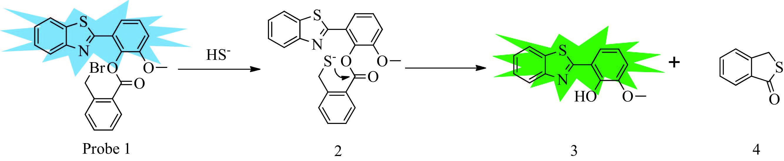
Herein, we reported a fluorescent probe using 2-2 (hyroxyphe- nyl) benzothiazole (HBT) , a typical ESIPT molecule, as fluorescent reporter, and 4- (bromomethyl) benzoate as trap group, which showed fast response to H2S through cascade reaction. As the hydroxyl group was protected by 2- (bromomethyl) benzoate, the HBT moiety of the probe showed enol-like fluorescence. The initial nucleophilic attack of H2S towards bromomethyl group would lead to an intermediate thiol, which was followed by a cyclization cascade reaction towards the adjacent ester carbonyl to release the HBT with keto emission. Thus, the detection of H2S was realized.
Probe 1 was synthesized using a simple procedure with HBT and 2- (bromomethyl) benzoic acid as starting materials, 1-ethyl-3- (3-dimethylaminopropyl) carbodiimide hydrochloride (EDCI) as a coupling reagent, and 4-dimethylaminopyridine (DMAP) as catalyst (Scheme 1) .
2- (2'-Hydroxy-3'-methoxyphenyl) benzothiazole (HMBT) : A solution of 2-aminothiophenol (0.3 mL, 4.2 mmol) and o-vanillin (0.48 g, 3.15 mmol) in EtOH (10 mL) , aq. H2O2 (30%, 18.9 mmol) and aq HCl (37%, 9.45 mmol) was stirred at rt for 90 min. The solution was quenched by 10 mL H2O. The precipitate was filtered, dried under vacuum and recrystallized from EtOH to afford the desired product as a light brown solid (0.64 g, 79% yield) . 1H NMR (CDCl3, 400 MHz, ppm) : d 12.75 (s, 1H) , 8.01 (d, 1H, J = 7.6 Hz) , 7.91 (d, 1H, J = 7.2 Hz) , 7.51 (t, 1H, J = 6.8 Hz) , 7.42 (t, 1H, J = 7.2 Hz) , 7.33 (dd, 1H, J1 = 1.2Hz, J2 = 8.0Hz) , 6.99 (dd, 1H, J1 = 1.2Hz, J2 = 8.0Hz) , 6.91 (t, 1H, J =8.0 Hz) , 3.96 (s, 1H) .
Probe 1: To a solution of EDCI (0.346 g, 1.5 mmol) in CH2Cl2 (20 mL) was added 2-bromodomethylbenzoic acid (0.312 g, 1.5 mmol) followed by DMAP (0.012 g, 0.1 mmol) and HMBT (0.346 g, 1 mmol) . The mixture was stirred at room temperature for 12 h and then filtered, washed with water. The filtrate was concentrated to afford the crude product, then purified by silica gel column chromatography (PE:CH2Cl2 = 1:1) to afford compound 1 as a white solid (0.24 g, 40%) . 1H NMR (400 MHz, CDCl3, ppm) : d 8.41 (d, 1H, J = 4.0 Hz) , 7.93-7.99 (m, 2H) , 7.83 (d, 1H, J = 4.0 Hz) , 7.34-7.56 (m, 6H) , 7.15 (d, 1H, J = 4.0 Hz, ) , 4.94-5.14 (d, 2H) , 3.90 (s, 3H) . 13C NMR (100 MHz, CDCl3) : 164.2, 162.3, 153.0, 152.1, 140.0, 139.8, 138.1, 135.5, 134.0, 133.3, 132.3, 132.0, 130.9, 129.0, 128.7.128.5.128.2.127.7.126.9.126.3.125.8.125.3.123.4.122.1, 121.1, 121.5, 121.4, 114.2, 69.6, 56.4, 44.1, 31.0. HRMS: calcd.: 454.010, found: 454.011.
The proposed sensing mechanism is shown in Fig. 1. As the hydroxyl group is protected by 2- (bromomethyl) benzoate, the excited state intramolecular proton transfer (ESIPT) process was forbidden. As a result, the HBT moiety of probe 1 shows enol-like fluorescence. The initial nucleophilic attack of H2S towards bromomethyl group would lead to an intermediate thiol, which is followed by a cyclization cascade reaction towards the adjacent ester carbonyl to release the HBT. Upon irradiation, the resulting HBT generated the excited state intramolecular proton transfer (ESIPT) tautomer, which shows keto emission. To identify the proposed sensing mechanism, probe 1 was treated with excess NaHS and Et3N in CH3CN. After the reaction, the HBT was released, with the formation of cyclization product 4. All products were separated and confirmed with 1H NMR (Figs. S1 and S2 in Supporting information) .
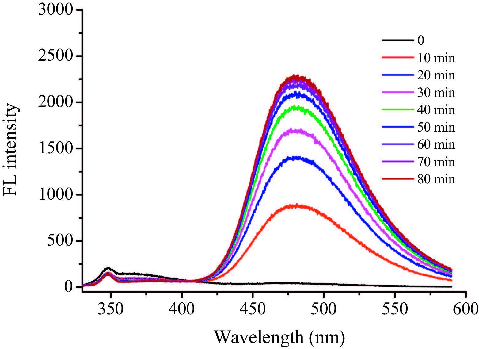
We first tested the absorption spectra of probe 1 in HEPES buffer. However, the obvious variation of the absorption spectra of 1 in 12 h suggested that it was unstable in HEPES buffer, probably due to its poor solubility in pure water (Fig. S3 in Supporting information) . Then we used 50% ethanol as co-solvent, and the stability of 1 was improved (Fig. S4 in Supporting information) . As shown in Fig. S5 in Supporting information, with the addition of 200 mmol/L NaHS in ethanol/HEPES buffer (1:1, v/v, 20 mmol/L, pH 7.4) , the emission at 350 nm (which is attributed to the enol form) decreased, followed by a new peak appearing at 484 nm (which is attributed to the keto form) . However, the fluorescence at 484 nm was quite weak, as the intramolecular hydrogen bond is strongly disturbed in polar solvents [39]. We speculated that the ESIPT would be enhanced in the nonpolar core of CTAB micelles. Probe 1 was stable in HEPES buffer (20 mmol/L, pH 7.4) containing 1 mmol/L CTAB (Fig. S6 in Supporting information) . As we expected, upon addition of NaSH to probe 1, the original emission at 350 nm decreased, and a significant fluorescence enhancement at 484 nm of approximately 60-fold was observed (Fig. 2) . In contrast, an enhancement of only 5-fold was observed in ethanol/ HEPES buffer (1:1, v/v, 20 mmol/L, pH 7.4) (Fig. S5) . The results indicated that ESIPT process was greatly enhanced with the assistance of CTAB micelles. Therefore, the following studies were carried out in HEPES buffer (20 mmol/L, pH 7.4, containing 1 mmol/L CTAB) . The probe 1 (10 mmol/L) upon reaction with NaHS (2 mmol/L) in HEPES buffer (20 mmol/L, pH 7.4, containing 1 mmol/L CTAB) exhibited a pseudo first order reaction kinetics with rate constant k = 0.59 min-1 (t1/2 = 1.17 min) , indicating the fast response of 1 towards H2S (Fig. S7 in Supporting information) .
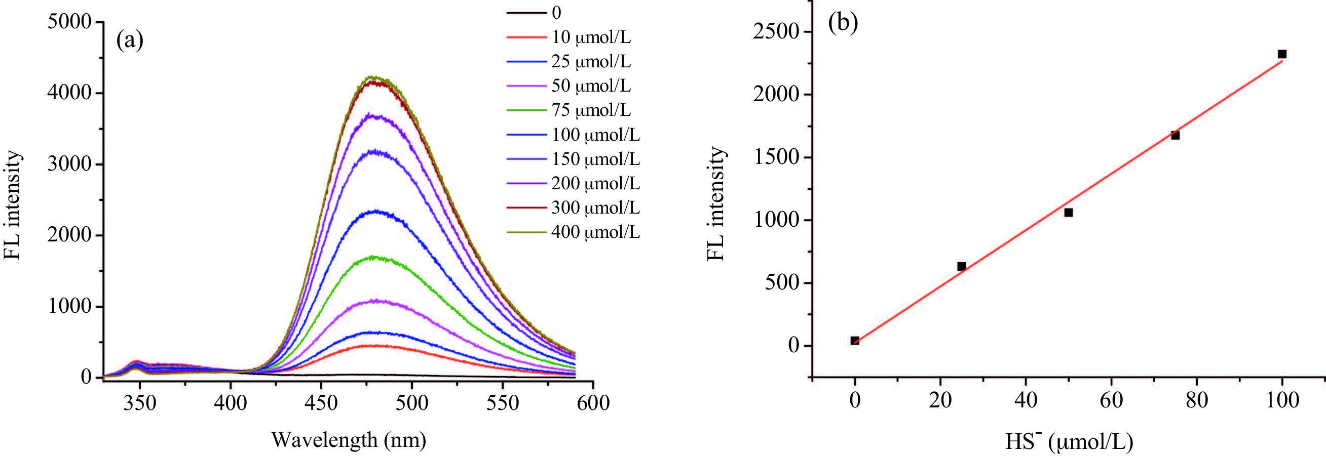
Subsequently, quantitative response of probe 1 in HEPES buffer (20 mmol/L, pH 7.4, containing 1 mmol/L CTAB) towards H2S was estimated. As shown in Fig. 3a, with the increasing concentrations of NaHS, the original emission at 350 nm decreased, and a significant fluorescence enhancement at 484 nm was observed. Moreover, the fluorescence intensity of 484 nm showed linear relationship with NaHS concentrations ranging from 0 to 100 mmol/L, suggesting the potential application for quantitative determination of H2S (Fig. 3b) . The detection limit for HS- was estimated to be 0.50 mmol/L (S/N = 3) , which is much lower than the concentration required to cause physiological response [40].
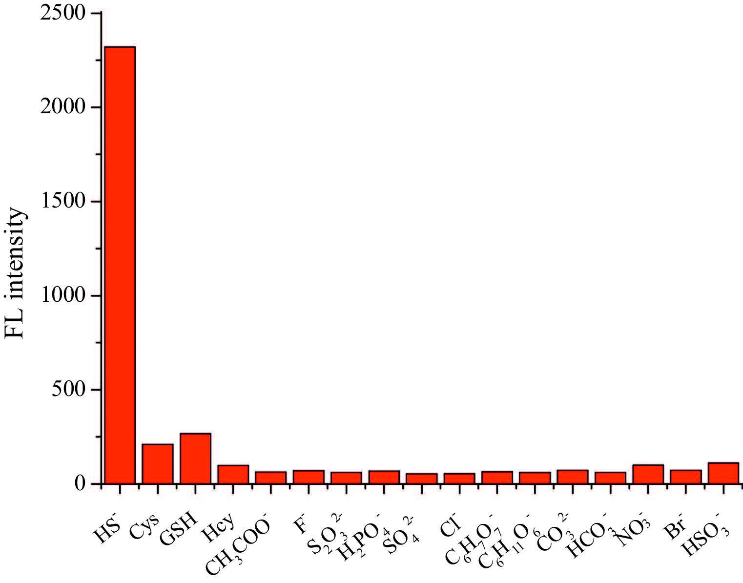
To evaluate the selectivity of the probe to H2S, emission spectra changes upon addition of 20 equivalents of different interfering species, such as cysteine (Cys) , glutathione (GSH) , homocysteine (Hcy) , sodium acetate (CH3COONa) , sodium fluoride (NaF) , sodium persulfate (Na2S2O4) , sodium dihydrogen phosphate (NaH2PO4) , sodium sulfate (Na2SO4) , sodium chloride (NaCl) , sodium tartrate (C4H4Na2O6) , sodium gluconate (C6H11NaO7) , sodium carbonate (Na2CO3) , sodium bicarbonate (NaHCO3) , sodium nitrate (NaNO3) , sodium bromide (NaBr) , sodium hydrogen sulfite (NaHSO3) were studied. As shown in Fig. 4, in most cases, little change in emission intensity was observed. In contrast, a great enhancement in emission intensity was only observed when adding NaHS. Thus, these results demonstrated that probe 1 has a high selectivity towards H2S.
We further examined the capability of probe 1 to H2S in living cells (Fig. 5) . After incubated with probe 1 (20 mmol/L) for 45 min in culture medium, HeLa cells showed almost no fluorescence in green channel. By contrast, when HeLa cells were pretreated with NaHS (200 mmol/L) before incubated with probe 1 (20 mmol/L) for 45 min, the obvious fluorescence was observed. It revealed that probe 1 has potential for visualizing H2S levels in living cells.

In conclusion, we designed and synthesized a novel fluorescent probe 1 for the detection of H2S based on ESIPT mechanism with the assistance of CTAB. As the hydroxyl group is protected by 2- (bromomethyl) benzoate, probe 1 showed weak enol-like fluorescence at 350 nm. The 2- (bromomethyl) benzoate group showed fast response to H2S through cascade reaction and released free HBT, which showed strong fluorescence at 484 nm in CTAB micelles. The emission at 484 nm showed linear relationship with the H2S concentration at 0-100 mmol/Lwith the detection limit of 0.50 mmol/L. The high selectivity and sensitivity of our probe to H2S may give new insights for the development of fluorescent probe to selectively detect H2S in biological systems.
Supplementary data associated with this article can be found, in theonlineversion, at http://dx.doi.org/10.1016/j.cclet.2016.04.023.
C. Szabó. Hydrogen sulphide and its therapeutic potential[J]. Nat. Rev. Drug. Discov., 2007, 6: 917-935. doi: 10.1038/nrd2425
M. Ishigami, K. Hiraki, K. Umemura. A source of hydrogen sulfide and a mechanism of its release in the brain,[J]. Antioxid. Redox Signal., 2009, 11: 205-214. doi: 10.1089/ars.2008.2132
G.A. Benavides, G.L. Squadrito, R.W. Mills. Hydrogen sulfide mediates the vasoactivity of garlic[J]. Proc. Natl. Acad. Sci. U.S.A., 2007, 104: 17977-17982. doi: 10.1073/pnas.0705710104
S. Singh, D. Padovani, R.A. Leslie, T. Chiku, R. Banerjee. Relative contributions of cystathionine b-synthase and g-cystathionase to H2S biogenesis via alternative trans-sulfuration reactions[J]. J. Biol. Chem., 2009, 284: 22457-22466. doi: 10.1074/jbc.M109.010868
S. Singh, R. Banerjee. PLP-dependent H2S biogenesis[J]. Biochim. Biophys. Acta, 2011, 1814: 1518-1527. doi: 10.1016/j.bbapap.2011.02.004
K. Eto, T. Asada, K. Arima, T. Makifuchi, H. Kimura. Brain hydrogen sulfide is severely decreased in Alzheimer's disease[J]. Biochem. Biophys. Res. Commun., 2002, 293: 1485-1488. doi: 10.1016/S0006-291X(02)00422-9
P. Kamoun, M.-C. Belardinelli, A. Chabli, K. Lallouchi, B. Chadefaux-Vekemans. Endogenous hydrogen sulfide overproduction in Down syndrome[J]. Am. J. Med. Genet. A, 2003, 116: 310-311.
W. Yang, G.D. Yang, X.M. Jia, L.Y. Wu, R. Wang. Activation of KATP channels by H2S in rat insulin-secreting cells and the underlying mechanisms[J]. J. Physiol. (Lond.), 2005, 569: 519-531. doi: 10.1113/jphysiol.2005.097642
S. Fiorucci, E. Antonelli, A. Mencarelli. The third gas:H2S regulates perfusion pressure in both the isolated and perfused normal rat liver and in cirrhosis[J]. Hepatology, 2005, 42: 539-548. doi: 10.1002/(ISSN)1527-3350
L.M. Siegel. A direct microdetermination for sulfide[J]. Anal. Biochem., 1965, 11: 126-122. doi: 10.1016/0003-2697(65)90051-5
E. Fischer. Formation of methylene blue in response to hydrogen sulfide[J]. Ber. Dtsch. Chem. Ges., 1983, 16: 2234-2236.
U. Hannestad, S. Margheri, B. Sö rbo. A sensitive gas chromatographic method for determination of protein associated sulfur[J]. Anal. Biochem., 1989, 178: 394-398. doi: 10.1016/0003-2697(89)90659-3
J. Furne, A. Saeed, M.D. Levitt. Whole tissue hydrogen sulfide concentrations are orders of magnitude lower than presently accepted values[J]. Am. J. Physiol. Regul. Integr. Comp. Physiol., 2008, 295: 1479-1485. doi: 10.1152/ajpregu.90566.2008
L.Y. Niu, Y.Z. Chen, H.R. Zheng. Design strategies of fluorescent probes for selective detection among biothiols[J]. Chem. Soc. Rev., 2015, 44: 6143-6160. doi: 10.1039/C5CS00152H
X. Zhou, S. Lee, Z.C. Xu, J. Yoon. Recent progress on the development of chemosensors for gases[J]. Chem. Rev., 2015, 115: 7944-8000. doi: 10.1021/cr500567r
L.Y. Niu, Y.S. Guan, Y.Z. Chen. BODIPY-based ratiometric fluorescent sensor for highly selective detection of glutathione over cysteine and homocysteine[J]. J. Am. Chem. Soc., 2012, 134: 18928-18931. doi: 10.1021/ja309079f
V.S. Lin, C.J. Chang. Fluorescent probes for sensing and imaging biological hydrogen sulfide[J]. Curr. Opin. Chem. Biol., 2012, 16: 595-601. doi: 10.1016/j.cbpa.2012.07.014
C.C. Zhao, X.L. Zhang, K.B. Li. Fö rster resonance energy transfer switchable self-assembled micellar nanoprobe:ratiometric fluorescent trapping of endogenous H2S generation via fluvastatin-stimulated upregulation[J]. J. Am. Chem. Soc., 2015, 137: 8490-8498. doi: 10.1021/jacs.5b03248
X. Wang, J. Sun, W.H. Zhang. A near-infrared ratiometric fluorescent probe for rapid and highly sensitive imaging of endogenous hydrogen sulfide in living cells[J]. Chem. Sci., 2013, 4: 2551-2556. doi: 10.1039/c3sc50369k
Z.S. Wu, Y.L. Feng, B. Geng, J.Y. Liu, X.J. Tang. Fluorogenic sensing of H2S in blood and living cells via reduction of aromatic dialkylamino N-oxide[J]. RSC Adv., 2014, 4: 30398-30401. doi: 10.1039/C4RA03677H
H.J. Peng, Y.F. Cheng, C.F. Dai. A fluorescent probe for fast and quantitative detection of hydrogen sulfide in blood[J]. Angew. Chem. Int. Ed., 2011, 50: 9672-9675. doi: 10.1002/anie.201104236
S. Chen, Z.J. Chen, W. Ren, H.W. Ai. Reaction-based genetically encoded fluorescent hydrogen sulfide sensors[J]. J. Am. Chem. Soc., 2012, 134: 9589-9592. doi: 10.1021/ja303261d
K.J. Wu, G.Q. Li, Y. Li, L.X. Dai, S.L. You. Reaction-based genetically encoded fluorescent hydrogen sulfide sensors[J]. Chem. Commun., 2011, 47: 493-495. doi: 10.1039/C0CC01769H
C.R. Liu, J. Pan, S. Li. Capture and visualization of hydrogen sulfide by a fluorescent probe[J]. Angew. Chem. Int. Ed., 2011, 50: 10327-10329. doi: 10.1002/anie.201104305
C.R. Liu, B. Peng, S. Li. Reaction based fluorescent probes for hydrogen sulfide[J]. Org. Lett., 2012, 14: 2184-2187. doi: 10.1021/ol3008183
K. Sasakura, K. Hanaoka, N. Shibuya. Development of a highly selective fluorescence probe for hydrogen sulfide[J]. J. Am. Chem. Soc., 2011, 133: 18003-18005. doi: 10.1021/ja207851s
F.P. Hou, L. Huang, P.X. Xi. A retrievable and highly selective fluorescent probe for monitoring sulfide and imaging in living cells[J]. Inorg. Chem., 2012, 51: 2454-2460. doi: 10.1021/ic2024082
X.W. Cao, W.Y. Lin, K.B. Zheng, L.W. He. A near-infrared fluorescent turn-on probe for fluorescence imaging of hydrogen sulfide in living cells based on thiolysis of dinitrophenyl ether[J]. Chem. Commun., 2012, 48: 10529-10531. doi: 10.1039/c2cc34031c
T.Y. Liu, Z.C. Xu, D.R. Spring, J.N. Cui. A lysosome-targetable fluorescent probe for imaging hydrogen sulfide in living cells[J]. Org. Lett., 2013, 15: 2310-2313. doi: 10.1021/ol400973v
S.D. Liu, L.W. Zhang, P.P. Zhou. HBT-based chemosensors for the detection of fluoride through deprotonation process:experimental and DFT studies[J]. RSC Adv., 2015, 5: 19983-19988. doi: 10.1039/C4RA13532F
L.H. Geng, X.F. Yang, Y.G. Zhong, Z. Li, H. Li. "Quinone-phenol" transduction activated excited-state intramolecular proton transfer:a new strategy toward ratiometric fluorescent probe for sulfite in living cells[J]. Dyes Pigm., 2015, 120: 213-219. doi: 10.1016/j.dyepig.2015.04.016
S. Sahana, G. Mishra, S. Sivakumar, P.K. Bharadwaj. A 2-(2[Prime or minute]-hydroxyphenyl)benzothiazole (HBT)-quinoline conjugate:a highly specific fluorescent probe for Hg2+ based on ESIPT and its application in bioimaging[J]. Dalton Trans., 2015, 44: 20139-20146. doi: 10.1039/C5DT03719K
S. Goswami, A. Manna, S. Paul. FRET based ‘red-switch’ for Al3+ over ESIPT based ‘green-switch’ for Zn2+:dual channel detection with live-cell imaging on a dyad platform[J]. RSC Adv., 2014, 4: 34572-34576. doi: 10.1039/C4RA05714G
X.F. Yang, Q. Huang, Y.G. Zhong. A dual emission fluorescent probe enables simultaneous detection of glutathione and cysteine/homocysteine[J]. Chem. Sci., 2014, 5: 2177-2183. doi: 10.1039/c4sc00308j
Q.S. Liu, C.L. Zhang, X.Q. Wang. Benzothiazole-pyimidine-based BF2 complex for selective detection of cysteine[J]. Chem. Asian J., 2016, 11: 202-206. doi: 10.1002/asia.v11.2
C.C. Zhao, X.A. Li, F.Y. Wang. Target-triggered NIR emission with a large stokes shift for the detection and imaging of cysteine in living cells[J]. Chem. Asian J., 2014, 9: 1777-1781. doi: 10.1002/asia.v9.7
S. Goswami, S. Das, K. Aich. A chemodosimeter for the ratiometric detection of hydrazine based on return of ESIPT and its application in live-cell imaging[J]. Org. Lett., 2013, 15: 5412-5415. doi: 10.1021/ol4026759
D.P. Murale, H. Kim, W.S. Choi, D.G. Churchill. Highly selective excited state intramolecular proton transfer (ESIPT)-based superoxide probing[J]. Org. Lett., 2013, 15: 3946-3949. doi: 10.1021/ol4017222
S.M. Aly, A. Usman, M. AlZayer. Solvent-dependent excited-state hydrogen transfer and intersystem crossing in 2-(20-Hydroxyphenyl)-Benzothiazole[J]. J. Phys. Chem. B, 2015, 119: 2596-2603. doi: 10.1021/jp508777h
A.R. Lippert, E.J. New, C.J. Chang. Reaction-based fluorescent probes for selective imaging of hydrogen sulfide in living cells[J]. J. Am. Chem. Soc., 2011, 133: 10078-10080. doi: 10.1021/ja203661j

Figure 2 Time-dependent fluorescence response of probe 1 (10 mmol/L) upon addition of NaHS (200 mmol/L) in HEPES buffer (20 mmol/L, pH 7.4, containing 1 mmol/L CTAB) . λex = 310 nm, λem = 484 nm. Slits: 5/5 nm.

Figure 3 (a) Fluorescence spectra of probe 1 (10 mmol/L) upon addition of NaHS (0-400 mmol/L) in HEPES buffer (20 mmol/L, pH 7.4, containing 1 mmol/L CTAB) . (b) Fluorescence response of probe 1 at 484 nm to NaHS concentration (0-100 mmol/L) . Spectra were recorded after incubation with different concentrations of NaHS for 1 h. λex = 310 nm, λem = 484 nm. Slits: 5/5 nm.

Figure 4 Fluorescence response of probe 1 (10 mmol/L) upon addition of various species in HEPES buffer (20 mmol/L, pH 7.4, containing 1 mmol/L CTAB) . λex = 310 nm, λem = 484 nm. Slits: 5/5 nm.

Figure 5 Images of H2S in HeLa cells using probe 1 (20 mmol/L) at 37 ℃. (a) Fluorescence and (b) bright-field images of HeLa cells incubated with probe 1 for 45 min. (c) Fluorescence and (d) bright-field images of HeLa cells incubated with NaHS (200 mmol/L) for 30 min and further incubated with probe 1 for 45 min. Scale bar: 50 mm.

 扫一扫看文章

扫一扫关注我们
 下载:
下载:

 下载:
下载:
 下载:
下载: