Login In
Application of Three-Dimensional Optical Tomography for in Vivo Bioimaging
- Corresponding author: WANG Zhenxin, wangzx@ciac.ac.cn
Figures(4)
Citation:
LI Zhuheng, ZHANG Hua, LIU Dianjun, WANG Zhenxin. Application of Three-Dimensional Optical Tomography for in Vivo Bioimaging[J]. Chinese Journal of Applied Chemistry,
;2018, 35(12): 1411-1419.
doi:
10.11944/j.issn.1000-0518.2018.12.180186

Figures(4)


Cassidy P J, Radda G K. Molecular Imaging Perspectives[J]. J R Soc Interface, 2005,2(3):133-144.
Signore A, Mather S J, Piaggio G. Molecular Imaging of Inflammation/Infection:Nuclear Medicine and Optical Imaging Agents and Methods[J]. Chem Rev, 2010,110(5):3112-3145. doi: 10.1021/cr900351r
Maldiney T, Bessiere A, Seguin J. The in Vivo Activation of Persistent Nanophosphors for Optical Imaging of Vascularization, Tumours and Grafted Cells[J]. Nat Mater, 2014,13(4):418-426.
Saadatpour Z, Rezaei A, Ebrahimnejad H. Imaging Techniques:New Avenues in Cancer Gene and Cell Therapy[J]. Cancer Gene Ther, 2017,24(1):1-5. doi: 10.1038/cgt.2016.61
Yun S H, Kwok S J J. Light in Diagnosis, Therapy and Surgery[J]. Nat Biomed Eng, 2017,1(6)0008.
Boisselier E, Astruc D. Gold Nanoparticles in Nanomedicine:Preparations, Imaging, Diagnostics, Therapies and Toxicity[J]. Chem Soc Rev, 2009,38(6):1759-1782. doi: 10.1039/b806051g
Biju V. Chemical Modifications and Bioconjugate Reactions of Nanomaterials for Sensing, Imaging, Drug Delivery and Therapy[J]. Chem Soc Rev, 2014,43(3):744-764.
Gao X, Li C. Nanoprobes Visualizing Gliomas by Crossing the Blood Brain Tumor Barrier[J]. Small, 2014,10(3):426-440.
Smith B R, Gambhir S S. Nanomaterials for in Vivo Imaging[J]. Chem Rev, 2017,117(3):901-986.
Wang C, Wang Z, Zhao T. Optical Molecular Imaging for Tumor Detection and Image-Guided Surgery[J]. Biomaterials, 2018,157(1):62-75.
Wei Q, Wei A. Optical Imaging with Dynamic Contrast Agents[J]. Chem Eur J, 2011,17(4):1080-1091.
Adhi M, Duker J S. Optical Coherence Tomography-Current and Future Applications[J]. Curr Opin Ophthalmol, 2013,24(3):213-221. doi: 10.1097/ICU.0b013e32835f8bf8
Drexler W, Liu M, Kumar A. Optical Coherence Tomography Today:Speed, Contrast, and Multimodality[J]. J Biomed Opt, 2014,19(7)071412. doi: 10.1117/1.JBO.19.7.071412
Manen L, Dijkstra J, Boccara C. The Clinical Usefulness of Optical Coherence Tomography During Cancer Interventions[J]. J Cancer Res Clin Oncol, 2018,144(10):1967-1990. doi: 10.1007/s00432-018-2690-9
Ntziachristos V, Tung C H, Bremer C. Fluorescence Molecular Tomography Resolves Protease Activity in Vivo[J]. Nat Med, 2002,8(7):757-761. doi: 10.1038/nm729
Zou W, Wang J, Hu D. A Reconstruction Approach in Wavelet Domain for Fluorescent Molecular Tomography via Rotated Sources Illumination[J]. Biomed Eng OnLine, 2015,1486. doi: 10.1186/s12938-015-0080-y
Zhou Y, Guang H, Pu H. Unmixing Multiple Adjacent Fluorescent Targets with Multispectral Excited Fluorescence Molecular Tomography[J]. Appl Opt, 2016,55(18):4843-4849. doi: 10.1364/AO.55.004843
Tang Q, Nagaya T, Liu Y. 3D Mesoscopic Fluorescence Tomography for Imaging Micro-Distribution of Antibody-Photon Absorber Conjugates During Near Infrared Photoimmunotherapy in Vivo[J]. J Control Release, 2018,279:171-180. doi: 10.1016/j.jconrel.2018.04.027
Wang G, Hoffman E A, McLennan G. Systems and Methods for Bioluminescent CT Reconstruction: US, 20040249260A1[P]. 2004-12-09.
Darne C, Lu Y, Sevick-Muraca E M. Small Animal Fluorescence and Bioluminescence Tomography:A Review of Approaches, Algorithms and Technology Update[J]. Phys Med Biol, 2014,59(1):R1-R64.
Zhang S, Leng C, Liu H. Fast In Vivo Bioluminescence Tomography Using a Novel Pure Optical Imaging Technique[J]. J Innov Opt Heal Sci, 2017,10(3)1750003. doi: 10.1142/S1793545817500031
Gao Y, Wang K, Jiang S. Bioluminescence Tomography Based on Gaussian Weighted Laplace Prior Regularization for in Vivo Morphological Imaging of Glioma[J]. IEEE Trans Med Imaging, 2017,36(11):2343-2354. doi: 10.1109/TMI.2017.2737661
Qin C, Feng J, Zhu S. Recent Advances in Bioluminescence Tomography:Methodology and System as Well as Application[J]. Laser Photonics Rev, 2014,8(1):94-114. doi: 10.1002/lpor.201280011
Robertson R, Germanos M S, Li C. Optical Imaging of Cerenkov Light Generation from Positron-Emitting Radiotracers[J]. Phys Med Biol, 2009,54(16):N355-N365. doi: 10.1088/0031-9155/54/16/N01
Li C, Mitchell G S, Cherry S R. Cerenkov Luminescence Tomography for Small-Animal Imaging[J]. Opt Lett, 2010,35(7):1109-1111. doi: 10.1364/OL.35.001109
Li C, Di K, Bec J. X-ray Luminescence Optical Tomography Imaging:Experimental Studies[J]. Opt Lett, 2013,38(13):2339-2341. doi: 10.1364/OL.38.002339
Lun M C, Zhang W, Li C. Sensitivity Study of X-ray Luminescence Computed Tomography[J]. Appl Opt, 2017,56(11):3010-3019. doi: 10.1364/AO.56.003010
Liu M, Zheng S, Zhang X. Cerenkov Luminescence Imaging on Evaluation of Early Response to Chemotherapy of Drug-Resistant Gastric Cancer[J]. Nanomedicine, 2018,14(1):205-213. doi: 10.1016/j.nano.2017.10.001
Dothager R S, Goiffon R J, Jackson E. Cerenkov Radiation Energy Transfer(CRET) Imaging:A Novel Method for Optical Imaging of PET Isotopes in Biological Systems[J]. PLoS One, 2010,5(10)e13300. doi: 10.1371/journal.pone.0013300
Cao X, Chen X, Kang F. Intensity Enhanced Cerenkov Luminescence Imaging Using Terbium-Doped Gd2O2S Microparticles[J]. ACS Appl Mater Interfaces, 2015,7(22):11775-11782. doi: 10.1021/acsami.5b00432
Tamura R, Pratt E C, Grimm J. Innovations in Nuclear Imaging Instrumentation:Cerenkov Imaging[J]. Semin Nucl Med, 2018,48(4):307-308. doi: 10.1053/j.semnuclmed.2018.03.002
Smith A M, Mancini M C, Nie S. Bioimaging:Second Window for in Vivo Imaging[J]. Nat Nanotechnol, 2009,4(11):710-711. doi: 10.1038/nnano.2009.326
Ntziachristos V. Going Deeper than Microscopy:The Optical Imaging Frontier in Biology[J]. Nat Methods, 2010,7(8):603-614. doi: 10.1038/nmeth.1483
Ballou B, Ernst L A, Waggoner A S. Fluorescence Imaging of Tumors in Vivo[J]. Curr Med Chem, 2005,12(7):795-805. doi: 10.2174/0929867053507324
Yu G. Near-Infrared Diffuse Correlation Spectroscopy in Cancer Diagnosis and Therapy Monitoring[J]. J Biomed Opt, 2012,17(1)010901. doi: 10.1117/1.JBO.17.1.010901
Shang Y, Li T, Yu G. Clinical Applications of Near-Infrared Diffuse Correlation Spectroscopy and Tomography for Tissue Blood Flow Monitoring and Imaging[J]. Physiol Meas, 2017,38(4):R1-R26.
Namikawa T, Sato T, Hanazaki K. Recent Advances in Near-infrared Fluorescence-Guided Imaging Surgery Using Indocyanine Green[J]. Surg Today, 2015,45(12):1467-1474. doi: 10.1007/s00595-015-1158-7
Haque A, Faizi S H, Rather J A. Next Generation NIR Fluorophores for Tumor Imaging and Fluorescence-Guided Surgery:A Review[J]. Bioorg Med Chem, 2017,25(7):2017-2034. doi: 10.1016/j.bmc.2017.02.061
Zhang R R, Schroede A B, Grudzinksi J J. Beyond the Margins:Real-Time Detection of Cancer Using Targeted Fluorophores[J]. Nat Rev Clin Oncol, 2017,14(6):347-364. doi: 10.1038/nrclinonc.2016.212
Reineck P, Gibson B C. Near-Infrared Fluorescent Nanomaterials for Bioimaging and Sensing[J]. Adv Opt Mater, 2017,5(2)1600446. doi: 10.1002/adom.v5.2
Zhao J, Zhong D, Zhou S. NIR-Ⅰ-to-NIR-Ⅱ Fluorescent Nanomaterials for Biomedical Imaging and Cancer Therapy[J]. J Mater Chem B, 2018,6(3):349-365. doi: 10.1039/C7TB02573D
Antaris A L, Chen H, Cheng K. A Small-Molecule Dye for NIR-Ⅱ Imaging[J]. Nat Mater, 2016,15(2):235-242.
Park Y I, Lee K T, Suh Y D. Upconverting Nanoparticles:A Versatile Platform for Wide-Field Two-Photon Microscopy and Multi-modal in Vivo Imaging[J]. Chem Soc Rev, 2015,44(6):1302-1317. doi: 10.1039/C4CS00173G
Liu J, Bu W, Shi J. Chemical Design and Synthesis of Functionalized Probes for Imaging and Treating Tumor Hypoxia[J]. Chem Rev, 2017,117(9):6160-6224. doi: 10.1021/acs.chemrev.6b00525
Chen X J, Zhang X Q, Liu Q. Nanotechnology:A Promising Method for Oral Cancer Detection and Diagnosis[J]. J Nanobiotechnology, 2018,16(1)52. doi: 10.1186/s12951-018-0378-6
Liu F, He X, Liu L. Conjugation of NaGdF4 Upconverting Nanoparticles on Silica Nanospheres as Contrast Agents for Multi-modality Imaging[J]. Biomaterials, 2013,34(21):5218-5225. doi: 10.1016/j.biomaterials.2013.03.058
Liu F, He X, Lei Z. Cancer Theranostics:Facile Preparation of Doxorubicin-Loaded Upconversion@Polydopamine Nanoplatforms for Simultaneous in Vivo Multimodality Imaging and Chemophotothermal Synergistic Therapy[J]. Adv Healthcare Mater, 2015,4(3):559-568.
Tian R, Zhang H, Chen H. Uncovering the Binding Specificities of Lectins with Cells for Precision Colorectal Cancer Diagnosis Based on Multimodal Imaging[J]. Adv Sci, 2018,5(6)1800214. doi: 10.1002/advs.v5.6
Huang D, Swanson E A, Lin C P. Optical Coherence Tomography[J]. Science, 1991,254(5035):1178-1181. doi: 10.1126/science.1957169
Schmitt J M, Knuttel A, Yadlowsky M. Optical-Coherence Tomography of a Dense Tissue:Statistics of Attenuation and Backscattering[J]. Phys Med Biol, 1994,39(10):1705-1720. doi: 10.1088/0031-9155/39/10/013
Fercher A F, Drexler W, Hitzenberger C K. Optical Coherence Tomography-Principles and Applications[J]. Rep Prog Phys, 2003,66(2):239-303. doi: 10.1088/0034-4885/66/2/204
Li Y, Choi W J, Qin W. Optical Coherence Tomography Based Microangiography Provides an Ability to Longitudinally Image Arteriogenesis in Vivo[J]. J Neurosci Methods, 2016,274(1):164-171.
Wi J S, Park J, Kang H. Stacked Gold Nanodisks for Bimodal Photoacoustic and Optical Coherence Imaging[J]. ACS Nano, 2017,11(6):6225-6232. doi: 10.1021/acsnano.7b02337
Oldenburg A L, Toublan F, Suslick K. Magnetomotive Contrast for in Vivo Optical Coherence Tomography[J]. Opt Express, 2005,13(17):6597-6614. doi: 10.1364/OPEX.13.006597
John R, Rezaeipoor R, Adie S G. In Vivo Magnetomotive Optical Molecular Imaging Using Targeted Magnetic Nanoprobes[J]. Proc Natl Acad Sci USA, 2010,107(18):8085-8090. doi: 10.1073/pnas.0913679107
Tang P, Jiang X, Wang Y. Plasmonic Nanoprobe of (Gold Triangular Nanoprism Core)/(Polyaniline Shell) for Real-Time Three-Dimensional pH Imaging of Anterior Chamber[J]. Anal Chem, 2017,89(18):9758-9766. doi: 10.1021/acs.analchem.7b01623
Jiang X, Tang P, Gao P. Gold Nanoprobe-Enabled Three-Dimensional Ozone Imaging by Optical Coherence Tomography[J]. Anal Chem, 2017,89(4):2561-2568. doi: 10.1021/acs.analchem.6b04785
Xi L, Satpathy M, Zhao Q. HER-2/neu Targeted Delivery of a Nanoprobe Enables Dual Photoacoustic and Fluorescence Tomography of Ovarian Cancer[J]. Nanomedicine, 2014,10(3):669-677. doi: 10.1016/j.nano.2013.11.004
Wu Z, Wang X, Yu J. Synchronization-Based Clustering Algorithm for Reconstruction of Multiple Reconstructed Targets in Fluorescence Molecular Tomography[J]. J Opt Soc Am A Opt Image Sci Vis, 2018,35(2):328-335. doi: 10.1364/JOSAA.35.000328
Suff N, Waddington S N. The Power of Bioluminescence Imaging in Understanding Host-Pathogen Interactions[J]. Methods, 2017,127(1):69-78.
Mezzanotte L, van't Root M, Karatas H. In Vivo Molecular Bioluminescence Imaging:New Tools and Applications[J]. Trends Biotechnol, 2017,35(7):640-652. doi: 10.1016/j.tibtech.2017.03.012
Wang G, Cong W, Durairaj K. In Vivo Mouse Studies with Bioluminescence Tomography[J]. Opt Express, 2006,14(17):7801-7809. doi: 10.1364/OE.14.007801
Shi J, Udayakumar T S, Xu K. Bioluminescence Tomography Guided Small-Animal Radiation Therapy and Tumor Response Assessment[J]. Int J Radiat Oncol Biol Phys, 2018,S0360/3016(18):30182-2.
Ma X, Hui H, Jin Y. Enhanced Immunotherapy of SM5-1 in Hepatocellular Carcinoma by Conjugating with Gold Nanoparticles and Its in Vivo Bioluminescence Tomographic Evaluation[J]. Biomaterials, 2016,87(1):46-56.
Li M T, Wang Y C, Liu M. Multimodality Reporter Gene Imaging:Construction Strategies and Application[J]. Theranostics, 2018,8(11):2954-2973. doi: 10.7150/thno.24108
Spinelli A E, Ferdeghini M, Cavedon C. First Human Cerenkography[J]. J Biomed Opt, 2013,18(2)020502. doi: 10.1117/1.JBO.18.2.020502
Balkin E R, Kenoyer A, Orozco J J. In Vivo Localization of 90Y and 177Lu Radioimmunoconjugates Using Cerenkov Luminescence Imaging in a Disseminated Murine Leukemia Model[J]. Cancer Res, 2014,74(20):5846-5854. doi: 10.1158/0008-5472.CAN-14-0764
Madru R, Tran T A, Axelsson J. 68Ga-Labeled Superparamagnetic Iron Oxide Nanoparticles(SPIONs) for Multi-modality PET/MR/Cherenkov Luminescence Imaging of Sentinel Lymph Nodes[J]. Am J Nucl Med Mol Imaging, 2014,4(1):60-69.
Hu Z, Chi C, Liu M. Nanoparticle-Mediated Radiopharmaceutical-Excited Fluorescence Molecular Imaging Allows Precise Image-Guided Tumor-Removal Durgery[J]. Nanomedicine:NBM, 2017,13(4):1323-1331. doi: 10.1016/j.nano.2017.01.005
Hu Z, Qu Y, Wang K. In Vivo Nanoparticle-Mediated Radiopharmaceutical-Excited Fluorescence Molecular Imaging[J]. Nat Commun, 2015,67560. doi: 10.1038/ncomms8560
Volotskova O, Sun C, Stafford J H. Efficient Radioisotope Energy Transfer by Gold Nanoclusters for Molecular Imaging[J]. Small, 2015,11(32):4002-4008. doi: 10.1002/smll.201500907
Bernhard Y, Collin B, Decréau R A. Redshifted Cherenkov Radiation for in Vivo Imaging:Coupling Cherenkov Radiation Energy Transfer to Multiple Förster Resonance Energy Transfers[J]. Sci Rep, 2017,745063. doi: 10.1038/srep45063
Pratt E C, Shaffer T M, Zhang Q. Nanoparticles as Multimodal Photon Transducers of Ionizing Radiation[J]. Nat Nanotechnol, 2018,418(13):418-426.
Jöbsis F F. Noninvasive, Infrared Monitoring of Cerebral and Myocardial Oxygen Sufficiency and Circulatory Parameters[J]. Science, 1977,198(4323):1264-1267. doi: 10.1126/science.929199
Smith A M, Mancini M C, Nie S. Second Window for in Vivo Imaging[J]. Nat Nanotechnol, 2009,4(11):710-711. doi: 10.1038/nnano.2009.326
Zhu B, Godavarty A. Near-Infrared Fluorescence-Enhanced Optical Tomography[J]. Biomed Res Int, 2016,20165040814.
Owens E A, Henary M, El Fakhri G. Tissue-Specific Near-infrared Fluorescence Imaging[J]. Acc Chem Res, 2016,49(9):1731-1740. doi: 10.1021/acs.accounts.6b00239
Cornelissen A J M, van Mulken T J M, Graupner C. Near-infrared Fluorescence Image-Guidance in Plastic Surgery:A Systematic Review[J]. Eur J Plast Surg, 2018,41(3):269-278. doi: 10.1007/s00238-018-1404-5
Ding F, Zhan Y, Lu X. Recent Advances in Near-infrared Ⅱ Fluorophores for Multifunctional Biomedical Imaging[J]. Chem Sci, 2018,9(19):4370-4380. doi: 10.1039/C8SC01153B
Donghui PAN , Yuping XU , Xinyu WANG , Lizhen WANG , Junjie YAN , Dongjian SHI , Min YANG , Mingqing CHEN . Preparation and in vivo tracing of 68Ga-labeled PM2.5 mimetic particles for positron emission tomography imaging. Chinese Journal of Inorganic Chemistry, 2024, 40(4): 669-676. doi: 10.11862/CJIC.20230468
Jinlong YAN , Weina WU , Yuan WANG . A simple Schiff base probe for the fluorescent turn-on detection of hypochlorite and its biological imaging application. Chinese Journal of Inorganic Chemistry, 2024, 40(9): 1653-1660. doi: 10.11862/CJIC.20240154
Siyi ZHONG , Xiaowen LIN , Jiaxin LIU , Ruyi WANG , Tao LIANG , Zhengfeng DENG , Ao ZHONG , Cuiping HAN . Targeting imaging and detection of ovarian cancer cells based on fluorescent magnetic carbon dots. Chinese Journal of Inorganic Chemistry, 2024, 40(8): 1483-1490. doi: 10.11862/CJIC.20240093
Liang TANG , Jingfei NI , Kang XIAO , Xiangmei LIU . Synthesis and X-ray imaging application of lanthanide-organic complex-based scintillators. Chinese Journal of Inorganic Chemistry, 2024, 40(10): 1892-1902. doi: 10.11862/CJIC.20240139
Qi Wang , Yicong Gao , Feng Lu , Quli Fan . Preparation and Performance Characterization of the Second Near-Infrared Phototheranostic Probe: A New Design and Teaching Practice of Polymer Chemistry Comprehensive Experiment. University Chemistry, 2024, 39(11): 342-349. doi: 10.12461/PKU.DXHX202404141
Xin MA , Ya SUN , Na SUN , Qian KANG , Jiajia ZHANG , Ruitao ZHU , Xiaoli GAO . A Tb2 complex based on polydentate Schiff base: Crystal structure, fluorescence properties, and biological activity. Chinese Journal of Inorganic Chemistry, 2024, 40(7): 1347-1356. doi: 10.11862/CJIC.20230357
Zhiwen HUANG , Qi LIU , Jianping LANG . W/Cu/S cluster-based supramolecular macrocycles and their third-order nonlinear optical responses. Chinese Journal of Inorganic Chemistry, 2025, 41(1): 79-87. doi: 10.11862/CJIC.20240184
Jinghan ZHANG , Guanying CHEN . Progress in the application of rare-earth-doped upconversion nanoprobes in biological detection. Chinese Journal of Inorganic Chemistry, 2024, 40(12): 2335-2355. doi: 10.11862/CJIC.20240249
Peng ZHOU , Xiao CAI , Qingxiang MA , Xu LIU . Effects of Cu doping on the structure and optical properties of Au11(dppf)4Cl2 nanocluster. Chinese Journal of Inorganic Chemistry, 2024, 40(7): 1254-1260. doi: 10.11862/CJIC.20240047
Laiying Zhang , Yinghuan Wu , Yazi Yu , Yecheng Xu , Haojie Zhang , Weitai Wu . Innovation and Practice of Polymer Chemistry Experiment Teaching for Non-Polymer Major Students of Chemistry: Taking the Synthesis, Solution Property, Optical Performance and Application of Thermo-Sensitive Polymers as an Example. University Chemistry, 2024, 39(4): 213-220. doi: 10.3866/PKU.DXHX202310126
Jiakun BAI , Ting XU , Lu ZHANG , Jiang PENG , Yuqiang LI , Junhui JIA . A red-emitting fluorescent probe with a large Stokes shift for selective detection of hypochlorous acid. Chinese Journal of Inorganic Chemistry, 2024, 40(6): 1095-1104. doi: 10.11862/CJIC.20240002
Zishuo Yi , Peng Liu , Yan Xu . Fluorescent “Chameleon”: A Popular Science Experiment Based on Dynamic Luminescence. University Chemistry, 2024, 39(9): 304-310. doi: 10.12461/PKU.DXHX202311079
Ming ZHENG , Yixiao ZHANG , Jian YANG , Pengfei GUAN , Xiudong LI . Energy storage and photoluminescence properties of Sm3+-doped Ba0.85Ca0.15Ti0.90Zr0.10O3 lead-free multifunctional ferroelectric ceramics. Chinese Journal of Inorganic Chemistry, 2024, 40(4): 686-692. doi: 10.11862/CJIC.20230388
Xianggui Kong , Wenying Shi . Comprehensive Chemical Experimental Design of Optically Encrypted Materials. University Chemistry, 2025, 40(3): 355-362. doi: 10.12461/PKU.DXHX202406067
Peiran ZHAO , Yuqian LIU , Cheng HE , Chunying DUAN . A functionalized Eu3+ metal-organic framework for selective fluorescent detection of pyrene. Chinese Journal of Inorganic Chemistry, 2024, 40(4): 713-724. doi: 10.11862/CJIC.20230355
Lin Song , Dourong Wang , Biao Zhang . Innovative Experimental Design and Research on Preparing Flexible Perovskite Fluorescent Gels Using 3D Printing. University Chemistry, 2024, 39(7): 337-344. doi: 10.3866/PKU.DXHX202310107
Han ZHANG , Jianfeng SUN , Jinsheng LIANG . Hydrothermal synthesis and luminescent properties of broadband near-infrared Na3CrF6 phosphor. Chinese Journal of Inorganic Chemistry, 2025, 41(2): 349-356. doi: 10.11862/CJIC.20240098
Jun LUO , Baoshu LIU , Yunchang ZHANG , Bingkai WANG , Beibei GUO , Lan SHE , Tianheng CHEN . Europium(Ⅲ) metal-organic framework as a fluorescent probe for selectively and sensitively sensing Pb2+ in aqueous solution. Chinese Journal of Inorganic Chemistry, 2024, 40(12): 2438-2444. doi: 10.11862/CJIC.20240240
Zijuan LI , Xuan LÜ , Jiaojiao CHEN , Haiyang ZHAO , Shuo SUN , Zhiwu ZHANG , Jianlong ZHANG , Yanling MA , Jie LI , Zixian FENG , Jiahui LIU . Synthesis of visual fluorescence emission CdSe nanocrystals based on ligand regulation. Chinese Journal of Inorganic Chemistry, 2025, 41(2): 308-320. doi: 10.11862/CJIC.20240138
Zhuoya WANG , Le HE , Zhiquan LIN , Yingxi WANG , Ling LI . Multifunctional nanozyme Prussian blue modified copper peroxide: Synthesis and photothermal enhanced catalytic therapy of self-provided hydrogen peroxide. Chinese Journal of Inorganic Chemistry, 2024, 40(12): 2445-2454. doi: 10.11862/CJIC.20240194
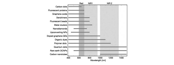
NPs:Nanoparticles; QDs:Quantum dots; DCNPs:Downconversion nanoparticles
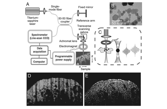
(A)Schematic of the experimental setup; (B)TEM of the MNPs used in the study; (C)Close-up of the sample arm with focusing lens, electromagnet, and specimen showing axial magnetomotive displacements of MNPs and corresponding change in the OCT interferogram.In vivo (D)MM-OCT and (E)OCT images of livers from MNP-injected mice.In (D) MM-OCT signal(green channel) is superposed on the OCT signal(red channel)
 DownLoad: